Characterising negative regulation
of CD8
+
T cell function in tolerance
and exhaustion
Mayura Vivek Wagle
June 2019
A thesis submitted for the degree of Doctor of Philosophy of The
Australian National University
© Copyright by Mayura Vivek Wagle, 2019
vi
Acknowledgements
Completing this PhD has been one of the most challenging yet highly rewarding experiences of my life. I could not have survived this rollercoaster of a journey with its highs and lows without the support and guidance of the following people.
I would firstly like to thank my supervisors Ian Parish and Chris Goodnow for their mentorship and for giving me the opportunity to undertake these projects at ANU. Thank you to Ian for introducing me to the complex fields of tolerance and exhaustion, for guiding me through the early daunting animal experiments and training me to be a thorough scientist. This thesis and the publications would not have been possible without the hard work, patience and commitment you provided as a supervisor. I would like to thank Chris for his support throughout this journey, for providing key guidance on the projects and sharing his scientific enthusiasm. Thank you also to my PhD panel members- David Tsharke and Ian Cockburn for their valuable advice throughout the PhD.
Next, I would like to thank the many amazing members of the Immunogenomics lab at JCSMR. Thank you to Debbie, Lisa, Yovina, Brigette and Sarp for helping me with my experiments and to other members including Michelle, Anselm, Kesuike, Mehmet, Manu, Yogesh, Jin, Zarah, Sofia and many others for your support and company. Thank you also to Immunogenomics lab at the Garvan Institute for welcoming me into the lab while I wrote my thesis. The experimental results included in this thesis would not have been possible without the great facilities at ANU. I would like to thank the wonderful staff at the flow cytometry unit at JCSMR- Mick and Harpreet, thank you for maintaining the machines so well, being very helpful and approachable with our FACS issues and for truly caring about our work. Also thank you to the APF staff, especially Barb and Christine who meticulously looked after all of the mice in the containment suites. Also a special mention to the administrative and support staff for the HDR students at ANU and JCSMR- thank you for all your prompt replies with our many queries throughout the degree and during the stressful submission time.
hours and fun times at all the conferences. I would also like to thank the broader JCSMR community, especially to all the students in the building who helped set up JARs and participated in the activities; it was an incredible experience to undertake during my PhD. I would especially like to thank my JCSMR family- Sarp, Ilenia, Emilie, Mayank, Dandan and others for your friendship and support- all of you made those weekend experiments much more bearable with your company and our weekend road trips certainly brought timely relief from the thesis.
Next, I would like to thank all my friends in Canberra. To my housemates Sarah and Alice, for always checking in on me during the late nights spent in the lab and creating a warm home to come back to. Also thank you to Jason, Jin, Manab, Alex and Monique for your friendship and support at different times of this journey. Thank you also to my Canberra and Sydney friends- Nishthaa, Christine, Rushika, Angelica, Anna and many others for always making the time to meet and support me through these years. Thank you also to my Singapore friends- Kavita, Christina and Stephen for your constant motivation and encouragement with the thesis writing.
I would like to thank my parents and Mihir for always supporting my ambitions and looking out for me throughout this tough journey. Thank you for driving to Canberra several times with short notice, providing week-long meals and always ensuring I maintained balance during this period. Thank you also to my new family- Jean aunty, Sujith uncle and Gerry, thanks for your constant support and motivation throughout the thesis. Special thanks to the grandaunts Elizabeth and Mary, for always checking in on my thesis during our visits, your encouragement certainly kept me going.
viii
Abstract
CD8+ T cells play a vital role in the immune system by clearing pathogen-infected tissues, however self-reactive CD8+ T cells that escape thymic selection pose the danger of causing autoimmune disease. These self-reactive CD8+ T cells are controlled in the periphery by tolerance mechanisms, which inhibit their function (anergy) or induce apoptosis (deletion). However, the specific molecular pathways crucial for negatively regulating self-reactive CD8+ T cells are not well elucidated. A
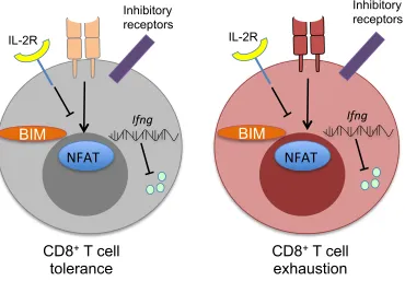
distinct form of negative regulation called “exhaustion” occurs within chronically stimulated effector CD8+ T cells during cancer and chronic infection. Due to the phenotypical similarities between CD8+ T cell tolerance and exhaustion, we aimed to understand if common underlying molecular pathways regulate these states.
The pro-apoptotic protein BIM is important in deletion of self-reactive CD8+ T cells, however the transcriptional control of Bim induction has been unclear. In Chapter 2, we assessed the contribution of the transcription factor FOXO3 in deletion of self-reactive CD8+ T cells given its role in Bim induction and cell death in effector and exhausted CD8+ T cells. While FOXO3 protein underwent activatory dephosphorylation during tolerance, FOXO3-deficient CD8+ T cells maintained the ability to induce BIM expression and undergo deletion. This result indicated that FOXO3 plays distinct roles in cell death of tolerant versus effector CD8+ T cells.
To further characterise CD8+ T cell tolerance pathways, in Chapter 3, we investigated whether the ubiquitin ligase adaptor NDFIP1, which is crucially required for CD4+ T cell anergy, influences CD8+ T cell tolerance. In a model of peptide-induced anergy, Ndfip1-deficient CD8+ T cells aberrantly expanded and differentiated into effector cells against high dose exogenous antigen, likely driven by increases in TCR signaling. In contrast, NDFIP1 was dispensable for peripheral deletion to low-dose exogenous antigen, and had little impact upon effector responses to acute infection. These results showed the importance of NDFIP1 in regulating CD8+ T cell tolerance and indicated that CD8+ T cell deletion and anergy are molecularly separable checkpoints.
While CD8+ T cell exhaustion appears distinct from tolerance, the transcriptional regulator EGR2 is commonly expressed between these states. In Chapter 4, we showed that exhausted CD8+ T cells in chronic LCMV infection expressed elevated levels of EGR2 compared to functional effectors. Loss of Egr2 severely disrupted terminal CD8+ T cell exhaustion in a cell intrinsic manner, with RNA-Seq results indicating a global enrichment of the exhaustion “stem cell” gene set in Egr2-deficient cells. Strikingly, the genes regulated by Egr2 during exhaustion appeared distinct from those controlled by Egr2 during T cell tolerance, suggesting that EGR2 is repurposed during T cell exhaustion.
x
Table of contents
Declaration ... iii
Statement of Contribution ... iv
Acknowledgements ... vi
Abstract ... viii
Table of contents ... x
List of figures ... xiv
List of tables ... xvi
Abbreviations ... xvii
CHAPTER 1: INTRODUCTION ... 1
1.1: CD8+ T cell biology ... 1
1.1.2: CD8+ T cell activation and differentiation ... 6
1.1.3: Negative regulation of CD8+ T cells ... 18
1.2: CD8+ T cell tolerance ... 20
1.2.1: Central Tolerance ... 21
1.2.2: Peripheral tolerance ... 24
1.2.3: CD8+ T cell tolerance outcomes ... 28
1.2.4: Molecular regulation of CD8+ T cell tolerance ... 34
1.3: CD8+ T cell exhaustion ... 42
1.3.1: Overview of CD8+ T cell exhaustion ... 42
1.3.2: Phenotype of exhausted CD8+ T cells ... 44
1.3.3: Evolutionary basis of exhaustion ... 49
1.3.4: Factors driving CD8+ T cell exhaustion ... 49
1.4: Comparing tolerance and exhaustion ... 57
1.5: Research questions ... 61
1.5.1. Does the same transcription factor control BIM-dependent death in tolerant versus effector cells? ... 61
1.5.2. Are similar molecular pathways engaged to enforce CD8+ T cell anergy versus deletion? ... 61
1.5.3. Are the same transcriptional regulatory pathways engaged downstream of TCR signaling during tolerance and exhaustion? ... 62
CHAPTER 2: FOXO3 is differentially required for CD8
+T cell death
during tolerance versus immunity ... 63
2.1: Abstract ... 64
2.3: Methods ... 66
2.3.1: Mice and mouse infection ... 66
2.3.2: T cell preparation for adoptive transfer ... 66
2.3.3: Flow cytometric analysis ... 67
2.3.4: Statistical analysis ... 67
2.4: Results ... 68
2.4.1: Elevated phosphorylation of FOXO proteins within CD8+ T cells in immunity versus tolerance ... 68
2.4.2: Foxo3MmR1/MmR1 mutant effector OT-I cells are partially resistant to T cell contraction ... 71
2.4.3: FOXO3 is dispensable for BIM induction and cell death during peripheral CD8+ T cell tolerance ... 71
2.5: Discussion ... 74
2.6: Acknowledgements ... 75
2.7: Conflict of Interest ... 75
2.8: References ... 76
CHAPTER 3: The ubiquitin ligase adaptor NDFIP1 selectively
enforces a CD8
+T cell tolerance checkpoint to high dose antigen 79
3.1: Abstract ... 803.2: Introduction ... 80
3.3: Experimental procedures ... 82
3.3.1: Mice ... 82
3.3.2: Peptide injections, rapamycin treatment and mouse infection ... 82
3.3.3: T cell preparation for adoptive transfer ... 82
3.3.4: EdU treatment and staining ... 83
3.3.5: Flow cytometric analysis ... 83
3.3.6: Phosphoflow and cytokine staining experiments ... 84
3.3.7:In vivo cytotoxicity assay ... 84
3.3.8: Histology ... 84
3.3.9: Statistical analysis ... 85
3.4: Results ... 85
3.4.1: Ndfip1 is dispensable for CD8+ T cell deletional tolerance to a pancreatic self-antigen ... 85
3.4.2: Ndfip1-deficiency disrupts CD8+ T cell tolerance to a pancreatic self-antigen in the context of higher self-antigen doses ... 88
3.4.3: Tolerogen concentration governs Ndfip1 restraint of CD8+ T cell expansion and differentiation ... 88
3.4.4: Ndfip1 limits TCR signaling in anergic CD8+ T cells ... 92
3.4.5: Ndfip1 restrains CD8+ T cell expansion and effector differentiation during continuous antigen exposure ... 95
xii
3.5: Discussion ... 98
3.6: Acknowledgements ... 100
3.7: Author contributions ... 100
3.8: Declarations of Interests ... 101
3.9: References ... 101
CHAPTER 4: The anergy-associated transcription factor EGR2 is
re-purposed to promote terminal CD8
+T cell exhaustion during
chronic viral infection ... 114
4.1: Abstract ... 115
4.2: Introduction ... 115
4.3: Methods ... 117
4.3.1: Mouse strains, adoptive transfer and infections ... 117
4.3.2: Bone marrow chimeras ... 118
4.3.3: CTV labeling, flow cytometric analysis and cell sorting ... 118
4.3.4: Phosphoflow and cytokine staining experiments ... 119
4.3.5: Antibodies and tetramers used for flow cytometric analysis ... 119
4.3.6: RNAseq analysis ... 120
4.3.7: ChIP-seq analysis ... 121
4.3.8: Statistical analysis ... 122
4.4: Results ... 122
4.4.1: EGR2 expression is elevated in chronic relative to acute LCMV infection ... 122
4.4.2: EGR2 is selectively expressed within TCF1+ memory-like exhausted T cells ... 125
4.4.3: EGR2 expression is maintained by chronic antigen encounter ... 127
4.4.4: EGR2 ablation cell intrinsically disrupts CD8+ T cell exhaustion ... 128
4.4.5: EGR2 controls inhibitory receptor expression and terminal exhaustion . 131 4.4.6: EGR2 broadly controls the terminal exhaustion gene program ... 132
4.4.7: EGR2 is re-routed away from anergy gene targets during T cell exhaustion to directly regulate exhaustion-associated genes ... 133
4.5: Discussion ... 139
4.6: Acknowledgements ... 143
4.7: Author contributions ... 143
4.8: References ... 144
CHAPTER 5- FINAL DISCUSSION ... 174
5.1: Introduction ... 174
5.2: Context-specific roles of molecular checkpoints in tolerance versus immunity ... 176
5.4: Building better mouse models to address unresolved questions in
tolerance ... 179 5.5: A question of semantics: is there a need to develop better nomenclature around T cell exhaustion and tolerance? ... 182 5.6: Significance of thesis findings and implications in the clinic. ... 184
xiv
List of figures
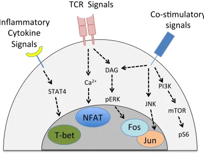
Figure 1.1. T cell signaling during effector differentiation……….……..12
Figure 1.2. Multiple fates of CD8+ T cell tolerance ………..31
Figure 1.3. Progressive differentiation towards an exhausted state …..………..48
Figure 1.4. Comparison of molecular pathways in CD8+ T cell tolerance and
exhaustion ………...………..59
Figure 2.1. Akt-dependent FOXO phosphorylation is elevated in effector versus
tolerised OT-I cells ………...70
Figure 2.2. FOXO3 is dispensable for BIM induction and cell death during OT-I
peripheral deletion ………..………..73
Figure 3.1. NDFIP1 limits autoimmune pathology after high antigen dose
stimulation………...87
Figure 3.2. Ndfip1 mutant OT-I cells resist tolerance to high antigen doses………...90
Figure 3.3. NDFIP1 restrains OT-I TCR signaling after high dose peptide
treatment……….... 93
Figure 3.4. NDFIP1 controls OT-I anergy to sustained antigen……….97
Figure 3.S1. Ndfip1 is dispensable for peripheral tolerance in RIP-OVAhi mice…...107
Figure 3.S2: Expression of apoptosis regulators and cytokines by Ndfip1 mutant OT-I
cells………109
Figure 3.S3. JUNB and CD25 expression levels are unchanged by Ndfip1 loss….110
Figure 3.S4. Ndfip1 loss does not affect OT-I expansion and differentiation during acute Listeria infection………111
Figure S5. NDFIP1 loss has little effect on CD8+ T cell effector and memory
differentiation during acute LCMV infection………113
Figure 4.1. EGR2 expression is elevated within TCF1+ memory-like exhausted cells
Figure 4.2. Egr2 expression is lost as cells undergo terminal exhaustion………….126
Figure 4.3. EGR2 expression is maintained by chronic antigen………...130
Figure 4.4. Egr2 loss blocks terminal exhaustion in the absence of CD4+ T cell
help………135
Figure 4.5. Global block in the terminal exhaustion gene program within EGR2
deficient cells………….………...138
Figure 4.6. EGR2 directly regulates a non-overlapping set of genes in exhaustion
versus anergy.………141
Figure 4.S1. CD8+ T cell phenotype in LCMV-Cl13 infected WT vs cKO mice…….153
Figure 4.S2. The virus-specific CD4+ T cell response within LCMV-Cl13 infected WT
versus cKO mice……..………...154
Figure 4.S3. Disruption of CD8+ T cell exhaustion by Egr2 loss is cell intrinsic……155
Figure 4.S4. CD8+ T cell expansion and phenotype in CD4 depleted, LCMV-Cl13
infected WT vs cKO mice.………..157
Figure 4.S5. EGR2 ChIP-seq analysis of CD8+ T cells isolated from LCMV-Cl13
infection……….159
Figure 5.1. Differential downstream functions of transcription factor EGR2 in CD8+ T cell tolerance and exhaustion………180
Figure 5.2. Proposed nomenclature system for negatively regulated CD8+ T cell
xvi
List of tables
Table 4.S1- significantly differentially expressed genes within Egr2 cKO CD8+ T cells relative to WT control cells (FDR<0.05)………...160
Table 4.S2 - HOMER de novo TF binding motifs identified within EGR2 ChIP-seq
binding peaks………...170
Table 4.S3 - Directly bound EGR2 targets during CD8+ T cell exhaustion…………171
Abbreviations
AIRE: Autoimmune Regulator ANOVA: Analysis of variance ANU: Australian National University APC: Antigen Presenting Cell APF: Australian Phenomics Facility Arm: Armstrong
AxV: Annexin V
BATF: Basic Leucine Zipper transcription factor Bcl2: B cell lymphoma 2
Bcl6: B cell lymphoma 6 BCR: B cell Receptor
BLIMP-1: B-lymphocyte-induced maturation protein 1 BM: Bone Marrow
BSA: Bovine Serum Albumin CD: Cluster of Differentiation c.f.u: colony forming units
ChIP: Chromatin Immunoprecipitation cKO: conditional Knockout
Cl13: Clone 13
CNS: Central Nervous System CTL: cytotoxic T lymphocyte CTV: Cell trace Violet
CXCR5: Chemokine CXC motif receptor 5 DAG: Diacylglycerol
DC: Dendritic Cell
DGK: Diacylglycerol Kinase
DMEM: Dubelcco’s Modified Eagle Media DNA: Deoxyribonucleic Acid
xviii EOMES: Eomesodermin
FACS: Fluoroscent activated cell sorting FCS: Fetal calf serum
FOXO3a: Forkhead box O3 FOXP3: Forkhead box P3 GFP: Green fluorescent protein GP: Glycoprotein
GZMB: Granzyme B HA: Heamagglutinin HEL: Hen Egg Lysozyme
HIV: Human Immunodeficiency Virus ICAM-1: Intracellular Adhesion Molecule-1 IFN: Interferon
IL: Interleukin
IRF4: Interferon regulatory factor 4 i.p: intra peritoneal
IP3: Inositol triphosphate
i.v: intra venous
ITIM: Immuno Tyrosine Inhibitory Motif KLRG1: Killer Cell Lectin Like Receptor G1 LAG3: Lymphocyte activation gene 3 LCMV: Lymphocytic choriomeningitis virus LM: Listeria monocytogenes
MFI: Mean fluorescence intensity
MHC I: Major Histocompatibility Complex Class I MHC II: Major Histocompatibility Complex Class II min: minutes
ml: milli litres
MS: Multiple Sclerosis
mTEC: medullary Thymic Epithelial Cells mTOR: mammalian Target of Rapamycin Ndfip1: NEDD4 family interacting protein 1
Down-regulated protein 4
NFAT: Nuclear Factor of Activated T cells OVA: Ovalbumin
PAMPs: Pathogen Associated Molecular Patterns PBS: Phosphate Buffered Saline
PD-1: Programmed Death 1 p.f.u: plaque forming units PGE2: Prostaglandin E2
PIP3: Phosphatidylinositol (3,4,5)-triphosphate
PI3K: Phosphoinositide-3 Kinase PLN: Pancreatic Lymph node
PRRs: Pathogen Recognition Receptors RA: Rheumatoid Arthritis
RIP: Rat insulin promoter RNA: Ribonucleic acid
RPMI 1640: Roswell Park Memorial Institute 1640 SCID: Severe Combined Immunodeficiency SLE: Systemic Lupus Erythematosus
SIV: Simian Immunodeficiency Virus T1D: Type I Diabetes
TCR: T cell receptor TF: Transcription Factor Th: T helper
TIM3: T cell immunoglobulin and mucin-domain containing-3 TLR: toll like receptors
TNFα: Tumour necrosis factor alpha TRAs: Tissue Restricted Antigens Treg: regulatory T cells
1
CHAPTER 1: INTRODUCTION
1.1: CD8+ T cell biology
1.1.1: Overview of the immune system
Living organisms have evolved to thrive in challenging external environments. A key factor for survival is the ability to protect oneself against pathogens or harmful microorganisms. Defense mechanisms collectively known as the organism's immune system have been evolving from the beginning of life. For successful protection, the immune system must be able to detect potential pathogens in the environment and mount an appropriate response against the pathogen.
The immune response of multicellular animals is multilayered and broadly classified into two arms - the innate and the adaptive immune systems. Each system consists of a network of cell types, which coordinate to recognise and combat pathogens. At the molecular level, immune cells express receptors to recognise conserved pathogen-associated molecular structures, as well as molecular moieties called antigens. When activated against a pathogenic antigen, the immune system generates an effective response to eliminate the pathogen through diverse cytotoxic and neutralizing mechanisms.
The innate immune system
In most organisms, the first line of defense includes physical barriers such as the skin and mucosal membranes, which limit entry of pathogens into the body. Upon breach of these barriers, multiple cell types forming the "innate immune system" such as the epithelium and phagocytes can be immediately activated due to their strategic location near these physical barriers. The innate immune system is crucial for rapid pathogen recognition and limiting pathogen spread. Phagocytes of the innate immune system comprise Dendritic Cells (DCs), macrophages and granulocytes, including neutrophils, eosinophils, and basophils. These cells are equipped with pattern recognition receptors (PRRs) to detect conserved pathogen-associated molecular patterns (PAMPs) such as bacterial cell wall components and viral nucleic acids (1). Activation of the PRRs leads to the generation of effector functions in the innate immune cells, such as the production of cytokines, degranulation of toxic molecules and phagocytosis. The latter allows the innate immune cells to ingest pathogens or infected cells with the aim to degrade the pathogen. This also enables "presentation" of pathogenic antigens on the surface of certain innate immune cells, which are classified as antigen presenting cells (APCs) (2). APCs are equipped with the task of processing pathogen-associated proteins in the phagosome and “present” pathogen-derived peptide fragments on their cell surface for recognition by cells of the adaptive immune system. Hence the innate immune system is vital for both quick responses to infection, and activation of the adaptive immune system through the production of cytokines and the presentation of antigen by APCs. However, it has multiple drawbacks, which necessitate the requirement for the adaptive immune system.
3
infection, in most cases the level of response generated against previously encountered pathogens does not change. This is an evolutionary disadvantage in the arms race against rapidly evolving pathogens. These factors potentially contributed to the development of the adaptive immune system in vertebrates.
The adaptive immune system
The adaptive immune system responds to specific molecular antigens and, hence, can confer pathogen-specific life-long protection or “immunity”. Greek philosopher Thucydides was the first to observe and record the features of adaptive immunity in the Athens plague of 430 B.C. In The History of the Peloponnesian War, he commented, “the same man was never attacked twice - never at least fatally” (3). The adaptive immune system consists of cells called lymphocytes that possess highly specialized antigen recognition capacity.
specialised system has its own disadvantages.
The nature of antigen recognition by the variable lymphocyte receptors is dependent on the chemical bonds formed between the antigen and the receptor, which determine the “affinity” of the interaction (8,9). Unlike the PRRs on innate immune cells, lymphocyte antigen receptors do not inherently recognise fixed molecular structures from bacterial cell walls or viral nucleic acids. This is because the unique lymphocyte receptor sequences are generated through randomized gene recombination events (10) as opposed to PRRs, which have conserved sequences. However, the diversity of lymphocyte receptor specificities thus generated can recognise any molecular moiety of a certain affinity regardless of whether they are host- or pathogen-derived, posing a risk of generating responses against self-tissues. Why does the immune system allow for the stochastic formation of lymphocyte receptors if it implies that the system has an equal chance of harming the host it has evolved to protect? The answer likely lies in the evolutionary advantage of a highly variable structure. The vast repertoire that randomized gene recombination provides outweighs the risk of generating self-specificities. Furthermore, the immune system employs multiple safeguards to mitigate the risk posed by self-reactive cells. First, the majority of lymphocytes bearing "self" specific receptors are either deleted from the repertoire, or functionally inactivated, via multiple immune "tolerance" processes (11) (described in the next section). Second, activation of adaptive immune cells typically requires signals from innate immune cells that are only produced upon innate immune recognition of an active infection, thus preventing the activation of self-reactive adaptive immune cells that may otherwise occur in the absence of infection (1). Lastly, the activation of lymphocytes is a multi-step process with each step stringently controlled by molecular networks acting as checkpoints to ensure lymphocytes are only activated against infectious stimulus (12,13). In this way, the immune system is able to usually keep any rogue self-reactive cells under control and limit their damage to the host.
Lymphocyte subsets
5
subjected to self-tolerance processes, their activation pathways are distinct due to differences in the structures of their lymphocyte receptors and hence the nature of the antigen that they recognize. B cells develop in the bone marrow and recognise native extracellular antigenic structures through their BCRs, such as repeating carbohydrates or intact proteins. Upon activation during an infection, pathogen-specific B cells can secrete their BCR as soluble proteins, known as antibodies, which can bind to the pathogen and induce multiple downstream effector responses to promote pathogen clearance. Hence B cells are crucial in clearing pathogens from the host circulation, however they are less effective at targeting intracellular pathogens residing in the cytoplasm of infected cells.
T cells detect and control intracellular infections through recognition of their specific antigens: small peptide fragments derived from proteins within the cell. In the 1970s, Doherty and Zinkernagel discovered that the TCR was in fact restricted to recognise antigen presented by the Major Histocompatibility Complex (MHC) molecules (14). Following this discovery, it was established that T cells carried either a CD4 or CD8 co-receptor to stabilize their interactions with MHC Class II (“MHC II”) and MHC Class I (“MHC I”) molecules respectively (15,16). These MHC molecules differ in their expression patterns and the nature of the peptide antigen presented in their grove. MHC II is almost exclusively expressed on APCs and usually presents extracellular phagocytized peptide antigens of typically 10-12 amino acids (2). MHC I, on the other hand, is expressed on most of the body’s cells and typically presents peptide antigens of 8-10 amino acids from intracellular pathogens or proteins (17). This dichotomy reflects the different functions of the responding CD4+ and CD8+ T cells. CD4+ T cells, known as “helper T cells”, are a highly variable subset, which regulate the function of B cells and CD8+ T cells through secretion of cytokines and expression of immunoregulatory cell surface receptors. CD8+ T cells, or "killer T
cells", are the primary responders against intracellular infections and have the ability to directly kill pathogen-infected cells. As CD8+ T cells comprise the subject of this
thesis, the remainder of the introduction will focus on this cell type.
self and foreign proteins processed by the proteasome within the cell. Thus, the continuous presentation of peptides on MHC I allows CD8+ T cells to survey the dynamic intracellular proteome within a target cell and scan for potential pathogen-derived peptides. Upon activation, CD8+ T cells produce cytotoxic enzymes called granzymes and the pore-forming toxin perforin, which are both released upon antigen recognition on an infected target cell to initiate programmed cell death or apoptosis. This cytotoxic machinery, along with other factors such as cytokines and death receptors, allows the activated CD8+ T cells to directly kill pathogen-infected cells.
For this reason, activated CD8+ T cells are known as Cytotoxic T lymphocytes (CTLs)
(18).
While CD8+ T cells are crucial in clearing intracellular pathogens, they can cause bystander tissue damage and also have the power to recognise endogenous self-antigens presented on MHC I. Thus, if inappropriately activated, CD8+ T cells have the potential to cause high levels of damage to host tissue. For instance, self-reactive CD8+ T cells can cause autoimmune disease by inappropriately targeting uninfected self-tissue independent of the presence of infection (19). Additionally, pathogen-specific CD8+ T cells can fatally damage whole organs during widely disseminated infections if their activity is not restrained (20–22). Hence regulation of CD8+ T cell activation by molecular checkpoints is vital to ensure optimal responses are generated against infection with minimal damage to the host. Exploring these molecular checkpoints in CD8+ T cell biology is the central aim of this thesis. However, in order to understand the pathways that limit CD8+ T cell immunity, it is important to first delineate the molecular pathways involved in the activation and differentiation of a functional CTL response.
1.1.2: CD8+ T cell activation and differentiation
Prior to activation, the repertoire of CD8+ T cell clones continuously circulates
7
lymphoid tissues (23). Naïve T cells receive homeostatic survival signals from frequent low avidity interactions with self-MHC and through the cytokine interleukin-7 (IL-7), which binds to the IL-7R expressed by naïve cells (24,25). Naïve CD8+ T cells do not acquire effector functions by default, and in the case of an infection must be provided with the correct signals for their activation by innate immune cells. Complete activation of CD8+ T cells requires extrinsic signals from the innate immune system,
which are then integrated intrinsically within the T cells to enable effector cell differentiation.
T cell activation by DCs
Activation of naïve CD8+ T cells first requires activation of innate immune cells by the invading pathogen. While B cells and macrophages can also act as APCs, it was shown in mixed leukocyte reactions that DCs are the most potent stimulators of T cell activation (26). Named after their characteristic dendrite-like projections, DCs are the key APC required for activation of naïve CD8+ T cells, as they provide context-specific activation signals to CD8+ T cells. To understand the specific role of DCs in contributing to the CD8+ T cell response, many genetic models were created to deplete DC populations in vivo, either using mice lacking the Transcription Factor (TF) Batf3 (27,28) or genetically expressing Diptheria toxin receptor (DTR) under the CD11c (29), CD205 (30) BDCA2 (31), and Clec9A (32) promoters whereby delivery of the Diptheria toxin would result in selective loss of DC subsets expressing the above genes. The resulting loss of DCs (or specific DC subsets) in the above models led to a defective CTL response to a variety of infection and immune challenge models (27–32). The reliance on DCs for CD8+ T cell activation and effector differentiation is due to a number of reasons such as their efficient activation in most infections, the unique ability to “cross-present” antigen and acting as a non-redundant source of key cytokines such as interferons (IFN).
tissue-restricted pathogens. This steady state migration is especially important given that naïve CD8+ T cells do not have access to peripheral tissues.
An important feature of the immune response is the ability to mount an efficient response tailored to the pathogen at hand. Along with providing information on the antigens present within a tissue, DCs are also able to provide the T cell with information on the type of pathogenic infection occurring in the peripheral site. The signals from the specific pathogenic infection result in the activation of the DCs, facilitating their transition into an “immunogenic state” capable of supporting effector T cell differentiation during infection (34,35). Immunogenic DCs, increase their migration to the nearest lymph nodes to activate the adaptive immune system, upregulate co-stimulatory molecules and cytokine production as well as activating other innate immune cells during infection. Costimulatory receptors, such as CD80 and CD86 (36,37), are upregulated after PRR activation and these play important roles in sending positive differentiation signals to the T cells that they interact with. Along with PRR stimulation, inflammatory cytokines, such as Type I interferons (IFN), that are produced by other innate immune cells and infected tissues can also act on the DCs to further boost expression of co-stimulatory receptors (38). Depending on the type of infection and hence specific PRR activated, DCs produce cytokines that can inform the adaptive system about the infection at hand. In regards to intracellular pathogens, bacterial infections induce IL-12 production by DCs (39,40), while viral infections trigger production of IL-12 as well as IFNα/β (41) – both of these signals are crucial for appropriate CD8+ T cell activation in these respective infections. The activation and signaling of PRRs also enhances sampling and endocytosis of pathogenic antigen by DCs, leading to more efficient antigen presentation on MHC molecules to the naive T cell pool (42–44). Hence, given their presence throughout tissues, rapid activation in infection, and ability to provide the correct activation signals to T cells, DCs serve as excellent activators of the immune system. However, they are specifically crucial for CD8+ T cell activation due to their ability to
“cross-present” antigens.
9
encounter pathogenic antigen by phagocytosis, they have no obvious way to present foreign antigen on MHC I and induce a response. To address this dilemma, certain subsets of DCs are uniquely endowed with the capacity to present exogenous antigen on MHC I in a process called “cross-presentation” (45). This deviation from the classical MHC presentation pathway is crucial for activation of CD8+ T cells against infections and tumor antigens, which are not intracellularly expressed within DCs (45–47). All CD8+ DCs with XCR1 expression (commonly referred to as the
cDC1 cells) are able to cross-present antigen to CD8+ T cells (48–50). This subset
stems from a common lineage controlled by the TF BATF3, which is crucial for the differentiation of cross-presenting DCs (27,51). Deletion of BATF3 in mouse models led to the crippling of the CTL response due to loss of the entire cross-presenting DC lineage (27). To specifically test the role of cross-presentation in the activation of CD8+ T cells, a recent study utilised mice with deficiency in the WDFY4 protein, a factor required for cross-presentation. In this model, despite the presence of the BATF3+ DC populations, the CD8+ T cell response to tumors and virus infection was defective due to the inability of DCs to cross-present antigen (52). Thus, DCs are uniquely specialised to license a naïve CD8+ T cell for effector differentiation within secondary lymphoid organs.
the CD28-CD80/86 interaction, CD8+ T cells are also activated by multiple redundant co-stimulatory pathways, such 4-1BB-41BBL (61) as well as CD27-CD70 (62). Thus, multiple redundant interactions are involved in CD8+ T cell activation; this redundancy likely evolved as a way to reduce possible immune evasion mechanisms by pathogens. In addition to co-stimulatory molecules, inflammatory cytokines such as IL-12 (63) or Type I IFN (64) released from DCs or other cell types, in addition to autocrine or paracrine IL-2 produced by the nascent effector T cell response (65), can directly act on the responding cells to enhance effector differentiation. The combination of cytokine and co-stimulatory signals encountered ultimately sum to both trigger effector differentiation of naive CD8+ T cells and determine the magnitude of the effector CD8+ T cell response (66).
T cell signaling
Productive CD8+ T cell activation ultimately hinges on the capacity of CD8+ T cells to convert the extracellular signals received (antigen, cytokines and co-stimulatory signals) into intracellular signaling events that direct effector differentiation. For this reason, insights into the signaling events engaged during effector differentiation have played a crucial role in shaping our understanding of the differentiation process. The TCR-antigen interaction, costimulatory receptor engagement and cytokine signals all engage both overlapping and independent signaling cascades in the T cell that culminate in the activation of transcription factors. Depending on the signals received, the respective transcription factors work together to cause the global gene changes required for the transition of naïve CD8+ T cells into functional CTLs appropriate for the infection context. While there are numerous complex signaling pathways involved in T cell activation, the most relevant ones, in addition to the broad principles underlying signaling in T cells, are discussed below and summarised in Figure 1.1.
11
triphosphate (IP3) and Diacylglycerol (DAG) molecules (68). IP3 induces
calcium-signaling which activates the key transcription factor NFAT (69,70). Many in vitro experiments use calcium ionophores to activate NFAT in T cells (68,71,72), illustrating the dependence of this transcription factor on calcium signaling. DAG produced from PIP3 hydrolysis is responsible for initiating numerous pathways, and
its function varies depending on context. One of the key functions of DAG is to induce Ras activation (73) and, ultimately, phosphorylation of ERK, which is a hallmark of TCR signaling (74,75). Importantly, ERK signaling contributes to activation of the TF Fos, a key component of the AP-1 TF complex that plays important roles in T cell activation (76) (see below). Thus, antigen signaling from TCR results in the induction of NFAT and Fos activity, however these pathways alone are not sufficient to completely activate the T cells and turn on effector genes.
Co-stimulatory and cytokine signals are equally important in the activation of T cells. This is a key point of regulation, as it ensures that CD8+ T cell activation will only optimally occur in the presence of an active infection. These signals critically contribute to T cell activation in a number of ways. Firstly, costimulatory signals, along with the TCR, are important for activating the JNK pathway, which is required for the activation of the JUN and FOS TFs that cooperatively make up 1 (77). AP-1, in collaboration with NFAT, is important for activating many key effector genes, including cytokines (78). Furthermore, co-stimulatory interactions induce the Phosphoinositide 3 Kinase (PI3K) pathway, which in turn activates the kinase Akt, and the mammalian Target of Rapamycin (mTOR) (79). The Akt/mTOR axis plays essential roles in enhancing the metabolism of the T cell by, for example, augmenting protein translation (80,81). This is a particularly important function of this pathway, as profound metabolic shifts are required to support T cell differentiation. To convert a CD8+ T cell from its naïve resting state to an army of effector cells loaded with
cytotoxic machinery requires drastic changes in CD8+ T cell physiology and changes
in cellular respiration (82). mTOR activation is crucial in enabling a metabolic switch in activated CD8+ T cells from using oxidative phosphorylation to aerobic glycolysis -
JNK
Inflammatory
Cytokine
Signals
TCR Signals
Co-s9mulatory
signals
NFAT
T-bet
Fos
Jun
STAT4
DAG
pERK
Ca
2+PI3K
mTOR
[image:31.595.99.497.154.455.2]pS6
Figure 1.1: T cell signaling during effector differentiation: Productive activation
of naïve CD8+ T cells into effector cells requires TCR signals, cytokine signals and
co-stimulatory signals from the immunogenic APC. This figure summarises the downstream pathways triggered by the different signals and the final transcription
13
These factors can then fuel synthesis of key effector proteins, as well as support growth and proliferation (86). Furthermore, mTOR signaling increases surface expression of glucose and amino acid transporters, which in turn allow greater nutrient uptake into the cell to compensate for inefficient energy generation by aerobic glycolysis (86,79,87,88).
While TCR and co-stimulatory signals are necessary for activation of naïve CD8+ T
cells, inflammatory cytokines secreted by the DC (and surrounding innate immune cells) further aid T cell differentiation into effector cells (89). The presence of IL-12 during infection can induce expression of the transcription factor T-BET in CD8+ T cells by binding to the IL-12R and signaling through STAT4 (90,91). T-BET is an important transcription factor that contributes to the effector function of CD8+ T cells(90,92) and is described in detail in the following sections. Additionally extracellular receptor interactions that likely occur during the T cell-DC interaction, such as Notch signaling, also affect acquisition of effector functions by CD8+ T cells by controlling downstream transcription factors (93,94).
Thus, the signaling events triggered by the TCR, co-stimulatory, and cytokine pathways, in addition to other extracellular signals, converge to activate transcription factors, which in turn determine the fate of the T cell in a manner appropriate to the infection at hand. While most CD8+ T cells receiving these signals during an infection will differentiate into effector cells, a sub-population of cells persists beyond the clearance of infection and differentiates into memory cells. The bifurcation of cell fate between effector and memory cells begins early during activation and is controlled by various molecular factors discussed below.
Effector and Memory CD8+ T cell differentiation
The goal of the CD8+ T cell response is to firstly produce sufficient numbers of robust
effector cells to clear the pathogen at hand, and to secondly form long-term memory to combat future invasion by the same pathogen. The typical CD8+ T cell response to
and self-renew to maintain a stable population in the absence of antigen, and rapidly differentiate and re-expand upon pathogen re-encounter. Hence given that the cellular properties of effector and memory cells are distinct from each other, a highly regulated differentiation process ensures that the correct proportion of effector CD8+ T cells either undergo terminal differentiation or persist as memory cells. This decision to either form a short-lived effector cell or persist as a memory cell begins early during effector differentiation (95,96) , and is controlled by a suite of extensively studied molecular cues. This section will cover the molecular pathways that control effector cell expansion and persistence through to memory.
Naïve CD8+ T cells undergo rapid proliferation following activation, with expansion capacity reaching up to 1000-fold over the initial naïve precursor frequency. However, not all T cell clones expand to similar levels, and proliferation is controlled by multiple factors including precursor frequency of clones specific for the antigen and pMHC affinity (6,97–99), as well as pathways downstream of TCR and co-stimulatory receptors such as mTOR signaling, cytokine activation of STAT1/4 and potentially Notch signaling (100). Multiple TCRs can be specific for a given antigen at different affinities and, although CD8+ T cells of lower avidities are recruited into the effector program, they undergo less proliferation due to competition with the high-affinity clones for the peptide-MHC complexes (101,102). This allows preferential expansion of the high-affinity TCR populations during effector differentiation in a process driven by the transcription factors IRF4 and BATF. IRF4 is induced downstream of NFAT and AP-1 activation in an antigen-dependent manner, with its expression directly proportional to the strength of TCR signaling (103). IRF4 enables high-affinity clones to undergo the metabolic switch to aerobic glycolysis and catabolic metabolism that is required to fuel effector cell expansion; this allows high affinity cells to become the dominant clonal population of effector cells (103). BATF is also induced downstream of TCR signalling, and it forms a complex with IRF4 and JUN proteins to initiate transcription of T-bet, Eomes and the cytokine receptor genes that control effector development (104).
15
both inflammatory chemokine receptors and specific integrins to enable homing to inflamed and infected tissues (23,107,108). Once they reach infected sites, they are able to mobilise effector functions upon target cell recognition. Downregulation of CD62L is linked to PI3K-mTOR activity, and this serves as a checkpoint ensuring that a cell’s migratory capacity and the metabolic changes required for effector function are acquired in parallel (109). PI3K-mTOR-dependent regulation of CD62L levels is achieved through PI3K dependent proteolytic shedding, and through suppression of the transcription factor KLF2. KLF2 induces expression of a number of receptors that promote recirculation (including CD62L), meaning that KLF2 inhibition facilitates CD8+ T cell migration into the tissue (110–112).
In order to achieve rapid expansion, tissue migratory capacity and cytolytic function, effector CD8+ T cells undergo a unique transcriptional and epigenetic differentiation program that results in global changes in gene expression (100,113–117). This differentiation program allows CD8+ T cells to perform effector functions, including secretion of inflammatory cytokines such as Interferon γ (IFNγ), Tumour Necrosis Factor α (TNFα) and Interleukin-2 (IL-2) (118) . Effector cells also gain cytotoxic functions to enable target cell killing by apoptosis, including release of granzymes and perforin, and ligation of the FAS receptor on target cells via FASL (119,120). Many of these gene changes are initiated by transcription factors such as T-BET and EOMES, which induce the Ifng, Gzmb and Prf1 genes (121,122). In order to reinforce and stabilize effector differentiation, certain TFs create positive feedback loops. T-BET provides an example of such a positive loop in effector CD8+ T cells. The expression, and thus function, of T-BET is controlled by the amount of inflammatory signals in the environment, specifically IL-12 (90). T-BET acts as a rheostat: measuring inflammation and driving the terminal differentiation of effector cells accordingly in a threshold dependent manner (90). T-BET promotes effector differentiation by inducing other TFs that reinforce the effector gene program. T-BET promotes effector function, in part, by inducing the transcription factor ZEB2, which allows CD8+ T cells to commit to the effector lineage and die after antigen clearance
induction of a related factor, ID2 (127), and optimal expansion and effector differentiation. These examples illustrate how a single TF (T-BET) can trigger a cascade of positive feedback loops that reinforce CTL differentiation and expansion.
Effector cells become terminally differentiated if they excessively proliferate and differentiate. Terminally differentiated cells have a poor capacity to persist (90), and once the infection is cleared, 90-95% of the effector CD8+ T cell population
(predominantly comprising terminally differentiated cells) undergoes apoptotic cell death (128). The crucial factors responsible for cell death during contraction are the pro-apoptotic BH3-only Bcl2 family members BIM (129) and, to a lesser extent, PUMA (130). The pathways that trigger BIM-dependent death are still being characterised. Many studies have suggested that transcriptional induction of BIM expression by the transcription factor FOXO3 plays an important role in BIM-dependent contraction. FOXO transcription factors are excluded from the nucleus and degraded upon phosphorylation by Akt (131). Conversely, upon loss of cytokine signaling, FOXO3 is dephosphorylated, leading to protein accumulation, nuclear translocation and access to target genes. In vitro experiments in several B and T cell lines (132,133) have indicated that FOXO3 directly binds to the Bim promoter, where it then induces expression of Bim mRNA (134) and subsequent death of the cells. In vivo models have confirmed a role for FOXO3 in contraction using mouse models with a T cell-intrinsic deficiency in FOXO3. These mice exhibited an accumulation of effector CD8+ T cells due to reduced apoptosis, and diminished Bim expression, during the T cell contraction phase in both LCMV and LM-OVA infections (135,136). However, it remains unclear whether FOXO3 directly controls Bim expression in vivo. Recent studies have presented contrasting evidence about the binding of FOXO3 to the BIM promoter (137), as mutation of the FOXO3 binding sites within the Bim promoter did not hinder BIM expression or apoptosis. Hence, other pathways, such as loss of Erk-mediated repression of BIM expression and protein levels (138– 140,75) upon pathogen clearance, could also contribute to BIM-dependent death during T cell contraction.
17
(141,142) and IL-7R (95,143) can help to identify these early populations of short-lived effector cells and memory precursors, and these markers have enabled tracking of these populations in several infection models. While there are multiple hypotheses explaining how these T cell populations arise (100), the “decreasing potential” model is backed by multiple lines of evidence. The decreasing potential model claims that the differentiation of naïve CD8+ T cells into either effector or memory precursors is
dependent on the balance in their expression of effector versus memory associated transcription factors. This model suggests that the more effector cells differentiate, the more they lose memory potential (100). This is postulated to occur due to increased expression of effector-specific TFs that specifically silence competing memory-associated TFs. However, the changes in TF expression are also compatible with other models of memory differentiation, such as binary fate determination by asymmetric cell division. There is likely a stochastic component to this differentiation process, with the small fraction of cells that are, by chance, exposed to less of the factors that promote terminal differentiation (such as inflammatory cytokines) retaining more memory potential. For example, CD8+ T cells exposed to high levels of IL-12 will up-regulate T-bet expression, while those clones that encounter less IL-12 will maintain expression of the opposing transcription factor,
Eomes, aiding subsequent persistence and differentiation into memory cells (144). Multiple transcription factor pairs oppose each other to promote either a memory or short-lived effector fate. These pairs include Id2 and Id3 (126,127,145), Zeb1 and
Zeb2 (123,124,146), T-bet and Eomes (144,147–149), Stat4 and Stat3 (150–152), and Blimp-1 and Bcl6 (125,150,153–157), with the former molecule involved in effector development and the latter in memory differentiation. The process of short-lived effector and memory commitment is also controlled at the epigenetic level, with the memory genes being shut down in effector cells as they commit to that lineage (158). On the other hand, memory cells do not repress the effector genes but have bivalent modifications on the associated histones, allowing the cells to differentiate back into effector cells for subsequent infections (158,159). Hence the fate of effector and memory cells is tightly regulated by extrinsic factors, which control the intrinsic regulatory machinery.
effector cell counterparts. First, they do not rely on antigen for their survival (160), but instead depend upon the homeostatic cytokines IL-7 (143,161) and IL-15 (162) for survival and self-renewal. Second, they produce greater amounts of IL-2 (163), and can rapidly give rise to effector cells upon antigen reencounter. In particular, memory cells undergo epigenetic changes that facilitate rapid re-expression of effector genes (114,115,164,165). Memory cells exhibit heterogeneity, with specialised subsets such as long-lived central memory cells (TCM), which reside in the lymphoid organs
and exhibit greater proliferative capacity, and short-lived effector memory cells (TEM),
which patrol tissues and can launch immediate responses to pathogen reinfection, but are less proliferative upon restimulation (163,166). TCM become enriched over
time, likely largely due to preferential survival and homeostatic proliferation of the TCM
population (98,167,168). Recently, a new subset of memory cells called tissue resident memory cells (TRM) was defined, with this subset restricted to the tissue site
of primary infection; this subset plays non-redundant roles in protection against re-infection at a specific tissue site (169,170). Each of these subsets is characterised by a unique combination of transcription factors, which drives their differentiation and maintains their identity and “stemness”. In general, long-lived circulating central memory CD8+ T cells are characterised by expression of multiple transcriptional regulators, including Eomes (149), Bcl6 (156), Id3 (126,145), Foxo1 (171,172), Tcf7
(173) and Zeb1 (146). In contrast, recent data indicate that TRM appear to have a
distinct transcriptional program from circulating memory cells (174–177). While decades of research have thoroughly characterised the network of transcription factors in effector-memory differentiation, the TF networks that dampen the CD8+ T cell response to chronic antigen are less well characterised.
1.1.3: Negative regulation of CD8+ T cells
While a highly coordinated effector and memory response is required to contain infection and prevent reinfection, a completely unrestrained CD8+ T cell response
poses danger to the host. Hence, there is a need for negative regulation and checkpoints in CD8+ T cell activation. T cell clones that recognise self-antigen need
self-19
reactive cells. Second, negative regulatory mechanisms are required during infection to protect the host against fatal collateral tissue damage. The negative regulatory pathways engaged to limit such immunopathology typically dampen the inflammatory function of chronically stimulated effector cells in a process often called T cell “exhaustion”. These two states of CD8+ T cell differentiation are driven by chronic antigen recognition, which induces negative regulatory pathways that suppress CD8+
1.2: CD8+ T cell tolerance
The vast diversity of lymphocyte receptor specificities in the adaptive immune system means an almost unlimited spectrum of antigens can be recognised, however this implies that B and T cells can also recognise self-antigens. This introduces a dilemma in the functioning of the adaptive immune system, as there is a possibility of generating cytotoxic responses against host tissues. To mitigate this risk, multiple layers of regulation, collectively referred to as immunological tolerance mechanisms, have evolved to control self-reactive lymphocytes. These mechanisms act in an antigen-specific manner, such that typically only T cells specific to self and steady-state antigens are subjected to these tolerance processes. The tolerance mechanisms are usually subdivided into two main categories: central tolerance mechanisms, which purge self-reactive cells from the repertoire during lymphocyte development, and peripheral tolerance mechanisms, which restrain those self-reactive cells that escape central tolerance.
21
pathways could become potential drug targets for the treatment of CD8+ T cell-mediated autoimmune disease.
1.2.1: Central Tolerance
In his clonal selection theory, Macfarlane Burnet proposed that clones recognizing self-antigens would be purged from the repertoire before they entered the periphery (4). In subsequent decades, this hypothesis was validated with the identification of “central tolerance” pathways that delete self-reactive B cells and T cells during their development in bone marrow and thymus respectively. To ensure that the bulk of self-reactive cells are rapidly eliminated before they can pose a risk to the host, these pathways operate immediately after lymphocyte receptor rearrangement. In the case of T cells, TCR rearrangement occurs within the thymus after immature Pre-T cells formed in the bone marrow migrate to the thymus. Due to the stochastic nature of the TCR gene recombination process, a diverse lymphocyte receptor repertoire of up to 1015 unique specificities is predicted to be generated in the thymus (179,180). While random receptor recombination can generate self-reactive clones, it can also generate useless clones that cannot interact with MHC. Thus, to ensure the TCRs are both functional and not self-reactive, the final receptor products need to be selected for their ability to interact with self-MHC molecules, and selected against recognition of self-antigens. For this reason, two selection processes have evolved within central tolerance: one to ensure that only T cells with TCRs capable of binding to MHC are retained (positive selection), and one to actively eliminate cells that are self-reactive (negative selection) (181).
negative selection include the medullary TECs (mTECs) and DCs, which present an array of self-antigens (183). While mTECs directly present self-antigens that they express, DCs have been shown to cross-present antigens to CD8+ T cell precursors (184,185). Negative selection can occur in both the medulla and the cortex regions of the thymus, and is manifested in two “waves” of selection. The first wave deletes immature thymocytes that can strongly recognise self-antigens presented in the thymus, and the second wave is important in generation of Tregs from mature CD4+
self-reactive thymocytes (186,187). While T cells are selected against ubiquitously expressed self-antigens within the thymus (188), it was initially hypothesized that T cells would not be tolerised against tissue restricted antigens (TRA) during thymic selection, and that peripheral tolerance mechanisms exclusively controlled T cells specific for TRAs. However, this dogma was challenged by multiple studies. Firstly, it was shown that DCs from the periphery could pick up antigen from peripheral tissues and migrate to the thymus for subsequent negative selection of TRA-specific CD8+ T cells (189). Secondly, early studies indicated promiscuous mRNA expression of TRAs in thymic mTECs, such as insulin, alpha-fetoprotein and myelin proteins (190– 192). The source of this promiscuous expression was eventually attributed to the transcription factor AIRE, which is responsible for a leaky expression system that turns on expression of different sets of tissue-restricted genes in each mTEC (193,194). While DCs do not express AIRE, they are able to cross-present AIRE-dependent TRAs derived from mTECs and thereby participate in negative selection (184,195). AIRE is likely not responsible for controlling all of the observed promiscuous TRA expression, and recently the transcription factor FEZF2 (196) was also shown to contribute to promiscuous TRA expression within mTECs. The importance of these transcription factors in thymic negative selection and immune tolerance has been highlighted in AIRE deficient mice and humans, which succumb to multi-organ autoimmune diseases (193,197–199).
23
also contribute to thymic deletion, as a lack of both Bim and Puma resulted in the onset of autoimmune disease, presumably by overwhelming peripheral tolerance processes (201).
Despite the comprehensive negative selection process in the thymus, negative selection is “leaky” and self-reactive CD8+ T cells do escape into the periphery.
Self-reactive T cells have been observed in the peripheral blood of healthy individuals at comparable levels to patients with autoimmune disease (202–206). More recently, tetramer enrichment of self-antigen specific T cells revealed that male antigen H-Y specific CD8+ T cell numbers were present in both male and female adults despite the presence of H-Y as a self-antigen in males (207). The number of tetramer+ cells present in males was a third of the number found in females, indicating that thymic selection only purged ~70% of the repertoire specific for H-Y antigen. This finding suggested that, at least in the case of the H-Y antigen, thymic selection was incapable of deleting all self-reactive T cells from the repertoire.
on tolerance of H-Y specific T cells in males also demonstrated the presence of unlimited TCR specificities in healthy controls against all possible mutagenic variations of a Hepatitis B viral peptide, emphasizing that a broad TCR repertoire was present in most individuals (207). Thus, to maintain this vast T cell repertoire in the periphery while simultaneously restraining the rogue self-reactive T cells that are an inevitable consequence of this diversity, the immune system has developed several “peripheral” tolerance mechanisms.
1.2.2: Peripheral tolerance
Peripheral tolerance mechanisms identify self-reactive T cells by selectively tolerising cells that recognise their cognate antigen in the steady state. This is based on the presumption that antigen encountered in the steady state (ie. in absence of inflammation or danger signals) is likely enriched for self-antigens, and hence any responding T cells are likely self-reactive. Furthermore, non-self antigens presented in the steady state are likely not harmful to the host, and an immune response against such steady-state persistent antigens could thus result in immunopathology and be detrimental to the host. As a result, antigen recognition by naïve peripheral CD8+ T cells in a non-inflammatory context typically fails to result in effector differentiation, and even if the responding cells do acquire effector characteristics, their functions are greatly muted compared to those adopted during the response to an active infection. These regulatory outcomes are imposed on the self-reactive CD8+ T cells through tolerogenic interactions with DCs, and suppression of activation by regulatory T cells (Tregs) (212,213). Similar peripheral tolerance mechanisms are responsible for limiting aberrant CD8+ T cell responses to benign foreign antigens
derived from food (214), gut bacteria (215) and a growing fetus (216).
25
antigens were genetically engineered for transgenic expression under the control of tissue-specific promoters, such that they would be recognised by the immune system as surrogate “self-proteins” in their respective tissue of expression. The TCRs that recognised immunodominant epitopes within these model antigens were typically well characterised, with TCR transgenic mice often available for the specific model antigen of interest. These included CD8+ TCR transgenic mice specific for well
characterised MHC I peptides, such as “Clone 4 (CL4)” mice bearing CD8+ T cells
specific for the HA533–541 peptide (223), “P14” mice with T cells specific for the GP33-41
peptide (224), and “OT-I” mice with cells specific for the OVA257-264 peptide (225).
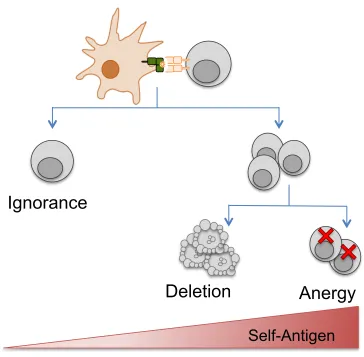
the tissue-specific promoter, model antigen and strength of antigen expression varied between mouse strains. These differences translated into different experimental outcomes, with the above studies often observing one of three different CD8+ T cell tolerance outcomes: ignorance, anergy or deletion (233,234). These tolerance outcomes are are largely guided by how T cells interact with steady-state DCs bearing self-antigen.
Regulation of peripheral tolerance by dendritic cells
Peripheral tolerance in CD8+ T cells is typically induced by presentation of
self-antigen by APCs in the absence of infection or pathology. In the steady state, DCs are in a quiescent state however they still continuously survey the environment for antigens. In particular, the CD8α+ subset of DCs actively captures apoptotic cells in the steady state and cross-presents captured proteins to self-reactive CD8+ T cells in the lymph nodes (235–237). The role of DCs in antigen cross-presentation has also been highlighted in transgenic tolerance models such as RIP-mOVA and RIP-OVAhi, where expression of membrane bound or soluble OVA respectively is limited to the pancreas, yet CD8+ T cells specific for OVA peptide are able to recognise antigen in the draining pancreatic lymph nodes due to cross-presentation by the CD8α+ DC subset (238). DCs are extremely crucial to steady state tolerance induction in CD8+ T cells, as several in vivo and in vitro experiments have shown loss of DCs through targeted depletion (239) or specific loss of cross-presentation capacity in the DCs results in loss of CD8+ T cell tolerance (237). The importance of DCs in inducing tolerance was further illustrated by the Ins-HA transgenic model, where the model antigen Haemaglutinnin (HA) from the Influenza virus is expressed under the RIP locus. Infection of adult mice with Influenza did not result in diabetes induction (240), as all HA-specific CD8+ T cells were tolerised by steady state DCs. However, when these experiments were carried out at 3 weeks of age, prior to the appearance of cross-presenting DCs within the pancreatic lymph nodes, Influenza infection led to diabetes induction due to the presence of endogenous HA specific CD8+ T cells


![Zwitterionic 1 {(1E) [(4 hydroxyphenyl)iminio]methyl}naphthalen 2 olate: crystal structure and Hirshfeld surface analysis](data:image/gif;base64,R0lGODlhAQABAIAAAP///wAAACH5BAEAAAAALAAAAAABAAEAAAICRAEAOw==)