www.fm.viamedica.pl
Address for correspondence: H.A.M. Saleh, Assistant Prof., Department of Anatomy, King Abdulaziz University, P.O Box 80316, Jeddah 21589, Saudi Arabia, tel./fax: 00966 2 6231774, e-mail: [email protected]
Vanilloid receptor type 1-immunoreactive
nerves in the rat urinary bladder and primary
afferent neurones: the effects of age
H.A.M. Saleh
Department of Anatomy, King Abdulaziz University, Jeddah, Saudi Arabia
[Received 17 February 2006, Revised 12 May 2006, Accepted 12 May 2006]
The vanilloid receptor (VR1) is a molecular integrator of various painful stimuli, including capsaicin, acid and high temperature. VR1 protein functions both as a receptor for capsaicin and a transducer of noxious thermal stimuli. In addition, VR1 is well characterised at the terminals of sensory nerves involved in the pain pathway. VR1 is also expressed in a capsaicin-sensitive and peptide-containing sub-population of primary sensory nerves.
Indirect immunohistochemistry was used to examine the distribution of nerves immunoreactive (ir) for VR1 in the base of the urinary bladder and in the neu-rones of the lumbosacral dorsal root ganglia (L1-L2 and L6-S1) of young adult (3 months) and aged (24 months) male rats. Semi-quantitative estimations of nerve densities were assessed and quantitative studies were also used to examine the effects of age on the percentage of VR1-ir dorsal root ganglion neurones. The bladder base in young adults showed dense VR1-ir fibres within the urothe-lium and in the subepitheurothe-lium and fibres ranging from sparse to moderate in number in the muscle coat. In comparison to the young animals, the aged rats showed sparse to moderate densities of VR1-ir nerves in the subepithelium and sparse fibres in the muscle layers. In the lumbosacral dorsal root ganglia the percentage of VR1-ir neuronal profiles showed a significant reduction from (mean ± SEM) 17.8 ± 2% in the young adult to 12 ± 1.6 in the aged rats.
The present findings suggest that the effects of VR1 on bladder function (noci-ception and reflex micturition) are influenced by age and the reduction with age of VR1-ir neurones in the dorsal root ganglia could also have important implica-tions for the micturition reflex.
Key words: VR1, immunohistochemistry, sensory neurones, dorsal root ganglia
INTRODUCTION
The vanilloid receptor type 1 (VR1) protein func-tions both as a receptor for capsaicin and as a trans-ducer of noxious stimuli [19]. VR1 is a non-selective cation channel expressed in about half the dorsal root ganglion neurones [9, 30]. The VR1-expressing sub-population of primary sensory neurones includes
the majority of small and medium-sized cells that synthesise neuropeptides, such as substance P (SP) or calcitonin gene-related peptide (CGRP) and car-bohydrates that bind isolectin B4 (IB4) [1, 19]. In
vit-ro, VR1-expressing cells can be activated by protons
findings showing that mice lacking the VR1 gene do not respond to vanilloids and have reduced sensitiv-ity to noxious heat and low pH confirmed that VR1 is responsible for vanilloid susceptibility and contrib-utes to heat and proton sensitivity in vivo [14].
There has been experimental and clinical evidence to show the importance of VR1 in the lower urinary tract [5]. In humans this receptor has been detected in nerve endings of primary sensory neurones, smooth muscle and connective tissue cells [22]. Moreover, VR1 has also been detected in the urothe-lium of the rat urinary bladder [4]. A large propor-tion of sensory fibres innervating the urinary blad-der are sensitive to capsaicin [7, 26, 31, 32].
Consistent with the nociceptive nature of capsa-icin-responsive primary afferents, intravesical instil-lation of capsaicin has been shown to produce burn-ing pain in humans [25] and pain-related behav-iour [20] and induction of spinal c-fos expression in animals [11]. On the other hand, capsaicin-sensitive bladder afferents also appear to participate in mic-turition control. Intravesical capsaicin application induces detrusor contractions and reduces the blad-der volume threshold for reflex micturition in hu-mans [26] and rats [20]. In addition, capsaicin-sensi-tive bladder afferents contribute to a lowering of the volume threshold for reflex micturition in ex-perimental animals subjected to bladder inflamma-tion [24] or chronic spinal cord injury [15].
Previous studies have revealed that aged rats showed slight reductions in the densities of CGRP, SP and nitric oxide synthase innervation in all re-gions of the rat urinary bladder [28, 29]. In addi-tion, in the lumbosacral dorsal root ganglia (DRG) the percentage of all of these peptides and NOS--immunoreactive (ir) neuronal profiles showed a significant reduction with age [29]. Moreover, attrition of both sympathetic neurones in the ma-jor pelvic ganglion of the rat and sympathetic in-nervation of the urinary tract [35] has been ob-served with increasing age. These changes in sym-pathetic innervation imply that a decrease in neu-ronal control of the urinary tract may take place with age and it is therefore important to investi-gate whether VR1 innervation is also affected by age. However, to date there have been no studies on the effects of ageing on VR1-ir neurones in the rat urinary bladder and the primary afferent neu-rones (L1-L2, L6-S1).
Therefore this study aimed to:
1. examine the distribution of VR1 in the male rat urinary bladder;
2. compare the distribution of VR1-ir axons between the urinary bladder of young adult (3 months) and aged (24 months) rats;
3. quantify the VR1-ir primary sensory neurones L1--L2 and L6-S1 DRG in both young and aged rats.
MATERIAL AND METHODS
Eight white male Wistar rats aged 3 months (young adult) and 24 months (aged) (n = 4 for each age group), maintained under conditions of constant tem-perature, humidity and lighting, were used in this study. The rats were deeply anaesthetised on a hal-othane anaesthetising unit (Fluorovac 240 V) by in-halation of 5% halothane in oxygen administered at 3 L/min. The animals were terminally anaesthetised by injecting 2 ml of Euthatal (20 mg/ml pentobarb-itone sodium; Rhône Mérieux, Dublin) into the liver. A 2 ml syringe containing 1 ml Heparin (Multihep-arin, 5000 U/ml; CP Pharmaceuticals) and 1 ml of 1% sodium nitrite (Fisons) was injected into the left ven-tricle. This was followed by perfusion of fixative solu-tion of 4% paraformaldehyde in 100 mM sodium phosphate-buffered saline (PBS) intracardially at a high-er pressure (116 ml/min). The DRG (L1, L2, L6 and S1) as well as the urinary bladder were dissected out and postfixed in 4% paraformaldehyde for 2 hours. After being rinsed in PBS, the tissues were stored overnight at 4°C in PBS containing 20% sucrose.DRG were isolated by cutting the roots as close to the ganglion as possible. The base of the urinary blad-der (which contains the trigone) was dissected out from the rest of the bladder tissues and the proce-dure for immunohistochemistry was followed. The base of the urinary bladder was selected for the study as it is a highly innervated region compared to the body and the dome (apex) regions [28, 35]. Trans-verse sections, including the whole bladder lumen, urothelium, muscular and adventitial layers, were collected from the lower end of the bladder base. Serial (10 mm) sections were collected onto slides coat-ed with poly-L-lysine (Sigma, UK) and allowcoat-ed to dry for 30 minutes. The sections were then incubated in PBS containing 5% normal swine serum (1 h), rinsed in PBS + 0.5% bovine serum albumin (BSA).
BSA and mounting in Vectashield antifade moun-tant. Control slides (sections were treated by omitting the primary antibody) showed no immun-ostaining. Finally, the slides were examined with a fluorescence microscope.
The density of the VR1-immunofluorescent neu-ral elements was assessed semi-quantitatively by assigning values on the basis of a (–) to (+ + +) scale, where (–) represents lack of VR1-ir, (+) very sparse innervation, (+ +) sparse to moderate inner-vation and (+ + +) dense innerinner-vation. Regarding DRG neurones, only those with clearly visible nuclei and nucleoli were counted. Counts of all VR1-ir neu-rones and non-immunostained cells were made from three equally spaced (50 mm) sections for each young and aged rat DRG. This protocol was used to avoid counting any individual neurone twice. The average number of VR1-ir cells (Mean ± S.E.M) per ganglia was then calculated. The ratio of labelled/unlabelled neuronal profiles was presented as a percentage [6, 23]. In addition, the percentage was determined of VR1-ir cells in all young and all aged (L1, L2, L6 and S1) DRG. Statistical comparison between each level of young and aged DRG was performed using analysis of variance (ANOVA). Moreover, comparisons be-tween total young and total aged VR1-ir cells were made using the unpaired t test. Values of p < 0.05 were considered statistically significant.
RESULTS
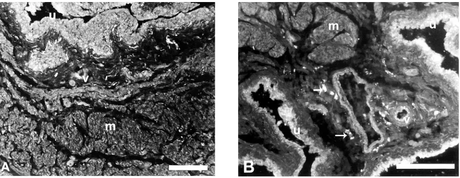
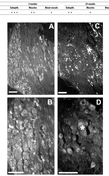
Age-related pigment was observed in all sections of the aged bladder. These pigments could be dif-ferentiated from the bright green FITC
immunofluo-rescence by the highly intense granular brownish-yellow autofluorescence (Figs. 1B, 2C, D) which did not mask the immunofluorescence.
Nerve fibres were distinguishable from the back-ground autofluorescence by the bead-like appearance of their varicosities and their bright fluorescence, which enabled the densities of immunoreactive nerves of different parts of the urinary bladder to be esti-mated semi-quantitatively in both age groups by as-signing values on the basis of – to + + + (Table 1).
Urinary bladder base
Intraepithelial VR1-ir nerve fibres that ramified from subepithelial axons were often observed in the base of the urinary bladder in both age groups (Fig. 1A). These VR1-ir axons were slightly varicose. An increase in the number of subepithelial varicosi-ties (running just under the epithelium) occurred more commonly among young animals. The lamina propria of the bladder base showed axons of similar shape with varicosities these were fewer in number. In the muscle layers most of the smooth muscle bun-dles were innervated by VR1-ir axons (Fig. 1A, B). There were many varicosities, which ran parallel to and inside the muscle bundles. These varicosities were also observed along most of the blood vessels (arteries and large veins) located in submucosal and muscular connective tissues. They had small varicos-ities and ran in a longitudinal and circular manner in the adventitial layer of the vessels. Serial sections of the bladder base showed no differences in the dis-tribution of VR1 immunostaining between the trig-one and other regions of the bladder base. While
Table 1. Semi-quantitative estimation of VR1-ir nerve densities in the urinary bladder base in young (3-month-old) and old
(24-month-old) rats; +: sparse; + +: moderate; + + +: dense
3 months 24 months
Region Subepith. Muscles Blood vessels Subepith. Muscles Blood vessels
BASE + + + + + + + + + +
VR1-ir varicosities in the mucosa are free terminal endings close to the epithelial cells, in the muscular layer they encroach into a shallow groove in the sur-face of the smooth muscle fibres.
It is noteworthy that the overall pattern of in-nervation of the young rat urinary bladder base decreased with age (Table 1). The young rat sub-epithelial region of the urinary bladder showed dense VR1-ir nerve axons compared to only mod-erate innervations in the aged rat tissues. In addi-tion, the muscular layers of the young rat urinary bladder base revealed moderate innervations of VR1. On the other hand, the aged rat bladder base showed only sparse VR1-ir nerve axons. However, blood vessels in both young and aged animals showed no remarkable changes in VR1 immunore-activity. It was also observed that the aged rat blad-der had long VR1-ir axons (varicosities) in the sub-epithelial region of the base, but these were short and less dense in the muscle layers (Figs. 1A, B).
Dorsal root ganglia
VR1-ir neuronal profiles were present in all sec-tions of young and aged DRG (L1-L2, L6-S1). With-in the dorsal root ganglia VR1-ir was restricted to small (15–25 µm) and medium (25–40 µm) sized neurons. The uniformity of immunostain-ing within a perikaryon varied in young and aged animals. VR1-ir neurons occurred in a variety of patterns. Small cells were usually intensely stained, with the immunoreactivity homoge-neously distributed within the cytoplasm (Figs. 2A, B, D). On the other hand, the immunostaining in the medium cells was usually weaker, whereby the immunoreactivity exhibited a patchy staining around the nucleus.
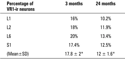
The quantitative data of the present study re-vealed that the mean percentage of young rat L1,
L2, L6 and S1 DRG neurones immunoreactive for VR1-ir showed a significant (p < 0.05) decrease with age (Table 2) in all DRG levels. L6 DRG showed the highest percentage of VR1-ir cells in both young (20%) and aged (13.4%) rats. Moreover, the mean percentage of VR1-ir cells in all aged DRG samples (12 ± 1.6) showed significant decreases (p < 0.05) compared to the mean (17.8 ± 2) in all young rat DRG (Table 2).
DISCUSSION
In this study we examined the distribution of VR1 in the male rat urinary bladder and lumbosacral (L1--L2 and L6-S1) DRG in both young and aged rats. These DRG were specifically chosen for study on the basis of previous observations which have shown that they are the main segmental levels of primary affer-ent neurones innervating the rat urinary bladder, the majority of which run in the pelvic nerve to L6-S1 [21]. In the present study, however, we found that the bladder base and its lumbosacral primary affer-ents were innervated by VR1-ir fibres.
The distribution pattern of nerve axons within the walls of the bladder base of the 3-months old rats observed in the present study is similar to that previously reported in young rats and young guin-ea pigs [2]. In addition, the distribution of the VR1-ir fibres underneath the epithelium and around the smooth muscle cells was similar to that of sensory neuropeptides such as SP CGRP in the rat bladder [4, 27, 29, 31, 32].
Changes in neuronal circuitry could lead to per-turbation of the micturition reflex during ageing. The sub-epithelial localisation of VR1 fibres is consistent with the nociceptive function of VR1-expressing pri-mary sensory neurones, whereas the intimate con-nection between VR1-expressing nerves and smooth muscle fibres suggests that the former play a role in mechanical sensitivity. As VR1 is not mechanosensi-tive, co-expression with a mechanotransducer could be another explanation.
Previous study has revealed the occurrence of VR1-ir nerve varicosities in shallow grooves on the surface of smooth muscle cells, suggesting a func-tional relationship between these profiles. In plain electron micrographs of the bladder such coupling has been interpreted as a possible neuromuscular junction between autonomic nerve fibres and smooth muscle cells [17]. Since at the periphery VR1 seems to be expressed exclusively in primary sensory neurones [33, 34], our results are suggestive that this coupling may have a mainly sensory function.
Table 2. Age-related changes in the percentage of L1, L2,
L6 and S1 DRG neurones immunoreactive to VR1 in both young adult and aged rats; *p < 0.05
Percentage of 3 months 24 months VR1-ir neurons
L1 16% 10.2%
L2 18% 11.9%
L6 20% 13.4%
S1 17.4% 12.5%
As previous data indicate that capsaicin-sensitive bladder afferents are activated by changes in the bladder volume [12, 13, 26, 34], it is reasonable to assume that the coupling between VR1-ir nerve end-ings and smooth muscle cells may act as a mechan-oreceptor. However, the couplings may also serve as an area where sensory neuropeptides bind to neu-rokinin receptors which modulate detrusor smooth muscle cell contractility [3].
In the mucosa VR1-ir varicosities occurred as free terminal endings close to the base membrane of the epithelial cells or between the latter. Free terminal endings have previously been found in the human bladder mucosa [16], some of them showing immu-noreactivity to SP [36]. As free nerve endings are usually associated with nociception [21], our mor-phological findings further strengthen the putative role of VR1-expressing nerve terminals in pain per-ception at the surface of the bladder.
The downregulation of VR1-ir cell profiles in the aged rat DRG raises the possibility that the direct nociceptive function of VR1 and/or the sen-sation or the transmission of sensory information from the urinary bladder may be compromised as age progresses. The present finding of a signifi-cant decrease with age in the percentage of VR1-ir neurons in (L1, L2, L6 and S1) DRG confirms previ-ous reports on the effects of age on the percent-age of SP-ir and CGRP-ir neurons in the rat DRG [6, 29]. This appears to be in line with the distri-bution of VR1-ir in unmyelinated C fibres [8], sug-gesting that the nociceptive function of VR1 may change with age.
Although the exact physiological role of VR1 in the nerves of the bladder is still uncertain, pre-vious studies in humans [12, 13] and rats [4] have shown that intravesical administration of vanilloids decrease the mechanosensitivity of bladder pri-mary afferents. In addition, intravesical vanilloids abolish reflex bladder contractions triggered by ice-cold water instillation [18]. This reflex is depen-dent on sensory input conveyed by C-fibres [10] and becomes overactive in certain pathological conditions of the human bladder [18]. However, VR1 is not sensitive to pressure [9] and under nor-mal conditions only responds to temperatures above 43°C [10]. Therefore the loss of mechanical and cold sensitivity after intravesical vanilloid ap-plication can hardly be attributed to VR1 desensi-tisation. It is, however, possible that vanilloids in-terfere with the function of other receptors be-sides VR1. As intravesical vanilloids do not induce
terminal afferent degeneration [4], a P2X3 (ATP--activated receptors involved in pain sensation) functional impairment, to occur, should be attrib-uted to post-translational changes induced by the increase of intracellular Ca2+ concentration that follows VR1 activation [4]. In addition, Ca2+ might also interfere with the function of other specific mechanosensitive ion channels already identified in capsaicin-sensitive fibres [7]. However, the con-tribution of these mechanoreceptors to bladder control is yet unknown. Alternatively, a general decrease in the bioelectrical activity of bladder pri-mary afferent fibres after vanilloid instillation may contribute to the loss of mechanical and thermal sensitivity. Recent results indicate that capsaicin induces a VR1 activity-dependent block of differ-ent ionic currdiffer-ents, including voltage-gated Na+ currents in DRG cells innervating the rat colon and voltage-gated Ca2+ currents in cultured DRG cells [32]. As both Na+ and Ca2+ currents play a crucial role in the generation of the action potential in noci-ceptive primary sensory neurones [18], the vanil-loid-induced reduction of these currents may de-crease the bladder sensory input to the spinal cord.
CONCLUSION
In conclusion, the present study showed that VR1-ir fibres are mainly peptidergic and form var-icose plexuses in the mucosa and in the muscu-lar layer in the base of the rat urinary bladder. The sub-epithelial localisation is consistent with the nociceptive function of VR1-expressing pri-mary sensory neurons. As capsaicin-sensitive bladder afferents are involved in nociception and reflex micturition control, the numerous free ter-minal nerve endings expressing VR1 in the mu-cosa seem more adequate to accomplish the former function. However, the reduction in VR1-ir nerve densities in the aged bladder base suggests that the nociceptive function of VR1 has been affected with age. In the DRG VR1 was found to belong almost exclusively to the peptide-contain-ing sub-population of primary afferents. Howev-er, the reduction with age in the VR1-ir DRG neu-rons could also have important implications for micturition control.
REFERENCES
2. Alm P, Zygmunt PK, Iselin C, Larsson B, Uvelius B, Wern-er S, AndWern-ersson K (1995) Nitric oxide synthase-immu-noreactive, adrenergic, cholinergic, and peptidergic nerves of the female rat urinary tract: a comparative study. J Anatom Nerv Syst, 56: 105–114.
3. Andersson KE (1999) Advances in the pharmaco-logical control of the bladder. Exp Physiol, 84: 195– –213.
4. Avelino A, Cruz F (2000) Peptide immunoreactivity and ultrastructure of the rat urinary bladder nerve fibers after topical desensitization by capsaicin or resinifera-toxin. Aut Neu, 86: 58–67.
5. Avelino A, Cruz C, Nagy I, Cruz F (2002) Vanilloid re-ceptor 1 expression in the rat urinary tract. Neuro-science, 109: 787–798.
6. Bergman E, Carlsson K, Liljeborg A, Manders E, Hökfelt T, Ulfhake B (1999) Neuropeptides, nitric oxide syn-thase and GAP-43 in B4-binding and RT97-immunore-active primary sensory neurons: normal distribution pattern and changes after peripheral nerve transec-tion and aging. Brain Res, 832: 63–83.
7. Birder LA, Kanai AJ, de Groat WC, Kiss S, Nealen ML, Burke NE (2001) Vanilloid receptor expression suggests a sensory role for urinary bladder epithelial cells. Proc Natl Acad, 98: 133–196.
8. Buck SH, Burks TS (1986) The neuropharmacology of capsaicin: a review of some recent observations. Phar-macol Rev, 38: 179.
9. Caterina M.J, Schumacher MA, Tominaga M, Rosen TA, Levine JD, Julius D (1997) The capsaicin receptor: a heat-activated ion channel in the pain pathway. Na-ture, 389: 816–824.
10. Cheng CL, Chai CY, de Groat WC (1997) Detrusor-sphincter dyssynergia induced by cold stimulation of the urinary bladder of rats. Am J Physiol, 272: R1271– –R1282.
11. Cruz F, Avelino A, Coimbra A (1996) Desensitization follows excitation of bladder primary afferents by in-travesical capsaicin, as shown by c-fos activation in the rat spinal cord. Pain, 64: 553–557.
12. Cruz F, Guimarães M, Silva C, Reis M (1997) Suppres-sion of bladder hyperreflexia by intravesical resinifera-toxin. Lancet, 350: 640–641.
13. Cruz F, Avelino A, Cruz C, Nagy I (2000) Distribution of vanilloid receptor (VR1) immunoreactive fibers in the rat urinary tract. Soc Neurosci, 26: 1692. 14. Davis JB, Gray J, Gunthorpe J, Hatcher JP, Davey PT,
Overend P, Harries MH, Latcham J, Claphams C, Atkinson K, Hughes SA, Rances K, Grau E, Harper AJ, Pugh PL, Rogers DC, Bingham S, Randall A, Shear-down SA (2000) Vanilloid receptor-1 is essential for inflammatory thermal hyperalgesia. Nature, 405: 183–187.
15. De Groat WC, Kawatani M, Hisamitsu T, Cheng CL, Ma CP, Thor K, Steers W, Roppolo JR (1990) Mecha-nisms underlying the recovery of urinary bladder func-tion following spinal cord injury. J Auton Nerv Syst, 30: S71–S78.
16. Dixon JS, Gilpin CJ (1987) Presumptive sensory axons of the human urinary bladder: a fine structural study. J Anat, 151: 199–207.
17. Gabella G (1995) The structural relations between nerve fibres and muscle cells in the urinary bladder of the rat. J Neurocytol, 24: 159–187
18. Geirsson G, Lindstrom S, Fall M (1999) The blad-der cooling reflex and the use of cooling as stimu-lus to the lower urinary tract. J Urology, 162: 1890–1896.
19. Guo A, Vulchanova L, Wang J, Li X, Elde R (1999) Im-munocytochemical localization of the vanilloid recep-tor 1 (VR1): relationship to neuropeptides, the P2X3 purinoreceptor and IB4 binding sites. Eur J Neurosci, 11: 946–958.
20. Ishizuka O, Mattiasson A, Andersson KE (1995) Urody-namic effects of intravesical resiniferatoxin and capsa-icin in conscious rats with and without outflow ob-struction. J Urol, 154: 611–616.
21. Keast JR, De Groat WC (1992) Segmental distribution and peptide content of Primary afferent neurons in-nervating the urogenital organs and colon of male rats. J Com Neurol, 319: 615–623.
22. Lazzeri M, Vannucchic MG, Zardoc C, Spinellib M, Benefortia P, Turinia D, Faussone-Pellegrinic M (2004) Immunohistochemical evidence of vanilloid receptor 1 in normal human urinary bladder. Eur Urol, 46: 792–798.
23. Lee SE, Shen H, Taglialatela G, Chung JM, Chung K (1998) Expression of nerve growth factor in the dorsal root ganglion after peripheral nerve injury. Brain Res, 796: 99–106.
24. Lecci A, Giuliani S, Tramontana M, Santicioli P, Criscuoli M, Dion S, Maggi, CA (1998) Bladder distension and acti-vation of the efferent function of sensory fibres: simi-larities with the effect of capsaicin. Br J Pharmacol, 124: 259–266.
25. Maggi CA, Barbanti G, Santicioli P, Beneforti P, Misuri D, Meli A, Turini D (1989) Cytometric evidence that cap-saicin-sensitive nerves modulate the afferent branch of micturition reflex in humans. J Urol, 142: 150–154. 26. Maggi CA, Lecci A, Santicioli P, Bianco ED, Giuliani S (1992) Cyclophosphamide cystitis in rats, involvement of capsaicin-sensitive primary afferents. J Auton Nerv Syst, 38: 201–208.
27. Mattiasson, Ekblad, E, Sundler F, Uvelius B (1985) Or-igin and distribution of neuropeptide Y-, vasoactive intestinal polypeptide- and substance P-containing nerve fibers in the urinary bladder of the rat. Cell Tis-sue Res, 239: 141–146.
28. Mohammed HA, Santer RM (2000) Sensory peptider-gic innervation of the lower urinary tract in young adult and aged rats. J Anat, 196: 136.
30. Michael GJ, Priestley JV (1999). Differential expression of the mRNA for the vanilloid receptor subtype 1 in cells of the adult rat dorsal root and nodose ganglia and its downregulation by axotomy. J Neurosci, 19: 1844–1854.
31. Sharkey KA, Williams RG, Schultzberg M, Dockray GJ (1983) Sensory substance P innervation of the uri-nary bladder: possible site of action of capsaicin in causing urine retention in rats. Neuroscience, 3: 861– –868.
32. Su HC, Wharton J, Polak JM, Muldery PK, Ghatei MA, Gibson SJ, Terenghi G, Morrison JFB, Ballesta J, Bloom SR (1986) Calcitonin gene-related peptide immunore-activity in afferent neurons supplying the urinary tract: combined retrograde tracing and immunohistochem-istry. Neuroscience, 18: 727–747.
33. Szallasi A (1995) Autoradiographic visualization and pharmacological characterization of vanilloid
(capsai-cin) receptors in several species, including man. Acta Physiol Scand, 629: 1–68.
34. Szallasi A, Nilsson S, Farkas-Szallasi T, Blumberg PM, Hokfelt T, Lundberg JM (1995) Vanilloid (capsaicin) receptors in the rat: distribution in the brain, regional differences in the spinal cord, axonal transport to the periphery, and depletion by systemic vanilloid treat-ment. Brain Res, 703: 175–183.
35. Warburton A, Santer RM (1994) Sympathetic and sen-sory innervation of the urinary tract in young adult and aged rats: a semi-quantitative histochemical and im-munohistochemical study. Histochem J, 26: 127–133. 36. Wakabayashi Y, Tomoyoshi T, Fujimiya M, Arai R,