JOURNAL OFVIROLOGY, May 2003, p. 6029–6040 Vol. 77, No. 10
0022-538X/03/$08.00⫹0 DOI: 10.1128/JVI.77.10.6029–6040.2003
Copyright © 2003, American Society for Microbiology. All Rights Reserved.
EBNA2 and Activated Notch Induce Expression of BATF
Lisa M. Johansen,
1† Christopher D. Deppmann,
1Kimberly D. Erickson,
2‡ William F. Coffin III,
2Tina M. Thornton,
1Sean E. Humphrey,
1Jennifer M. Martin,
2and Elizabeth J. Taparowsky
1*
Department of Biological Sciences, Purdue University, West Lafayette, Indiana 47907-1392,1and Department of Molecular,
Cellular and Developmental Biology, University of Colorado, Boulder, Colorado 803092
Received 14 June 2002/Accepted 20 February 2003
The immortalization of human B lymphocytes by Epstein-Barr virus (EBV) requires the virus-encoded transactivator EBNA2 and the products of both viral and cellular genes which serve as EBNA2 targets. In this study, we identified BATF as a cellular gene that is up-regulated dramatically within 24 h following the infection of established and primary human B cells with EBV. The transactivation ofBATF is mediated by EBNA2 in a B-cell-specific manner and is duplicated in non-EBV-infected B cells by the expression of mammalian Notch proteins. In contrast to other target genes activated by EBNA2, theBATFgene encodes a member of the AP-1 family of transcription factors that functions as a negative regulator of AP-1 activity and as an antagonist of cell growth. A potential role for BATF in promoting EBV latency is supported by studies in which BATF was shown to negatively impact the expression of a BZLF1reporter gene and to reduce the frequency of lytic replication in latently infected cells. The identification ofBATFas a cellular target of EBV provides important new information on how programs of viral and cellular gene expression may be coordinated to promote viral latency and control lytic-cycle entry.
Epstein-Barr virus (EBV) is causally associated with Bur-kitt’s lymphoma (BL), nasopharyngeal carcinoma, unusual T-cell lymphomas, and other lymphoproliferative diseases (re-viewed in references 35 and 59). EBV has a restricted host range and primarily infects CD21⫹B lymphocytes and epithe-lial cells (reviewed in references 37 and 59). EBV establishes a latent infection in B cells in which the viral episome persists and expresses proteins that promote indefinite B-cell prolifer-ation (immortalizprolifer-ation) (reviewed in reference 35). Latency in cultured B cells is associated with the expression of six nuclear proteins (EBNAs). Of these, only EBNA1, EBNA2, EBNA3A, and EBNA3C are essential for immortalization (5, 18, 73). In addition to the EBNAs, three integral membrane proteins are expressed during latent infection (latent membrane proteins [LMPs]). Of these, only LMP1 is required for B-cell immor-talization, and it serves as the major transforming protein en-coded by the EBV genome (3, 34, 75).
EBNA2 is one of the first viral gene products to be expressed after EBV infection and plays a crucial role in the process by transcriptionally activating viral genes encoding EBNA1, -3A, -3B, -3C, LMP1, and itself (reviewed in reference 35). In ad-dition to viral targets, EBNA2 can enhance or repress the transcription of cellular genes whose encoded proteins contrib-ute to the alterations in cellular growth that are associated with EBV infection. EBNA2 does not bind DNA directly but in-stead activates gene expression by interacting with the cellular CBF1 (RBP-J) protein complex (17, 21, 42, 74, 83) to mask its transcriptional repressor function (25). The ability of cellular Notch proteins to activate gene transcription is also facilitated
by its interaction with CBF1 (72; reviewed in references 2 and 51), implying that there may be overlap between the genes induced by Notch signaling and those up-regulated during EBV latency (26). In support of this, constitutively active Notch can substitute for EBNA2 in B-cell immortalization (16, 22). Interestingly, of the identified cellular genetic targets of EBNA2 in B cells (8, 30, 32, 38, 69), onlyCD21is a common target for up-regulation by EBNA2 and activated Notch (71). In this paper, we report the novel finding that the human gene encoding BATF is up-regulated in B lymphocytes follow-ing infection with EBV. BATF is a member of the AP-1/ATF superfamily of basic leucine zipper transcription factors and forms heterodimers with the Jun proteins to bind preferentially to AP-1 consensus sites (9, 10). In vitro and in vivo studies have demonstrated that BATF:Jun heterodimers have a reduced transcriptional activity relative to Fos:Jun heterodimers and thus inhibit the expression of AP-1 target genes and retard AP-1-mediated cell growth (10, 79). BATF is expressed pre-dominantly in hematopoietic tissues, suggesting a tissue-spe-cific function for BATF as a negative regulator of AP-1 activ-ity.
The induction ofBATFgene expression by EBV is mediated by EBNA2 and is duplicated in non-EBV-infected B cells by an activated form of the Notch protein. Interestingly, the induc-tion of BATF by EBNA2 and Notch is B cell specific, suggest-ing that additional B-cell transcription factors (or signalsuggest-ing events) are required for BATF gene expression. The docu-mented role of AP-1 in the transactivation of several EBV genes, includingBZLF1 (reviewed in references 35 and 68), provides a context for investigating the biological significance of BATF induction in EBV-infected cells. Our studies demon-strate that BATF negatively impacts the expression of a
BZLF1reporter gene and reduces the efficiency of lytic-cycle induction. The identification of BATFas a target of EBNA2 activity early in infection provides important new insight into how coordinated programs of viral and cellular gene
expres-* Corresponding author. Mailing address: Department of Biological Sciences, Purdue University, West Lafayette, IN 47907-1392. Phone: (765) 494-7978. Fax: (765) 496-2536. E-mail: [email protected].
† Present address: CombinatoRx, Inc., Boston, MA 02118. ‡ Present address: Department of Microbiology, University of Col-orado Health Sciences Ctr., Denver, CO 80262.
6029
on November 8, 2019 by guest
http://jvi.asm.org/
sion can positively impact viral latency by decreasing the num-ber of cells entering the lytic cycle in response to lytic-cycle induction.
MATERIALS AND METHODS
Cell culture and EBV infections.BJAB (EBV-negative B-cell lymphoma) (36) and 721 (in vitro immortalized lymphoblastoid cell line) (65) cells were main-tained in RPMI 1640 medium supplemented with 10% fetal bovine serum (FBS)
and 100 U of penicillin/ml–100g of streptomycin/ml (P/S). DG75
(EBV-neg-ative B-cell lymphoma) (4), Raji (EBV-positive BL) (ATCC CCL86), B95-8 (EBV-positive marmoset B-cell line) (62), and HH514 (EBV-positive BL clone derived from the P3HR1 clone of Jijoye BL) (56) cells were maintained in RPMI 1640 medium supplemented with 10% calf serum and P/S. RPMI 2650 cells (ATCC CCL30) were cultured in minimum essential medium–10% FBS–P/S, and HeLa cells (ATCC CCL2) were grown in basal modified Eagle medium– 10% FBS–P/S. Primary human B lymphocytes were isolated from 200 ml of whole blood by Ficoll-Hypaque gradient, followed by positive selection with bovine serum albumin (63), and were cultured in RPMI 1640 medium–10% FBS–P/S. All medium components were obtained from Gibco-BRL Life Tech-nologies.
Virus supernatants were prepared from B95-8 and HH514 cells as described
previously (11). BJAB cells at a density of 2⫻105cells/ml were incubated with
virus supernatant for 3 h at 37°C with continuous rocking. Infected cells were harvested by centrifugation and cultured in complete RPMI 1640 medium. The efficiency of infection was monitored 72 h postinfection by EBNA staining (58) and/or by immunoblotting for LMP1 expression as described previously (47). Primary B lymphocytes were infected with B95-8 virus as described above and cultured for 5 days prior to analysis.
RNA analysis.Total RNA was prepared from cells as described previously
(53), and poly(A⫹) mRNA was isolated by using the FastTrack 2.0 kit
(Invitro-gen). RNA hybridizations were performed as described previously (53) by using
2g of poly(A⫹) mRNA and32P-labeled cDNA probes forBATF(10) and
human-actin(Clontech) mRNA. Reverse transcription (RT) involved 1g of
total RNA, 50 pmol of oligo(dT) primer, and RT reagents purchased from Gibco-BRL. The PCR was performed with 1/10 volume of each RT reaction and
20 pmol of 5⬘(GGAGCAGTCCCTCTGCACC) and 3⬘(CAGTTCCTCTGTG
AGCTGC) BATF primers in 50l containing 1⫻buffer (Promega), 4 mM
MgCl2, and 5 U ofTaqpolymerase (Promega). Thirty cycles of denaturation at
94°C for 1 min, annealing at 60°C for 30 s, and extending at 72°C for 30 s, with the final extension performed for 5 min, defined each amplification reaction.
Amplification with 5⬘(CGAGCACGGCATCGTCACC) and 3⬘(GTCAGGCA
GCTCGTAGCTC)-actinprimers served as the control. Semiquantitative PCR
was performed as described previously (54) by using the PCR Mimic construc-tion kit (Clontech) to generate a heterologous DNA standard amplified by the
BATFprimers described above. An equal amount of mimic DNA was added to
each BATF reaction. Amplification was performed in the presence of radiola-beled nucleotide and quantified by densitometry. The results were normalized by using the signal from the internal mimic.
Plasmids.Flag EBNA2, a simian virus 40-driven EBNA2 expression vector, and p288, an simian virus 40-driven LMP1 expression vector, have been de-scribed previously (43, 80). The CBF1-chloramphenicol acetyltransferase (CAT) reporter gene and the pPDL151 and pPDL152 plasmids encoding CBF1-inter-acting and noninterCBF1-inter-acting EBNA2 proteins, respectively, were obtained from P. Ling (42, 43). mNotch IC expresses a constitutively active mouse Notch1 protein and was a gift of J. Nye (50). Human Notch1 expression plasmids for activated
Notch1 (NIC), an NICvariant appended with a strong nuclear localization signal
(NLS), and an NICNLS protein deleted for the CBF1 interaction domain (⌬R)
were obtained from T. Capobianco (29). pDCRBATFexpresses an
amino-ter-minal, hemagglutinin antigen (HA)-tagged BATF protein (9). pCS2⫹MT-BATF
was generated as part of this study to express a BATF protein tagged at the N
terminus with seven tandem Myc epitopes (MT). pCS2⫹MT-BATF(1-49)
en-codes a nonfunctional derivative of BATF lacking the basic leucine zipper re-gion. CMV-c-Jun-HA was a gift from S. Rhodes (Indiana University-Purdue University at Indianapolis). BMRF1-CAT and pCMV-Z were obtained from S. Kenney (81). Luciferase derivatives of the ZII and mutant ZII (mut ZII) CAT reporter genes described in Ruf and Rawlins (60) were generously provided by I. Ruf. The pCMV-lacZ and pRL normalization vectors used in the transient expression studies were obtained from S. Konieczny (Purdue University) and Promega, respectively.
Gene transfer and expression analysis.A total of 2⫻107BJAB cells in a
volume of 400l of RPMI 1640 medium were electroporated at 200 V and 975
F with 20g of the indicated plasmid DNA and transferred to complete
medium for 48 h. RNA was prepared from 107cells. Protein extracts were
prepared from the remaining cells, normalized for protein content, and resolved by sodium dodecyl sulfate (SDS)–10% polyacrylamide gel electrophoresis (PAGE). Proteins were transferred to a nitrocellulose membrane and immuno-blotted by using the transfer, blocking, hybridization, and wash conditions de-scribed in reference 57 and a 1:500 dilution of monoclonal EBNA2 antibody (Novocastra Laboratories) or a 1:5,000 dilution of LMP1 antiserum as primary antibody. Immune complexes were detected by using peroxidase-conjugated goat anti-mouse immunoglobulin G (IgG) or anti-rabbit IgG (Vector Laboratories), enhanced chemiluminescence (Amersham Pharmacia Biotech), and autoradiog-raphy. Gene transfer to the RPMI 2650 and HeLa cell lines utilized standard
Ca2PO4-DNA transient transfection as described previously (53). Cell extracts
were prepared, normalized to the-galactosidase activity of pCMV-lacZ, and
assayed for CAT activity as described previously (53). A total of 2⫻106DG75
cells in 250l were electroporated at 310 V and 950F with the amounts of
DNA indicated in the legend to Fig. 6. Forty-eight hours after transfer of extracts to complete medium, luciferase activities were measured and normalized by using a dual-activity luciferase kit (Promega). All reporter gene assays were performed a minimum of three times in duplicate.
BATF protein expression analysis.Total protein was prepared from DG75 and 721 cells by lysis in standard radioimmunoprecipitation assay (RIPA) buffer plus protease inhibitors (Sigma) and normalized for protein content by a Bio-Rad protein assay. Total protein from BJAB cells mock infected or infected with
B95-8 virus was harvested by lysis in 1.5⫻SDS-PAGE sample buffer, separated
by SDS–15% PAGE, transferred to a polyvinylidene difluoride membrane, and blotted by following the procedure described in reference 57. A 1:250 dilution of affinity-purified rabbit anti-BATF antiserum or a 1:1,000 dilution of LMP1 an-tiserum was used as primary antibody. Immune complexes were detected by using a 1:5,000 dilution of peroxidase-conjugated goat mouse IgG or anti-rabbit IgG (Vector Laboratories), SuperSignal West Dura extended-duration substrate (Pierce), and autoradiography. The BJAB blot was stripped by incu-bating it for 30 min at room temperature in 2% (wt/vol) SDS–62.5 mM Tris-HCl
(pH 6.8)–100 mM-mercaptoethanol and then reprobed as described above with
a 1:5,000 dilution of clone C4 mouse monoclonal-actin antibody (ICN) and
goat anti-mouse IgG secondary antibody.
Electrophoretic mobility shift assays (EMSA). Complementary double-stranded oligonucleotides containing a consensus AP-1 binding site (Promega)
were labeled by using T4 DNA polynucleotide kinase and [␥-32P]ATP (6,000
Ci/mmol; NEN Life Science Products). Nuclear extracts were prepared from
HH514 cells and 721 cells as described previously (39). A total of 10g of
[image:2.603.87.238.73.249.2]nuclear extract was incubated with 40 fmol (1.0⫻105cpm) of probe DNA on ice
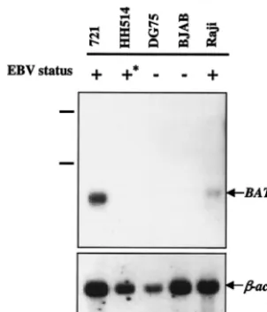
FIG. 1. Expression ofBATFmRNA in EBV-positive (⫹) and neg-ative (⫺) human B cell lines.⫹*, presence of a transformation-defec-tive EBV episome. Poly(A⫹) mRNA was hybridized with a
radiola-beled probe forBATFmRNA (upper panel) as described in Materials and Methods. The filter was stripped and reprobed for-actinmRNA as a control (lower panel). The migration of the 18S and 28S rRNA in a total RNA sample resolved in parallel is indicated to the left of the upper panel.
on November 8, 2019 by guest
http://jvi.asm.org/
for 15 min in 20l of buffer containing 10 mM HEPES (pH 7.9), 50 mM KCl,
0.1 mM EDTA, 5 mM dithiothreitol, 5 mM MgCl2, 10% glycerol, 1g of bovine
serum albumin, and 2g of poly(dI-dC) (Sigma). Binding specificity was assayed
by using a 100-fold molar excess of cold AP-1 or nonspecific E-box competitor DNA (positive strand, AGTGTTAATCCCCAGTTGTTTTTGAAGTTG; neg-ative strand, CAACTTCAAAAACAACTGGGGATTACT). For the supershift
experiments, the incubation with AP-1 DNA was followed by the addition of 4l
of affinity-purified rabbit polyclonal anti-BATF antiserum (K. Williams and E.
Taparowsky, unpublished data) or 4l of preimmune rabbit serum (PI) and a
further incubation on ice for 15 min. Protein/DNA complexes were resolved on a 4% native polyacrylamide gel in Tris-glycine buffer (25 mM Tris, 190 mM glycine, 1 mM EDTA, pH 8.5) and visualized by autoradiography.
Coimmunoprecipitation assays.DG75 cells were electroporated as described
above with 10g of pCS2⫹MT-BATF, 10 g of CMV–c-Jun–HA, or both
plasmids; transferred to complete medium; and incubated for 36 h, after which the cells were lysed in standard RIPA buffer plus protease inhibitors (Sigma) and normalized for protein content. A fraction of each normalized extract was re-solved by SDS–12.5% PAGE, transferred to a nitrocellulose membrane (Bio-Rad), and immunoblotted with a 1:1,000 dilution of Myc monoclonal anti-body 9E10 (Developmental Studies Hybridoma Bank) or anti-HA monoclonal antibody (12CA; Boehringer Mannheim) by using the transfer, blocking, hybrid-ization, and wash conditions outlined in reference 57. Immune complexes were detected by incubating extracts for 60 min with a 1:5,000 dilution of peroxidase-conjugated goat anti-mouse IgG (Vector Laboratories) and visualized with
Su-perSignal chemiluminescence reagent (Pierce) and autoradiography. The re-maining volume of each extract was incubated with anti-HA monoclonal antibody bound to protein A-Sepharose (Amersham Pharmacia Biotech) for 90 min at 4°C. Immunoprecipitates were washed extensively with RIPA buffer plus
protease inhibitors, and proteins were eluted by boiling for 5 min in 1.5⫻SDS
sample buffer. Proteins were resolved by SDS–12.5% PAGE, transferred to a nitrocellulose membrane, and immunoblotted with anti-HA antibody as de-scribed above. The blot was stripped by incubating it for 30 min at 60°C in 2%
(wt/vol) SDS–62.5 mM Tris-HCl (pH 6.8)–100 mM-mercaptoethanol and
re-probed as described above by using the 9E10 anti-Myc antibody.
Immunofluorescence analysis.HH514 cells were electroporated with 5g of plasmid DNA encoding MT-tagged wild-type BATF (MT-BATF) or a nonfunc-tional derivative of BATF [MT-BATF(1-49)] in which the bZIP dimerization domain had been deleted. After 24 h in complete medium, EBV’s lytic cycle was
induced with 20 ng of 12-O-tetradecanoylphorbol-13-acetate (TPA)/ml–3.5 mM
sodium butyrate. Seventy-two hours after electroporation, cells were washed
three times in phosphate-buffered saline and applied to microscope slides (104
cells/l) and dried. Cells were fixed in acetone (⫺20°C) and were costained with
a 1:10 dilution of anti-gp350/220 mouse monoclonal antibody (a gift from L. Hutt-Fletcher) and a 1:200 dilution of anti-mouse Alexa Fluor 594-conjugated secondary antibody (Molecular Probes) and with a 1:50 dilution of anti-Myc antibody (A14; Santa Cruz Biotechnology) and a 1:50 dilution of anti-rabbit fluorescein isothiocyanate-conjugated secondary antibody (Sigma). Slides were
[image:3.603.92.489.75.369.2]mounted by using VECTASHIELD with DAPI (4⬘,6⬘-diamidino-2-phenylindole;
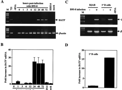
FIG. 2. BATFmRNA is induced in human B cells following infection with EBV. (A) RT-PCR analysis of total RNA isolated at the indicated times (hours) from BJAB cells infected with B95-8 EBV or transformation-defective HH514 EBV. Amplification of BATF from the 721 B-cell line provided a positive control for expression. No RNA (⫺RNA) and RNA from mock-infected BJAB cells (mock) served as negative controls. ØX174 HaeIII DNA (M) served as a molecular weight marker. Each RT reaction was PCR amplified with primers for-actinto control for sample integrity and amount. (B) Semiquantitative RT-PCR of the RT samples shown in panel A was performed as described in Material and Methods. The fold increase inBATFmRNA expression was quantified following the densitometry of ethidium bromide-stained gels. The experiment was performed three independent times with error bars indicating the standard error of the mean for each averaged set of values. (C) Primary (1° B) cells were isolated from human blood and mock infected (⫺) or infected with B95-8 EBV (⫹) as described in Materials and Methods. Total RNA from the primary B cells or control BJAB cells was isolated and examined for BATF mRNA expression by RT-PCR (upper panel). The amplification of -actin served as the positive control. A no-RNA (⫺RNA) reaction served as the negative control. ØX174 HaeIII DNA (M) provided a marker for molecular weight. (D) Semiquantitative RT-PCR was performed on the 1° B-cell samples, and the fold increase inBATF mRNA was quantified as described for panel B.
VOL. 77, 2003 EBNA2 TRANSACTIVATESBATF 6031
on November 8, 2019 by guest
http://jvi.asm.org/
Vector Laboratories), viewed with a Nikon Eclipse E800 fluorescence
micro-scope at a 40⫻lens objective, and photographed at 100⫻magnification with a
Cooke SensiCam Digital camera and SlideBook software.
RESULTS
EBV infection inducesBATFmRNA and protein in human B cells.TheBATFgene is expressed predominantly in human hematopoietic tissues and in the BL cell line Raji (9, 10). The elevated level of BATF expression in Raji, an EBV⫹ B-cell line, prompted us to investigate ifBATFgene transcription is a general feature of EBV-infected cells. Poly(A⫹) mRNA pre-pared from a panel of human B-cell lines was analyzed by Northern blot hybridization. As shown in Fig. 1, the two cell lines harboring a fully functional EBV episome (Raji and 721) expressedBATFwhile the lines negative for EBV (BJAB and DG75) did not. Interestingly,BATFmRNA was not detected in HH514 cells, a human BL cell line harboring a transforma-tion-defective strain (HH514) of EBV, indicating that events associated with transformation by EBV are necessary to induce endogenousBATFexpression.
To test ifBATFmRNA expression is induced in human B cells in response to EBV infection, BJAB cells were infected with viral supernatant from B95-8 cells (harboring a transform-ing strain of EBV). Controls included BJAB cells infected with virus supernatant prepared from HH514 cells or mock infected with medium only. The efficiency of viral infection was moni-tored by immunostaining for EBNAs or LMP1 and was esti-mated to be 50 to 80% for all groups (data not shown). At specific times following infection, total RNA was isolated and analyzed by RT-PCR for the presence of BATF mRNA. As early as 24 h postinfection, BATFmRNA was induced and
remained at an elevated level for the duration of the experi-ment (Fig. 2A). In contrast,BATFmRNA was not detected in mock-infected cells or in cells infected with HH514 virus. Du-plicate samples were analyzed by using semiquantitative RT-PCR, and results showed a 25-fold induction ofBATFmRNA 24 h postinfection (Fig. 2B).
In an effort to relate the EBV-induced expression ofBATF
observed in BJAB cells to the process of EBV infection in vivo, primary B cells were purified from human peripheral blood, treated with B95-8 virus supernatant, and cultured for 5 days prior to the preparation of RNA. Whereas noninfected pri-mary B cells do not express detectable levels ofBATFmRNA, cells infected with B95-8 virus rapidly induceBATFgene tran-scription (Fig. 2C). Semiquantitative RT-PCR was used to estimate the induction at 15 times the normalBATFlevel (Fig. 2D). These results provide evidence that transcription of the
BATF gene is an early response of B lymphocytes to EBV infection in vivo.
The induction of BATF transcripts observed in EBV-in-fected cells should reflect an increase in BATF protein. Ex-tracts prepared from DG75 (BATF⫺) and 721 (BATF⫹) cells (Fig. 3A) or from BJAB cells mock infected and infected with B95-8 virus (Fig. 3B) were resolved by SDS-PAGE and immu-noblotted with a polyclonal rabbit antiserum to BATF (see Materials and Methods for details). The results show expres-sion of BATF protein in the 721 and B95-8 samples. Reprob-ing the BJAB blot with an antibody to -actin demonstrated equal loading of the samples; probing a parallel blot with an antibody to LMP1 confirmed the infection of the BJAB cells with EBV.
BATFis a cellular target of the EBNA2 transactivator. In-fection with B95-8, the prototypical transforming strain of EBV, resulted in a dramatic up-regulation of BATFmRNA and protein. In contrast, infection with the nontransforming HH514 strain had no effect onBATFgene expression (Fig. 2). The HH514 virus is nontransforming due to a deletion of the latent genes EBNA2 and EBNA-LP (18). While EBNA2 is absolutely required for the immortalization of B cells by EBV (17), EBNA-LP plays an ancillary role in latency by enhancing EBNA2 function (19, 49, 52, 67). EBNA2 is a well character-ized transactivator that regulates both viral and cellular genes (reviewed in references 35, 37, and 66). Among the viral genes activated by EBNA2 isLMP1(1, 12, 76), which encodes the major transforming protein of the EBV genome (3, 34, 75). Thus, cells infected with HH514 virus do not express LMP1. To test the possibility that EBNA2 and/or LMP1 is responsible for
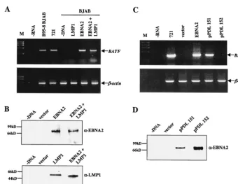
BATFmRNA induction in B cells, expression vectors for the proteins were introduced into BJAB cells by electroporation. After 48 h, total RNA was prepared and analyzed by RT-PCR. As shown in Fig. 4A and B,BATFmRNA was induced in cells expressing EBNA2 but not in cells expressing LMP1 alone. These data identify the EBNA2 transactivator as both neces-sary and sufficient to induceBATFgene expression in human B cells.
EBNA2 possesses a potent transcription activation domain but lacks a DNA binding domain (6, 7). EBNA2 exerts its transcriptional influence on viral and cellular target genes by interacting with a cellular sequence-specific DNA binding pro-tein called CBF1 or RBP-J(17, 21, 42, 74, 83). To examine if the induction ofBATFgene expression relies on the
interac-FIG. 3. BATF protein is detected in EBV-infected cells. (A) Total protein from 721 (EBV⫹) and DG75 (EBV⫺) cells was immunoblot-ted with anti-BATF antiserum as described in Materials and Methods to detect endogenous BATF protein expression. (B) Total protein from BJAB cells mock infected or infected with B95-8 virus was im-munoblotted as described for panel A to detect endogenous BATF protein (top panel). To ensure equal loading of the samples, the membrane was stripped and reprobed with anti--actin antibody (mid-dle panel). An immunoblot performed in parallel was probed for LMP1 expression to confirm viral infection of the B95-8 sample (lower panel). The migration of molecular weight markers is indicated to the left of BATF blots.
on November 8, 2019 by guest
http://jvi.asm.org/
tion of EBNA2 with CBF1, an expression vector for wild-type EBNA2 (pDL151) and a matched vector for an EBNA2 pro-tein containing a mutation that abolishes binding to CBF1 (pDL152) were expressed in BJAB cells. RT-PCR results show that BATF mRNA was induced only by the fully functional EBNA2 protein (Fig. 4C). Immunoblot analysis of cell extracts prepared in parallel demonstrate that both EBNA2 proteins were expressed at roughly equivalent levels in the cells (Fig. 4D). These data link the induction ofBATFgene expression with the formation of an EBNA2/CBF1 complex and suggest that BATF may be a direct cellular target for regulation by EBNA2.
BATFis a putative target for regulation by the Notch sig-naling pathway.Having identified the viral EBNA2 transacti-vator as an inducer ofBATFgene expression in B cells, we next sought to identify an endogenous regulator of BATF gene expression. The Notch family of proteins is comprised of trans-membrane receptor proteins that are processed into nuclear-targeted transcriptional regulators following activation (re-viewed in references 2 and 51). In recent years, a number of studies have shown that activated Notch functions in a way similar to that of EBNA2 to positively impact cellular gene expression via an interaction with CBF1 (26, 45, 70, 72) and
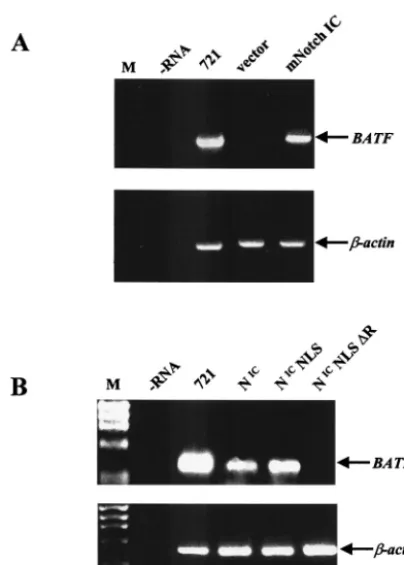
can function as a substitute for EBNA2 in B-cell immortaliza-tion (16, 22). To examine if Notch activates BATFgene ex-pression, a constitutively activated derivative of Notch1 (mNotch IC) was expressed in BJAB cells. Efficient transacti-vation occurred in cells expressing mNotch IC but not in con-trol cells (Fig. 5A). An equivalent level of activation was ob-served when the experiment was repeated with a constitutively active form of mouse Notch2, the Notch family member that predominates in B cells (27, 78) (data not shown).
In order to test if the Notch-mediated activation ofBATF
relies on the interaction with CBF1, the experiment was re-peated with Notch IC and variants were appended with a strong nuclear import signal (NLS) or appended with an NLS with a deletion (⌬R) of the CBF1 interaction motif. Although all three proteins were expressed equivalently (data not shown),BATFmRNA induction was observed with only Notch IC and Notch IC NLS and not with Notch IC NLS⌬R (Fig. 5B). These data strongly suggest that nuclear Notch activates
BATFin B cells and that this activation depends upon an intact CBF1 interaction domain.
[image:5.603.122.462.70.332.2]Induction ofBATFgene expression by EBNA2 and Notch is B cell specific.Our results implicate CBF1 as a DNA binding protein that is important to the transcriptional regulation of
FIG. 4. TheBATFgene is a target for transactivation by EBNA2. (A) BJAB cells were electroporated as described in Materials and Methods with 10g of each indicated plasmid DNA (20g of total DNA in each group). Total RNA was isolated after 48 h and analyzed forBATF expression and control-actingene expression by RT-PCR. RNA isolated from EBV-infected BJAB cells (B95-8 BJAB) and 721 B cells provided positive controls. A no-RNA reaction (⫺RNA) and a control BJAB reaction (⫺DNA) served as the negative controls. ØX174HaeIII DNA (M) provided a marker for molecular weight. (B) Immunoblot analysis of EBNA2 and LMP1 protein expression in cell extracts prepared from BJAB cells electroporated as described for panel A with the indicated expression plasmids. The migration of molecular mass protein markers is indicated to the left of each autoradiography gel. (C) BJAB cells were electroporated as described for those in panel A with two EBNA2 expression plasmids (EBNA2, pPDL151) and a plasmid directing the production of a nontransactivating EBNA2 variant (pPDL152). Total RNA was isolated after 48 h and analyzed forBATFand-actinmRNA expression by RT-PCR. RNA from 721 cells served as the positive control. A no-RNA (⫺RNA) reaction and RNA from BJAB cells electroporated with empty vector DNA served as negative controls. ØX174 HaeIII DNA (M) provided a molecular weight marker. (D) Immunoblot analysis of EBNA2 protein expression in the indicated groups analyzed forBATFgene expression as for that in panel C. The migration of molecular mass protein markers is indicated to the left of the autoradiography gel.
VOL. 77, 2003 EBNA2 TRANSACTIVATESBATF 6033
on November 8, 2019 by guest
http://jvi.asm.org/
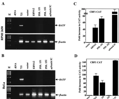
theBATFgene. CBF1 is a member of a highly conserved and ubiquitously expressed family of proteins that mediate the ac-tivation or repression of gene transcription depending on the cofactors available for interaction (25, 28, 30, 82; reviewed in reference 51). To examine the role of CBF1 in BATF gene regulation, we selected HeLa cells, a cell line that has been used previously to investigate CBF1 function (45), and RPMI 2650 cells, a human nasal epithelial cell line. Both cell types express CBF1, but not BATF (data not shown). DNA precip-itates containing expression vectors for the indicated EBNA2 proteins or Notch IC were introduced into both cell types with the goal of activating endogenous BATFmRNA expression. To ensure that the introduced activators functioned as ex-pected, a CAT reporter gene controlled by CBF1 binding sites was included in each precipitate. After 48 h, the cells were harvested and processed for the extraction of RNA and pro-tein. As shown in Fig. 6A and B, BATFmRNA was not de-tected in any of the groups even though the CBF1 CAT re-porter was efficiently activated by wild-type EBNA2 and constitutively active Notch1 (Fig. 6C and D). We concluded from these experiments that CBF1 and its interacting transac-tivator(s) play an important role in the up-regulation ofBATF
gene transcription in B cells but that transcription activation via CBF1 is not sufficient to activateBATFgene expression in non-B-cell types.
Protein complexes containing BATF bind to target AP-1 sites. Previous studies have shown that BATF forms het-erodimers with the Jun family of bZIP proteins to bind AP-1 DNA (9, 10). To demonstrate that BATF is part of the AP-1 DNA binding activity in EBV-infected B cells, nuclear extracts were prepared from 721, a cell line that carries a fully trans-forming EBV episome and expresses BATF, and HH514, a cell line that carries a nontransforming EBV variant in which BATF expression is undetectable. EMSA with 32P-labeled
AP-1 DNA as a probe revealed a significantly higher level of AP-1 DNA binding activity in extracts prepared from the cells expressing BATF(Fig. 7A). The AP-1 binding observed was specific since it was competed by unlabeled AP-1 DNA and not by consensus E-box DNA to which basic helix-loop-helix tran-scription factors bind. The presence of BATF in the AP-1 DNA complex from 721 cells was established by adding anti-BATF antiserum or control PI to the binding reactions. As expected, anti-BATF antiserum supershifted the AP-1 com-plex derived from 721 cells but not from the non-BATF-ex-pressing HH514 cells (Fig. 7A).
The ability to detect BATF-containing AP-1 DNA com-plexes in nuclear extracts prepared from EBV-positive B cells prompted us to examine if BATF forms heterodimers with the c-Jun protein in B cells. DG75 B cells were electroporated with vectors expressing Myc-tagged BATF and/or HA-tagged c-Jun. Cell extracts were prepared, and c-Jun protein complexes were immunoprecipitated with anti-HA antibody. Following resolu-tion by SDS-PAGE, the proteins were immunoblotted with anti-HA antibody to detect c-Jun and then anti-Myc antibody to detect BATF protein. Control immunoblots in which whole-cell extracts were incubated with HA and Myc anti-bodies established protein mobilities and antibody specificity (data not shown). The results of this experiment revealed that BATF is present in c-Jun precipitates prepared from cells coexpressing both proteins (Fig. 7B). We concluded that the intracellular environment of human B cells supports the for-mation of BATF:c-Jun heterodimers that bind AP-1 target DNA.
BATF modulates EBV’s lytic cycle.The transcriptional ac-tivity of AP-1 is known to be essential for the transition from EBV latency to lytic replication in cultured B lymphocytes (reviewed in reference 35). A standard method of lytic-cycle induction in vitro employs treatment with the phorbol ester TPA to stimulate intracellular signaling events involving the activation of protein kinase C and AP-1 (reviewed in reference 68). Consensus AP-1 sites are found within the promoter re-gions of a number of immediate-early viral genes and must be occupied to achieve full transcriptional activation of the genes (reviewed in references 35 and 68). Thus, the accumulation of a molecular inhibitor of AP-1 activity is predicted to promote a delay in the onset of the lytic cycle.
[image:6.603.61.263.68.350.2]To investigate the potential of BATF to modulate immedi-ate-early events in the EBV lytic cycle, we tested the impact of ectopic BATF expression on a reporter gene containing the ZII element from the promoter ofBZLF1, a viral gene encod-ing a transactivator of early-EBV-lytic gene expression. Previ-ous studies have shown that ZII binds an AP-1 complex of
FIG. 5. TheBATFgene is a target for transactivation by Notch. (A) BJAB cells were electroporated as described in Fig. 3 with 10g of the indicated plasmid DNA. After 48 h, total RNA was isolated and analyzed forBATFand control-actingene expression by RT-PCR. RNA from 721 B cells served as a positive control. A no-RNA (⫺RNA) reaction and RNA from BJAB cells electroporated with empty vector DNA served as negative controls. ØX174HaeIII DNA (M) was the molecular weight marker. (B) The experiment shown in panel A was repeated with activated human Notch 1 (NIC), NICwith a
nuclear import signal (NICNLS), and NICNLS with a deletion (⌬R) of
the CBF1 interaction domain. RNA for 721 B cells served as a positive control. A no-RNA reaction (⫺RNA) served as the negative control.
on November 8, 2019 by guest
http://jvi.asm.org/
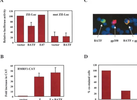
unknown composition (60) and that AP-1 binding is required for full activation of theBZLF1gene (14; reviewed in refer-ence 68). Electroporation of the EBV⫺ BATF⫺ B-cell line DG75 with the ZII-luciferase reporter resulted in efficient ac-tivation, which was reduced by 40% following the cointroduc-tion of BATF (Fig. 8A). The repression was specific for BATF since it was not duplicated by E47, an E-box binding transcrip-tion factor. In additranscrip-tion, the effect of BATF depended upon the AP-1 site in ZII since the basal level of expression of a ZII construct containing a point mutation in the AP-1 site (mut ZII) was unaffected by coexpressed BATF. These results indi-cate that BATF exerts a negative regulatory impact on the AP-1 complexes involved in transcriptionally activating the
BZLF1gene.
The BZLF1gene encodes a dimerizing transactivator, re-ferred to as ZEBRA, Zeta (Zta), or Z, that shows functional and structural similarity to AP-1 family members (13). Z binds AP-1-related DNA sites in the promoters of a number of viral genes, including the early-lyticBMRF1gene (24). To test the possibility that BATF, in addition to inhibiting Z production, also interferes with the efficient activation of Z target genes, we
[image:7.603.105.483.71.386.2]examined the transactivation of a BMRF1 reporter gene in HeLa cells. BMRF1-CAT is not expressed in HeLa cells unless activated by Z (24, 55) (Fig. 8B). When assayed in the presence of Z and BATF, no reduction in CAT expression was observed, demonstrating that in this cell system, BATF does not impair the efficiency with which Z activatesBMRF1gene expression. The studies described above suggest that the induction of BATF in latently infected B cells may influence the lytic switch by modulating the efficient production of Z. In order to exam-ine if BATF expression inhibits the efficiency of lytic-cycle induction in B cells, HH514 cells (EBV⫹, BATF⫺) were elec-troporated with an expression plasmid for Myc-tagged BATF or for a Myc-tagged BATF variant rendered nonfunctional by a deletion of the leucine zipper dimerization domain. HH514 cells are permissive for viral replication, and the lytic cycle can be induced by TPA-butyrate treatment. Twenty-four hours fol-lowing electroporation, the cells were treated with TPA-bu-tyrate for 48 h, at which time the cultures were analyzed by immunofluorescence and individual cells were scored for BATF expression, for the expression of the viral late protein gp350, and for the coexpression of both proteins (Fig. 8C). The
FIG. 6. Induction ofBATFmRNA expression is B cell specific. The adherent RPMI 2650 cell line (A and C) and the HeLa cell line (B and D) were transfected as described in Materials and Methods with 5g of the indicated activators, 5g of the CBF-1 CAT reporter, and 2g of a-galactosidase expression plasmid. Forty-eight hours following transfection, the cells were harvested for the preparation of RNA and protein. (A and B) RNA was analyzed by RT-PCR forBATF. As a control, the same RT samples were analyzed for the expression of-actinmRNA by PCR. RNA from 721 B cells provided a positive control. A no-RNA (⫺RNA) reaction and total RNA from cells transfected with empty vector DNA served as the negative controls. ØX174HaeIII DNA (M) was the molecular weight marker. Results indicate no activation ofBATFgene expression in either cell line. (C and D) Protein extracts were normalized to-galactosidase activity and assayed for CAT as described in Materials and Methods. The transfection was performed a minimum of three independent times to arrive at the average activities presented. Error bars indicate the standard errors of the means.
VOL. 77, 2003 EBNA2 TRANSACTIVATESBATF 6035
on November 8, 2019 by guest
http://jvi.asm.org/
efficiency of lytic-cycle induction (gp350⫹cells/total cells) was 11% for each group, and the efficiency of gene transfer (BATF⫹cells/total cells) was between 3 and 4% on average and was independent of the input plasmid. Importantly, the number of BATF- and gp350-costained cells was 32% of the number of BATF(1-49)- and gp350-costained cells (Fig. 8D), indicating that BATF expression inhibits lytic-cycle entry in response to induction with TPA-butyrate. Thus, expression of BATF in EBV-positive lymphoblastoid cells may play a crucial role in the maintenance of latency by modulating lytic-cycle-promoting AP-1 activity.
DISCUSSION
EBV establishes a latent infection in B cells in which a precise program of viral gene expression maintains the viral
episome and orchestrates cellular changes leading to cell im-mortalization and transformation (reviewed in reference 35). The role of the EBNA2 protein in transactivating the expres-sion of EBV latency genes, including theLMP1oncogene, has been well studied (reviewed in reference 66). Only recently, however, has the role of EBNA2 as an activator of cellular gene expression been fully appreciated and the efforts to iden-tify EBNA2-regulated cellular genes been revitalized (32, 33, 69; reviewed in reference 35). In most cases, the genes acti-vated by EBNA2 encode proteins that function directly, or indirectly, to activate B cells and promote cell cycle progres-sion (8, 38). In this paper, we have identified theBATFgene as a cellular target of the EBNA2 transactivator. In contrast to other cellular genes up-regulated early following infection, the
BATF gene encodes a protein that functions as a negative regulator of AP-1 activity and an antagonist of cell growth (10, 79). Thus, our results are the first to link infection of B cells with EBV to a transcriptional regulatory pathway that inhibits cell growth.
TheBATFgene is expressed predominantly in mammalian hematopoietic tissues (10, 79). In addition to the dramatic increase inBATFgene expression observed in EBV-infected B cells, increasedBATFexpression has also been observed fol-lowing receptor engagement on T helper cells (79) and the infection of T cells with human T-cell leukemia virus type 1 (20). Most recently, BATFmRNA was shown to be elevated following the activation of NF-B in pre-B cells (41). Given the similarity between the molecular mechanism of transcription activation by EBNA2 and the Notch proteins (23, 26, 61, 70), we were able to establish thatBATFmRNA is induced in B cells by nuclear-localized Notch proteins containing an intact CBF1 interaction domain. Interestingly, EBNA2 and Notch activation of the BATF gene is B cell specific even though CBF1 is expressed ubiquitously (reviewed in reference 51) and a simple CBF1 reporter gene is responsive to these transacti-vators in non-B-cell types (45). Cell specificity has been de-scribed for the EBNA2-mediated activation ofLMP1(12, 76), suggesting that a major factor in EBNA2 inducibility is the collaboration of EBNA2 with additional B-cell transcription factors and/or cellular signaling events. In this regard, CBF2/ AUF and the Ets domain transcription factors PU.1/Spi1 and Spi-B have been identified as additional DNA binding proteins that interact with EBNA2 response elements (15, 31, 40). These factors, plus the molecules activated in response to other stimuli associated with the induction ofBATFgene ex-pression in T and B lymphocytes (41, 79), will provide the starting point for future studies to identify EBNA2 collabora-tors.
The humanBATFgene has been cloned, and although the 5⬘
flanking region directs the B-cell-specific expression of a CAT reporter gene (48), the expression of this reporter is not re-stricted to EBV-infected B cells and is not induced to higher levels by EBNA2 or Notch (N. Meyer and E. Taparowsky, unpublished observations). There is a near-consensus CBF1 binding site within the first intron of the BATF gene, and future experiments will examine if this region (or additional extra- or intragenic regions) function together with the 5⬘
flanking region to confer inducibility. It remains a formal pos-sibility that the induction ofBATFgene expression by EBNA2 and Notch is indirect and requires the de novo synthesis of a
FIG. 7. BATF is a component of AP-1 DNA binding activity in B cells. (A) Nuclear extracts were prepared from HH514 (BATF⫺) and
721 (BATF⫹) B cells as described in Materials and Methods and used
for EMSA with a32P-labeled AP-1 DNA probe. The major complex
formed in the control (c) reactions (1) was competed by the addition of unlabeled AP-1 DNA but not by adding an unrelated DNA se-quence (E-box). Addition of PI or anti-BATF antiserum (BATF) to the reaction resulted in a supershift (2) only in the extracts from 721 cells. Ab, antibody. (B) Coimmunoprecipitation of BATF:c-Jun com-plexes from B cell extracts. DG75 cells were electroporated as de-scribed in Materials and Methods with 10g of an expression plasmid for HA-tagged c-Jun, 10g of an expression plasmid for a Myc-tagged BATF, both plasmids, and empty vector DNA (where needed) to adjust the total DNA to 20g. After 36 h, the cells were lysed and protein complexes were immunoprecipitated (IP) with HA anti-serum (␣-HA). The proteins were resolved by SDS-PAGE and immu-noblotted (IB) initially with␣-HA and then with anti-Myc antiserum (␣-Myc) to detect BATF. Results indicate the presence of coimmuno-precipitated BATF only in extracts from DG75 cells expressing both proteins.
on November 8, 2019 by guest
http://jvi.asm.org/
second protein(s). Evidence to support this possibility was gen-erated by using EBNA2-inducible B-cell lines where, in the presence of a protein synthesis inhibitor, BATFmRNA was induced, but not to the level observed in the absence of inhib-itor (G. Laux, personal communication). A similar observation has been described for the induction of the C-MYCgene by EBNA2 (32).
Although the precise mechanism(s) through which EBNA2 and Notch activateBATFgene expression remains to be de-termined, the accumulation of this AP-1 inhibitor in EBV-infected B cells is intriguing. There have been numerous stud-ies on the importance of AP-1 transcription complexes during the transition from EBV latency to lytic replication (reviewed
in references 35 and 68). In particular, AP-1 activity is essential for the expression ofBZLF1(14), the lytic-cycle switch gene encoding Z, a transactivator that coordinates the expression of the immediate-early viral genes required for productive infec-tion (reviewed in references 35 and 68). A cis-acting DNA element referred to as ZII mediates the AP-1 induction of
BZLF1 expression and is occupied by proteins prior to and following the treatment of B cells with lytic-phase inducers (14). While the composition of the complexes bound to ZII is still under investigation, there is clear evidence pointing to occupancy by a c-Jun heterodimer during latency and either an ATF1 or ATF2 dimer in cells actively expressing Z (44, 46, 77). We found that the high basal activity of a ZII-luciferase
re-FIG. 8. Overexpression of BATF inhibitsBZLF1gene expression and reduces the frequency of lytic-cycle induction. (A) Five micrograms of a luciferase reporter gene driven by the ZII element of theBZLF1promoter (ZII-Luc) or a matched reporter gene containing a mutated ZII element (mut ZII-Luc) were electroporated into DG75 cells along with 5g of expression plasmids for the indicated proteins and 2g of pRL DNA. After 48 h, cell extracts were prepared, normalized to the control luciferase activity from pRL, and assayed for ZII-directed luciferase expression. The experiment was repeated three times in duplicate, and the average luciferase values are expressed relative to the levels in the ZII-Luc/vector-alone group which were set at 100. Error bars indicate the standard errors of the means. (B) HeLa cells were transfected as described in the legend to Fig. 5 with a Z-responsive BMRF1-CAT reporter gene (5g) and 5g of the indicated expression plasmids. The experiment was performed three times, the CAT activities were averaged, and results were expressed relative to the basal level of BMRF1-CAT expression that was set to 1.0. Error bars indicate the standard errors of the means. (C) Immunofluorescence of HH514 cells electroporated with BATF or BATF(1-49) expression vectors and induced with TPA-butyrate (see Materials and Methods for details). Shown is an example of a field of cells stained for BATF (green), gp350/220 (red), or both BATF and gp350/220 (yellow). (D) Captured images were used to score the number of gp350/220-positive cells (red) for the lytic cycle, BATF- and BATF(1-49)-positive cells (green), total cells (DAPI), and gp350/220- and BATF-costained or gp350/220- and BATF(1-49)-costained cells (yellow). The number of costained cells in the BATF-transfected population was expressed as a percentage of the number of costained cells in the BATF(1-49)-transfected population. These results are representative of three experiments with similar results.
VOL. 77, 2003 EBNA2 TRANSACTIVATESBATF 6037
on November 8, 2019 by guest
http://jvi.asm.org/
[image:9.603.65.519.80.413.2]porter gene in DG75 cells (presumably the result of the con-stitutive expression of AP-1 family activators in these immor-talized EBV⫺B cells) is reduced by 40% following expression of BATF. This suggests that BATF forms dimers with endog-enous AP-1 activators, depleting the pool of AP-1 dimers with full transactivation potential. Additional experiments are re-quired to identify the partners of BATF during viral latency and to test if these heterodimers bind the ZII element. Re-gardless of the outcome of these studies, the significant reduc-tion inBZLF1promoter activity observed in the presence of BATF suggests that, during lytic-cycle induction, BATF delays accumulation of the levels of Z necessary to activate immedi-ate-early lytic-gene expression.
To obtain evidence that BATF can play a role in regulating the lytic-phase switch in vitro, expression vectors for wild-type and nonfunctional BATF were used to transfect HH514 cells and the lytic program was induced by treatment with TPA-butyrate. By counting the number of cells coexpressing the lytic-cycle protein gp350/220 and either BATF or BATF(1-49), we observed 60% fewer gp350/220-positive cells in the BATF-expressing population than in the BATF(1-49)-BATF-expressing pop-ulation. Thus, BATF expression decreased the number of TPA-butyrate-induced HH514 cells in the lytic cycle. Based on our knowledge of BATF, we predicted that BATF would in-terfere with the AP-1 transcriptional activity triggered by the treatment of cells with phorbol ester tumor promoters. As we have shown, one target of BATF-mediated inhibition of AP-1 activity is the viralBZLF1gene. However, the ability of BATF to change the composition and overall function of cellular
AP-1 activity suggests that the expression of additional genes also will be affected.
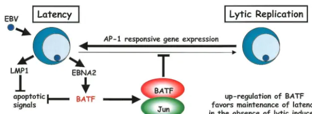
The findings presented here are the first to describe a cel-lular gene product induced immediately following EBV infec-tion that has the potential to modulate viral latency by reduc-ing the efficiency of lytic-cycle induction. The model we present in Fig. 9 provides an explanation as to why B cells expressing BATF resist entry into the lytic phase unless appropriately induced. It also considers that the subversion of cellular apo-ptosis that is associated with EBV latency and replication may be the result of the collaborative actions of antiapoptotic sig-naling by LMP1 and the negative regulation of proapoptotic gene expression by BATF. Certainly the observations that BATF is induced following NF-B activation (41) and that AP-1 activity is associated with apoptosis in some cell types (reviewed in reference 64) are consistent with this role. While validation of the model will rely on future studies aimed at characterizing the composition of BATF protein complexes and identifying the genetic targets of these complexes in B cells, we are encouraged by the potential of cellular proteins like BATF to control the in vivo progression of EBV-associ-ated human disease.
ACKNOWLEDGMENTS
We thank Al Zullo for technical assistance with the Notch experi-ments and Gerhard Laux for his generous exchange of unpublished information.
L.M.J. was supported by a predoctoral fellowship from the Ameri-can Heart Association. C.D.D. and S.E.H. are predoctoral trainees of PHS T32 CA09634 and GM08298, respectively. This work was sup-FIG. 9. Proposed function of BATF as a positive modulator of EBV latency and as a deterrent to lytic-cycle entry. The EBNA2 protein is expressed immediately following infection and utilizes its transactivator function to trigger cell proliferation as well as the direct induction of BATF, which functions in the context of a BATF: Jun heterodimer to restrict cell growth, inhibit AP-1 target gene expression, and, together with LMP1, block apoptosis. Exposure to an appropriate lytic-cycle inducer (e.g., TPA) enhances intracellular AP-1 activity through new protein synthesis (Fos) and the posttranslational modification of resident AP-1 family members (Jun). The resultant sustained increase in AP-1 activity is sufficient to overcome the BATF inhibition of multiple AP-1 target genes (includingBZLF1) and promote production of the immediate-early transactivators required for lytic-gene expression.
on November 8, 2019 by guest
http://jvi.asm.org/
[image:10.603.135.448.70.185.2]ported by National Institutes of Health grants AI01537 (J.M.M.), CA64610 (J.M.M.), and CA78264 (E.J.T.).
REFERENCES
1. Abbot, S. D., M. Rowe, K. Cadwallader, A. Ricksten, J. Gordon, F. Wang, L. Rymo, and A. B. Rickinson.1990. Epstein-Barr virus nuclear antigen 2 induces expression of the virus-encoded latent membrane protein. J. Virol.
64:2126–2134.
2. Artavanis-Tsakonas, S., M. D. Rand, and R. J. Lake.1999. Notch signaling:
cell fate control and signal integration in development. Science284:770–776.
3. Baichwal, V. R., and B. Sugden.1989. Transformation of Balb/c 3T3 cells by
the BNLF-1 gene of Epstein-Barr virus. Oncogene2:461–467.
4. Ben-Bassat, H., N. Goldblum, S. Mitrani, T. Goldblum, J. M. Yoffey, M. M. Cohen, Z. Bentwich, B. Ramot, E. Klein, and G. Klein.1977. Establishment in continuous culture of a new type of lymphocyte from a “Burkitt-like”
malignant lymphoma (line D.G.-75). Int. J. Cancer19:27–33.
5. Cohen, J., F. Wang, J. Mannick, and E. Kieff.1989. Epstein-Barr virus nuclear protein 2 is a key determinant of lymphocyte transformation. Proc.
Natl. Acad. Sci. USA86:9558–9562.
6. Cohen, J., and E. Kieff.1991. An Epstein-Barr virus nuclear protein 2 domain essential for transformation is a direct transcriptional activator.
J. Virol.65:5880–5885.
7. Cohen, J., F. Wang, and E. Kieff.1991. Epstein-Barr virus nuclear protein 2 mutations define essential domains for transformation and transactivation.
J. Virol.65:2545–2554.
8. Cordier, M., A. Calender, M. Billaud, U. Zimber, G. Rousselet, O. Pavlish, J. Banchereau, T. Tursz, G. W. Bornkamm, and G. M. Lenoir.1990. Stable transfection of Epstein-Barr virus (EBV) nuclear antigen 2 in lymphoma cells containing the EBV P3HR1 genome induces expression of B-cell
acti-vation molecules CD21 and CD23. J. Virol.64:1002–1013.
9. Dorsey, M. J., H.-J. Tae, K. G. Sollenberger, N. T. Mascarenhas, L. M. Johansen, and E. J. Taparowsky.1995. B-ATF: a novel human bZIP protein that associates with members of the AP-1 transcription factor complex.
Oncogene11:2255–2265.
10. Echlin, D., H.-J. Tae, N. Mitin, and E. J. Taparowsky.2000. B-ATF functions as a negative regulator of AP-1 mediated transcription and blocks cellular
transformation by Ras and Fos. Oncogene19:1752–1763.
11. Erickson, K. D., and J. M. Martin.1997. Early detection of the lytic LMP-1 protein in EBV-infected B-cells suggests its presence in the virion. Virology
234:1–13.
12. Fahraeus, R., A. Jansson, A. Ricksten, A. Sjoblom, and L. Rymo.1990. Epstein-Barr virus-encoded nuclear antigen 2 activates the latent membrane protein promoter by modulating the activity of a negative regulatory
ele-ment. Proc. Natl. Acad. Sci. USA87:7390–7394.
13. Farrell, P. J., D. T. Rowe, C. M. Rooney, and T. Kouzarides.1989. Epstein-Barr virus BZLF1 transctivator specifically binds to a consensus AP-1 site
and is related to c-fos. EMBO J.8:127–133.
14. Flemington, E., and S. H. Speck.1990. Identification of phorbol ester re-sponse elements in the promoter of the Epstein-Barr virus putative lytic
switch geneBZLF1. J. Virol.64:1217–1226.
15. Fuentes-Panana, E. M., R. Peng, G. Brewer, J. Tan, and P. D. Ling.2000. Regulation of the Epstein-Barr C promoter by AUF1 and the cyclic AMP
protein kinase A signaling pathway. J. Virol.74:8166–8175.
16. Gordadze, A. V., R. Peng, J. Tan, G. Liu, R. Sutton, B. Kempkes, G. W. Bornkamn, and P. D. Ling.2001. Notch1IC partially replaces EBNA2
func-tion in B cells immortalized by Epstein-Barr virus. J. Virol.75:5899–5912.
17. Grossman, S., E. Johannsen, X. Tong, R. Yalamanchili, and E. Kieff.1994. The Epstein-Barr virus nuclear protein 2 transactivator is directed to re-sponse elements by the Jk recombination signal binding protein. Proc. Natl.
Acad. Sci. USA91:7568–7572.
18. Hammerschmidt, W., and B. Sugden.1989. Genetic analysis of
immortaliz-ing functions of Epstein-Barr virus in human B lymphocytes. Nature340:
393–397.
19. Harada, S., and E. Kieff.1997. Epstein-Barr virus nuclear protein LP stim-ulates EBNA-2 acidic domain-mediated transcriptional activation. J. Virol.
71:6611–6618.
20. Hasegawa, H., Y. Utsunomiya, K. Kishimoto, Y. Tange, M. Yasukawa, and S. Fujita.1996. SFA-2, a novel bZIP transcription factor induced by human T-cell leukemia virus type 1, is highly expressed in mature lymphocytes.
Biochem. Biophys. Res. Commun.222:164–170.
21. Henkel, T., P. D. Ling, S. D. Hayward, and M. G. Peterson.1994. Mediation of Epstein-Barr virus EBNA-2 transactivation by recombination
signal-bind-ing protein J. Science265:92–95.
22. Ho¨felmayr, H., L. J. Strobl, G. Marschall, G. W. Bornkamn, and U. Zimber-Strobl.2001. Activated Notch1 can transiently substitute for EBNA2 in the maintenance of proliferation of LMP1-expressing immortalized B cells.
J. Virol.75:2033–2040.
23. Ho¨felmayr, H., L. J. Strobl, C. Stein, G. Laux, G. Marschall, G. W. Bornkamm, and U. Zimber-Strobl.1999. Activated mouse Notch1 transac-tivates Epstein-Barr virus nuclear antigen 2-regulated viral promoters. J.
Vi-rol.73:2770–2780.
24. Holley-Guthrie, E. A., E. B. Quinlivan, E.-C. Mar, and S. Kenney.1990. The
Epstein-Barr virus (EBV) BMRF 1 promoter for early antigen (EA-D) is regulated by the EBV transactivators, BRLF1 and BZLF1, in a cell-specific
manner. J. Virol.64:3753–3759.
25. Hsieh, J. J.-D., and S. D. Hayward.1995. Masking of the CBF1/RBP-J
transcriptional repression domain by Epstein-Barr virus EBNA2. Science
268:560–563.
26. Hsieh, J. J.-D., T. Henkel, P. Salmon, E. Robey, M. G. Peterson, and S. D. Hayward.1996. Truncated mammalian Notch1 activates CBF1/RBPJk-re-pressed genes by a mechanism resembling that of Epstein-Barr virus
EBNA2. Mol. Cell. Biol.16:952–959.
27. Hsieh, J. J.-D., D. E. Nofziger, G. Weinmaster, and S. D. Hayward.1997. Epstein-Barr virus immortalization: Notch2 interacts with CBF1 and blocks
differentiation. J. Virol.71:1938–1945.
28. Hsieh, J. J., S. Zhou, L. Chen, D. B. Young, and S. D. Hayward.1999. CIR, a corepressor linking the DNA binding factor CBF1 to the histone
deacety-lase complex. Proc. Natl. Acad. Sci. USA96:23–28.
29. Jeffries, S., and A. J. Capobianco.2000. Neoplastic transformation by Notch
requires nuclear localization. Mol. Cell. Biol.20:3928–3941.
30. Jochner, N., D. Eick, U. Zimber-Strobl, M. Pawlita, G. W. Bornkamm, and B. Kempkes.1996. Epstein-Barr virus nuclear antigen 2 is a transcriptional
suppressor of the immunoglobulingene: implications for the expression of
the translocated c-mycgene in Burkitt’s lymphoma cells. EMBO J.15:375–
382.
31. Johannsen, E., E. Koh, G. Mosialos, X. Tong, E. Kieff, and E. Grossman.
1995. Epstein-Barr virus nuclear protein 2 transactivation of the latent
mem-brane protein 1 promoter is mediated by Jand PU.1. J. Virol.69:253–262.
32. Kaiser, C., G. Laux, D. Eick, N. Jochner, G. W. Bornkamm, and B. Kempkes.
1999. The proto-oncogene c-mycis a direct target gene of Epstein-Barr virus
nuclear antigen 2. J. Virol.73:4481–4484.
33. Kaiser, C., O. vonStein, G. Laux, and M. Hoffmann.1999. Functional genomics in cancer research: identification of target genes of the Epstein-Barr virus nuclear antigen 2 by subtractive cDNA cloning and high-through-put differential screening using high-density agarose gels. Electrophoresis
20:261–268.
34. Kaye, K. M., K. M. Izumi, and E. Kieff.1993. Epstein-Barr virus latent membrane protein 1 is essential for B-lymphocyte growth transformation.
Proc. Natl. Acad. Sci. USA90:9150–9154.
35. Kieff, E.1996. Epstein-Barr virus and its replication, p. 1109–1162.In Fun-damental virology, B. N. Fields, D. M. Knipe, and P. M. Howley (ed.). Lippincott-Raven, Philadelphia, Pa.
36. Klein, G., T. Lindahl, M. Jondal, W. Leibold, J. Menezes, K. Nilsson, and C. Sundstrom.1974. Continuous lymphoid cell lines with characteristics of B cells (bone-marrow-derived), lacking the Epstein-Barr virus genome and
derived from three human lymphomas. Proc. Natl. Acad. Sci. USA71:3283–
3286.
37. Knecht, H., C. Berger, A. S. Al-Homsi, C. McQuain, and P. Brousset.1997.
Epstein-Barr virus oncogenesis. CRC Crit. Rev. Oncol.-Hematol.26:117–135.
38. Knutson, J. C.1990. The level ofc-fgrRNA is increased by EBNA2, an
Epstein-Barr virus gene required for B-cell immortalization. J. Virol.64:
2530–2536.
39. Lassar, A. B., R. L. Davis, W. E. Wright, T. Kadesch, C. Murre, A. Voronova, D. Baltimore, and H. Weintraub.1991. Functional activity of myogenic HLH
proteins requires hetero-oligomerization with E12/E47-like proteinsin vivo.
Cell66:305–315.
40. Laux, G., B. Adam, L. J. Strobl, and F. Moreau-Gachelin.1994. The Spi1/ PU.1 and Spi-B ets family transcription factors and the recombination signal
binding protein RBP-Jinteract with an Epstein-Barr nuclear antigen 2
responsivecis-element. EMBO J.13:5624–5632.
41. Li, J., G. W. Peet, D. Balzarano, X. Li, P. Massa, R. W. Barton, and K. Marcu.2001. Novel NEMO/IB kinase and NF-B target genes at the pre-B
to immature B cell transition. J. Biol. Chem.276:18579–18590.
42. Ling, P. D., D. R. Rawlins, and S. D. Hayward.1993. The Epstein-Barr virus immortalizing protein EBNA-2 is targeted to DNA by a cellular
enhancer-binding protein. Proc. Natl. Acad. Sci. USA90:9237–9241.
43. Ling, P. D., J. J.-D. Hsieh, I. K. Ruf, D. R. Rawlins, and S. D. Hayward.1994. EBNA-2 upregulation of Epstein-Barr virus latency promoters and the cel-lular CD23 promoter utilizes a common targeting intermediate, CBF1. J.
Vi-rol.68:5375–5383.
44. Liu, P., S. Liu, and S. H. Speck.1998. Identification of a negativecis-element
within the ZII domain of the Epstein-Barr virus lytic switchBZLF1gene
promoter. J. Virol.72:8230–8239.
45. Lu, F. M., and S. E. Lux.1996. Constitutively active human Notch1 binds to the transcription factor CBF 1 and stimulates transcription through a pro-moter containing a CBF 1-responsive element. Proc. Natl. Acad. Sci. USA
93:5663–5667.
46. MacCallum, P., L. Karimi, and L. J. Nicholson.1999. Definition of the transcription factors which bind the differentiation responsive element of the
Epstein-Barr virusBZLF1Z promoter in human epithelial cells. J. Gen.
Virol.80:1501–1512.
47. Martin, J., and B. Sugden.1991. Transformation by the oncogenic latent membrane protein correlates with its rapid turnover, membrane localization,
and cytoskeletal association. J. Virol.65:3246–3258.
VOL. 77, 2003 EBNA2 TRANSACTIVATESBATF 6039
on November 8, 2019 by guest
http://jvi.asm.org/
48. Meyer, N. P., L. M. Johansen, H.-J. Tae, P. P. Budde, K. L. Williams, and E. J. Taparowsky.1998. Genomic organization of human B-ATF, a target for
regulation by EBV and HTLV-1. Mamm. Genome9:849–852.
49. Nitsche, F., A. Bell, and A. Rickinson.1997. Epstein-Barr virus leader pro-tein enhances EBNA-2-mediated transactivation of latent membrane propro-tein
1 expression: role for the W1W2repeat domain. J. Virol.71:6619–6628.
50. Nye, J. S., R. Kopan, and R. Axel.1994. An activatedNotchsuppresses neurogenesis and myogenesis but not gliogenesis in mammalian cells.
De-velopment120:2421–2430.
51. Osborne, B., and L. Miele.1999. Notch and the immune system. Immunity
11:653–663.
52. Peng, R., J. Tan, and P. D. Ling.2000. Conserved regions in the Epstein-Barr virus leader protein define distinct domains required for nuclear localization
and transcriptional cooperation with EBNA2. J. Virol.74:9953–9963.
53. Peters, M. A., K. G. Sollenberger, T.-L. Kao, and E. J. Taparowsky.1997. A
minimal regulatory region maintains constitutive expression of themaxgene.
Mol. Cell. Biol.17:1037–1048.
54. Price, T., J. Altken, and E. R. Simpson.1992. Relative expression of aro-matase cytochrome p450 in human fetal tissues as determined by competitive
polymerase chain amplification. J. Clin. Endocrinol. Metab.74:879–883.
55. Quinlivan, E. B., E. A. Holley-Guthrie, M. Norris, D. Gutsch, S. L. Bachen-heimer, and S. C. Kenney.1993. Direct BRLF1 binding is required for cooperative BZLF1/BRLF1 activation of the Epstein-Barr virus early
pro-moter,BMRF1.Nucleic Acids Res.21:1999–2007.
56. Rabson, M., L. Heston, and G. Miller.1983. Identification of a rare Epstein-Barr virus variant that enhances early antigen expression in Raji cells. Proc.
Natl. Acad. Sci. USA80:2762–2766.
57. Ramocki, M. B., S. E. Johnson, M. A. White, C. L. Ashendel, S. F. Konieczny, and E. J. Taparowsky.1997. Signaling through MAP kinase and Rac/Rho does not duplicate the effects of activated Ras on skeletal myogenesis. Mol.
Cell. Biol.17:3547–3555.
58. Reedman, B. M., and G. Klein.1973. Cellular localization of an Epstein-Barr virus (EBV)-associated complement-fixing antigen in producer and
non-producer lymphoblastoid cell lines. Int. J. Cancer11:499–520.
59. Rickinson, A. B., and E. Kieff.1996. Epstein-Barr virus, p.2397–2446.In
Fields virology, B. N. Fields, D. M. Knipe, and P. M. Howley (ed.). Lippin-cott-Raven, Philadelphia, Pa.
60. Ruf, I. K., and D. R. Rawlins.1995. Identification and characterization of ZIIBC, a complex formed by cellular factors and the ZII site of the
Epstein-Barr virusBZLF1promoter. J. Virol.69:7648–7657.
61. Sakai, T., Y. Taniguchi, K. Tamura, S. Minoguchi, T. Fukuhara, L. J. Strobl, U. Zimber-Strobl, G. W. Bornkamm, and T. Honjo.1998. Functional re-placement of the intracellular region of the Notch1 receptor by Epstein-Barr
virus nuclear antigen 2. J. Virol.72:6034–6039.
62. Sample, J., L. Young, B. Martin, T. Chatman, E. Kieff, A. Rickinson, and E. Kieff.1990. Epstein-Barr viruses types 1 and 2 differ in their EBNA-3A,
EBNA-3B, and EBNA-3C genes. J. Virol.64:4084–4092.
63. Severson, C. D., D. L. Burg, D. E. Lafrenz, and T. L. Feldbush.1987. An
alternative method of panning for rat B lymphocytes. Immunol. Lett.15:
291–295.
64. Shaulian, E., and M. Karin.2002. AP-1 as a regulator of cell life and death.
Nat. Cell. Biol.4:131–136.
65. Shimizu, Y., D. E. Geraghty, B. H. Koller, H. T. Orr, and R. DeMars.1988.
Transfer and expression of three cloned human non-HLA-A,B,Cclass I
major histocompatibility complex genes in mutant lymphoblastoid cells.
Proc. Natl. Acad. Sci. USA85:227–231.
66. Sinclair, A. J., and P. J. Farrell.1992. Epstein-Barr virus transcription
factors. Cell Growth Diff.3:557–563.
67. Sinclair, A. J., I. Palmero, G. Peters, and P. J. Farrell.1994. EBNA-2 and
EBNA-LP cooperate to cause G0to G1transition during immortalization of
resting human B lymphocytes by Epstein-Barr virus. EMBO J.13:3321–3328.
68. Speck, S. H., T. Chatila, and E. Flemington.1997. Reactivation of
Epstein-Barr virus: regulation and function of theBZLF1gene. Trends Microbiol.
5:399–405.
69. Spender, L. C., G. H. Cornish, B. Rowland, B. Kempkes, and P. J. Farell.
2001. Direct and indirect regulation of cytokine and cell cycle proteins by
EBNA-2 during Epstein-Barr virus infection. J. Virol.75:3537–3546.
70. Strobl, L. J., H. Ho¨felmayr, C. Stein, G. Marschall, M. Brielmeier, G. Laux, G. W. Bornkamm, and U. Zimber-Strobl.1997. Both Epstein-Barr viral nuclear antigen 2 (EBNA2) and activated Notch1 transactivate genes by
interacting with the cellular protein RBP-J. Immunobiology198:299–306.
71. Strobl, L. J., H. Ho¨felmayr, G. Marschall, M. Brielmeier, G. W. Bornkamm, and U. Zimber-Strobl.2000. Activated Notch modulates gene expression in
B cells similarly to Epstein-Barr viral nuclear antigen 2. J. Virol.74:1727–1735.
72. Tamura, K., Y. Taniguchi, S. Minoguchi, T. Sakai, T. Tun, T. Furukawa, and T. Honjo.1995. Physical interaction between a novel domain of the receptor
Notch and the transcription factor RBP-J/Su(H). Curr. Biol.5:1416–1423.
73. Tomkinson, B., E. Robertson, and E. Kieff.1993. Epstein-Barr virus nuclear proteins EBNA-3A and EBNA-3C are essential for B-lymphocyte growth
transformation. J. Virol.67:2014–2025.
74. Waltzer, L., C. Logeat, C. Brou, A. Israel, A. Sergeant, and E. Manet.1994. The human J kappa recombination signal sequence binding protein (RBP-J kappa) targets the Epstein-Barr virus EBNA2 protein to its DNA responsive
elements. EMBO J.13:5633–5638.
75. Wang, D., D. Liebowitz, and E. Kieff.1985. An EBV membrane protein expressed in immortalized lymphocytes transforms established rodent cells.
Cell43:831–840.
76. Wang, F., S. Tsang, M. Kurilla, J. Cohne, and E. Kieff.1990. Epstein-Barr virus nuclear antigen 2 transactivates the latent membrane protein (LMP1).
J. Virol.64:3407–3416.
77. Wang, Y.-C. J., J.-M. Huang, and E. A. Montalvo.1997. Characterization of
proteins binding to the ZII element in the Epstein-Barr virusBZLF1
pro-moter: transactivation by ATF1. Virology227:323–330.
78. Weinmaster, G., V. J. Roberts, and G. Lemke.1992.Notch2: a second
mammalianNotchgene. Development116:931–941.
79. Williams, K. L., I. Nanda, G. E. Lyons, C. T. Kuo, M. Schmid, J. M. Leiden, M. H. Kaplan, and E. J. Taparowsky.2001. Characterization of murine BATF: a negative regulator of activator protein-1 activity in the thymus. Eur.
J. Immunol.31:1620–1627.
80. Yalamanchili, R., X. Tong, S. Grossman, E. Johannsen, G. Mosialos, and E. Kieff.1994. Genetic and biochemical evidence that EBNA 2 interaction with a 63-kDa cellular GTG-binding protein is essential for B lymphocyte growth
transformation by EBV. Virology204:634–641.
81. Zhang, Q., D. Gutsch, and S. Kenney.1994. Functional and physical inter-action between p53 and BZLF1: implications for Epstein-Barr virus latency.
Mol. Cell. Biol.14:1929–1938.
82. Zhou, S., M. Fujimuro, J. J. Hseih, L. Chen, and S. D. Hayward.2000. A role
for SKIP in EBNA2 activation of CBF1-repressed promoters. J. Virol.74:
1939–1947.
83. Zimber-Strobl, U., L. J. Strobl, C. Meitinger, R. Hinrichs, T. Sakai, T. Furukawa, T. Honjo, and G. W. Bornkamm.1994. Eppstein-Barr virus nu-clear antigen 2 exerts its transactivating function through interaction with
recombination signal binding protein RBP-J kappa, the homologue of
Dro-sophilaSuppressor of Hairless. EMBO J.13:4973–4982.