R E S E A R C H
Open Access
Detection of viable
Plasmodium
ookinetes
in the midguts of
Anopheles coluzzi
using
PMA-qrtPCR
Tibebu Habtewold
1,2*, Zoe Groom
1,3, Luc Duchateau
2and George K. Christophides
1Abstract
Background:Mosquito infection with malaria parasites depends on complex interactions between the mosquito immune response, the parasite developmental program and the midgut microbiota. Simultaneous monitoring of the parasite and bacterial dynamics is important when studying these interactions. PCR based methods of genomic DNA (gDNA) have been widely used, but their inability to discriminate between live and dead cells compromises their application. The alternative method of quantification of mRNA mainly reports on cell activity rather than density. Method:Quantitative real-time (qrt) PCR in combination with Propidium Monoazide (PMA) treatment (PMA-qrtPCR) has been previously used for selectively enumerating viable microbial cells. PMA penetrates damaged cell membranes and intercalates in the DNA inhibiting its PCR amplification. Here, we tested the potential of PMA-qrtPCR to discriminate between and quantify live and deadPlasmodium bergheimalarial parasites and commensal bacteria in the midgut of Anopheles coluzziiCoetzee & Wilkerson 2013 (formerlyAn. gambiaeM-form).
Results:By combining microscopic observations with reverse transcriptase PCR (RT-PCR) we reveal that, in addition to gDNA, mRNA from dead parasites also persists inside the mosquito midgut, therefore its quantification cannot accurately reflect live-only parasites at the time of monitoring. In contrast, pre-treating the samples with PMA selectively inhibited qrtPCR amplification of parasite gDNA, with about 15 cycles (Ct-value) difference between PMA-treated and control samples. The limit of detection corresponds to 10Plasmodiumookinetes. Finally, we show that the PMA-qrtPCR method can be used to quantify bacteria that are present in the mosquito midgut.
Conclusion:The PMA-qrtPCR is a suitable method for quantification of viable parasites and bacteria in the midgut of Anophelesmosquitoes. The method will be valuable when studying the molecular interactions between the mosquito, the malaria parasite and midgut microbiota.
Keywords:Anopheles coluzzii, Propidium monoazide,Plasmodium, Midgut invasion, Commensal microbiota
Background
The first hours after ingestion of an infectious blood meal by the mosquito vector are key for sucssesful infection with malaria parasites. This period of the Plasmodium lifecycle involves gametogenesis, fusion of the male and female gametes, zygote development and differentiation to motile ookinete, ookinete traversal of the peritrophic membrane and the midgut epithelium and transformation to oocyst. Importantly, this period is marked by a major
drop of parasite numbers. The rate of parasite losses dur-ing the ookinete to oocyst transition in An. gambiae is shown to reach 50, 41 and 69 folds for P. berghei [1], P. yoelii [2, 3] and P. falciparum [4], respectively.
Inside the mosquito blood bolus,Plasmodiumparasites are found in close proximity with the mosquito midgut microbiota. The Anopheles mosquito midguts harbour numerous genera of microbiota, including Pseudomonas, Aeromonas, Asaia, Comamonas, Elizabethkingia, Entero-bacter, Klebsiella,Pantoea,Serratiaand others [5]. These microbiota proliferate dramatically and reach a peak at approximately 24–30 h post blood meal, before falling back to pre-blood meal levels [6]. The microbiota in the mosquito midgut play a significant role in malaria * Correspondence:[email protected]
1Department of Life Sciences, Imperial College London, London, UK 2
Department of Comparative Physiology and Biometrics, University of Ghent, Ghent, Belgium
Full list of author information is available at the end of the article
transmission dynamics. It has been shown that Plasmo-diuminfection intensity is increased when mosquitoes are treated with antibiotics and this phenotype is reversed when microbiota are reconstituted (e.g. [7, 8]). Interaction of the microbiota with Plasmodium development appears to be exclusive to certain strains of gram-negative bacteria [9]. Currently, the mechanisms underlying the apparent bacterial inhibition of Plasmodium development are not fully elucidated; they are thought to involve parasite killing both directly and through induction of mosquito immune responses [9–12]. Induction of immune responses either through the gut microbiota or through direct parasite recognition plays a major role in vector competence.
Microscopic examination of mosquito midgut epithe-lium remains the gold standard for determining Plas-modium infection intensity. Dissection and microscopic examination of mosquito midguts is typically performed 5–7 days after infection, in order to allow the blood meal bolus to clear and the oocyst to increase in size becoming detectable. However, by this time, the gut bacterial population and mosquito innate immune activ-ity have subsided to a normal state. Thus, microscopy does not allow the simultaneous assessment of the para-site, bacterial and immune response dynamics. Quantita-tive real-time PCR (qrtPCR) of genomic DNA (gDNA) are currently used as a fast, sensitive, and specific mo-lecular tool for the detection and quantification of path-ogens, including Plasmodium parasites in the mosquito [13, 14]. However, false-positive results due to amplifica-tion of gDNA from dead cells are a major drawback. In environmental microbiology, an alternative method in-volves mRNA-based qrtPCR for selective detection of viable cells [15, 16]. This technique takes advantage of the rapid mRNA degradation compared to gDNA. However, mRNA does not accurately represent bacterial cell density but mostly refers to bacterial cell growth and proliferation. A diagnostic technique that precludes detection of DNA from dead cells involves pre-treatment of samples with propidium monoazide (PMA) and subsequent PCR ana-lysis of gDNA [17]. This method is referred as viability PCR (v-PCR). PMA has high affinity to double stranded DNA and, upon photolysis using bright white light, reacts strongly with hydrocarbons of the bound DNA inducing permanent modification and rendering it inaccessible to DNA polymerase [18]. The technique exploits the fact that PMA not only binds to free floating DNA but also the DNA of dead cells as it penetrates the damaged or permeable membranes of dead cells but not the intact membranes of viable cells. The PMA-qrtPCR technique has been increasingly used for the selective detection of viable bacteria [19, 20], fungi [21] and protozoa [22]. Here, we evaluated the potential of the PMA-qrtPCR technique to simultaneously quantify viable Plasmodium parasites and bacterial microbiota in the mosquito midgut. This
method could be used to investigate the interactions between the mosquito vector, the gut microbiota and the Plasmodium parasite during the first critical hours of mosquito infection.
Methods Ethics statement
The protocol for infecting mice withP. bergheiandP. yoelii was approved and carried out at the Imperial College London under the UK Home Office License PPL70/7185 awarded in January 2010.
Mosquito colonies and maintenance
The Anopheles gambiae strain N’gousso M-form (a laboratory-strain colonized in 2006 from field mosquitoes collected around Yaoundé, Cameroon), now formally named as Anopheles coluzzii Coetzee & Wilkerson 2013 [23], were used in these experiments. The mosquitoes were reared and maintained at 27 °C, 70 % relative humid-ity, subject to a 12 h light/dark cycle. Adult mosquitoes were fed on 10 % sucrose cotton pads.
Plasmodium strains and maintenance
Green Florescent Protein (GFP)-Plasmodium berghei (ANKA 2.34 strain) [24] or GFP-Plasmodium yoelii(17X strain) [25] parasite lines were used throughout this study. The parasite lines were maintained by serial passage in 8–12 week-old female TO mice (Harlan, UK).
Mosquito infection with plasmodium
Female mosquitoes, 3–5 days old, were infected with Plasmodium parasites using two methods: (1) direct feeding on gametocytemic mice as previously described [26] or (2) using the membrane feeder system with ooki-netes. For the ookinete culture, mice were injected with P. berghei or P. yoelii infected 3 days after phenyl hy-drazine treatment to encourage reticulocyte formation. 3 days post infection, 1 ml of blood was drawn from the mice and immediately transferred to a vented tissue culture flask containing 30 mL 80 % (v/v) complete ookinete culture medium (RPMI1640 (Sigma)). The cul-ture was incubated at 21 °C in air for 24 h (48 h forP. yoelii). The number of mature ookinetes was determined with a haemocytometer. Blood meal serum, containing approximately 800 ookinetes/μl, was offered to mosqui-toes for feeding.
Injection of mosquitoes with ookinetes
in all the above methods were maintained at 21 °C and 24 °C, respectively.
Dissecting mosquito midgut and harvesting gut content
Midgut contents of infected mosquitoes were dissected on ice cold PBS + BSA (2 %). For harvesting the gut contents, a midgut was placed in 20 μl of PBS and a longitudinal incision was made on the gut to draw out its contents into the PBS. The resulting sheet of midgut tissue and the gut contents were used in subsequent microscopic examination or nucleic acid extraction.
Preparation of samples for microscopic examination
Midgut sheets were immunostained with Pbs28 antibody conjugated with 13.1-Cy3 dye for 30 min on ice and then examined under florescent microscopy to detect or enumerate dead and viable ookinetes. Dead ookinetes were negative for GFP. Alternatively, midguts were subjected to dual staining using propidium monoazide (PMA) and Syber Green (Sb) I dyes. Cells with compro-mised membranes (considered as inactive) were positive for red florescence arising from PMA, whereas the viable cells remained green.
Total RNA (tRNA) extraction
Total RNA was extracted from whole mosquito or mid-gut content. To extract tRNA from a whole mosquito, 10 mosquitoes were transferred into 2 ml Safe-Lock RNA/DNA extraction RNase free tube containing 90 μl lysis buffer (3 mg/ml lysozyme in TE) and ~25 ng acid washed glass beads. The mixture was then incubated in a thermostatic mixer for 10 min at 37 °C at 300 rpm, and for an additional 10 min after adding 10μl protein-ase K. Buffer RLT (350 μl) was added to the mixture, and then homogenised using Precellys® 24 tissue hom-ogeniser. The homogenate was spiked with 106 copies of control RNA template transcribed from the plasmid pAW109 (GeneAmp® RNA kit) as an internal standard. Total RNA was also extracted from the midgut content of mosquito. In this case, the midgut was emptied into 20μl PBS. The gut content together with PBS was trans-ferred to an RNase free tube containing the lysis buffer (80 μl). After incubation (see above), the RLT buffer (350μl) was added to the mixture and the mixture was spiked with control RNA template. For all types of samples in RLT buffer, QIAGEN RNeasy® Mini kit was used to extract tRNA following the kit protocol.
Sample preparation for treatment with PMA
In vitroookinete culture, bacterial culture from mosquito gut content, midgut epithelial tissue of mosquitoes, gut content and whole mosquitoes samples were prepared for PMA treatment as follows. The total volume of in vitro ookinete culture, containing a known number of mature
ookinetes, was adjusted to 500 μl to prepare for PMA treatment. Bacterial culture was prepared for the PMA treatment as follows. The midgut bolus was removed from mosquitoes that had obtained a naive bloodmeal 24 h before the dissection and was directly inoculated into LB medium (5 ml) and incubated over night at 37 °C. A 500 μl aliquot of the bacterial culture was removed and heated at 100 °C for 10 min before PMA treatment. The control sample was left unheated. Ookinetes embedded in the midgut epithelia tissues were prepared for PMA treat-ment. First, midguts were dissected (n= 10) on ice cold PBS + PMA and were transferred into 500 μl ice cold PBS-BSA (2 %) after removing blood boluses. The blood-meal bolus in PBS + BSA was transferred into a separate tube for PMA treatment. To prepare whole mosquitoes for PMA treatment, first a longitudinal incision was made in the abdomen to expose the interior including the mid-gut that was dissected onto ice cold PBS + BSA. Then an additional longitudinal incision was made into the midgut. All the mosquito tissue, including the blood meal bolus, was transferred to a tube and the volume was adjusted to 500μl with PBS-BSA. Finally, the sample was placed on a rocker for 1 h in the cold and the circular sheath was carefully removed with fine forceps.
PMA treatment and genomic DNA (gDNA) extraction
Procedures for PMA treatment described previously [19, 27] were used with minor modification. Briefly, PMA stock solution (20 mM in 20 % dimethyl sulfoxide; Bio-tium Inc., Hayward, CA) was added at a concentration of 200 mM. The sample-PMA mixture was incubated in the dark for 20 min. To cross link PMA to DNA molecules, the samples were placed horizontally on ice on a shaker and exposed for 5 min to a 650 W halogen lamp at a dis-tance of 20 cm. Genomic DNA extraction was performed on the PMA treated samples using a QIAGEN DNeasy® Blood & Tissue kit. After PMA treatment, the sample was spun at 3000 rpm for 10 min at a cold temperature, and the supernatant was replaced by 180μl of lysis buffer from the kit. This mixture was incubated for 30 min at room temperature and then for 30 min at 55 °C after adding 20 μl proteinase K. The mixture was then homogenised using Precellys® 24 tissue homogeniser. Before proceeding to the gDNA extraction, the homogenate was spiked with 106 copies of control cDNA synthesised from RNA tem-plate transcribed from the plasmid pAW109 (GeneAmp® RNA kit) as an internal standard. The QIAGEN DNeasy® Blood & Tissue kit protocol was followed to extract gRNA.
Reverse transcription polymerase chain reaction (RT-PCR)
bolus at specified time points. RT-PCR was performed using the QIAGEN One-step RT-PCR method which en-sures a high specificity and sensitivity and minimises sample loss and contamination. Briefly, tRNA samples (25μl) were added to an RT-PCR master mix containing 5x OneStep RT-PCR Enzyme Buffer 10 μl, dNTP mix (10 mM) 2μl, 5x Q-Solution 10μl, gene specific primers (10 μM) 0.5μl each and One-step RT-PCR Enzyme mix 2μl to make a final volume of 50μl. The primers and their sequences used for amplification of the SOAP gene in both the parasite species were Forward, TCGAAGGAG CAAGGAAAAATTCC and Reverse, ATGAACAGCTA CATTCTTCGGTC. The amplified product was a 438 bp fragment. Exogenous pAW109 RNA (106), co-extracted with the sample RNAs, served as an invariant control. The GeneAmp® RNA kit primers for pAW109 control RNA were used to amplify a product of 308 bp, primer DM151, GTCTCTGAATCAGAAATCCTTCTATC and Primer DM152, ATGTCAAATTTCACTGCTTCATCC were also used. The reaction mixture was first incubated at 50 °C for 30 min to reverse transcribe tRNA, followed by heating at 95 °C for 15 min to activate DNA Polymer-ase, deactivate Omniscript and Reverse TranscriptPolymer-ase, and denature the cDNA template. Initial heating was followed by 30 cycles of 94 °C for 30 s, 56 °C for 30 s and 72 °C for 30 s. The final elongation stage was carried out at 72 °C for 4 min. RT-PCR products were analysed by agarose 1 % gel electrophoresis.
Real-time PCR (QRT-PCR)
A series of qrtPCR assays were performed on the gDNA that was extracted fromIn vitroookinete culture, bacter-ial culture from the mosquito gut content, the midgut epithelial tissue of mosquitoes, the gut content and the whole mosquito to determine the relative abundance of Plasmodium genes or gut bacterial genes. Set primer pairs used to amplify the SOAP, CTRP or GFP gene
were SOAP Forward CCAAAACAACAGGCCAAGAG
and SOAP Reverse AACATCGGCCAATGGATTAC, CTRP Forward TGCAATGATGTTTGTGGTGATTT and CTRP Reverse TGGTGATACATTTCTGGTTCTTATT CTT, GFP Forward CCTGTCCTTTTACCAGACAACCA and GFP Reverse GGTCTCTCTTTTCGTTGGGATCT. Universal 16S bacterial primers sets (357f CTCCTACGG GAGGCAGCAG and 519r GTTTACCGCGGCAGCTG) were used to amplify a 162 bp fragment. The PCR reactions were performed in a total volume of 20μl, containing 2μl gDNA, 10μl of 2x SYBR®premix Ex Taq (Takara), 0.2μM of each forward and reverse primer specific to target genes or internal standard and 0.4 μl Rox reference dye (50x). Amplification and detection of the florescence signal was carried out using an Applied Biosystems 7500 Fast Real-Time PCR system. The PCR cycling program consisted of an initial denaturation at 95 °C for 20 s, followed by
40 cycles of 95 °C for 3 s and 60 °C for 30 s. Each target was quantified in duplicate and the values were normalized by the data obtained withAn. gambiaerRNA gene (AgS7) as internal standards. The primer sequences for the internal standard AgS7 gene were: forward, GTGC GCGAGTTGGAGAAGA and reverse, ATCGGTTTG GGCAGAATGC.
A standard curve was generated after qrtPCR analysis on a serial dilution of ookinetes fromin vitro culture to calculate the limit of detection (LoD) and limit of quantifi-cation (LoQ) for PMA-qrtPCR techniques as described in a previous study [28].
Results
Microscopic and RT-PCR detection of ookinetes in the blood bolus
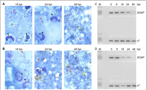
Direct microscopic examination of smears of midgut contents in P. berghei (Fig. 1a) and P. yoelii (Fig. 1b) infected A. gambiae mosquitoes confirmed that both zygotes and mature ookinetes were present in the gut lumen at 18 h post infection (hpi), while mature ooki-netes were only observed at 24 hpi. All the ookiooki-netes were physically cleared from the gut lumen by 48 hpi. However, RT-PCR performed on total RNA extracted from the mosquito midgut content showed that tran-scripts of the ookinete-specific gene SOAP were detect-able until 48 hpi (Fig. 1c). The physical as well as molecular detection of P. yoelii ookinetes in the mos-quito gut content were considerably more transient compared toP. berghei. This is expected as the P. yoelii infected mosquitoes were kept at a higher temperature (24 °C) compared toP. berghei (21 °C). MatureP. yoelii ookinetes were observed in midgut contents harvested at 18 hpi, but only ookinete remains were detected at 24 hpi. As with P. berghei, P. yoelii SOAP transcripts were still detectable at 24 hpi and only cleared by 48 hpi (Fig. 1d). These data indicate that ookinete-specific tran-scripts persist in the blood bolus even in the absence of viable ookinetes.
Persistence of ookinete mRNA in the mosquito midgut and hemolymph
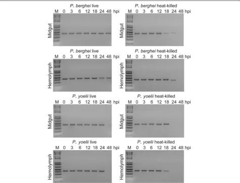
to P. yoeliiookinetes (Fig. 2). This difference is attribut-able to the difference in temperature between these two infection models and consequently the slower metabolic rate of mosquitoes at 21 °C (P. berghei) compared to 24 °C (P. yoelii). For heat-killed ookinetes, SOAP tran-scripts persisted in mosquito tissues up to 24 hpi and 18 hpi for P. berghei and P. yoelii ookinetes, respect-ively, whereas transcripts from live ookinetes were detect-able until 48 hpi. The slow mRNA decay rate suggests that transcript abundances measured by PCR methods are not fully reflective of viable parasites.
PMA-induced inhibition of DNA amplification from dead parasites and bacteria
Propidium monoazide (PMA) has been increasingly used for the treatment of microbiological samples to exclude PCR signals from non-viable cells, but this has not been tested to date in investigations involving Plasmodium. We confirmed that PMA can stain deadP. berghei ooki-netes prepared from an ookinete culture and heat-killed at 42 °C for 30 min before incubating them with PMA (Fig. 3a). Next, we stained ookinetes sampled directly from an overnight culture that was thought to include
both live and dead cells. The ookinetes were incubated with a mixture of PMA and SYBR green that also stains nucleic acids but, unlike PMA, actively enters live cells. Fluorescent microscopic observations confirmed that some ookinetes are stained only with PMA, while most of the ookinetes are stained only with SYBR green (Fig. 3b). We applied the same protocol to An. gambiae midguts tissues infected 24 h earlier withP. berghei. The results showed that SYBR and PMA could be used to discriminate between live and dead parasites, respect-ively (Fig. 3c). Bacteria present in the blood meal bolus were also differentially stained with SYBR and PMA (Fig. 3d).
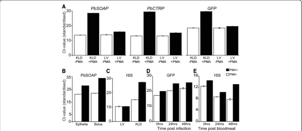
[image:5.595.56.540.89.386.2]of An. gambiaegDNA and amplification of the S7 gene served as a heterologous internal standard. The results showed that PMA treatment significantly inhibited the qrtPCR detection of SOAP,CTRP orGFPgenes in dead ookinetes (Fig. 4a). We observed a similar inhibitory
effect of PMA on qrtPCR assays for bacterial 16S rRNA gene in the gDNA from total culturable bacteria in the mosquito midgut content (Fig. 4b).
Next, we tested the PMA inhibitory effect on parasite detection in mosquitoes by comparing the level of Fig. 2SOAPmRNA persistence in theAn. coluzziimidgut and hemolymph. Agarose gel electrophoresis images ofPlasmodium SOAPgene transcrips inAn. coluzziimosquitoes. Heat-killed or liveP. bergheiorP. yoeliiookinetes were delivered to mosquitoes either by addition to the blood meal or injection into the haemolymph. RT-PCR assays were carried our on whole mosquito RNAs prepared at 0, 3, 6, 12, 18, 24 and 48 h post ookinete ingestion or injection (hpi)
[image:6.595.61.537.89.451.2] [image:6.595.59.540.593.686.2]reduction of qrtPCR signal (increase in Ct-value) in the midgut contents and midgut epithelial tissues of mos-quitoes that had been infected 24 h earlier with P. ber-ghei parasites. The qrtPCR analysis of thePbSOAPgene showed an increased Ct-value in gut content or gut epi-thelial tissue samples that were treated with PMA when compared to controls (Fig. 4c). The Ct-value was higher for PMA treated samples at 24 h pi compared to 0 h pi, but there was no further increase from 24 h to 48 h, suggesting the PMA treatment inhibited DNA amplifica-tion from dead ookinetes and also from those ookinetes that were eventually killed.
Detection and quantification of ookinetes using PMA qrt-PCR
The efficiency of PMA-qrtPCR for detecting and quanti-fying Plasmodium ookinetes was tested using 10-fold serial dilutions of P. bergheiookinetes cultured in vitro, from 2.5×10⁶ to 2.5×10⁻¹. Samples were spiked with equal volumes of mosquito tissue homogenates before proceeding to gDNA extraction. In two independent PMA-qrtPCR experiments, the resulting Ct-values were standardized to the An. gambiae S7 gene in the corre-sponding experiment. Averaged Ct-values correcorre-sponding
to each dilution were plotted against the ookinete num-bers in the respective dilution. Standard curves were linear for the serial dilution range (Fig. 5). The limit of quantification (LoD) for the PMA-qrtPCR was ~25 ookinetes.
Fig. 4PMA-qrtPCR investigation of the inhibition of DNA amplification in killed ookinetes or bacteria during PCR.aAverage cycle threshold (ct-value) forPbSOAPPbCTRP andPbGFPCONgenes in heat inactivated (KLD) or live (LV) ookinetes from overnight gametocyte culture that were pre-treated with PMA. Inactivated and live ookinetes without PMA were used as a control. Samples, before DNA extraction, were spiked with an equal volume ofAnopheles coluzziihomogenate with the aim to use the mosquito S7 gene as a heterologous internal standard.
bAverage ct-values for thePbGFP gene in the midgut contents or gut epithelium tissue from mossquitoes 24 h pi withP. bergheiparasite.
cAverage ct-values for bacterial 16S rRNA gene from heat inactivated samples and live bacteria cultured from mosquito midgut contents (24 h after bloodmeal). Samples without PMA treatment served as a control.dCt-values forPbGFP genes from gDNA samples that were extracted from mosquitoes that obtanedP. bergheiookinetes in their bloodmeal.eCt-values for 16S rRNA genes in gDNA extracted from mosquitoes that obstained naïve bloodmeal.An. gambiae S7gene served as a heterologous internal standard for all the PMA-qrtPCR reactions that were performed using samples that were isolated from mosquito tissues. Error bars represent standard deviation from at least two independent assays. Each qrPCR experiment repeated three times
[image:7.595.57.539.90.299.2] [image:7.595.305.539.519.677.2]Discussion and Conclusions
In this study, we performed a series of experiments aimed to establish an efficient PCR based method to detect and quantify the success of Plasmodium parasite infection of the mosquito midgut. The majority ofPlasmodium ooki-netes are killed during the first 32 h of their life in the mosquito midgut. Dead parasites limit the use of standard PCR based methods to quantify live ookinete numbers during that time due to amplification of nucleic acids from both dead and viable parasites. An alternative method that addresses this limitation is quantification of mRNA that is often used as a viability marker on the basis of its rapid degradation and therefore considered to be a good indica-tor of live cells [29–32]. However, our data show that Plasmodiumtranscripts exclusively expressed in ookinetes and required for midgut invasion [33, 34] persist in mos-quito midguts at least 24 h beyond the termination of their expression or after ookinete death. Therefore the detection and quantification of ookinetes transcripts using qrtPCR cannot exclude dead parasites, and mRNA origin-ating from non-dead ookinetes in the midgut killed by multiple factors could lead to an overestimation of the number of ookinetes. Numerous studies have demon-strated that RNA can persist for up to a week in dead cells [35, 36]. Due to indecisiveness of discrimination of viable cells by this technique, the use of mRNA based qrtPCR is more adapted for gene expression studies than estimating of the bacterial abundance [37].
To circumvent the indecisiveness of mRNA-based techniques, a combination of pretreatment of samples with PMA and nucleic acid detection methods have been developed as an alternative strategy [38]. This tecnique is based on DNA detection of cells with intact cell/wall membranes, the viability qPCR (v-qPCR). The PMA selectively enters dead cells and interacts with DNA preventing its amplification during PCR. Until now, the PMA-qrtPCR technique has not been tested to date in investigations involvingPlasmodiumviability in the mos-quito vector. We demonstrate that PMA can selectively enter dead ookinetes both from in vitro cultures and in vivo, while dual staining of the mosquito midgut contents with SYBR green I (SGI) and PMA confirmed that PMA enters exclusively into dead bacterial cells. Subsequent qrtPCR of PMA treated samples effectively differentiates viable from non-viable ookinetes, and the same effect is observed on bacteria isolated from mosquito midguts.
There are several examples of successful application of the PMA-qrtPCR technique including quantification of viable oocysts ofCryptosporidium parvum[22] and tro-phozoites and cysts of Acanthamoeba castellani [39]. The technique has been also successfully used to quantify viable microorganisms in environmental [40–42], food [43–45] and pathological samples [46, 47]. It has been shown that the limit of detection of the PMA-qrtPCR
method was at least equivalent to that obtained by using CFU values for the gram-negative bacteria Legionella pneumophila [48]. Here we show that the technique can detect as few as 10 and accurately quantify as few as 25 ookinetes.
Therefore, the PMA-qrtPCR technique can allow accur-ate and simultaneous monitoring of viable Plasmodium ookinetes and midgut microbiota cells during the time of mosquito midgut invasion. The ookinetes and microbiota data obtained by this method can be also used to assess the dynamics and magnitude of mosquito immune re-sponses at a time of infection.
Abbreviations
PMA:Propidium Monoazide; PCR: Polymerase chain reaction; RT-PCR: Reverse transcriptase PCR; qrtPCR: Quantitative real-time PCR; DNA: Deoxyribonucleic acid; RNA: Ribonucleic acid; v-PCR: Viability PCR; SOAP: Secreted ookinete adhesive protein; PbSOAP:Plasmodium bergheiSOAP; CTRP: Circumsporozoite and TRAP related protein; GFP: Green fluorescent protein; Ct: Threshold cycle; LoD: Limit of detection.
Competing interest
The authors declare that they have no competing interests.
Authors’contribution
Conceived and designed the experiments: TH, GKC. Performed the experiments: TH, ZG. Analyzed the data: TH, GKC. Contributed reagents/ materials/analysis tools: TH, GKC, LD. Wrote the manuscript: TH, ZG, GKC, LD. All authors read and approved the final manuscript.
Acknowledgments
We thank Katarzyna Sala for technical support and Valerie Ukegbu for supply of parasite cultures. The work was supported through grants by the Wellcome Trust, WT093587MA and the BBSRC Project BB/K009338/1.
Author details
1Department of Life Sciences, Imperial College London, London, UK. 2Department of Comparative Physiology and Biometrics, University of Ghent, Ghent, Belgium.3Costello Medical Consulting, Cambridge, UK.
Received: 10 August 2015 Accepted: 11 September 2015 References
1. Alavi Y, Arai M, Mendoza J, Tufet-Bayona M, Sinha R, Fowler K, et al. The dynamics of interactions betweenPlasmodiumand the mosquito: a study of the infectivity ofPlasmodium bergheiandPlasmodium gallinaceum, and their transmission by Anopheles stephensi,Anopheles gambiaeandAedes aegypti. Int J Parasitol. 2003;33(9):933–43. Epub 2003/08/09. PubMed. 2. Ghosh A, Edwards MJ, Jacobs-Lorena M. The Journey of the Malaria Parasite
in the Mosquito: Hopes for the New Century. Parasitol Today. 2000;16(5):196–201. doi:10.1016/s0169-4758(99)01626-9.
3. Vaughan JA, Hensley L, Beier JC. Sporogonic development ofPlasmodium yoelii in five anopheline species. J Parasitol. 1994;80(5):674–81. Epub 1994/10/01. PubMed.
4. Vaughan JA, Noden BH, Beier JC. Population dynamics ofPlasmodium falciparumsporogony in laboratory-infected Anopheles gambiae. J Parasitol. 1992;78(4):716–24. Epub 1992/08/01. PubMed.
5. Gendrin M, Christophides GK. The Anopheles Mosquito Microbiota and Their Impact on Pathogen Transmission, Anopheles mosquitoes - New insights into malaria vectors. Manguin PS, editor. http://www.intechopen. com/books/anopheles-mosquitoes-new-insights-into-malaria-vectors/the- anopheles-mosquito-microbiota-and-their-impact-on-pathogen-transmission. 2013.
7. Dong Y, Das S, Cirimotich C, Souza-Neto JA, McLean KJ, Dimopoulos G. EngineeredAnophelesImmunity toPlasmodiumInfection. PLoS Pathog. 2011;7(12), e1002458. doi:10.1371/journal.ppat.1002458.
8. Garver LS, Bahia AC, Das S, Souza-Neto JA, Shiao J, Dong Y, et al.Anopheles Imd pathway factors and effectors in infection intensity-dependent anti-Plasmodium action. PLoS Pathog. 2012;8(6):e1002737. doi:10.1371/journal.ppat.1002737. Epub 2012/06/12. PubMed PMID: 22685401; PubMed Central PMCID: PMCPMC3369948.
9. Cirimotich CM, Dong Y, Clayton AM, Sandiford SL, Souza-Neto JA, Mulenga M, et al. Natural Microbe-Mediated Refractoriness to Plasmodium Infection in Anopheles gambiae. Science. 2011;332(6031):855–8. doi:10.1126/science.1201618. 10. Luckhart S, Vodovotz Y, Cui L, Rosenberg R. The mosquitoAnopheles
stephensilimits malaria parasite development with inducible synthesis of nitric oxide. Proc Natl Acad Sci U S A. 1998;95(10):5700–5.
11. Molina-Cruz A, DeJong RJ, Charles B, Gupta L, Kumar S, Jaramillo-Gutierrez G, et al. Reactive oxygen species modulateAnopheles gambiaeimmunity against bacteria andPlasmodium. J Biol Chem. 2008;283(6):3217–23. doi:10.1074/jbc.M705873200. Epub 2007/12/11. PubMed.
12. Weiss B, Aksoy S. Microbiome influences on insect host vector competence. Trends Parasitol. 2011;27(11):514–22. doi:10.1016/j.pt.2011.05.001.
13. Bourgeois N, Boutet A, Bousquet PJ, Basset D, Douard-Enault C, Charachon S, et al. Comparison of three real-time PCR methods with blood smears and rapid diagnostic test inPlasmodiumsp. infection. Clin Microbiol Infect. 2010;16(8):1305–11. doi:10.1111/j.1469-0691.2009.02933.x.
14. Farcas GA, Soeller R, Zhong K, Zahirieh A, Kain KC. Real-Time Polymerase Chain Reaction Assay for the Rapid Detection and Characterization of Chloroquine-ResistantPlasmodium falciparumMalaria in Returned Travelers. Clin Infect Dis. 2006;42(5):622–7. doi:10.1086/500134.
15. Hellyer TJ, DesJardin LE, Hehman GL, Cave MD, Eisenach KD.
Quantitative analysis of mRNA as a marker for viability ofMycobacterium tuberculosis. J Clin Microbiol. 1999;37(2):290–5. Epub 1999/01/16. PubMed PMID: 9889206; PubMed Central PMCID: PMCPMC84288. 16. Hellyer TJ, DesJardin LE, Teixeira L, Perkins MD, Cave MD, Eisenach KD.
Detection of viableMycobacterium tuberculosisby reverse transcriptase-strand displacement amplification of mRNA. J Clin Microbiol. 1999;37(3):518–23. Epub 1999/02/13. PubMed PMID: 9986805; PubMed Central PMCID: PMCPMC84447.
17. Cenciarini-Borde C, Courtois S, La Scola B. Nucleic acids as viability markers for bacteria detection using molecular tools. Future Microbiol. 2009;4(1):45–64. doi:10.2217/17460913.4.1.45. Epub 2009/02/12. PubMed.
18. Nocker A, Sossa-Fernandez P, Burr MD, Camper AK. Use of propidium monoazide for live/dead distinction in microbial ecology. Appl Environ Microbiol. 2007;73(16):5111–7. doi:10.1128/aem.02987-06. Epub 2007/06/26. PubMed PMID: 17586667; PubMed Central PMCID: PMCPMC1951001. 19. Nocker A, Cheung CY, Camper AK. Comparison of propidium monoazide
with ethidium monoazide for differentiation of live vs. dead bacteria by selective removal of DNA from dead cells. J Microbiol Methods. 2006;67(2):310–20. doi:10.1016/j.mimet.2006.04.015. Epub 2006/06/07. PubMed.
20. Pan Y, Breidt Jr F. Enumeration of viable Listeria monocytogenescells by real-time PCR with propidium monoazide and ethidium monoazide in the presence of dead cells. Appl Environ Microbiol. 2007;73(24):8028–31. doi:10.1128/aem.01198-07. Epub 2007/10/16. PubMed PMID: 17933922; PubMed Central PMCID: PMCPMC2168130.
21. Vesper S, McKinstry C, Hartmann C, Neace M, Yoder S, Vesper A. Quantifying fungal viability in air and water samples using quantitative PCR after treatment with propidium monoazide (PMA). J Microbiol Methods. 2008;72(2):180–4. doi:10.1016/j.mimet.2007.11.017. Epub 2007/12/28. PubMed.
22. Brescia CC, Griffin SM, Ware MW, Varughese EA, Egorov AI, Villegas EN. Cryptosporidium propidiummonoazide-PCR, a molecular biology-based technique for genotyping of viableCryptosporidiumoocysts. Appl Environ Microbiol. 2009;75(21):6856–63. doi:10.1128/aem.00540-09. Epub 2009/09/ 15. PubMed PMID: 19749067; PubMed Central PMCID: PMCPMC2772443. 23. Coetzee M, Hunt RH, Wilkerson R, Della Torre A, Coulibaly MB, Besansky NJ.
Anopheles coluzziiandAnopheles amharicus, new members of the Anopheles gambiae complex. Zootaxa. 2013;3619:246–74. Epub 2013/01/01. PubMed.
24. Franke-Fayard B, Trueman H, Ramesar J, Mendoza J, van der Keur M, van der Linden R, et al. APlasmodium bergheireference line that constitutively expresses GFP at a high level throughout the complete life cycle. Mol Biochem Parasitol.
2004;137(1):23–33. doi:10.1016/j.molbiopara.2004.04.007. Epub 2004/07/29. PubMed.
25. Ono T, Tadakuma T, Rodriguez A.Plasmodium yoelii yoelii17XNL constitutively expressing GFP throughout the life cycle. Exp Parasitol. 2007;115(3):310–3. doi:10.1016/j.exppara.2006.09.008. Epub 2006/10/20. PubMed PMID: 17049517; PubMed Central PMCID: PMC1779952. 26. Habtewold T, Povelones M, Blagborough AM, Christophides GK.
Transmission blocking immunity in the malaria non-vector mosquito Anopheles quadriannulatusspecies A. PLoS Pathog. 2008;4(5):e1000070. doi:10.1371/journal.ppat.1000070. Epub 2008/05/24. PubMed PMID: 18497855; PubMed Central PMCID: PMCPMC2374904.
27. Alvarez G, Gonzalez M, Isabal S, Blanc V, Leon R. Method to quantify live and dead cells in multi-species oral biofilm by real-time PCR with propidium monoazide. AMB Express. 2013;3(1):1. doi:10.1186/2191-0855-3-1. Epub 2013/01/08. PubMed PMID: 23289803; PubMed Central PMCID: PMCPMC3549832.
28. Saadati N, Abdullah MP, Zakaria Z, Sany SB, Rezayi M, Hassonizadeh H. Limit of detection and limit of quantification development procedures for organochlorine pesticides analysis in water and sediment matrices. Chem Cent J. 2013;7(1):63. doi:10.1186/1752-153x-7-63. Epub 2013/04/09. PubMed PMID: 23561579; PubMed Central PMCID: PMCPMC3630005.
29. Alifano P, Bruni C, Carlomagno M. Control of mRNA processing and decay in prokaryotes. Genetica. 1994;94(2–3):157–72. doi:10.1007/BF01443430. 30. Mendum TA, Sockett RE, Hirsch PR. The detection of Gram-negative
bacterial mRNA from soil by RT-PCR. FEMS Microbiology Lett. 1998;164(2):369–73. http://femsle.oxfordjournals.org/content/164/2/369. 31. Uyttendaele M, van Boxstael S, Debevere J. PCR assay for detection of the
E. coli O157:H7 eae-gene and effect of the sample preparation method on PCR detection of heat-killed E. coli O157:H7 in ground beef. Int J Food Microbiol. 1999;52(1–2):85–95. Epub 1999/11/26. PubMed.
32. Vettraino AM, Sukno S, Vannini A, Garbelotto M. Diagnostic sensitivity and specificity of different methods used by two laboratories for the detection ofPhytophthora ramorumon multiple natural hosts. Plant Pathol. 2010;59(2):289–300. doi:10.1111/j.1365-3059.2009.02209.x.
33. Dessens JT, Siden-Kiamos I, Mendoza J, Mahairaki V, Khater E, Vlachou D, et al. SOAP, a novel malaria ookinete protein involved in mosquito midgut invasion and oocyst development. Mol Microbiol. 2003;49(2):319–29. Epub 2003/06/28. PubMed.
34. Oakley MS, Kumar S, Anantharaman V, Zheng H, Mahajan B, Haynes JD, et al. Molecular factors and biochemical pathways induced by febrile temperature in intraerythrocyticPlasmodium falciparumparasites. Infect Immun. 2007;75(4):2012–25. doi:10.1128/iai.01236-06. Epub 2007/02/07. PubMed PMID: 17283083; PubMed Central PMCID: PMCPMC1865691. 35. Chimento A, Cacciola SO, Garbelotto M. Detection of mRNA by
reverse-transcription PCR as an indicator of viability inPhytophthora ramorum. Forest Pathol. 2012;42(1):14–21. doi:10.1111/j.1439-0329.2011.00717.x. 36. Sheridan GEC, Masters CI, Shallcross JA, Mackey BM. Detection of mRNA
by Reverse Transcription-PCR as an Indicator of Viability inEscherichia coli Cells. Appl Environ Microbiol. 1998;64(4):1313–8. PubMed.
37. Postollec F, Falentin H, Pavan S, Combrisson J, Sohier D. Recent advances in quantitative PCR (qPCR) applications in food microbiology. Food Microbiol. 2011;28(5):848–61. doi:10.1016/j.fm.2011.02.008. Epub 2011/05/17. PubMed. 38. Wagner AO, Malin C, Knapp BA, Illmer P. Removal of Free Extracellular DNA from
Environmental Samples by Ethidium Monoazide and Propidium Monoazide. Appl Environ Microbiol. 2008;74(8):2537–9. doi:10.1128/aem.02288-07. 39. Fittipaldi M, Pino Rodriguez NJ, Adrados B, AgustÍ G, PeÑUela G, MoratÓ J,
et al. Discrimination of ViableAcanthamoeba castellaniTrophozoites and Cysts by Propidium Monoazide Real-Time Polymerase Chain Reaction. J Eukaryot Microbiol. 2011;58(4):359–64. doi:10.1111/j.1550-7408.2011.00557.x. 40. Nocker A, Richter-Heitmann T, Montijn R, Schuren F, Kort R. Discrimination
between live and dead cellsin bacterial communities from environmental water samples analyzed by 454 pyrosequencing. Int Microbiol. 2010;13(2):59–65. Epub 2010/10/05. PubMed.
41. Wagner AO, Malin C, Knapp BA, Illmer P. Removal of free extracellular DNA from environmental samples by ethidium monoazide and propidium monoazide. Appl Environ Microbiol. 2008;74(8):2537–9. doi:10.1128/aem.02288-07. Epub 2008/02/26. PubMed PMID: 18296534; PubMed Central PMCID: PMCPMC2293149.
doi:10.1128/aem.02895-10. Epub 2011/05/24. PubMed PMID: 21602375; PubMed Central PMCID: PMCPMC3127682.
43. Li B, Chen JQ. Development of a sensitive and specific qPCR assay in conjunction with propidium monoazide for enhanced detection of live Salmonella spp. in food. BMC Microbiol. 2013;13:273. doi:10.1186/1471-2180-13-273. Epub 2013/12/03. PubMed.
44. Liang N, Dong J, Luo L, Li Y. Detection of viableSalmonellain lettuce by propidium monoazide real-time PCR. J Food Sci. 2011;76(4):M234–7. doi:10.1111/j.1750-3841.2011.02123.x. Epub 2012/03/16. PubMed. 45. Banihashemi A, Van Dyke MI, Huck PM. Long-ampliconpropidium
monoazide-PCR enumeration assay to detect viableCampylobacterandSalmonella. J Appl Microbiol. 2012;113(4):863–73. doi:10.1111/j.1365-2672.2012.05382.x. Epub 2012/07/04. PubMed.
46. Yasunaga A, Yoshida A, Morikawa K, Maki K, Nakamura S, Soh I, et al. Monitoring the prevalence of viable and dead cariogenic bacteria in oral specimens and in vitro biofilms by qPCR combined with propidium monoazide. BMC Microbiol. 2013;13:157. doi:10.1186/1471-2180-13-157. Epub 2013/07/16. PubMed PMID: 23848601; PubMed Central PMCID: PMCPMC3717283.
47. Rogers GB, Cuthbertson L, Hoffman LR, Wing PA, Pope C, Hooftman DA, et al. Reducing bias in bacterial community analysis of lower respiratory infections. ISME J. 2013;7(4):697–706. doi:10.1038/ismej.2012.145. Epub 2012/ 11/30. PubMed PMID: 23190732; PubMed Central PMCID: PMCPMC3603400. 48. Chang B, Taguri T, Sugiyama K, Amemura-Maekawa J, Kura F, Watanabe H.
Comparison of ethidium monoazide and propidium monoazide for the selective detection of viableLegionellacells. Jpn J Infect Dis. 2010;63(2):119–23. Epub 2010/03/25. PubMed.
Submit your next manuscript to BioMed Central and take full advantage of:
• Convenient online submission
• Thorough peer review
• No space constraints or color figure charges
• Immediate publication on acceptance
• Inclusion in PubMed, CAS, Scopus and Google Scholar
• Research which is freely available for redistribution