1
A STUDY OF CIRCLE OF WILLIS WITH ITS
VARIATIONS AND ITS CLINICAL
CORRELATIONS
A Dissertation submitted toThe Tamil Nadu Dr. M. G. R. Medical University,
Chennai.
In partial fulfillment of the requirements for the degree of
M.D. DEGREE EXAMINATION BRANCH – XXIII (ANATOMY)
GOVERNMENT STANLEY MEDICAL COLLEGE AND HOSPITAL
CHENNAI – 600 001.
THE TAMILNADU DR. M.G.R. MEDICAL UNIVERSITY
CHENNAI
2
CERTIFICATE
This is to certify that the dissertation work on “A STUDY OF CIRCLE OF WILLIS WITH ITS VARIATIONS AND ITS CLINICAL CORRELATIONS” is a bonafide research work done by Dr. V. SHANTHI, post graduate (2015-2018) in the Department of Anatomy, Govt. Stanley Medical College and Hospital, Chennai under my direct guidance and supervision, in partial fulfillment of the regulations laid down by the TAMILNADU DR. M.G.R. MEDICAL UNIVERSITY Chennai for the award of M.D. Anatomy (Branch XXIII) degree examination to be held in APRIL 2018.
Prof. Dr. S.Ponnambala Namasivayam, Dr. T. Vasantha Kumar, M.S.,
M.D., D.A., D.N.B., Professor and Head of the
Department The Dean Department of Anatomy
Stanley Medical College Stanley Medical College,
3
DECLARATION
I hereby declare that this dissertation entitled in “A STUDY OF CIRCLE OF WILLIS WITH ITS VARIATIONS AND ITS
CLINICAL CORRELATIONS” was written by me in the Department
of Anatomy, Government Stanley Medical College and Hospital, Chennai under the guidance and supervision of Prof. Dr. T. VASANATHA
KUMAR, M.S., Professor and Head of the Department of Anatomy,
Government Stanley Medical College and Chennai – 600 001.
This dissertation is submitted to The Tamilnadu Dr. M.G.R. Medical University, Chennai in partial fulfilment of the university regulations for the award of Degree of M.D., Anatomy (Branch XXIII) Examination to be held in April 2018.
Date:
4
ACKNOWLEDGEMENT
I wish to express my sincere thanks & gratitude to Prof. Dr. S. Ponnambala Namasivayam, M.D., D.A., D.N.B., Dean, Stanley Medical College and Hospital, Chennai – 1 for having permitted me to utilise the facilities in this college for the conduct of the study.
My heartfelt thankfulness, gratitude and gratefulness to Dr. T. Vasantha Kumar, M.D., Professor and Head of the Department of Anatomy, Government Stanley Medical College, Chennai for his invaluable guidance, motivation and persistent support, encouragement and for providing all necessary arrangement to make the study a reality.
I sincerely thank Dr. S. Chitra, M.S., (Retd. Professor & HOD, Department of Anatomy), for her invaluable guidance, motivation and encouragement.
My sincere thanks to Dr. S.Balasubramaniam, D.C.H., M.D.,
5
I am grateful to Dr. C. Amarnath, M.D., RD., Professor and Head of the Department of Radiology, Govt. Stanley Medical College, Chennai-1 and his faculty for their help in radiological study.
I sincerely thank Dr. J.Thilagavathi, M.S., Associate Professor and Dr. K.Sujatha, M.S., Associate Professor, Dr. B.Anbumalar, M.D., Senior Asst. Professor Dr. J.K. Raja, M.D., Dr. S. Elizabeth Priyadarisini, M.D., Dr. M.Anuradha M.D., Dr.Adline Misba, M.D., Dr. E. Anitha, M.D., Dr. B. Ramkumar, M.D., Dr. M.R.Manimegalai, M.D., Dr. F.Stelina Sophia Dina, M.D., Assistant Professors for their valuable suggestions and constant encouragement throughout the study.
I also specialy thank earnestly my colleague Dr. J.Senthilkumar and my seniors Dr.C.Sasikala, M.D., Dr.S.Manonmani, MD., and my juniors Dr.P.J.Seeja, Dr.S.Sivakumari, Dr. P.Maharathi, Dr. Mohammed Samiullah for their help rendered to me during the study.
I am also thankful to lab technicians Smt.K.Rajalakshmi, Smt. E.Jayanthi, and departmental staffs, Thiru.C.Birammaiah, Thiru.A.Kadar Basha, Thiru.M.Jagadeesan, Mr. Srinivasan and Tmt. Susila for helping me in carrying out the study. It gives me great pleasure in preparing this dissertation and I take this opportunity to thank everyone who made this possible.
6
CONTENTS
S.NO. TITLE PAGE NO.
1 INTRODUCTION 1
2 ANATOMICAL AND
EMBRYOLOGICAL CONSIDERATIONS 5
3 AIM OF THE STUDY 16
4 REVIEW OF LITERATURE 19
5 MATERIALS AND METHODS 39
6 OBSERVATION 44
7 DISCUSSION 77
8 CONCLUSION 92
8
INTRODUCTION
The brain is a vital organ which requires rich blood supply to sustain its ongoing activities. Irreversible brain damage occurs when the blood supply is interrupted for few minutes, which results in loss of consciousness (Neeta Kulkarni 3rd edit). Approximately 15% of the
total blood supply reaches the brain and approximately 20% of the oxygen utilization is consumed by the adult brain (Parthapratim Pradhan et al 2009). The blood flow to the brain is estimated to be around 800ml/min of brain tissue. The blood flow is faster in the grey matter (70-80ml/100g/min) than in white matter (30ml/100g/min). Blood flow of less than 15ml/100g/min leads to irreversible brain damage
(Gray’s Anatomy edit 40th).
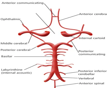
The brain receives its blood supply from four arterial trunks, two internal carotid arteries and two vertebral arteries. The internal carotid artery arises from the common carotid artery at the level of upper border of thyroid cartilage. The vertebral artery is a branch of first part of subclavian artery (Ranganathan T.S edit 3rd ).
9
the arteries together with their branches lie within the subarachnoid space called interpeduncular cistern at the base of the brain. It is a ring like network of blood vessels which is essential for the perfusion of brain. The two internal carotid arteries contribute 80% of the blood supply to the brain and the two vertebral arteries contribute 20% of the blood supply (Grand W 1999). Each of the internal carotid artery branches to form anterior and middle cerebral arteries which supply blood to the brain. The ring is completed by the communicating arteries which connect the anterior and posterior part of the circle. In about 60% of cases, anatomical variations in the Circle of Willis may be found
(Battacharji SK 1967, Craig Hacking).
10
Under normal condition, the blood flow in the communicating arteries are negligible. If a subject has an atypical circle (missing of one of the main artery or the communicating artery) or under pathological condition (partial or complete occlusion of cerebral or carotid vessels), the flow can be redirected to perfuse deprived areas (Vare AM 1970). It has been found that in 50% of the normal brain and in 80% of the dysfunctional brain, the Circle of Willis is often incomplete or underdeveloped. The most common morphological variations are absent vessels, hypoplastic vessels and extra vessels (Neeta Kulkarni edit 3rd ).
These variations affect the ability to maintain the blood flow through arterioles, which may increase the risk of stroke and transient ischemic attack in atherosclerotic patients (Riggs HE1963). Acute occlusion due to embolus (embolic stroke) or chronic occlusion due to stenosis of anyone of the vessel (carotid and other large vessel stenosis) occurs in patients with incomplete Circle of Willis (Alpers 1959). These problems are significant, as stroke is one of the leading cause of morbidity and death in elderly people.
11
12
ANATOMICAL AND EMBRYOLOGICAL
CONSIDERATIONS
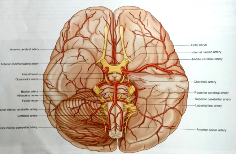
The Circle of Willis is one of the principal arterial anastomosis of the brain. It is formed by the anastomosis between the branches of cerebral part (supraclinoid part) of right and left internal carotid arteries and the terminal branches of basilar artery. Circle of Willis is responsible for brain’s collateral blood supply (Gray’s Anatomy 40th edit). Circle of Willis is composed of following arteries:
Internal Carotid Artery (left and right). Anterior Cerebral Artery (left and right). Anterior Communicating Artery.
Posterior Cerebral Artery (left and right).
Posterior Communicating Artery (left and right).
13
[image:13.612.130.511.350.599.2]The left and right posterior cerebral arteries arise from the basilar artery, which inturn is formed by the union of fourth part of left and right vertebral arteries. The posterior communicating artery is given off as a branch of the internal carotid artery just before it divides into its terminal branches- the anterior and middle cerebral arteries. The anterior cerebral artery forms the anterolateral portion of the Circle of Willis, while the middle cerebral artery does not directly contribute to the circle. The two anterior cerebral arteries communicate with each other through the anterior communicating artery (Gray’s Anatomy 40th edit).
14
The internal carotid arteries and their major branches (the internal carotid system or ‘anterior’ circulation) supply blood to the majority of the forebrain. The internal carotid artery arises from the bifurcation of the common carotid artery at the level of upper border of thyroid cartilage, ascends in the neck and enters the carotid canal of the temporal bone
(Romanes GJ Cuningham edit 15th).
Its subsequent course is said to have petrous, cavernous and intracranial part. The petrous part of the internal carotid artery ascends in the carotid canal, curves anteromedially and then superomedially above the cartilage that fills the foramen lacerum. It forms the anterior wall of the tympanic cavity and is separated from the tympanic cavity by a thin bony lamella that is cribriform in the young and partly filled in old age
(Ranganathan T.S edit 3rd). Anteriorly, it is separated from the
trigeminal ganglion by the thin plate of carotid canal. The artery is surrounded by venous plexus and by the autonomic plexus derived from the internal carotid branch of superior cervical ganglion. The petrous part gives two branches, one or two small caroticotympanic arteries and pterygoid artery (Gray’s Anatomy 40th edit).
15
through the dural roof of the sinus. The oculomotor, trochlear, ophthalmic and maxillary nerves are lateral to internal carotid artery within the lateral wall of cavernous sinus (Kamath S 1981) (fig 2). The cavernous part gives off small meningeal, cavernous, and inferior hypophyseal branches.
Fig 2 : Relation of internal carotid artery in cavernous sinus.
The internal carotid artery after piercing the duramater turns back below the optic nerve and gives off superior hypophyseal artery, runs between the optic nerve and the oculomotor nerve. It reaches the anterior perforated substance at the medial end of the lateral fissure. It terminates by dividing into anterior and middle cerebral arteries
16
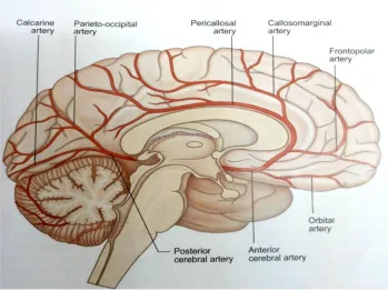
ANTERIOR CEREBRAL ARTERY:
The anterior cerebral artery is the smaller of the two terminal branches of the internal carotid arteries. According to Ranil De Silva 2016 Surgical nomenclature divides the vessel into three parts. A1-from the termination of internal carotid artery to the junction with the anterior communicating artery; A2-from the junction with the anterior communicating artery to the origin of callosomarginal artery; A3-distal to the origin of callosomarginal artery(fig 3).
The cortical branches supply the orbital surface of the frontal lobe and supply the olfactory cortex, gyrus rectus and medial orbital gyrus. They also supply the motor and somatosensory areas that represent the lower limb.
Frontal branches supply the corpus callosum, cingulate gyrus and paracentral lobule.
Parietal branches supply the precuneus and the surrounding areas. Central branches supply the rostrum of corpus callosum, the
septum pellucidum, anterior part of the putamen, head of the caudate nucleus and adjacent parts of the internal capsule
17
Fig 3: showing the course of anterior cerebral artery.
MIDDLE CEREBRAL ARTERY:
The middle cerebral artery is the larger terminal branch of the internal carotid artery. It divides into four parts.
M1-from the termination of the internal carotid artery to its bifurcation. This is known as sphenoidal segment.
M2 segment - running in the lateral (sylvian) fissure, also known as the insular or sylvian segment.
M3 segment- extends laterally from insula towards the cortex and is known as opercular part.
18
The middle cerebral artery runs at first in the lateral fissure, then posterosuperiorly on the insula and divides into branches. Middle cerebral artery gives off cortical and central branches (Vaibhav V 2016).
VERTEBRAL ARTERY:
The right and left vertebral arteries are derived from the first part of respective subclavian arteries, ascend through the neck in the foramina transversoria of the upper six cervical vertebrae and enter the cranial cavity through the foramen magnum close to the anterolateral aspect of the medulla. They converge medially and unite to form the basilar artery at the pontomedullary junction (Cunninghams manual 15th edit).
BASILAR ARTERY:
Basilar artery is a large median vessel formed by the union of the right and left vertebral arteries at the mid medullary level. It lies in the pontine cistern and follows the shallow median groove on the ventral pontine surface, extending to the upper border of the pons. It ends by dividing into right and left posterior cerebral arteries at a variable level behind the dorsum sellae, usually in the interpeduncular cistern
19
Fig 4:showing the vertebral and basilar network.
POSTERIOR CEREBRAL ARTERY:
The posterior cerebral artery is a terminal branch of the basilar artery. Surgical nomenclature divides into three parts (Ace Dodevski et al 2014).
P1-from the basilar bifurcation to the junction with the posterior communicating artery.
20
P3-the portion that runs in the calcarine fissure. The cortical branches supply the temporal, parieto-occipital lobes of the brain, the central branches supply the subcortical structures.
ANTERIOR COMMUNICATING ARTERY:
Anterior communicating artery is about 4mm in length and it connects the two anterior cerebral arteries.
POSTERIOR COMMUNICATING ARTERY:
The posterior communicating arteries are usually very small, arising from the internal carotid artery at the junction where it divides into anterior and middle cerebral arteries. Sometimes it is so large that the posterior cerebral artery is supplied via the posterior communicating artery rather than from the basilar artery (fetal posterior communicating artery) (Anuba Saha et al 2013)
EMBRYOLOGICAL CONSIDERATION:
The vascular system develops in two stages: Vasculogenesis and Angiogenesis. The embryological development of the circulatory system supplying blood to the brain begins with the formation of the six pairs of primitive branchial arteries at the 1.3mm embryonic stage
21
The brain vascular system arises from the perineural vascular plexus which sprout radially into the neuroepithelium. They subsequently branch off laterally in the subventricular zone, the subventricular plexus
(Mall FP 1905). Foxc1 gene is required for early stage telencephalic vascular development as it is expressed in endothelial cells and pericytes, as well as in the cranial mesenchyme surrounding the brain tube
(Streeter GL).
Most of the branches of the Circle of Willis are derived from the internal carotid artery which is embryologically formed from the third arch artery distal to the external carotid bud and cranial part of the dorsal aorta distal to the attachment of third arch artery (Langman embryology 12th edit).The ventral portion of the 2nd branchial artery disconnects from the dorsal aorta near the origin of the internal carotid artery and becomes the ventral pharyngeal artery. Then the ventral pharyngeal and the internal carotid artery fuse to form the common carotid artery.
22
The vertebral artery is a composite vessel and it is developed from four sources (Mall FP 1905).
The first part of the artery develops from the dorsal ramus of the intersegmental artery.
The second part of the artery develops from the enlargement of the post-costal anastomosis with the consequent regression of the stems of the upper six intersegmental arteries.
The third part of the artery develops from the spinal branch of the first cervical intersegmental artery.
The fourth part of the artery develops from the pre-neural division of the spinal branch which meets the corresponding branch of the opposite side at the caudal border of the pons to form the basilar artery.
23
AIM AND OBJECTIVES
The brain is richly supplied by blood through the vertebro-basilar and internal carotid arteries. The oxygen consumption of the neural tissue is very high (Neeta Kulkarni 3rdedit). Acute arrest of circulation
produces loss of consciousness in about approximately seven seconds. The cerebrovascular accidents (CVA) due to infarction, hemorrhage and embolism result in stroke, in which the entire half of the body or a limb or one half of the face is suddenly paralyzed (Klutmans et al 1999).
According to Kawther A 2007 ‘The persons with narrowing of cerebral arteries due to hypertension, atherosclerosis and diabetes are more prone to stroke’. The common causes of cerebral hemorrhage are aneurysms of major arteries, small arteriolar aneurysms due to hypertension and A-V malformations.
Anatomical variations in the Circle of Willis are more common. Under normal condition, the blood flow is redistributed to the ischemic area through the collateral circulation, but in pathological states like thrombo-embolism, aneurysms, there is failure of redistribution of blood flow. This leads to ischemia and necrosis of the affected area of the brain
24
knowledge of the formation of Circle of Willis and its variations are essential for proper diagnosis and treatment of the diseases (Iqbal S 2013, Hendrikse J 2005).
AIM:
To study the normal pattern of Circle of Willis both morphologically and radiologically.
To study the variations in the branching pattern of the Circle of Willis and to correlate it clinically.
OBJECTIVES:
1. To study the formation of the Circle of Willis under the following parameters in both morphological specimens and radiological images.
a. Complete or incomplete formation. b. Shape of Circle of Willis.
c. Normal or abnormal caliber of vessels. d. Symmetrical or asymmetrical formation.
2. To study the variations in the formation, branching pattern under the following parameters in both morphological and radiological images.
25 b. hypoplasia
c. accessory vessels and
d. anomalous origin in both morphology and radiology.
3. To study the incidence of variation of individual vessels, anterior and posterior part of Circle of Willis.
4. To study the clinical correlation of the Circle of Willis and to state its importance in cerebrovascular diseases.
26
REVIEW OF LITERATURE
MORPHOLOGICAL STUDY:
1. The morphology of Circle of Willis dates back to Hetrophilis, who discovered a structure which he called ‘Rete mirabile”. Galen
stated that the carotid arteries run in the neck and form rete mirabile after they enter the cranial cavity. Fallopius (1523-62), Casserius (1561-1616), Vesling (1598-1649) also described the formation of Circle of Willis in their studies.
2. Thomas Willis (1621-1675) gave the correct description of Circle
of Willis which supplies the brain and the surrounding structures.
3. According to Gray’s Anatomy 40th edit, 2016) 60% show
variations in the morphology of the Circle of Willis. Cerebral arteries and the communicating arteries, the anterior and the posterior may be absent, variably hypoplastic, double or even triple in number. The hemodynamic balance may be disturbed by variations in the caliber of the cerebral and the communicating arteries.
4. Neeta Kulkarni 3rd edit (2016) states that the Circle of Willis
27
brain, because it is the main collateral channel of both sides. It provides an alternate channel if any one of the arteries forming the Circle of Willis is blocked.
5. Cunningham manual of practical Anatomy 15th edit(1986)
28
FORMATION OF CIRCLE OF WILLIS:
6. The formation of Circle of Willis has been studied by many authors under various parameters. This includes complete or incomplete form, shape of the circle, caliber and symmetry of the circle. The incidence of ‘text book ‘ type of Circle of Willis range from 5% to 72% (Iqbal 2013). The incidence of variations in the morphological pattern of the Circle of Willis has been reported by various authors. This includes Kamath S -44%, Alpers et al-47.7%, Raja Reddy et al-46.7%, Stephen and John-48%, Macchi et al-59%, Hartkamp et al-58%.
VARIATIONS IN THE FORMATION AND BRANCHING
PATTERN OF THE VESSELS OF CIRCLE OF WILLIS:
7. E.Fawcett 1905 –The Circle of Willis: An examination of 700 specimens stated that the percentage of incomplete circle with asymmetrical and abnormal shape constitute 30.3% of the total specimens.
29
artery was more commonly found to be hypoplastic followed by P1 segment of posterior cerebral artery.
9. Kamath S 1981 “Observation on the length and diameter of
vessels forming the Circle of Willis” stated that hypoplasia was the more common variant among 100 fixed brain specimens. Abnormal narrow diameter occurred in 25 vessels of 24 circles and was most frequently seen in the posterior cerebral and posterior communicating arteries.
10. Bernard J. Alpers et al on their study “Circle of Willis in cerebral Vascular Disorders” stated that there is great variability in the normal configuration of the Circle of Willis. Among 350 normal brain specimens, normal circle were found in 52%. This study states that the collateral circulation appears to have greater significance in cerebral vascular disorders.
11.Raghavendra et al conducted morphological study on 50 adult
30
12. RanilD. De Silva et al on their study ‘Comparison of the
configuration of the posterior bifurcation of the posterior communicating artery between fetal and adult brains: A study of a Sri Lankan population’ measured the external diameter of posterior communicating artery and P1 segment of posterior cerebral arteries in 34 fetal brain specimens and 225 adult cadaver brain. This study states that the blood supply to the occipital lobe by the posterior cerebral artery and the internal carotid artery was seen in 25(59%) and 16(34%) of fetal brain respectively. The transitional configuration of the posterior communicating artery is equal in diameter to P1 segment of posterior communicating artery is seen in 5(7.4%) with P<0.0001.
13. A.Krishnamoorthy et al 2008 in their study ‘Morphometry of
31
assume significance in case of obstruction to the artery by an embolus.
14. Prathapratim Pradhan 2009 on his study ‘Morphological study of
Circle of Willis’ stated that variability of the anterior communicating artery were characterized by duplication, whereas the posterior communicating arteries showed morphological asymmetry. No aneurysm was found in any part of the arterial anastomosis or related arteries. Most anomalies were found on the left side(75%). However, variations were found almost equal in male(92.5%) and female (90%). The length of anterior communicating artery range from 2mm to 6mm, posterior communicating artery range from 11mm to 38mm and posterior cerebral artery range from 3mm to 11mm.
15. Hina Siddiqi et al 2010 ‘Variations in Cerebral Arterial Circle of
32
16. Nordon et al 2012 on their study “Variations in the brain circulation- Circle of Willis’ studied fifty cadavers on the Brazilian population”. The results were, 54% showed at least one variation common in the posterior circulation (88.5%) on the right side of the brain (59.7%), non-classical morphology was more common bilaterally and in the posterior circulation of the Circle of Willis(37%). The most common finding was the absence of the posterior communicating artery (32%), followed by posterior cerebral arteries originated from the internal carotid artery (18%). Accessory anterior cerebral arteries were present in 6%. This study concluded that variations were seen in atleast 54% and no difference is seen between races in the variations of brain circulation.
33
artery on the right and a posterior communicating artery arising from the posterior cerebral artery, connecting with the basilar artery.
18. Anubha Saha et al 2013 on their article, ‘Variation of Posterior Communicating Artery in Human Brain: A morphological study stated that among 60 specimens collected from adult donated cadavers, 38.2% of cases showed absent posterior communicating artery and 23.3% cases were found to be hypoplastic. This study concludes that exact knowledge of the variation of posterior communicating artery is essential not only to explain various neurological symptoms but also for successful micro-vascular surgery in this region.
19. S.Iqbal 2013 stated that the “majority of the circles showed anomalies. Among 50 randomly selected specimens, 52% were found to be anomalous. Hypoplasia was the most common anomaly reported and was found in 24% of the brain and posterior communicating artery was found to be more frequent. Accessory vessels (duplication/triplication) of anterior communicating artery were seen in 12% of the circles”.
34
specimens of human brain of both sexes, aged between 20-60years which were obtained from voluntary body donation. 62 out of 80 specimens (77.5%) have been found to be complete, symmetrical and having normal length and caliber. 7 specimens (6.25%) were incomplete and heptagonal in shape. This study states that most variations are seen in posterior communicating artery (10%) followed by anterior cerebral artery (6.25%) and anterior communicating artery (6.25%) and the most common type of variation is hypoplasia.
21. H.R.Sharada et al 2014 ‘Morphometric Analysis and Variations
of the Circle of Willis’ stated that out of fifty cadaveric brain specimens (35male and 15female), only 2 specimens showed variation and the rest showed normal pattern. The two variations include double anterior communicating artery and hypoplasia of posterior communicating artery.
22. Sushma R.K et al 2014 on their case report stated that both the
35
this network will lead to conditions like stroke by impairing the vital blood circulation.
23. According to Nebojsa N Stojanovic 2015 on his study ‘Characterization of the variations of Circle of Willis in the series of subject autopsy’, 56 subjects were studied, changes in anterior segment were found in 42.8%. Out of that, forty one percent were found in anterior communicating artery. In 16%, the A1 segment affected the symmetry of Circle of Willis. 10.9% of the fetal type of posterior communicating artery and 30.4% with the hypoplasia of anterior communicating artery.
24. Vaibhav V. Sande 2016 ‘Variations of the anterior part of Circle of
Willis in human cadavers’ a dissection study stated that variations of the anterior part of Circle of Willis are not quite rare. The most common variation seen was the absence anterior communicating artery. Among 30 specimens, 5 cases were reported to have anomalous. The anterior cerebral artery was absent in 2 cases and hypoplastic artery in one case.
36
brains showed complete Circle of Willis. The most common anomaly is the abnormal diameter of the arteries and is found to be more frequent in the posterior communicating artery. The second most common anomaly is the absence of the component vessels, the posterior communicating artery is again frequent. Another additional changes reported in this study were (i) the right posterior cerebral artery is thin and was dividing into slender branches after some distance (ii) one of its branches was joining the hyperplastic anterior choroidal artery and (iii) the posterior communicating artery was absent on both sides.
26. Narayana Rao reported hypoplastic posterior cerebral artery on the
37
RADIOLOGICAL STUDY:
27. Kalula N.T.Kayembe 1984, “Cerebral Aneurysms and Variations
in the Circle of Willis” studied about the variation of the Circle of Willis and aneurysms. The most numerous aneurysms were seen in anterior communicating artery (40.3%) followed by middle cerebral artery with 22.4%. There were slightly more aneurysms on the right side of the circle than the left. This study concludes that variations in the Circle of Willis play some role in the development of cerebral aneurysms.
28. Miralles et al 1995 on their study “The role of the Circle of Willis in carotid occlusion: assessment with phase contrast Magnetic Resonance Angiography and transcranial duplex” with 38 patients with internal carotid artery occlusion in symptomatic patients. The data suggested in the study is that collateral blood supply is enough to maintain normal hemispheric perfusion. The anterior communicating artery plays an key collateral pathway as non-functioning anterior communicating artery is associated with an increased risk of developing low flow infarcts.
29. Stock K.W 1996 on their study Anatomic evaluation of the Circle
38
sensitive technique for detecting the anatomy of the Circle of Willis. Maximum intensity projection images are more specific than source images and arterial segment with the diameter of atleast 1mm on the source images are almost always present and patent.
30. M J Krabbe-Hartkamp et al 1998 took one hundred and fifty
samples of Magnetic Resonance Angiography and concluded that 111 (74%) subjects demonstrated a complete anterior part of the circle, 78 (52%) demonstrated a complete posterior part of the circle, and 63 (42%) demonstrated an entirely complete Circle of Willis (complete anterior and posterior parts of the circle combined). The presence of an entirely complete Circle of Willis was slightly higher in younger persons and in women. Most vessel diameters were smaller in women, except for the diameter of the posterior communicating artery.
31. A.W.J.Hoksbergen 2000 stated that collateral pathway is essential
for maintaining the cerebral blood flow. Failure of this pathway was caused by hypofunctional communicating arteries.
32. Hsin Wen Chen et al 2004, “Magnetic Resonance Angiographic
39
Morphologic Study in 507 Cases” stated that the configuration of Circle of Willis may vary largely in general population. The prevalence of complete configuration of the circle is higher in younger persons as well as in female. This normal anatomical difference may be correlated to the development of cerebrovascular disease.
33. Jeroen Hendriskse et al 2005 on their study “Distribution of cerebral blood flow in the Circle of Willis” measured the volume blood flow in the basilar artery and the internal carotid artery among 208 patients with atherosclerosis or risk factor for atherosclerosis. The internal carotid artery volume flow in subjects with a complete configuration of the Circle of Willis was 245ml/min±65 (standard deviation). The flow is considerably increased when there is a missing A1 segment. In the subjects with a unilateral or bilateral fetal type posterior cerebral artery, the internal carotid artery volume flow was increased and the basilar artery flow was decreased in comparison with the flow in subjects with no fetal-type Circle of Willis.
34. H.Tanaka et al 2006 studied variations in the pattern of Circle of
40
study also measured the total flow rates for the three variations. This study concludes that the variation in the Circle of Willis correlate significantly with relative contributions by the flow rates of the bilateral internal carotid and basilar arteries.
35. Yu Ming Chang 2007 on their study, “Anterior Cerebral Artery
A1 Segment Hypoplasia may contribute to A1 Hypoplasia Syndrome”- studied 280 patients with Magnetic Resonance Angiogram with aged 60-80years admitted with history of acute ischemic stroke. This study stated that anterior cerebral artery A1 segment hypoplasia is an uncommon fetal variant of the Circle of Willis. The frequency of this variation is 1-13% as derived from angiograms and autopsy reports. Impaired collateral blood flow through the Circle of Willis is a recognized risk factor for ischemic stroke. The A1 segment of the anterior cerebral artery is the principle supplier of anterior collateral blood flow and A1 hypoplasia may be responsible for acute ischemic stroke.
36. Kawther A. Hafez 2007, “Anatomical Variations of the Circle of
41
completeness of the circle were inspected. The study postulated that, the anterior part of the circle was complete in 70% of males and 75% of females and there is no significant difference between the sexes. The posterior part of the circle was complete in 48% males and 58% of females. The most common variant was the bilateral posterior communicating arteries, followed by unilateral posterior communicating artery. An entire complete circle was found only in 45% of the entire population and it was higher in the females than in the males. In addition, 10 cadaveric brain was dissected and among that, six cases showed normal circle , three cases showed unilateral fetal posterior communicating artery and one case showed absent posterior communicating artery.
37. Haripriya 2010 on their study “A Study of the Anatomical
42
Imaging is the best tool to show the collateral circulation and the anastomotic variants of the Circle of Willis.
38. Mohamed Abdelaziz Maaly 2011 studied 250 patients and stated
that complete anterior and posterior part of Circle of Willis were seen in 68.3% and 38.3% respectively. Complete classical circle were seen in 46.7%. The prevalence of entirely complete Circle of Willis was slightly higher in females and young than males and older subjects. This study concludes that statistically significant difference is found in vessel diameter in relation to age and sex.
39. R.Shane Tubbs 2013 on his review described the Circle of Willis
43
40. Alma Volijevica et al 2013 ‘Morphometric analysis of Willis
circle arteries’ stated that the arterial circle is the most important part of the collateral circulation to the brain. 100 angiograms of carotid system were studied. It was observed that larger diameters of blood vessel in Circle of Willis were observed in the younger subjects compared to older except the diameter of the internal carotid artery which was 0.2 to 0.3mm larger in the older subjects.
41. Bishwajeetsaikia et al 2014 on their study with both gross
dissection and with Magnetic Resonance Angiography, took 140 specimens and concluded that only 22.14% present with a complete Circle of Willis, out of which 20% were cadaveric specimen and 24% were in Magnetic Resonance Angiogram group.
42. ChuanyaQiu et al 2014, ‘Magnetic Resonance Angiogram study
44
segment of the anterior cerebral artery. This study concludes that Circle of Willis variation is a common phenomenon among the healthy subjects and MAGNETIC RESONANCE ANGIOGRAM could enable reflecting the physiological morphology of Circle of Willis in a comprehensive manner.
43. Ace Dodevki et al 2014 studied about the diameter of posterior cerebral artery and compared on both sides. The posterior cerebral artery was 1.74 ± 0.3mm on the right side and 1.98 ±0.4mm on the left side. The adult configuration was present in 37 specimens and fetal type was seen in 12 and transitional configuration was present in 4 of the patients. This study concluded that the frequency of fetal posterior cerebral artery was present in 22.64%.
44. Guangyu Zhu et al 2015 on their study of hemodynamics in the
45
45. Ayse Karatas et al 2015on their study with one hundred adult specimen who underwent Computerized Tomography images among Turkish population. Results of the study revealed 82% adult type, 17% fetal type and 1% transitional configurations. A complete polygonal structure was observed in 28% of cases and variations of the Circle of Willis were more common in the posterior portion. Hypoplasia was found to be the most common variation and was observed as a maximum in the posterior communicating artery.
46. Shankar Rao Naveen 2015,’Magnetic Resonance Angiographic
evaluation of Circle of Willis stated that out of 300 patients taken into study, complete circle was seen in 50 subjects(16.6%) slightly higher in females and younger subjects. Complete anterior circle was present in 77.3%. Most common anterior variant is type A with a prevalence of 66% and among posterior circle is type E. Overall, variants are slightly more common among the women in comparison to men.
47. Arjun Bahaddur 2016 stated that the prevalence of complete
46
MATERIALS AND METHODS
MATERIALS OF STUDY:
1. 50 embalmed adult human brain specimens were obtained and studied.
2. 50 Magnetic Resonance Angiography images were obtained and studied.
METHOD OF STUDY:
A. DISSECTION METHOD. B. RADIOLOGICAL METHOD.
DISSECTION METHOD:
47
Fig 5: Cadaveric specimen showing the Circle of Willis. AcoA- Anterior communicating artery. ACA-Anterior cerebral artery. MCA-Middle cerebral artery. PcoA- Posterior communicating artery. PCA- Posterior cerebral artery.
AcoA
ACA MCA
PcoA
Basilar A. PCA
48
The skull cap was removed after making a pencil mark on the skull not more than one cm above the orbital margins and the external occipital protuberance. Using chisel and hammer, vault of the skull was opened along the marked line and the skull cap is removed. The meninges are reflected and the brain is removed carefully by cutting the cranial nerves near their exit along the various foramina and spinal cord is detached from it below the level of medulla oblongata. The vertebral arteries, basilar artery and internal carotid arteries are traced. Then the Circle of Willis is exposed by opening the interpeduncular cisterns at the base of the brain (Cunninghams manual 15th edit). The base of the brain
including the brainstem with intact arterial circle was preserved in 10% formalin for 10 days. The Circle of Willis and its major branches were carefully dissected. The photos of Circle of Willis and their variations were taken and documented.
The Circle of Willis is present in the interpeduncular cistern at the base of the brain (Gray’s Anatomy 40th Edit.). The Circle of Willis is a
major collateral blood flow and principal anastomotic trunk to the brain connecting the vertebrobasilar and the internal carotid arteries.
49
cerebral vessels and less than 0.5mm for communicating arteries were considered as hypoplastic (Prasanna Veera Kumar 2016, S.Iqbal 2013)
RADIOLOGICAL METHOD:
The radiological study was conducted in the Department of Radiology, Stanley Medical College and Hospital from 2015-2017.The patients coming to the hospital as out patient as part of their health checkup were included in the study.
A total of 50 Magnetic Resonance Angiogram images were taken for the study with the proper informed consent from each individual. Clearance from the Institutional Ethical Committee was obtained before the start of study.
50
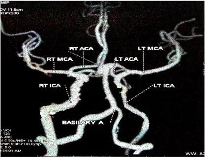
identification,the Circle of Willis is divided into anterior and posterior configuration(fig 6).
The following parameters of Circle of Willis were noted in this study from both Dissection and Radiological methods.
1. Complete or incomplete formation. 2. Shape of the Circle of Willis. 3. Normal or abnormal caliber.
4. Symmetrical or asymmetrical pattern.
5. Morphological variations like absent vessels, attenuation, duplication / triplication and abnormal origin were atudied.
51
OBSERVATION
MORPHOLOGICAL STUDY:
The internal carotid artery bifurcates at the medial end of the lateral sulcus into anterior and middle cerebral arteries. The middle cerebral artery runs in the sylvian fissure while the anterior cerebral artery runs in the longitudinal fissure and are connected by the anterior communicating artery (Vaibhav V et al 2016). The basilar artery which is formed by the union of two vertebral arteries at the pontomedullary junction and bifurcates into two posterior cerebral arteries (Ranganathan T S 3rd
edit). The anterior circulation and the posterior circulation are connected by the posterior communicating artery.
The anterior cerebral artery from the origin to the level until the junction of anterior commnunicating artery is considered circular part of A1 segment. The posterior cerebral artery from its origin from basilar artery until the level of junction of posterior communicating artery is considered circular part of P1 segment (Iman Singh 2014).
52
respectively (Gray’s Anatomy, S.Iqbal,BishwajeetSaikia). Among the 50 brain specimens, the morphology is considered under four parameters.
1. Complete form. 2. Symmetrical. 3. Normal caliber. 4. Polygonal in shape.
Out of 50 specimens, 29 specimens(58%) were found to have complete normal symmetrical configuration, normal caliber and polygonal shape. Other 21(42%) specimens were abnormal either incomplete or asymmetrical or hypoplastic. Abnormalities in the vertebral and basilar artery were not taken into consideration in this study.
Anomalous circle:
The present study shows 21(42%) anomalous circle and are described below.
I. INCIDENCE OF ABSENCE VESSELS:
53
specimen. The absence of anterior communicating artery was not encountered in the study (Table 1).
Name of the vessel No. of absent
vessel Percentage
Anterior cerebral artery(A1)
- -
Anterior
communicating artery
- -
Posterior cerebral artery(P1)
- -
Posterior
communicating artery
1 4.7
54
Fig 7: Absent posterior communicating artery.
II. INCIDENCE OF HYPOPLASTIC VESSELS:
Cerebral arteries of less than 1mm in external diameter while less than 0.5mm in diameter for communicating artery were considered hypoplastic (Gray’s Anatomy, S.Iqbal, Bishwajeet
Saikia).The common anomaly encountered is the hypoplasia of one or
55
other components of the Circle of Willis. Posterior communicating artery were found to be more frequent among hypoplastic vessels followed by posterior cerebral artery. The incidence of hypoplasia is depicted in Table 2.
Name of the vessel No. of hypoplastic
vessel
Percentage
Anterior cerebral artery (A1)
1 4.7
Anterior communicating artery
1 4.7
Posterior cerebral artery(P1)
3 14.28
Posterior communicating artery
8 38.09
56
The posterior communicating artery shows high percentage of hypoplasia (38.09%) among all the vessels taken for the study(fig 8).
Fig 8: Showing hypoplasia of Posterior communicating artery. Hypoplasia of Right
57
III. INCIDENCE OF ACCESSORY VESSELS:
The accessory vessels were present in the form of duplication or triplication of one of the component of Circle of Willis. Duplication of component vessels were seen while triplicate vessels were not found in any specimens. Out of 21 variations seen, 5 specimens showed accessory vessels in the form of duplication(fig 9,10). The anterior communicating artery were found to be more among all the vessels(19.04%). No duplication or triplication were seen in posterior part of Circle of Willis (Table 3)
Name of the vessel No. of accessory
vessel Percentage
Anterior cerebral
artery (A1) 1 4.7
Anterior
communicating artery 4 19.04
Posterior cerebral
artery(P1) - -
Posterior
communicating artery - -
58 Fig 9: Accessory Anterior cerebral artery
59
Fig 10:Accessory Anterior communicating artery.
60
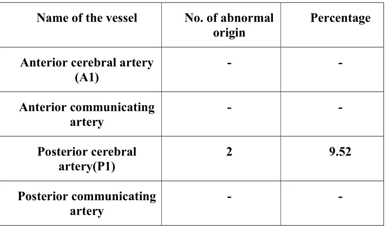
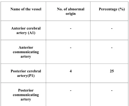
IV. INCIDENCE OF ANOMALOUS ORIGIN:
[image:60.612.128.523.379.611.2]The fetal origin of posterior cerebral artery from the internal carotid artery is common variant in the posterior part of the circle. Here the posterior cerebral artery is small, hypoplastic or even absent and inorder to compensate for the posterior circulation, the posterior cerebral artery is connected by a small communicating type of vessel. In this study, fetal origin of Posterior cerebral artery was found in 2 specimens(9.52%). (Fig 11)
Table 4: showing number of abnormal origin out of 21 specimens.
The other vessels show no abnormality(Table 4)
Name of the vessel No. of abnormal
origin Percentage
Anterior cerebral artery
(A1) - -
Anterior communicating
artery - -
Posterior cerebral
artery(P1) 2 9.52
Posterior communicating
61
Fig 11: Anomalous origin of Posterior cerebral artery
62
V. INCIDENCE OF VARIATION IN ANTERIOR AND POSTERIOR PART OF THE CIRCLE:
No./Percentage Anterior circle Posterior circle
Number 7 14
Percentage 33.33 66.67
63
Fig 12: Pie chart showing graphical representation of incidence of variations of anterior and posterior circle.
Antr circle 34%
Postr circle 66%
64
VI. VARIATION IN INDIVIDUAL VESSELS:
Name of the
vessels Absent vessels Hypoplasia Accesory vessels Anomalous origin Percentage (%)
Anterior cerebral
artery - 1 1 - 9.52
Anterior
communicating Artery
- 1 4 - 23.80
Posterior cerebral
artery - 3 - 2 23.80
Posterior
communicating Artery
1 8 - - 42.86
Table 6: showing overall incidence of variation of individual vessels.
65
Fig 13: Coloumn showing graphical representation of variations of individual vessels.
1
1 1
3 8 1 4 2 0 1 2 3 4 5 6 7 8 9
ACA AcoA PCA PcoA
Aplasia Hypoplasia accesory
66
Name of the variations Percentage of total variations
Absent vessels 4.7
Hypoplasia 61.90
Accesory vessels 23.80
Anomalous origin 9.52
67
Fig 14: Piechart showing percentage of total variations.
ACA 9%
AcoA 24%
PCA 24% PcoA
43%
68
Table 8 : showing percentage among total specimens.
The most common variation noted is the hypoplasia of the cerebral and communicating vessels with a percentage of 61.90%. The next common variation is presence of accessory vessels with 23.80%. Among all the four vessels, Posterior communicating artery were found to have more variations (42.85%). Hypoplasia of posterior communicating artery with vessel diameter less than 0.5mm is 38.09%. followed by posterior cerebral artery with 14.2%.(Table 8).
Name of the variations Percentage for total specimens(50)
Absent vessels 2
Hypoplasia 26
Accesory vessels 10
69
RADIOLOGICAL STUDY
Out of 50 cases, 34 cases were found to have normal, symmetrical and complete form of circle. The remaining 16 cases were found to be abnormal and showing one or more variations in the morphological patterns.
70
ANOMALOUS CIRCLE:
The sixteen specimen with anomalous circle were taken into consideration.
I.INCIDENCE OF ABSENT VESSELS:
In the present study, no case of absent vessels were reported in any of the vessel.
II.INCIDENCE OF HYPOPLASTIC VESSELS:
Name of the vessel No. of hypoplastic vessel Percentage (%)
Anterior cerebral artery
(A1) 7 43.75
Anterior
communicating artery - -
Posterior cerebral
artery(P1) 4 25
Posterior
communicating artery 1 6.25
71
72
73
The hypoplasia was found to be common in anterior cerebral artery in 7 cases with a percentage of 43.75%(fig 16, 17). The posterior cerebral artery was hypoplastic in 4 cases with a percentage of 25% and posterior communicating artery with one case with a percentage of 6.25%. The anterior communicating artery does not show any hypoplastic changes in this study.
Fig 18: Piechart showing incidence of hypoplastic vessels in MAGNETIC RESONANCE ANGIOGRAM.
ACA 59%
AcoA 0% PCA 33%
PcoA 8%
[image:73.612.130.536.256.530.2]74
75
III. INCIDENCE OF ACCESSORY VESSELS:
In the present study, no case of absent vessels were reported.
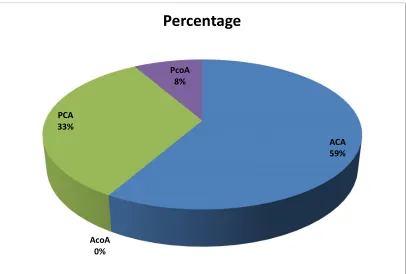
IV. INCIDENCE OF ANOMALOUS ORIGIN:
Name of the vessel No. of abnormal
origin Percentage (%)
Anterior cerebral
artery (A1) - -
Anterior communicating
artery
- -
Posterior cerebral
artery(P1) 4 25
Posterior communicating
artery
- -
[image:75.612.104.536.189.546.2]76
[image:76.612.128.545.185.504.2]The fetal origin of posterior cerebral artery from internal carotid artery was found in 4 cases with a percentage of 25%. The other arteries do not show any abnormal origin.
77
78
[image:78.612.131.521.146.530.2]79
V) INCIDENCE OF VARIATION IN ANTERIOR AND POSTERIOR PART OF THE CIRCLE OF WILLIS:
No./Percentage Anterior circle Posterior circle
Number of cases 7 9
[image:79.612.127.513.398.615.2]Percentage 43.75% 56.25%
Table 11: showing percentage of variation in anterior and posterior part of circle.
Fig 23: Pie chart showing graphical representation of incidence of variations of anterior and posterior circle.
Antr circle 44% Postr.circle
56%
80
VI. VARIATION IN INDIVIDUAL VESSELS:
Name of the vessels Absent vessels Hypoplasia Accesory vessels Anomalous origin
Percentag e (%)
(50 specimens)
Anterior cerebral
artery - 7 - - 43.75
Anterior
communicating Artery
- - - - -
Posterior cerebral
artery - 4 - 4 50
Posterior
communicating Artery
[image:80.612.89.547.145.492.2]- 1 - - 6.25
81
The overall variation in the anterior part of the circle is 43.75% and of the posterior circle is 56.35%. The common variation seen radiologically is hypoplasia of anterior cerebral artery followed by posterior cerebral artery. The anomalous origin of fetal posterior cerebral artery arising from internal carotid artery was seen in 4 cases with a percentage of 25%.(table 12)
Fig 24: Column showing graphical representation of incidence of variations among cerebral and communicating vessels.
7 4 1 4 5 0 1 2 3 4 5 6 7 8
ACA AcoA PCA PcoA
[image:81.612.129.537.258.586.2]82
Name of the variations Percentage of total variations
Absent vessels -
Hypoplasia 75
Accesory vessels -
[image:82.612.126.538.65.276.2]Anomalous origin 25
Table 13: showing percentage of total variations.
Fig 25: Pie chart showing percentage of individual variation in the circle of vessels.
ACA 44%
AcoA 0% PCA
50%
PcoA 6%
[image:82.612.129.535.351.596.2]83
84
DISCUSSION
85
[image:85.612.115.530.64.355.2]Table 14: Comparing the normal and variation percentage with present and other studies.
Fig 26:Line diagram comparing the percentage of variations.
0 10 20 30 40 50 60 70
Column1
% of total variations
Author name Normal pattern(%) Variations(%)
Alpers et al 52.3% 47.7%
Raja Reddy et al 53.3% 46.7%
Kamath S 56% 44%
Stephen and John 52% 48%
Macchi et al 41% 59%
Raghavendra et al 56% 44%
Hartkamp et al 42% 58%
[image:85.612.129.536.448.689.2]86
2. Raghavendra et al studied about Morphometric variation in the Circle of Willis in 50 adult brain specimens. Among the 50 specimens, 28 cases (56%) had a normal pattern of Circle of Willis while the remaining 22 cases(44%) showed variations. The common variation reported is the hypoplastic posterior communicating artery with a percentage of 31.8%. Of the 22 cases, 7 variations were seen in the anterior circulation and 15 variations were seen in the posterior circulation. In this study, variations in anterior circle was found in 7 cases and 14 cases showed variations in posterior circle (Table 15)
Variations Raghavendra et al (percentage) Present study (percentage)
Anterior circle 31.8 33.34
Posterior circle 68.2 66.67
Table no 15: Comparing the present study with that of other study.
Thus the finding of the present study is comparable with that of Raghavendra et al .
87
88
Variations S.Iqbal et al (in percentage)
Present study (in percentage)
Absent vessels 6% 2%
Hypoplasia 24% 26%
Accessory vessels 12% 10%
[image:88.612.164.524.65.293.2]Abnormal origin 10% 4%
Table no 16: Comparing percentage of variations in both studies.
Fig 27: Column comparing percentage of variations between S.Iqbal and present study. S.Iqbal Present study Column1 0 5 10 15 20 25 30 Absent
vessel Hypoplasia Accessory
vessels Abnormalorigin
[image:88.612.129.539.353.599.2]89
4.Hypoplasia or attenuation of either the cerebral or the communicating vessel were found to be common among the variations. Hypoplasia were found in 26% of the present study and were common among all other variation. Alpers et al reported an incidence of 27%, Kamath with an incidence of 24% and Fetterman and Moran with an incidence of 23%. The following table compare the incidence of hypoplasia of the present study with the other studies.
Name of the
author PCOA PCA segment) (P1 ACOA ACA(A1 segment)
Alpers et al 22% - 3% 2%
Riggs &
Rupp 53% 27.7% 9.25% 11.97%
Kamath 10% - 2% 2%
Present study 38.09% 14.2% 4.7% 4.7%
[image:89.612.127.503.287.526.2]90
[image:90.612.128.537.196.435.2]The wide range of variation is due to the different criteria used to measure hypoplastic vessels. Hypoplasia were more prominent in the posterior portions of the Circle of Willis. This finding in the present study is comparable with other studies.
Fig 28: Bar diagram comparing incidence of hypoplasia.
4. Accessory vessels in the form of duplication or triplication were found in the Circle of Willis. Among the accessory vessels, the incidence of duplication of Anterior communicating artery were 19.04% and the anterior cerebral artery were 4.7% in a total of five cases. The accessory vessels were not seen in posterior circulation. Apart from two normal anterior cerebral artery, it is also accompanied by a midline third anterior cerebral artery. This finding were comparable with S.Iqbal et al.
0 10 20 30 40 50 60
PcoA PCA AcoA ACA
91
5. The embryonic origin of the posterior cerebral artery from the internal carotid artery were found in 9.52% of the circle. Such a vessel is connected to the basilar artery by a small communicating artery.
Name of the author Incidence of embryonic origin of PCA from ICA(%)
S.Iqbal 10%
Riggs & Rupp 22%
Alpers et al 15%
Milenkovic et al 21%
[image:91.612.161.527.194.491.2]Present study 9.52%
Table No.18. Comparing the incidence of anomalous origin.
92
Fig 29. Column comparing embryonic origin of posterior cerebral artery.
6. The least common anomaly of the Circle of Willis was the absence of one of the cerebral or the communicating vessels. In the present study, absence of posterior communicating artery were seen in one case with a percentage of 4.7%. The reported incidence of absent vessels in the Circle of Willis in normal brain ranges from 0.6% (Alpers) to 17%(Windle BCA). The absent vessel is not seen in anterior part of the circle. Fawcett and Blachford reported an
Embryonic origin of PCAColumn1 Column2
0 5 10 15 20 25
10 22
15 21
93
incidence of 3.8% of absent vessels of posterior communicating arteries.
7. Cerebrovascular diseases such as stroke, thromboembolism, aneurysms together with their signs and symptoms depend upon the variations of the anatomical pattern of the Circle of Willis. The main collateral blood flow is through the communicating arteries and obstruction of the collateral pathway determines the severity of hemodynamic impairment(Yu-Ming Chuang). The state of circle becomes important in determining the adequacy of the brain circulation.
94
Fig no30: Right side Vertebral artery hypoplasia along with hypoplasia of Right ACA.
[image:94.612.139.510.433.657.2]95
Fig no32: Showing ectasia of left internal carotid artery
Variations Total no.of cases Variations in the Circle of
Willis 16
Vertebral artery
hypoplasia 5
Stenosis/Narrowing of
internal carotid artery 2
Ectasia of internal carotid
artery 1
Vertebrobasilar fenestrations 1
[image:95.612.166.490.386.610.2]96
Fig no33: Radiological image showing vertebrobasilar fenestrations.
97
10. The incidence of normal pattern of Circle of Willis radiologically shows wide variations among different studies. In the present study, out of 50 radiological images, 16 cases showed variation in the pattern of Circle of Willis with an percentage of 32%. The same parameters of the other study were compared in Table no 20.
Study Brains examined Normal pattern(%)
Krabbe-Hartkamp
et al 150 42%
Biswajeethsaikia et
al 70 24.18%
Haripriya and
Melani 50 32%
[image:97.612.126.493.225.469.2]Present study 50 68%
98
Fig 34; Column comparing normal pattern of Circle of Willis.
The prevalence of complete Circle of Willis was seen in 68% of the present study.
Normal patternColumn1 Column2 0
10 20 30 40 50 60 70
99
CONCLUSION
The arterial Circle of Willis was studied and analysed as per the parameters taken by dissection method and from radiological images.
The results were charted out, statistically analyzed and correlated with the finding of the existing studies. The findings are summarized below.
1. The normal parameter like complete formation, shape, normal caliber of vessels and symmetrical Circle of Willis were found in more than 55% of cases.
2. The variations in the Circle of Willis like absent vessels, hypoplasia of vessels and accessory vessels also found.
3. The posterior part of the circle were found to have more variation (66.67%) than the anterior part of the circle (33.34%) involving the posterior cerebral artery and posterior communicating artery.
4. Among the variations found, hypoplasia was the most common variation and hypoplasia was seen more commonly in posterior communicating artery followed by posterior cerebral artery .
100
101
BIBLOGRAPHY
1. A. Prasanna Veera Kumar, K.S.N.Prasad, ‘A Study of Variation of Circle of Willis, in the Adult Population in South India, IJCMR, vol 3, issue 4 May 2016.
2. A.W.J.Hoksbergen, ‘Collateral Configuration of the Circle of Willis’. Vol 31:1346-1351, year 2000.
3. Ace Dodevski , Dobrila TosovskaLazarova , NadicaMitreska , VjolcaAliji , ElizabetaStojovskaJovanovska. Posterior cerebral artery – variation in the origin and clinical significance., Sec. Med. Sci., XXXV 1, 2014.
4. Alma volijevica, Elvira Talovic, Esad Pepic, Amna Pleho Kapic ‘Morphometric analysis of Willis circle arteries’ vol 4(2) pg 77-82, 2013
5. Alpers BJ, Berry RG, Paddison RM. Anatomical studies of the Circle of Willis in normal brain. Arch NeurolPsychiat. 1959;81:409–18.
102
Artery in Human Brain: A morphological study.,J Med Sci 2013; 11:42-6.
7. AyseKaratas, GokmenCoban, CelalCinar, Ismail Oran, and AysunUz. Assessment of the Circle of Willis with Cranial Tomography Angiography.Med Sci Monit. 2015; 21: 2647–2652.
8. Battacharji SK, Hutchinson EC, McCall AJ. The Circle of Willis: The incidence of developmental abnormalities in normal and infracted brains. Brain. 1967;90:747–58.
9. Bernard J. Alpers, Richard G. Berry. Circle of Willis in cerebral vascular disorders -the anatomical structure. Arch Neurol. 1963;8(4):398-402
10. BishwajeetSaikia , Akash Handique , PranjalPhukan , DonboklangLynser , AmitavSarma et al Circle of Willis: Variant forms and their embryology using gross dissection and magnetic resonance angiography.International Journal of Anatomy and Research, vol 2(2):344-353.
103
12. Craig Hacking, Frank Gaillard,’Circle of Willis’-radiopedia
13. Dr. Arjun Bahaddur, Dr. Chandan G. “Anatomical Variants of Circle of Willisin South Indian Population: A Study by Using Magnetic Resonance Angiography” Volume 4 Issue 5, May 2015.
14. Fawcett E, Blachford JV. The Circle of Willis: An examination of 700 specimens. J Anat and Physiol. 1905;40:63–70.
15. Fetterman GH, Moran TJ. Anomalies of the Circle of Willis in relation to cerebral softening. Arch Pathol. 1941;32:251–57.
16. Gardner E, Gray DJ, O'Rahilly R. Anatomy a regional study of human structure. 3rd edition. W.B. Saunders Company; Philadelphia, Toronto, London: 1969; 60- 555.
17. Grand W, Hopkins LN. Variations in Clinical Anatomy. Vasculature of the Brain and Cranial Base. New York: Thieme; 1999.
104
19. H.R.Sharada, K.K.Chaitanya, Anil, R.Sherke, Krishniah, Gayathri, Ashish.k,’Morphometric Analysis and Variations of the Circle of Willis, Sch.J.App.Med.Sci.,2014;2(5E):1831-1834.
20. Haripriya M, Melani RS. “A Study of the Anatomical Variations of the Circle of Willis Using Magnetic Resonance Imaging. International Journal of Anatomical Sciences 2010, 1: 21-25.
21. Hendrikse J, van Raamt AF, van der Graaf Y, Mali WP, van der Grond J: Distribution of cerebral blood flow in the Circle of Willis. Radiology 2005; 235(1): 184-9.
22. Hina Siddiqi, Mohammad Tahir, Khalid Parvez Lone ‘Variations in Cerebral Arterial Circle of Willis in Adult Pakistani Population’ Journal of the college of Physician and Surgeons Pakistan 2013 vol23(9) pg615-619.