Comprehensive Mutational Analysis Reveals p6
Gag
Phosphorylation To
Be Dispensable for HIV-1 Morphogenesis and Replication
Benjamin Radestock,aIvonne Morales,a* Sheikh Abdul Rahman,aSonja Radau,bBärbel Glass,aRené Peiman Zahedi,bBarbara Müller,a Hans-Georg Kräusslicha
Department of Infectious Diseases, Virology, University of Heidelberg, Heidelberg, Germanya
; Leibniz-Institut für Analytische Wissenschaften, Dortmund, Germanyb
The structural polyprotein Gag of human immunodeficiency virus type 1 (HIV-1) is necessary and sufficient for formation of
virus-like particles. Its C-terminal p6 domain harbors short peptide motifs that facilitate virus release from the plasma
mem-brane and mediate incorporation of the viral Vpr protein. p6 has been shown to be the major viral phosphoprotein in
HIV-1-infected cells and virions, but the sites and functional relevance of p6 phosphorylation are not clear. Here, we identified
phos-phorylation of several serine and threonine residues in p6 in purified virus preparations using mass spectrometry. Mutation of
individual candidate phosphoacceptor residues had no detectable effect on virus assembly, release, and infectivity, however,
sug-gesting that phosphorylation of single residues may not be functionally relevant. Therefore, a comprehensive mutational
analy-sis was conducted changing all potentially phosphorylatable amino acids in p6, except for a threonine that is part of an essential
peptide motif. To avoid confounding changes in the overlapping
pol
reading frame, mutagenesis was performed in a provirus
with genetically uncoupled
gag
and
pol
reading frames. An HIV-1 derivative carrying 12 amino acid changes in its p6 region,
abolishing all but one potential phosphoacceptor site, showed no impairment of Gag assembly and virus release and displayed
only very subtle deficiencies in viral infectivity in T-cell lines and primary lymphocytes. All mutations were stable over 2 weeks of
culture in primary cells. Based on these findings, we conclude that phosphorylation of p6 is dispensable for HIV-1 assembly,
re-lease, and infectivity in tissue culture.
T
he final steps in human immunodeficiency virus type 1
(HIV-1) replication are budding and release of viral particles
from the host cell plasma membrane. The viral structural
polypro-tein Gag is sufficient for the production of virus-like particles
in
vitro
(
1
) and in tissue culture (
2
) and is essential for virus particle
formation in infected cells. Gag traffics to the plasma membrane,
where it assembles into a curved lattice and recruits other viral
proteins and genomic RNA. The assembly process culminates in
budding of immature enveloped virions from the plasma
mem-brane carrying a semispherical Gag shell (
3
–
5
). Concomitant with
budding, the viral protease (PR) cleaves Gag into its mature
sub-units, matrix (MA), capsid (CA), nucleocapsid (NC), and p6, as
well as two spacer peptides, SP1 and SP2, leading to maturation of
the infectious virion (
6
).
In the mature particle, MA lines the viral envelope and CA
forms the conical capsid shell surrounding the nucleoprotein
complex, in which NC condenses the viral RNA genome (
6
). The
C-terminal domain of HIV-1 Gag, p6, comprises the “late
do-mains” of the virus, small peptide motifs that interact with
com-ponents of the host cell endosomal complex required for transport
(ESCRT) (reviewed in references
6
,
7
and
8
). Recruitment of a
subset of the ESCRT machinery to the viral budding site mediates
membrane constriction and the abscission of the virus from the
host cell membrane. Late domains have been identified in Gag
proteins of retroviruses and in structural proteins of several other
enveloped viruses (
9
,
10
). In the case of HIV-1, a P(T/S)AP motif
and an LYPX
nLXXL motif interact with the ESCRT-I component
TSG101 and the ESCRT-associated protein ALIX, respectively. In
most cell lines, the P(T/S)AP motif is functionally dominant.
De-letion or mutation of this motif results in the accumulation of late
viral budding structures at the plasma membrane, connected by a
thin membrane tether to the host cell (
11
). Structural analyses of
HIV-1 budding sites and immature virus particles (
4
) and live-cell
imaging of ESCRT recruitment to HIV-1 budding sites (
12
,
13
)
have suggested temporal regulation to coordinate Gag assembly
and the formation of ESCRT complexes during HIV-1 assembly
and budding.
The p6 domain of HIV-1 comprises 52 amino acids; it is largely
unstructured (
14
,
15
) and lacks distinct features besides the highly
conserved late domains and motifs required for incorporation of
the viral accessory protein Vpr (
14
,
16
). However, p6 has been
shown to be posttranslationally modified by
monoubiquitinyla-tion (
17
) and phosphorylation (
18
), and p6 is the predominant
viral phosphoprotein in HIV-1 particles. Both types of
modifica-tion are well known to play regulatory roles, sometimes connected
in a sequential manner (reviewed, e.g., in reference
19
), allowing
their interplay and cross talk (
20
). Late-domain-carrying proteins
of other viruses, e.g., murine leukemia virus (
21
), Mason-Pfizer
monkey virus (
22
), Marburg virus (
23
), and vesicular stomatitis
virus (
24
), are also phosphorylated. Phosphorylation of HIV-1 p6
was found to occur at multiple sites and to involve serine,
threo-nine, and tyrosine residues (
18
); the exact sites of phosphorylation
have not been determined, however.
Received14 August 2012 Accepted18 October 2012
Published ahead of print31 October 2012
Address correspondence to Hans-Georg Kräusslich, Hans-Georg.Kraeusslich @med.uni-heidelberg.de.
* Present address: Ivonne Morales, Program in Physical Biology, NICHD/NIH, Bethesda, Maryland, USA.
Supplemental material for this article may be found athttp://dx.doi.org/10.1128 /JVI.02162-12.
Copyright © 2013, American Society for Microbiology. All Rights Reserved.
doi:10.1128/JVI.02162-12
on November 7, 2019 by guest
http://jvi.asm.org/
Several studies focused on individual potentially
phosphory-lated p6 residues, but the functional role of HIV-1 p6
phosphor-ylation remains largely enigmatic. Residue T23 of p6 can be
phos-phorylated by Erk kinase, and mutation of this residue was
reported to result in a late-domain-deficient phenotype, with viral
buds arrested at the plasma membrane and reduced bulk particle
release (
25
). More recently, Votteler and coworkers (
26
) reported
that an S40F mutation in p6 resulted in aberrant HIV-1 core
for-mation and reduced viral infectivity, while bulk release efficiency
was unaffected. A mechanistic connection between
phosphoryla-tion at either of these sites and HIV-1 release has not been
identi-fied, however. Since 13 of the 52 amino acids of p6 are potentially
phosphorylatable and phosphorylation at several sites has been
detected (
18
), mutational approaches are particularly difficult in
this case. Furthermore, many proteins carry redundant
phosphor-ylation sites (
27
), and mutation of individual residues thus may
not necessarily be conclusive. Finally, the p6 coding sequence
overlaps the
pol
reading frame in the HIV-1 genome, limiting the
options for changing p6 without generating potentially
confound-ing changes in
pol
.
Here, we made use of an HIV-1 variant in which the
gag
and
pol
reading frames were uncoupled by mutagenesis in order to
inves-tigate the role of p6 phosphorylation in infectious HIV-1 particle
formation in a comprehensive manner. Candidate individual
res-idues, multiple resres-idues, or all phosphorylatable residues except
T8, which is part of the PTAP motif essential for p6 function, were
changed to nonphosphorylatable amino acids. The resulting
mu-tant viruses were characterized for Gag phosphorylation status,
particle formation, and infectivity in different cell lines and
pri-mary cells. Unexpectedly, even mutation of almost all
phosphor-ylatable residues had only minor effects on virus release and
infec-tivity, indicating that phosphorylation of p6
Gagis dispensable for
HIV-1 replication.
MATERIALS AND METHODS
Tissue culture and PBMC isolation.HeLa TZM-bI (28), HeLa (29), and human embryonic kidney 293T (30) cells were grown in Dulbecco’s mod-ified Eagle’s medium (Invitrogen). T cells (C8166 [31] and MT-4 [32]) were cultivated in RPMI 1640 (Invitrogen). All media were supplemented with 10% fetal bovine serum (FBS) (Biochrom), 100 units/ml penicillin, 100 g/ml streptomycin (Pen/Strep; Biochrom), and 10 mM HEPES (Sigma-Aldrich). Peripheral blood mononuclear cells (PBMCs) were iso-lated from buffy coats of healthy blood donors by Ficoll density gradient centrifugation following a standard protocol. For each experiment, PBMCs obtained from three different donors were used. Before infection, 4⫻106cells were stimulated by growth in RPMI supplemented with 10g/ml interleukin-2 (IL-2) (Biomol) and 2g/ml phytohemagglutinin (PHA) (Sigma) for 3 days.
Plasmids and transfections.Infectious proviral constructs were based on plasmid pNL4-3 (33). Derivatives of pNL4-3 carrying single point muta-tions were produced by PCR-based site-directed mutagenesis using primers 5=GCCCCACCAGAAGAGGCCTTCAGGTTTGGGGAAG3= (S14A), 5= GGAAGAGACAACAATTCCCTCTCAGCCGCAGG3=(T23I), 5=GAAG AGACAACAACTCCCGCTCAGAAGCAGGAGCCG3=(S25A), and 5=G GCAGCGACCCCTCGTAACAATAAAGATAGGG3=(S51stop).
The plasmid pNL4-3unc, uncoupling the gagandpolopen reading frames (ORFs) of HIV-1, was constructed according to a previous report (34). A silent unique MluI restriction site was introduced into pNL4-3 at nucleotide position 1356 with respect to the start of thegagreading frame. Subsequently, an ApaI-MluI fragment was replaced by a 345-bp synthetic DNA fragment (GeneArt AG, Regensburg, Germany). In this fragment, the original slippery site and the stem-loop structure of the Gag-Pol
frameshift signal were destroyed by point mutations. In addition, a new functional frameshift signal was introduced upstream of thegagstop codon according to the method of Leiherer et al. (34). This mutagenesis procedure resulted in extension of thegagreading frame by 15 bp. Fur-thermore, a unique silent ClaI restriction site was introduced at nucleo-tide position 1225 with respect to the start of thegagreading frame. This resulted in construct pNL4-3uncwild type (wt) carrying three unique re-striction sites (XmaI, ClaI, and MluI) spanning the p6 region (Fig. 1D). Synthetic gene fragments (GeneArt AG, Regensburg, Germany) carrying mutations in potentially phosphorylated residues of p6 (Fig. 1D) were then cloned into pNL4-3uncwt to generate pNL4-3uncNterm (with ex-change of the XmaI-ClaI fragment), pNL4-3uncCterm (ClaI-MluI frag-ment), and pNL4-3uncFL (XmaI-MluI fragment). Detailed cloning pro-tocols and sequences of synthetic DNA fragments are available upon request.
Plasmids expressing enhanced green fluorescent protein (EGFP)-labeled Gag for live-cell imaging were constructed based on pCHIV and pCHIVeGFP(35). pCHIV
uncand pCHIVunceGFPwere constructed by replacing a SwaI fragment in pCHIV or pCHIVeGFPwith the corre-sponding SwaI fragment from pNL4-3unc. To construct pCHIVuncFL and pCHIVunceGFPFL, an ApaI-AgeI fragment comprising thegag re-gion of pNL4-3uncFL was inserted into pCHIVuncand pCHIVunceGFP. Virus production, p24 ELISA, and infectivity assays.293T cells were seeded at a density of 5⫻105/well (6-well plates) or 1.5⫻106/well (10-cm dishes). On the following day, the cells were transfected with 2g (6-well plate) or 10g (10-cm dishes) of the respective plasmid by a standard CaPO4method. At 30 h posttransfection (p.t.), the supernatants were harvested and cleared by centrifugation at 720⫻gand 4°C for 10 min or filtration through 0.45-m-pore-size filters. Virus production was quan-titated by an in-house enzyme-linked immunosorbent assay (ELISA) de-tecting the HIV-1 CA protein p24 (36). To analyze single-round infectiv-ity, 104HeLa TZM-bl indicator cells were incubated with serial 5-fold dilutions of supernatants. At 48 h p.i., luciferase activity in cell lysates was determined using the Steady-Glo Luciferase Assay System (Promega). Relative light units (RLU) were normalized to virus amounts as deter-mined by p24 ELISA. The 50% tissue culture infective dose (TCID50) on C8166 T cells was determined by endpoint titration. For this, 3⫻104 cells/well were seeded in 96-well plates and infected with serial 10-fold dilutions in 6-fold replicates as described by Reed and Muench (37).
For infection experiments, stimulated PBMCs from two different do-nors were mixed 3 days after isolation. Cell mixtures were grown in stim-ulation medium lacking PHA (infection medium). Cells (3⫻105) were seeded in 100l infection medium in 96-well V-bottom plates and in-fected with equal amounts of the respective virus preparations (0.1 ng p24). Starting at 24 h p.i., samples of supernatant (95l) were removed at the indicated time points and replaced by fresh prewarmed infection me-dium. The amounts of p24 in the supernatant samples were quantified by ELISA.
Release assay and Vpr incorporation.Tissue culture supernatants from 293T cells transfected with the respective proviral plasmids were harvested at 30 h p.t. and cleared by filtration through a 0.45-m filter, and particles were collected by ultracentrifugation through a 20% (wt/wt) sucrose cushion, sometimes followed by centrifugation through an Op-tiprep gradient (38). Cell lysates and particles were separated by SDS-polyacrylamide gel electrophoresis (PAGE) and transferred to a nitrocel-lulose membrane. Gag- and Vpr-derived proteins were detected by quantitative immunoblotting (LiCor) using polyclonal sheep antiserum raised against recombinant HIV-1 CA and polyclonal rabbit antiserum raised against synthetic HIV-1 Vpr, as well as secondary antibodies and protocols provided by the manufacturer. Band intensities were quantified using the LiCor Odyssey 3.0 software, and relative release was quantitated by dividing total amounts of CA-reactive bands in the particle fraction by the total CA-reactive bands in cell lysates and particles.
Phosphopeptide mass spectrometry analysis.Virus particles were purified from the tissue culture medium of transfected 293T cells (NL4-3,
on November 7, 2019 by guest
http://jvi.asm.org/
NL4-3unc, and mutants) or infected MT-4 T cells (NL4-3) by centrifuga-tion through a 20% (wt/wt) sucrose cushion, followed by ultracentrifuga-tion through an OptiPrep gradient as described previously (38). Through-out the purification procedure, samples were supplemented with a phosphatase inhibitor cocktail (Phosphatase Inhibitor Cocktail Set V; Calbiochem). For phosphopeptide analysis of NL4-3-derived proteins, the purity of virus preparations was analyzed by SDS-PAGE, followed by silver staining. Virus concentrations were determined by p24 ELISA. Sam-ples corresponding to 4g p24 were separated by SDS-PAGE (NuPAGE Bis-Tris 4 to 12% gel; Invitrogen). The positions of p6-containing bands were identified by parallel immunoblotting with a polyclonal antiserum raised against HIV-1 p6. The corresponding bands were excised from the gel and in-gel digested as described previously (39). For NL4-3uncwt and mutant virus samples, particles were produced from 293T cells trans-fected with the respective proviral plasmids and grown in the presence of the HIV-1 protease inhibitor lopinavir (2M). Purified particles were resuspended in 0.9% NaCl solution supplemented with Halt Phosphatase Inhibitor Cocktail (Thermo Scientific) according to the manufacturer’s instructions. Virus concentrations and purity were analyzed by SDS-PAGE, followed by silver staining. Samples corresponding to 2g Gag were separated by SDS-PAGE (NuPAGE Bis-Tris 4 to 12% gel; Invitro-gen) and processed as described above.
Peptides were extracted with 50l 0.1% trifluoroacetic acid and 50l 0.1% trifluoroacetic acid-acetonitrile (50:50) for 15 min each, and the two extracts were combined. NL4-3-derived phosphopeptides were enriched using titanium dioxide (TiO2) (40). Sample preparation was conducted as described previously (41). Samples were analyzed by nano-liquid chro-matography tandem mass spectrometry (LC–MS-MS) on an LTQ Orbitrap Velos (Thermo Scientific) mass spectrometer coupled to an Ultimate 3000 nano-high-performance liquid chromatograph (HPLC) (Thermo Scientific). Peptides were preconcentrated on a self-packed Syn-ergi HydroRP trapping column (100-m inner diameter; 4-m particle size; 100-Å pore size; 2-cm length) and separated on a self-packed Synergi HydroRP main column (75-m inner diameter; 2.5-m particle size; 100-Å pore size; 30-cm length) at 60°C and a flow rate of 270 nl/min using a binary gradient (A, 0.1% formic acid; B, 0.1% formic acid, 84% aceto-nitrile) ranging from 5% to 50% B in 50 min. MS survey scans were acquired from 350 to 2,000m/zin the Orbitrap at a resolution of 60,000 using the polysiloxanem/z445.120030 as lock mass (42). The five most intense signals were subjected to collision-induced dissociation (CID)-based MS-MS in the LTQ using a normalized collision energy of 35%, an activation time of 30 ms, and a dynamic exclusion of 10 s. Automatic gain control (AGC) values were set to 106for MS and 104for MS-MS scans.
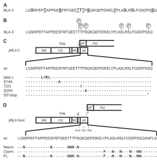
Raw data were processed using the Proteome Discoverer software V1.3 FIG 1Schematic representation of potential p6 phosphorylation sites and p6 sequences of mutated viruses used in this study. (A) Amino acid sequence of HIV-1NL4-3p6. Conserved residues among HIV-1 group M isolates (see Fig. S1 in the supplemental material) are shown in boldface. Residues that were predicted
to be phosphorylated by the NetPhos algorithm (http://www.cbs.dtu.dk/services/NetPhos/) are underlined (see Table S1 in the supplemental material). (B) Phosphorylated residues within NL4-3 p6 isolated from viral particles. The circled P symbols indicate residues in p6 that we identified by mass spectrometry as phosphorylated in virion-associated Gag products (see Table S2 in the supplemental material). (C) Scheme of thegagandpolORFs in the HIV-1NL4-3genome.
The arrow indicates the position of the Gag-Pol frameshift signal. Amino acid sequences of wt p6 and mutant derivatives used in this study are shown; mutated residues are indicated. (D) Scheme of thegagandpolORFs in the HIV-1NL4-3uncgenome. The arrow with an asterisk indicates the newly introduced frameshift
signal at the 3=end ofgag; the arrow with the cross indicates the mutated authentic frameshift signal. Restriction sites for XmaI, ClaI, and MluI were introduced to facilitate exchange of p6-encoding fragments. Amino acid sequences of the altered p6 and sequences of the mutated NL4-3uncvariants are shown below the
scheme; mutated residues are indicated.
Radestock et al.
on November 7, 2019 by guest
http://jvi.asm.org/
[image:3.585.129.448.62.387.2](Thermo Scientific) and searched against a Swiss-Prot virus database (14,986 target sequences; 8 February 2011) using Mascot (version 2.3.2) and Sequest with the following settings: 10-ppm MS tolerance, 0.4-Da MS-MS tolerance, trypsin as the enzyme allowing a maximum of 2 missed cleavage sites, and carbamidomethylation of cysteine as fixed modifica-tions and oxidation of methionine, as well as phosphorylation of S/T/Y, as variable modifications. Peptide Validator and phosphoRS (43) nodes were used to assess global false-discovery rates and phosphorylation site localization probabilities, respectively. The results were filtered for mass deviations ofⱕ4 ppm and high confidence (corresponding to a false-discovery rate of⬍1% with Peptide Validator).
Microscopy and single-virus tracking.HeLa cells were seeded at 1.5⫻ 104cells/well into 8-well glass bottom chamber slides (LabTek; Nunc). After 24 h of incubation at 37°C, the cells were transfected with equimolar amounts (500 ng DNA/well) of either pCHIVuncand pCHIVunceGFP, or pCHIVuncFL and pCHIVunceGFPFL, respectively, using Fugene 6 (Roche) according to the manufacturer’s instructions. At 20 h p.t., the cells were transferred into imag-ing buffer (25 mM Na-HEPES [pH 7.4], 137 mM NaCl, 2.7 mM KCl, 1 mM CaCl2, and 30 mM glucose), and live-cell total internal reflection fluorescence microscopy (TIR-FM) was performed.
The TIR-FM setup (Visitron) used here is based on a Zeiss Axiovert 200 M fluorescence microscope equipped with an alpha plan Fluar 100⫻/ 1.45 oil immersion objective and an electron-multiplying charge-cou-pled-device (EM-CCD) camera (Cascade II; Roper Scientific; 512 by 512 pixels). The details of the setup have been described previously (35). Im-age acquisition was performed using Metamorph V6.3r7. ImIm-age analysis, identification of individual assembly sites, and single-virus tracing were performed using an automated particle-tracking algorithm as described previously (44,45). Changes in GageGFPfluorescence intensity at individ-ual HIV-1 assembly sites were analyzed over time, and exponential assem-bly phases from multiple individual sites were aligned and averaged as described previously (45). The data were fitted to a saturating exponential function using GraphPad Prism 5 to determine average rate constants of HIV-1 assembly.
RESULTS
Prediction of potentially phosphorylated amino acids in HIV-1
p6.
To test whether phosphorylation of p6 is functionally relevant
for HIV-1 replication, we initially wanted to analyze the
conse-quences of mutating individual phosphorylatable residues within
p6 for viral replication. Phosphoamino acid analysis had revealed
that p6 is phosphorylated at multiple sites and that serine,
threo-nine, and tyrosine residues can be phosphorylated (
18
). Since 13
of the 52 amino acids of HIV-1
NL4-3p6 are potentially
phosphor-ylatable (
Fig. 1A
), we first selected appropriate candidate residues
for individual mutational analysis. Reasoning that functionally
relevant candidates should be (i) highly conserved, (ii) recognized
by cellular kinases, and (iii) detectably phosphorylated in HIV-1
p6, we first checked for conservation among different subtypes of
HIV-1 type M based on Gag sequence data obtained from the Los
Alamos database (
http://www.hiv.lanl.gov/
). As shown in
Fig. 1A
and Fig. S1 in the supplemental material, most phosphorylatable
residues in the p6 region are highly conserved among the subtype
consensus sequences. Second, we performed a search using the
NetPhos phosphorylation site prediction program (
http://www
.cbs.dtu.dk/services/NetPhos/
) to identify residues predicted to be
phosphorylated by known kinases based on their primary
se-quence context (
46
).
Figure 1A
summarizes the residues
consid-ered most likely to be phosphorylated based on the ranking by the
NetPhos algorithm (see Table S1 in the supplemental material).
This comprises residues T8, S14, T21, T23, S25, Y36, and S51.
Finally, we isolated p6-containing proteins from highly purified
HIV-1 particles to directly determine which residues were actually
phosphorylated. Tryptic fragments were analyzed by LC–MS-MS
following enrichment of phosphopeptides by titanium dioxide
af-finity chromatography (see Table S2 in the supplemental
mate-rial). Residues T22/23, S25, S40, S43, and S51 were repeatedly
detected as being phosphorylated in p6 or intermediate p6
pro-cessing products in virus preparations from transfected 293T or
infected MT-4 cells (
Fig. 1B
; see Table S2 in the supplemental
material). Only a subset of these residues was found to be
phos-phorylated in each individual sample, however (see Table S2 in the
supplemental material). Whether the different patterns observed
reflect cell-type-specific differences, dynamic changes, or simply
subtle technical differences in sample preparation and analysis
remains to be determined.
Effects of mutation of individual p6 residues.
Based on the
data summarized in
Fig. 1A
and
B
(see Figure S1 and Tables S1 and
S2 in the supplemental material), we selected residues S14, T23,
S25, and S51 for mutagenesis. Residue S40 was omitted because a
detailed analysis of an HIV-1 p6 mutant, S40F, had recently been
performed (
26
). Residue T8 was not included because it is part of
the essential PTAP motif (
11
), and residue S43 was not included
because an S43A mutation had no apparent effect on HIV-1
re-lease (
16
). The respective constructs are summarized in
Fig. 1C
;
S14 and S25 were changed to alanine, T23 to isoleucine, and S51 to
a stop codon. The last two mutations retained the amino acid
sequence in the overlapping
pol
reading frame, while the S14A and
S25A changes led to alterations from glutamate to glycine and
from leucine to arginine in residues 30 and 41 of Pol, respectively.
An HIV-1 variant in which the PTAP sequence was mutated to
LIRL [“late(
⫺
)”] in order to block recruitment of ESCRT-I (
47
)
was included as a control for impaired p6 function in HIV-1
par-ticle release (
Fig. 1C
).
293T cells were transfected with pNL4-3 or the mutated
pro-viral plasmids. Cell lysates and particles were harvested 30 h p.t.
and analyzed by quantitative immunoblotting using antiserum
against HIV-1 CA (
Fig. 2A
). The wt samples showed the expected
processing pattern, with mainly Gag and intermediate processing
products detected in the cell lysates and predominantly mature
CA in the particle fractions. The late(
⫺
) control virus displayed
reduced release efficiency. It also showed the increased amount of
the intermediate CA processing product CA-SP1 typically
ob-served upon impairment of p6 function (
47
). In contrast,
muta-tions at individual phosphorylatable residues in p6 did not result
in detectable alterations of Gag processing or particle release
com-pared to the wt. This visual impression was confirmed by
quanti-tative analysis of four individual experiments. With the exception
of the late(
⫺
) control, the relative release efficiencies of the
vari-ants did not significantly differ from that of wt virus (
Fig. 2B
). The
single-round infectivity of the released particles (normalized for
particulate antigen) was tested by titration on HeLa TZM-bl
indi-cator cells, which express luciferase driven by an HIV-1
Tat-de-pendent long terminal repeat (LTR) promoter. As shown in
Fig.
2C
, none of the p6 variants except the late(
⫺
) control showed a
significant decrease in relative infectivity compared to NL4-3 wt.
The late(
⫺
) virus was strongly reduced in relative infectivity, in
agreement with the observed impairment of proteolytic Gag
mat-uration.
Comprehensive mutational analysis of p6 phosphorylation
sites.
The results described so far demonstrate that
phosphoryla-tion of p6 at individual residue S14, S25, T23, or S51 is not
re-quired for HIV-1 particle release or infectivity. Conceivably,
on November 7, 2019 by guest
http://jvi.asm.org/
phorylation at residues not included in our analysis may still be
crucial, however. In addition, redundant phosphorylation may
occur at one or several possible acceptor sites, which cannot be
determined by mutagenesis of individual sites. We therefore
em-barked on a comprehensive mutational analysis of all
phosphor-ylatable residues in HIV-1 p6. Mutations in
p6
also affect the
se-quence of the overlapping
p6
*
pol
region and may, in addition,
influence frameshifting efficiency, thereby confounding the
inter-pretation of results. To address this problem, we made use of a
recently described HIV-1 derivative in which the
gag
and
pol
read-ing frames are uncoupled by mutagenesis of the authentic
frame-shift signal in conjunction with a short sequence duplication
in-troducing a functional frameshift signal directly upstream of the
gag
stop codon (
34
). In this context, mutations within
p6
do not
affect the primary sequence of Pol. Applying this strategy, we
gen-erated a
gag-pol
uncoupled variant of pNL4-3 (pNL4-3
unc)
(
Fig. 1D
). Silent unique restriction sites were introduced to
facil-itate the exchange of p6-encoding fragments.
Virus release and Gag proteolytic maturation of HIV-1
NL4-3uncwere compared to those of wt HIV-1 by transfecting 293T cells
with pNL4-3
uncand pNL4-3, followed by quantitative
immuno-blot analysis of cell lysates and viral particles. A slightly lower
electrophoretic mobility of Gag indicated the presence of five
ad-ditional amino acids at the C terminus of Gag of NL4-3
unc, while
Gag processing was not impaired by the introduced mutations
(
Fig. 3A
). Quantitative evaluation of immunoblot data confirmed
that there was no significant difference in relative virus release
between NL4-3 and NL4-3
unc(
Fig. 3B
). Viral infectivity was tested
by single-round infection of TZM-bl indicator cells (
Fig. 3C
) and
by endpoint titration on C8166 T cells (
Fig. 3D
). In accordance
with previous results (
34
), we observed a tendency toward slightly
reduced infectivity of NL4-3
uncin both assays, but the observed
reduction was not statistically significant.
We subsequently constructed a derivative of pNL4-3
unccarry-ing mutations in all possible phosphorylation sites in p6, with the
exception of T8, which is part of the essential PTAP late-domain
motif (termed pNL4-3
uncFL) (
Fig. 1D
). The remaining 11 serine
and threonine residues were changed to asparagine, and the single
tyrosine residue was changed to phenylalanine. These
conserva-tive exchanges were chosen to retain the chemical properties of the
original residues as much as possible. Besides an exchange of
phosphorylatable residues in the entire p6 domain, two additional
constructs were made, in which either six N-terminal or six
C-ter-minal phosphorylatable residues were exchanged (pNL4-3
uncNterm and pNL4-3
uncCterm, respectively) (
Fig. 1D
). The
respec-tive proviral plasmids were transfected into 293T cells, and cell
lysates and viral particles were collected and analyzed.
Phosphor-ylation of Gag proteins was determined by LC–MS-MS analysis of
Gag isolated from highly purified immature HIV-1
NL4-3unc(wt,
Nterm, Cterm, and FL). These analyses revealed no obvious
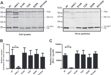
FIG 2Effects of single mutations in p6 on Gag processing, viral release, and infectivity. (A) Gag processing and particle release efficiency. Virus particles were prepared by ultracentrifugation from the tissue culture supernatant of 293T cells transfected with the indicated proviral plasmids. Cell lysates (left) and virus particles (right) were analyzed for Gag-derived products by quantitative immunoblotting (LiCor) using antiserum raised against recombinant HIV-1 CA. The positions of molecular mass standards are shown on the left, and Gag-derived proteins are indicated on the right. (B) Quantitative immunoblotting of cell lysates and particles shown in panel A was used to calculate the amount of CA released, normalized to total anti-CA reactive proteins as described in Materials and Methods. Mean values and standard deviations (SD) from four independent experiments are shown. (C) Single-round virus infectivity. 293T cells were transfected with the indicated pNL4-3 derivatives. Supernatants were harvested at 30 h p.t., and infectivity was quantitated by titration on TZM-bl reporter cells, as described in Materials and Methods. Values were normalized for virus amounts as determined by p24 ELISA. The mean values and SD from four independent experiments are shown.Pvalues in panels B and C were calculated using an unpaired Student’sttest (**,P⬍0.01; ***,P⬍0.001). Comparison of S14A, T23I, S25A, and S51stop to the wt yielded nonsignificantPvalues.
Radestock et al.
on November 7, 2019 by guest
http://jvi.asm.org/
[image:5.585.99.486.66.328.2]change in the phosphorylation status of the MA and CA domains
upon mutation of phosphorylatable residues within p6.
Phos-phorylation in the p6 domain itself was completely lost in the case
of the FL mutant, however. Consistent with the respective
muta-tions, the Nterm mutant virus retained only phosphorylation of
p6 residue S40 (close to the C terminus of p6), and the Cterm
variant retained phosphorylation at T22/23 and S25 (in the
N-ter-minal part of p6). No new phosphorylation sites within p6 were
detected in any of the variant Gag proteins, suggesting that
re-moval of phosphorylatable residues did not result in
compensa-tory phosphorylation at other sites.
Inspection of Gag processing patterns on immunoblots of cell
lysates and particles (
Fig. 4A
) showed no detectable processing
impairment for any of the variants in the particle fraction. A subtle
increase in the relative amount of CA-SP1 was observed in cells
expressing the Cterm and FL variants, while processing was
com-pletely unaffected for the Nterm variant (
Fig. 4A
). Particle release
was unaffected for all three variants compared to the wt (
Fig. 4B
).
Since the p6 domain mediates incorporation of the viral protein R
(Vpr) into the virion (
48
), we examined whether the extensive
mutations affected Vpr particle incorporation. As shown in
Fig. 4C
, Vpr incorporation was retained in all three variants.
Comparison of relative Vpr incorporation (
Fig. 4D
) revealed an
⬃
2-fold reduction of Vpr in the Nterm and FL viruses compared
to the wt. No difference in Vpr incorporation was observed
be-tween the wt and the Cterm variant despite mutation of three
serine residues (at positions 40, 43, and 47) that flank, or are part
of, the LRSLFG Vpr incorporation motif (
16
) in p6. These results
suggest that other regions of p6 outside the known incorporation
motif may also affect Vpr uptake by HIV-1.
To assess potential differences in viral assembly kinetics,
live-cell microscopy was performed for wt and FL virus. These
exper-iments made use of a previously established system where Gag is
labeled by insertion of EGFP between the MA and CA domains of
the protein (resulting in plasmid pCHIV
eGFP[
35
]). This construct
leads to the formation of fluorescently labeled viral particles with
wt efficiency and wt particle morphology if cotransfected together
with an equimolar amount of its unlabeled counterpart. Assembly
of individual virus particles can then be directly observed by
live-cell TIR-FM at the membranes of virus-expressing live-cells (
45
). The
gag-pol
regions of pNL4-3
uncand pNL4-3
uncFL were transferred
into the context of pCHIV
eGFP, and Gag assembly kinetics were
investigated in HeLa cells transfected with the respective
con-structs (
Fig. 5
; see Movies S1 and S2 in the supplemental material).
As previously observed for HIV
eGFP, cells expressing HIV
unceGFP
or HIV
unceGFPFL displayed a gradual accumulation of punctate
assembly sites at the plasma membrane over a period of 1 to 2 h
(
Fig. 5A
and Movies S1 and S2). Detailed analysis of assembly
kinetics over the exponential phase of Gag accumulation (
Fig.
5B
) yielded average rate constants of 0.0057
⫾
0.0035 s
⫺1for
HIV
unceGFPand 0.0045
⫾
0.0025 s
⫺1for HIV
unceGFPFL,
respec-tively. These rates are similar to the rate constant of 0.0043
⫾
0.00054 s
⫺1previously determined for HIV
eGFP(
45
). There was
no significant difference between the constructs carrying wt p6
or its FL variant, indicating that alteration of all but one
phos-phorylatable residue in p6 did not alter HIV-1 Gag assembly
rates. Of note, a variant with a mutation of T8 in the context of
a PTAP late-domain change to LIRL had been analyzed in a
previous study, which showed no alteration of Gag assembly
rates (
45
).
We proceeded to test the infectivity of the variants on TZM-bl
(
Fig. 6A
) and C8166 (
Fig. 6B
) cells. There was no reduction in
infectivity for NL4-3
uncNterm compared to wt NL4-3
uncin either
of these cell lines. A slight reduction was observed for NL4-3
uncFL
and NL4-3
uncCterm compared to wt NL4-3
uncin single-round
infectivity on TZM-bl cells. Subtle decreases in the infectious titers
of these variants on C8166 cells were not statistically significant,
however (
Fig. 6A
and
B
). Finally, the effect of the
p6
alterations on
HIV-1 replication capacity was assessed in primary T cells. Virus
par-ticles produced from transfected 293T cells were used to infect
hu-man PBMCs, and viral spread was monitored over a period of 13 days
by quantitation of HIV-1 antigen released into the culture medium.
FIG 3Effect of uncoupling the HIV-1gagandpolreading frames on Gag processing, viral release, and infectivity. (A) Gag processing and release. 293T cells were transfected with the indicated proviral plasmids. At 30 h p.t., cell lysates (left) and tissue culture supernatants (right) were harvested, and virus particles were prepared by ultracentrifugation from the supernatant. Samples were separated by SDS-PAGE, and Gag-derived proteins were detected by quantitative immu-noblotting (LiCor) using polyclonal antiserum raised against recombinant HIV-1 CA. The positions of molecular mass standards are shown on the left, and Gag-derived proteins are indicated on the right. (B) Results from the quantitative immunoblot analysis shown in panel A were used to calculate relative release efficiencies, as described in Materials and Methods. Mean values and SD from five independent experiments are shown. (C and D) Relative infectivities of tissue culture supernatants harvested from 293T cells transfected with the indicated proviral plasmids. (C) Single-round infectivity was determined by titration of virus on TZM-bl cells, as forFig. 2C. (D) Infectivity on C8166 T cells was determined by endpoint titration, as described in Materials and Methods. The values were normalized for the amount of virus, as determined by p24 ELISA. The graphs show means and SD of at least three independent experiments. (B to D) Nonsignificance (n.s.) was determined by an unpaired Student’sttest.
on November 7, 2019 by guest
http://jvi.asm.org/
[image:6.585.96.491.66.209.2]Figure 6C
shows the results of parallel infections of PBMCs from
three blood donors (always used as mixtures of PBMCs from two
donors) with three independent virus preparations of NL4-3
uncand
the three p6 variants. NL4-3
uncFL, as well as the variants with
altera-tions in either half of p6, replicated efficiently in PBMCs, reaching
titers similar to those of the NL4-3
unccontrol at day 7 p.i. Small but
significant reductions in titer compared to the NL4-3
unccontrol were
observed for variants NL4-3
uncCterm and NL4-3
uncFL at earlier time
points (day 3 or 5) in cultures containing PBMCs from donor B,
suggesting that these two variants may display a very subtle
donor-dependent delay in replication in primary cells. To determine
whether reversion of the introduced mutation(s) or recombination
of the short duplicated sequence occurred upon prolonged passage,
we analyzed the relevant region of the viral genomic RNA in tissue
culture media from infected PBMCs. RNA was extracted from the
culture medium collected from day 1 to day 13 p.i. and subjected to
reverse transcriptase PCR. No recombination products were detected
up to the peak of viral replication at day 7 p.i. (see Fig. S2 in the
supplemental material). Sequencing of the predominant band from
the sample taken at day 13 p.i. confirmed that all mutations
intro-duced into the
p6
region were still present. We thus conclude that the
growth properties determined in this study reflect the replication
phenotype of the respective mutated virus.
DISCUSSION
Based on our previous observation that p6 is the major
phospho-peptide of HIV-1 and that it can be phosphorylated at multiple
sites (
18
), we have identified several residues that are
phosphory-lated in virion-associated p6 by mass spectrometry. Individual
mutation of selected residues had no effect on virus assembly,
release, and infectivity, suggesting that phosphorylation of any
one of these residues may not be important. Phosphorylation sites
are often redundant (
27
), however, and alteration of several
po-tential phosphoacceptor sites may be required to reveal a popo-tential
phenotype. We therefore performed a comprehensive mutational
analysis by altering all potentially phosphorylatable residues in p6.
The only exception was residue T8, which was retained in all
con-structs as part of the functionally essential PTAP late-domain
mo-tif. Unexpectedly, changing 11 serine and threonine residues to
asparagine and the single tyrosine residue to phenylalanine had no
effect on virus release and only very minor effects on HIV-1
infec-tivity in two different cell lines and in primary cells. Passage of the
FIG 4Effect of multiple mutations in p6 on Gag processing, viral release, and Vpr incorporation into viral particles. (A) Gag processing and release efficacy. Virions were purified from tissue culture supernatants of 293T cells transfected with the indicated proviral plasmids by ultracentrifugation through a sucrose cushion. Samples were separated by SDS-PAGE, and Gag-derived products from cell lysates (left) or virus pellets (right) were analyzed by quantitative immunoblotting (LiCor) using antiserum raised against recombinant HIV-1 CA. The positions of molecular mass standards are shown on the left, and Gag-derived proteins are indicated on the right. (B) Relative particle release efficiencies. Quantitative immunoblots shown in panel A were used to determine the amounts of CA released, normalized to total anti-CA reactive proteins expressed, as described in Materials and Methods. The graph shows mean values and SD from five independent experiments. (C) Vpr incorporation. Virions were purified from tissue culture supernatants of 293T cells transfected with the respective plasmids and cultured in the presence of 2M lopinavir by ultracentrifugation through an OptiPrep gradient. Samples were separated by SDS-PAGE, and viral particles were analyzed by quantitative immunoblotting (LiCor) using the indicated antisera. The positions of molecular mass standards are shown on the left, and proteins are indicated on the right. (D) Quantification of Vpr incorporation. The relative ratio of Vpr to Gag was calculated based on integrated band intensities from quantitative immunoblots, as shown in panel C. The graph shows mean values and SD from three independent experiments. The values were normalized to the ratio determined for the respective wt control.
Radestock et al.
on November 7, 2019 by guest
http://jvi.asm.org/
[image:7.585.80.503.64.352.2]NL4-3
uncFL virus in PBMCs for
⬃
2 weeks did not lead to a
rever-sion of any of the introduced p6 mutations. Phosphopeptide
anal-ysis of mutant viruses confirmed the loss of phosphorylation in
the altered regions of p6 and yielded no evidence for
compensa-tory phosphorylation at other sites within Gag. We thus conclude
that the observed phosphorylation of HIV-1 p6 in infected cells
and virions is dispensable for HIV-1 particle formation,
matura-tion, and infectivity in tissue culture. We cannot formally exclude
the functional importance of a potential phosphorylation of p6
residue T8, which was not mutated in this study. However, our
mass spectrometry analyses of Gag phosphopeptides provided no
evidence for phosphorylation at the site.
Previous reports have described a phenotype for changes of
potential phosphorylation sites in HIV-1 p6, suggesting an
impor-tant contribution of p6 phosphorylation to HIV-1 replication in
tissue culture. This included residue T23, which was reported to
be phosphorylated by virion-associated Erk-2 kinase (
25
);
phos-phorylation of T23 was confirmed by our mass spectroscopy
anal-ysis. Hemonnot et al. reported that a T23A alteration impaired
virus release efficiency and Gag processing. Late viral budding
structures arrested at the plasma membrane were observed by
electron microscopy, reminiscent of the phenotype of known
ESCRT recruitment-deficient HIV-1 variants. Furthermore,
sin-gle-round virus infectivity was reported to be reduced (
25
). In
contrast, we did not observe a comparable phenotype for an
indi-vidual T23I mutation or for the change of all phosphorylatable
residues in the p6 N-terminal moiety (including T23) to
aspara-gine residues. No defect in virus assembly, release, Gag
polypro-tein processing, or infectivity was detected for either of these
vari-ants compared to wt HIV-1. The previously analyzed T23A
variant may have different properties from the T23I and T23N
variants analyzed here. Since none of the residues chosen to
re-place T23 could be phosphorylated, the potential differences are
clearly not related to phosphorylation, however. Phosphorylation
at T23 may thus reflect a bystander effect caused by recruitment of
Erk-2 to the viral assembly site. Alternatively, phosphorylation of
this and/or other p6 residues may be important under specific
conditions not reflected in common tissue culture analyses (e.g.,
cell-to-cell transmission or infection of special cell types), and this
will be the topic of future studies.
Votteler et al. (
26
) reported a processing and infectivity
phe-notype for an S40F variant of p6. This nonconservative mutation
was chosen to avoid potentially confounding changes in the
over-lapping
pol
region. The authors observed that this mutation
re-sulted in decreased infectivity of viral particles in single-round
infection and decreased replication efficiency in tissue culture.
This was accompanied by morphological aberrations of the
vari-ant particles. A defect in proteolytic maturation of CA correlated
with impaired formation of the mature conical core. In the
cur-rent study, we showed that S40 of p6 can indeed be
phosphory-lated. Based on the results presented here, we suggest, however,
that the pronounced phenotype reported for the S40F variant may
FIG 5Effect of HIV-1 Gag p6 full-length phosphorylation mutation on HIV-1 assembly kinetics. HeLa cells were cotransfected with an equimolar ratio of either pCHIVunc/pCHIVunc
eGFP(A, top row) or pCHIV
uncFL/pCHIVunc
eGFPFL (A, bottom row). TIR-FM imaging was initiated at 12 h p.t. Cells expressing detectable
amounts of GageGFPthat displayed only a low number of budding sites, detected as individual punctae at the plasma membrane, were selected for acquisition of
image series with a time resolution of 2 s per frame. Shown are selected frames from Movies S1 (top) and S2 (bottom) in the supplemental material corresponding to the indicated time points. The scale bars represent 10m. (B and C) Average Gag assembly rates. Individual assembly sites from 50 and 52 different cells expressing HIVunc
eGFP(B) or HIV unc
eGFPFL (C), respectively, were tracked, and exponential assembly phases were averaged as described in Materials and
Methods. The graphs show average intensities (black line) and SD (gray bars) from the indicated number (n) of individual sites. Assembly rate constants were determined by fitting the data to a saturating single exponential function (white line) using GraphPad Prism 5, yielding the indicated rate constants,k. rel.,
relative.
on November 7, 2019 by guest
http://jvi.asm.org/
[image:8.585.89.504.64.322.2]be largely due to the very different chemical nature of the
intro-duced phenylalanine residue. Exchange of all phosphorylatable
residues in the C-terminal part of p6 (including S40) had only a
minor effect on viral infectivity and CA processing. It is
conceiv-able that the minor effects observed for the Cterm and FL
con-structs are due to the alteration of S40. Determining the relative
contribution of any individual mutation would be difficult,
how-ever, given that the combination of six point mutations in this
region displayed only a subtle effect on the viral phenotype.
The finding that mutating approximately one-fifth of all p6
residues did not affect virus assembly kinetics or release and had
only a subtle effect on processing and infectivity was unexpected.
Since several of these residues are highly conserved among all
HIV-1 clades, we anticipated the FL variant would be
noninfec-tious, requiring restoration of one or several essential residues to
regain infectivity. However, sequence analysis of the HIV-1
uncFL
genome after 2 weeks of passage in tissue culture revealed no
re-version of mutated residues. On the other hand, HIV-1 genomes
undergo constant sequence alteration during viral replication
in
vivo
, and tolerance of a large proportion of amino acid exchanges
within an HIV-1 protein is not without precedent. Various studies
have shown accumulation of a high number of mutations in
HIV-1 occurring under selective drug pressure. For example,
HIV-1 isolated from a patient under antiretroviral therapy was
found to have acquired 19 nonsynonymous mutations in
PR
(cor-responding to one-fifth of the PR primary sequence) without
sig-nificantly lower viral titers (
49
). Conservation of p6 residues may
also be caused, at least in part, by requirements imposed by the
nearby frameshift signal and the overlapping
pol
reading frame.
This makes it difficult to draw firm conclusions from the fact that
a specific residue is conserved. The p6 domain appears to be
largely unstructured, with only a weak helical propensity (
14
,
15
).
This feature is common for proteins that, like p6, serve as
molec-ular adapters for several partners. Such flexibility may also
in-crease mutational tolerance, as long as the overall hydrophilic
pro-file is preserved. It is currently unclear whether p6 comprises any
additional functional regions besides the small peptide motifs
identified as binding Tsg101, ALIX, or Vpr. The experimental
ap-proach described here allows testing of this question by
conserva-tive mutagenesis of the entire p6 domain, except for its known
functional modules. If a general high sequence flexibility of p6
could be confirmed, it would open the possibility of inserting
lin-ear peptide motifs with specific functional properties, thereby
en-dowing lentiviral vectors with selected features.
ACKNOWLEDGMENTS
This work was supported in part by the Deutsche Forschungsgemein-schaft through the collaborative research grant SFB638 (project A9) and the Ministerium für Innovation, Wissenschaft und Forschung des Landes Nordrhein-Westfalen. H.-G.K. is an investigator of the excellence cluster CellNetworks (EXC81).
We thank Anke-Mareil Heuser and Maria Anders-O¨ sswein for expert technical assistance.
FIG 6Effects of multiple mutations in p6 on viral infectivity and replication kinetics in primary cells. Viral particles were harvested from tissue culture supernatants of 293T cells transfected with plasmid pNL4-3uncor its indicated derivatives and were tested for replication competence in cell lines and primary T
cells. (A and B) Relative infectivities in cell lines. (A) Single-round infectivity was assessed by titration of viral particles on TZM-bl cells as described in Materials and Methods. (B) Relative infectious titers on C8166 T cells were determined by endpoint titration as described in Materials and Methods. The values were normalized for virus amounts as determined by p24 ELISA. The graphs show means and SD of three independent experiments, normalized to the respective HIV-1NL4-3uncwt control. (A and B)Pvalues were calculated using an unpaired Student’sttest (n.s.,P⬎0.05; *,P⬍0.05; **,P⬍0.01). (C) Replication kinetics
in primary cells. PBMCs were isolated using buffy coats from healthy blood donors and stimulated for 3 days using PHA and IL-2. Subsequently, samples from two individual donors each were mixed, and virus was added (0.1 ng p24/3⫻105cells). Samples of tissue culture supernatants were collected at the indicated time
points, and virus production was measured by p24 ELISA. The data shown are mean values and SD from three independent experiments using independent virus preparations. In each experiment, three different combinations of PBMCs (donors A plus B, B plus C, and A plus C) were tested.
Radestock et al.
on November 7, 2019 by guest
http://jvi.asm.org/
[image:9.585.112.475.65.306.2]REFERENCES
1.Morikawa Y, Goto T, Sano K.1999. In vitro assembly of human immu-nodeficiency virus type 1 Gag protein. J. Biol. Chem.274:27997–28002. 2.Gheysen D, Jacobs E, de Foresta F, Thiriart C, Francotte M, Thines D,
De Wilde M.1989. Assembly and release of HIV-1 precursor Pr55gag virus-like particles from recombinant baculovirus-infected insect cells. Cell59:103–112.
3.Briggs JA, Simon MN, Gross I, Krausslich HG, Fuller SD, Vogt VM, Johnson MC.2004. The stoichiometry of Gag protein in HIV-1. Nat. Struct. Mol. Biol.11:672– 675.
4.Carlson LA, Briggs JA, Glass B, Riches JD, Simon MN, Johnson MC, Muller B, Grunewald K, Krausslich HG.2008. Three-dimensional anal-ysis of budding sites and released virus suggests a revised model for HIV-1 morphogenesis. Cell Host Microbe4:592–599.
5.Fuller SD, Wilk T, Gowen BE, Krausslich HG, Vogt VM.1997. Cryo-electron microscopy reveals ordered domains in the immature HIV-1 par-ticle. Curr. Biol.7:729 –738.
6.Sundquist WI, Krausslich HG.2012. HIV-1 assembly, budding, and maturation. Cold Spring Harb. Perspect. Med.2:a006924. doi:10.1101/ cshperspect.a006924.
7.Martin-Serrano J, Neil SJ.2011. Host factors involved in retroviral bud-ding and release. Nat. Rev. Microbiol.9:519 –531.
8.Weiss ER, Gottlinger H.2011. The role of cellular factors in promoting HIV budding. J. Mol. Biol.410:525–533.
9.Freed EO.2002. Viral late domains. J. Virol.76:4679 – 4687.
10. von Schwedler UK, Stuchell M, Muller B, Ward DM, Chung HY, Morita E, Wang HE, Davis T, HE GP, Cimbora DM, Scott A, Krausslich HG, Kaplan J, Morham SG, Sundquist WI.2003. The protein network of HIV budding. Cell114:701–713.
11. Gottlinger HG, Dorfman T, Sodroski JG, Haseltine WA.1991. Effect of mutations affecting the p6 gag protein on human immunodeficiency virus particle release. Proc. Natl. Acad. Sci. U. S. A.88:3195–3199.
12. Baumgartel V, Ivanchenko S, Dupont A, Sergeev M, Wiseman PW, Krausslich HG, Brauchle C, Muller B, Lamb DC.2011. Live-cell visu-alization of dynamics of HIV budding site interactions with an ESCRT component. Nat. Cell Biol.13:469 – 474.
13. Jouvenet N, Simon SM, Bieniasz PD.2011. Visualizing HIV-1 assembly. J. Mol. Biol.410:501–511.
14. Fossen T, Wray V, Bruns K, Rachmat J, Henklein P, Tessmer U, Maczurek A, Klinger P, Schubert U.2005. Solution structure of the human immunodeficiency virus type 1 p6 protein. J. Biol. Chem.280: 42515– 42527.
15. Stys D, Blaha I, Strop P.1993. Structural and functional studies in vitro on the p6 protein from the HIV-1 gag open reading frame. Biochim. Biophys. Acta1182:157–161.
16. Kondo E, Gottlinger HG.1996. A conserved LXXLF sequence is the major determinant in p6gag required for the incorporation of human immunodeficiency virus type 1 Vpr. J. Virol.70:159 –164.
17. Ott DE, Coren LV, Copeland TD, Kane BP, Johnson DG, Sowder RC, II, Yoshinaka Y, Oroszlan S, Arthur LO, Henderson LE.1998. Ubiquitin is covalently attached to the p6Gag proteins of human immunodeficiency virus type 1 and simian immunodeficiency virus and to the p12Gag pro-tein of Moloney murine leukemia virus. J. Virol.72:2962–2968. 18. Muller B, Patschinsky T, Krausslich HG. 2002. The
late-domain-containing protein p6 is the predominant phosphoprotein of human im-munodeficiency virus type 1 particles. J. Virol.76:1015–1024.
19. Gao M, Karin M.2005. Regulating the regulators: control of protein ubiquitination and ubiquitin-like modifications by extracellular stimuli. Mol. Cell19:581–593.
20. Guo Z, Kanjanapangka J, Liu N, Liu S, Liu C, Wu Z, Wang Y, Loh T, Kowolik C, Jamsen J, Zhou M, Truong K, Chen Y, Zheng L, Shen B. 2012. Sequential posttranslational modifications program FEN1 degrada-tion during cell-cycle progression. Mol. Cell47:444 – 456.
21. Oskarsson MK, Long CW, Robey WG, Scherer MA, Vande Woude GF. 1977. Phosphorylation and nucleic acid binding properties of m1 Molo-ney murine sarcoma virus-specific pP60gag. J. Virol.23:196 –204. 22. Yasuda J, Hunter E.1998. A proline-rich motif (PPPY) in the Gag
poly-protein of Mason-Pfizer monkey virus plays a maturation-independent role in virion release. J. Virol.72:4095– 4103.
23. Kolesnikova L, Mittler E, Schudt G, Shams-Eldin H, Becker S.2012. Phosphorylation of Marburg virus matrix protein VP40 triggers assembly
of nucleocapsids with the viral envelope at the plasma membrane. Cell. Microbiol.14:182–197.
24. Clinton GM, Guerina NG, Guo HY, Huang AS.1982. Host-dependent phosphorylation and kinase activity associated with vesicular stomatitis virus. J. Biol. Chem.257:3313–3319.
25. Hemonnot B, Cartier C, Gay B, Rebuffat S, Bardy M, Devaux C, Boyer V, Briant L.2004. The host cell MAP kinase ERK-2 regulates viral assem-bly and release by phosphorylating the p6gag protein of HIV-1. J. Biol. Chem.279:32426 –32434.
26. Votteler J, Neumann L, Hahn S, Hahn F, Rauch P, Schmidt K, Studtrucker N, Solbak SM, Fossen T, Henklein P, Ott DE, Holland G, Bannert N, Schubert U.2011. Highly conserved serine residue 40 in HIV-1 p6 regulates capsid processing and virus core assembly. Retrovirology8:11.
27. Roach PJ.1991. Multisite and hierarchal protein phosphorylation. J. Biol. Chem.266:14139 –14142.
28. Wei X, Decker JM, Liu H, Zhang Z, Arani RB, Kilby JM, Saag MS, Wu X, Shaw GM, Kappes JC.2002. Emergence of resistant human immuno-deficiency virus type 1 in patients receiving fusion inhibitor (T-20) mono-therapy. Antimicrob. Agents Chemother.46:1896 –1905.
29. Scherer WF, Syverton JT, Gey GO.1953. Studies on the propagation in vitro of poliomyelitis viruses. IV. Viral multiplication in a stable strain of human malignant epithelial cells (strain HeLa) derived from an epider-moid carcinoma of the cervix. J. Exp. Med.97:695–710.
30. Sena-Esteves M, Saeki Y, Camp SM, Chiocca EA, Breakefield XO.1999. Single-step conversion of cells to retrovirus vector producers with herpes sim-plex virus-Epstein-Barr virus hybrid amplicons. J. Virol.73:10426 –10439. 31. Salahuddin SZ, Markham PD, Wong-Staal F, Franchini G,
Kalyanara-man VS, Gallo RC.1983. Restricted expression of human T-cell leuke-mia-lymphoma virus (HTLV) in transformed human umbilical cord blood lymphocytes. Virology129:51– 64.
32. Miyoshi I, Taguchi H, Fujishita M, Niiya K, Kitagawa T, Ohtsuki Y, Akagi T.1982. Asymptomatic type C virus carriers in the family of an adult T-cell leukemia patient. Gann73:339 –340.
33. Adachi A, Gendelman HE, Koenig S, Folks T, Willey R, Rabson A, Martin MA.1986. Production of acquired immunodeficiency syndrome-associated retrovirus in human and nonhuman cells transfected with an infectious molecular clone. J. Virol.59:284 –291.
34. Leiherer A, Ludwig C, Wagner R.2009. Uncoupling human immuno-deficiency virus type 1 Gag and Pol reading frames: role of the transframe protein p6* in viral replication. J. Virol.83:7210 –7220.
35. Lampe M, Briggs JA, Endress T, Glass B, Riegelsberger S, Krausslich HG, Lamb DC, Brauchle C, Muller B. 2007. Double-labelled HIV-1 particles for study of virus-cell interaction. Virology360:92–104. 36. Wiegers K, Rutter G, Kottler H, Tessmer U, Hohenberg H, Krausslich
HG.1998. Sequential steps in human immunodeficiency virus particle maturation revealed by alterations of individual Gag polyprotein cleavage sites. J. Virol.72:2846 –2854.
37. Reed LJ, Muench H.1938. A simple method of estimating fifty per cent endpoints. Am. J. Epidemiol.27:493– 497.
38. Dettenhofer M, Yu XF.1999. Highly purified human immunodeficiency virus type 1 reveals a virtual absence of Vif in virions. J. Virol.73:1460 –1467. 39. Zahedi RP, Sickmann A, Boehm AM, Winkler C, Zufall N, Schonfisch B, Guiard B, Pfanner N, Meisinger C.2006. Proteomic analysis of the yeast mitochondrial outer membrane reveals accumulation of a subclass of preproteins. Mol. Biol. Cell17:1436 –1450.
40. Larsen MR, Thingholm TE, Jensen ON, Roepstorff P, Jorgensen TJ. 2005. Highly selective enrichment of phosphorylated peptides from pep-tide mixtures using titanium dioxide microcolumns. Mol. Cell. Proteom-ics4:873– 886.
41. Beck F, Lewandrowski U, Wiltfang M, Feldmann I, Geiger J, Sickmann A, Zahedi RP.2011. The good, the bad, the ugly: validating the mass spectrometric analysis of modified peptides. Proteomics11:1099 –1109. 42. Olsen JV, de Godoy LM, Li G, Macek B, Mortensen P, Pesch R,
Makarov A, Lange O, Horning S, Mann M.2005. Parts per million mass accuracy on an Orbitrap mass spectrometer via lock mass injection into a C-trap. Mol. Cell. Proteomics4:2010 –2021.
43. Taus T, Kocher T, Pichler P, Paschke C, Schmidt A, Henrich C, Mechtler K.2011. Universal and confident phosphorylation site localiza-tion using phosphoRS. J. Proteome Res.10:5354 –5362.
44. Godinez WJ, Lampe M, Worz S, Muller B, Eils R, Rohr K. 2009. Deterministic and probabilistic approaches for tracking virus particles in time-lapse fluorescence microscopy image sequences. Med. Image Anal. 13:325–342.
on November 7, 2019 by guest
http://jvi.asm.org/
45. Ivanchenko S, Godinez WJ, Lampe M, Krausslich HG, Eils R, Rohr K, Brauchle C, Muller B, Lamb DC.2009. Dynamics of HIV-1 assembly and release. PLoS Pathog.5:e1000652. doi:10.1371/journal.ppat.1000652. 46. Blom N, Gammeltoft S, Brunak S.1999. Sequence and structure-based
prediction of eukaryotic protein phosphorylation sites. J. Mol. Biol.294: 1351–1362.
47. Huang M, Orenstein JM, Martin MA, Freed EO.1995. p6Gag is required for particle production from full-length human immunodeficiency virus type 1 molecular clones expressing protease. J. Virol.69:6810 – 6818.
48. Kondo E, Mammano F, Cohen EA, Gottlinger HG.1995. The p6gag domain of human immunodeficiency virus type 1 is sufficient for the incorporation of Vpr into heterologous viral particles. J. Virol.69:2759 – 2764.
49. Kozisek M, Henke S, Saskova KG, Jacobs GB, Schuch A, Buchholz B, Muller V, Krausslich HG, Rezacova P, Konvalinka J, Bodem J.2012. Mutations in HIV-1 gag and pol compensate for the loss of viral fitness caused by a highly mutated protease. Antimicrob. Agents Chemother. 56:4320 – 4330.
Radestock et al.
on November 7, 2019 by guest
http://jvi.asm.org/