DIAGNOSTIC IMMUNOHISTOCHEMISTRY WITH TISSUE MICROARRAY TECHNIQUE – A PILOT STUDY ON NON
HODGKIN LYMPHOMA
Dissertation submitted to
THE TAMIL NADU DR. M. G. R MEDICAL UNIVERSITY CHENNAI- 600032
In partial fulfillment of the requirement for the degree of Doctor of Medicine (Branch III)
M. D. (PATHOLOGY)
DEPARTMENT OF PATHOLOGY TIRUNELVELI MEDICAL COLLEGE
TIRUNELVELI- 11
CERTIFICATE
This is to certify that the dissertation, ‘‘DIAGNOSTIC IMMUNOHISTOCHEMISTRY WITH TISSUE MICROARRAY
TECHNIQUE – A PILOT STUDY ON NON HODGKIN
LYMPHOMA’’, is a bonafide work done by Dr.G.KUMUDHINI PRIYA, Postgraduate student in the Department of Pathology,Tirunelveli Medical College Hospital, Tirunelveli for the award of Degree of M.D.(Branch III) Pathology under my guidance and supervision during the academic period of 2012-2015.
The DEAN
CERTIFICATE
This is to certify that the dissertation entitled, ‘‘DIAGNOSTIC IMMUNOHISTOCHEMISTRY WITH TISSUE MICROARRAY
TECHNIQUE – A PILOT STUDY ON NON HODGKIN
LYMPHOMA’’, done by Dr.G.KUMUDHINI PRIYA, Post graduate in Pathology (2012-2015), is a bonafide research work carried out under our direct supervision and guidance and is submitted to The Tamilnadu Dr. M.G.R. Medical University, Chennai, for M.D. Degree Examination in Pathology, Branch III, to be held in April 2015. This work done has not formed the basis for previous award of any degree.
Dr. J.Suresh Durai M.D. (Guide), Dr.K.Shantaraman M.D., Professor of Pathology, Professor and HOD,
Department of Pathology, Department of Pathology, Tirunelveli Medical College, Tirunelveli Medical College,
DECLARATION
I solemnly declare that the dissertation titled “DIAGNOSTIC IMMUNOHISTOCHEMISTRY WITH TISSUE MICROARRAY
TECHNIQUE – A PILOT STUDY ON NON HODGKIN
LYMPHOMA’’ is an original work done by me during the academic year of 2012 – 2015 at Tirunelveli Medical College, Tirunelveli, under the supervison and guidance of Dr.J.Suresh Durai M.D.
The dissertation is submitted to The Tamilnadu Dr.M.G.R. Medical University towards the partial fulfillment of requirements for the award of M.D. Degree (Branch III) in Pathology.
Place: Tirunelveli Dr. G.KUMUDHINI PRIYA
Date: Postgraduate Student,
M.D Pathology,
ACKNOWLEDGEMENT
To begin with, I thank the most powerful and compassionate, The Almighty for being with me always.
I take immense pleasure to acknowledge all those who have helped me to make this dissertation possible.
I am grateful to the Dean, Tirunelveli Medical College and Medical superintendent of the Tirunelveli Medical College Hospital for permitting me to undertake this study.
I express my profound sense of gratitude to Dr.K.Shantaraman M.D., my respected professor and Head of the Department of Pathology, Tirunelveli Medical College, Tirunelveli and my guide Dr.J.Suresh Durai M.D., for their unstinted guidance and motivation.
I immensely thank Dr.SithyAthiyaMunavarah M.D., Dr.S.Vallimanalan M.D., Dr.K.Swaminathan M.D., Dr.Arasi Rajesh M.D., Professors of pathology and all the Assistant professors for their constant support and encouragement. I profoundly thank all the other faculties for their valuable support.
I sincerely thank all the Technicians and other members of the department of pathology for their help and cooperation.
ABBREVIATIONS
AIDS - Acquired Immune Deficiency Syndrome. ALK - Anaplastic Lymphoma Kinase.
bcl - b cell lymphoma gene. CD - Cluster of Differentiation.
CLL - Chronic Lymphocytic Leukemia. DNA - Deoxyribo nucleic acid.
DLBCL - Diffuse Large B cell Lymphoma. EBV - Ebstein Barr virus.
ELISA - Enzyme Linked Immunosorbent Assay. EDTA - Ethylene diamine tetra acetic acid. HL - Hodgkin Lymphoma.
HHV - Human Herpes Virus.
HTLV - Human T cell Lymphotrophic Virus. H&E - Hematoxylin and Eosin.
IHC - Immunohistochemistry. Ig - Immunoglobulin.
NHL - Non Hodgkin Lymphoma.
PTLPD - Post Transplant Lymphoproliferative Disorder. PCR - Polymerase Chain Reaction.
SLE - Systemic Lupus Erythematosis. TMA - Tissue Microarray.
Tris - Trisodium.
CONTENTS
Sl.No. Title Page No.
1 AIM & OBJECTIVES 1
2 INTRODUCTION 2
3 REVIEW OF LITERATURE 4
4 MATERIALS AND METHODS 75
5 OBSERVATION & RESULTS 83
6 COLOUR PLATES
7 DISCUSSION 94
8 SUMMARY AND CONCLUSION 101
9 BIBLIOGRAPHY
10 ANNEXURES
LIST OF TABLES
LIST OF CHARTS
S.NO CONTENT PAGE NO
1 Distribution of cases 86
2 Classification of B-cell lymphoma after IHC analysis
88
S.NO
CONTENT PAGE NO
1 Comparison of histopathological examination
and immunohistochemical diagnosis
83
2 Distribution of cases 85
3 Classification of B-cell lymphoma after IHC
analysis
87
4 Pattern and grading of IHC staining 89
LIST OF COLOUR PLATES
FIGURE NUMBER
CONTENTS
Fig Ia & Ib Diffuse large B-cell lymphoma
Fig IIa & IIb Chronic lymphocytic leukemia/ Small lymphocytic lymphoma
Fig IIIa, IIIb & IIIc Mantle cell lymphoma
Fig IVa & IVb T-cell lymphoma
DIAGNOSTIC IMMUNOHISTOCHEMISTRY WITH TISSUE MICROARRAY TECHNIQUE – A PILOT STUDY ON NON
HODGKIN LYMPHOMA
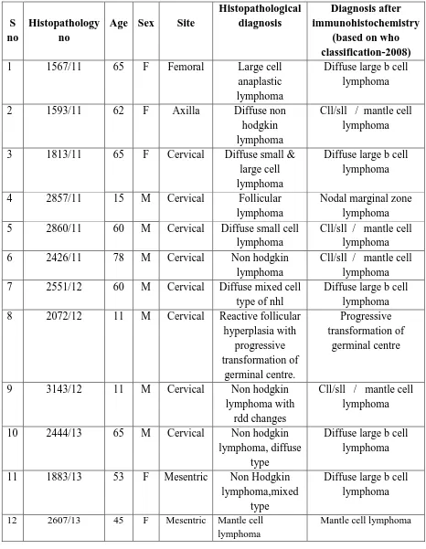
Abstract: Non-Hodgkin lymphoma (NHL) is a collective term for a heterogeneous group of lymphoproliferative malignancies with differing patterns of behaviour and responses to treatment. Incidence of lymphoma has doubled in the past 4 decades and continues to increase. In the context of lymphomas, immunohistochemistry is used to classify, sub-classify and predict prognosis of several classes of lymphomas. Tissue microarray facilitates the analysis of molecular alterations in thousands of tissue specimens in a massively parallel fashion. This study was conducted in Department of pathology, Tirunelveli medical college in which tissue micro array was prepared using manual method from lymph nodes of 21 cases among which 20 were diagnosed as Non-Hodgkin lymphoma by histomorphology. All the cases were subjected for immunohistochemical analysis using CD3, CD20, CD5, and CD10 from which 19 cases were taken for classification and analysis based on recent WHO classification of lymphomas and leukemias.
1
AIM & OBJECTIVES
To apply a panel of basic IHC markers for the diagnosis and classification of Non Hodgkin lymphoma.
To standardize the manual tissue microarray technique.
DIAGNOSTIC IMMUNOHISTOCHEMISTRY WITH TISSUE MICROARRAY TECHNIQUE – A PILOT STUDY ON NON
HODGKIN LYMPHOMA
Abstract: Non-Hodgkin lymphoma (NHL) is a collective term for a heterogeneous group of lymphoproliferative malignancies with differing patterns of behaviour and responses to treatment. Incidence of lymphoma has doubled in the past 4 decades and continues to increase. In the context of lymphomas, immunohistochemistry is used to classify, sub-classify and predict prognosis of several classes of lymphomas. Tissue microarray facilitates the analysis of molecular alterations in thousands of tissue specimens in a massively parallel fashion. This study was conducted in Department of pathology, Tirunelveli medical college in which tissue micro array was prepared using manual method from lymph nodes of 21 cases among which 20 were diagnosed as Non-Hodgkin lymphoma by histomorphology. All the cases were subjected for immunohistochemical analysis using CD3, CD20, CD5, and CD10 from which 19 cases were taken for classification and analysis based on recent WHO classification of lymphomas and leukemias.
2
INTRODUCTION
Lymphoid neoplasms include a diverse group of tumors of B-cell, T-cell, and NK-cell origin. In many instances the phenotype of the neoplastic cell closely resembles that of a particular stage of normal lymphocyte differentiation. Lymphomas are ranked 6thamong all cancers, worldwide (1). Incidence of lymphoma has doubled in the past 4 decades and continues to increase. Non-Hodgkin lymphoma (NHL) is a collective term for a heterogeneous group of lymphoproliferative malignancies with differing patterns of behaviour and responses to treatment. Non Hodgkin lymphoma is the fifth most common cancer in the world with an upsurge of incidence in India. Compared with Hodgkin disease, NHL is much less predictable and has a far greater predilection to disseminate to extranodal sites. The prognosis depends on the histologic type, stage, and treatment.
3
in the aforementioned circumstances, but also to classify, sub-classify and predict prognosis of several classes of lymphomas.
4
REVIEW OF LITERATURE
LYMPH NODE
Lymphoid organs constitute important component of our body. They are classified as central and peripheral lymphoid organs. The organs in which the lymphoid cells are primarily produced constitute the central lymphoid tissues while peripheral lymphoid organs are those in which the lymphoid cell differentiates, matures and processes the antigen. Bone marrow and thymus are the central lymphoid organs from which the lymphoid cells (B-lymphocytes and T-lymphocytes) are produced. Lymph nodes, spleen, MALT (Mucosa associated lymphoid tissue) constitutes peripheral lymphoid organs.
Among all the other peripheral lymphoid organs, lymph nodes are considered the most important because they actively participate in immune response. Lymph nodes are organised collection of lymphoid cells surrounded by a capsule and located in regions where they drain the lymphatic vessels. These include cervical, axillary, abdominal, pelvic and inguinal group of lymph nodes. They are bean shaped organs composed of dense collection of lymphoid cells. Normally, the lymph node measures about 2 to 20 mm with an average of 15mm in its longest dimension.(2,3)
5
palpable, soft, grey-pink and homogenous. They become palpable as a result of intense immune response, neoplastic transformation or metastatic deposit. The diameter of lymph node greater than 3cm with nodular whitish cut surface and firm consistency are the features suggestive of neoplastic transformation or metastatic deposit.
The characteristic features of lymph node architecture varies according to age and site based upon the exposure of lymphoid cells to different antigens. Normally, lymph nodes draining the head and neck region including the cervical and axillary lymph nodes show numerous lymphoid follicles with reactive germinal centres which are due to secondary antigenic stimulation. But, lymph nodes such as mesenteric nodes possess wide medullary cords and sinuses. The count of peripheral lymph nodes also varies significantly with no nodes in newborn, numerous in younger age groups and more numerous in the elderly(4).
6
The anatomy of lymph node includes cortex, medulla and para-cortical area which are formed by lymphoid cells and plasma cells in varying stages of maturation. The supporting framework is formed by the capsule, trabeculae, reticulinfibres and network of vesicular cells. The fibroblasts along with smooth muscle cells, nerve bundles, Schwann cells and blood vessels constitute capsule which extends into the underlying parenchyma as trabeculae.
The reticulinfibres which originate from reticulin cells are fine type 3 collagen fibrils, which form the main supporting framework of the lymph node. Reticulinfibres surround individual lymphoid cells, hence are not visualised under H&E stain. It can be well demonstrated by using silver impregnation technique (Gomori‘s technique).
CORTEX
7
primarily B-cells and tingible body macrophages with few scattered T lymphocytes. The germinal centres are cuffed off by small lymphocytes which form the mantle zone. The peripheral portion of the mantle zone is composed of cells that are loosely packed forming the marginal zone. The marginal zones are distinctly made out in the reactive follicles of spleen and usually are not so evident in the lymph nodes.
PARACORTEX
Paracortical areas are the T-cell packed zones in the lymph nodes and they are located deep beneath the cortex extending in between the lymphoid follicles. These areas are composed of T-cells in various stages of maturation (6), admixed with post capillary venules lined by high endothelial cells. In addition, there are interdigitating dendritic cells which serve as antigen presenting cells. They play an important role in evoking an immune response.
MEDULLARY AREA
8
CELLULAR COMPOSITION
The lymph node is formed by B-cell zone and T-cell zone containing cells in various stages of activation and differentiation. Germinal centres and lymphoid follicles are the B-cell zone whereas the paracortical area forms the T-cell zone. The medullary region is predominantly formed by the plasma cells.
The dark staining naive B cells that forms the primary follicles also accounts for the mantle zone of the secondary follicles (8). These are small cells with scant cytoplasm and increased nuclear cytoplasmic ratio. Centroblasts are large cells that form the dark zone in the germinal centres which express IgM on their surface. They differentiate into centrocytes which forms the light zone in the germinal centres.
9
The tingible body macrophages contain numerous DNA fragments formed secondary to cell death after bcl-2 gene gets shut off.
NORMAL LYMPH NODE AND IHC
The main application of immunohistochemistry in normal lymph node is to study the differential expression of the same antibody in various regions of the lymph node and assess the cellular composition and clone of the lymphoid cells. Studying the clone among various lymphoid populations helps us to differentiate reactive hyperplasia from neoplastic transformation of the lymph node, more importantly Non Hodgkin lymphoma.
10
The small lymphocytes that are scattered throughout the germinal centre show positive expression for CD3, CD5, CD7, CD43 and CD4/8 that gives a clue that they are reactive T cells. Tingible body macrophages stain with CD11b, CD35 and CD68 whereas dendritic reticulum cells stain with CD21, 35, C3b, C3d. (12)
The cells in the paracortical region stain with pan T cell markers like CD2, CD3, CD5 and CD7, CD43 and HLA-DR. Interdigitating dendritic cells show positive expression for S100, CD24, HLA- DR. Plasma cells are CD138(Syndecan 1) positive. Macrophages stain for lyzozyme, α1-antitrypsin, S100 protein and CD68. (13,14)
LYMPHOMA
11
proper selection of the investigations will facilitate correct diagnosis at a reasonable expense to the patient.
NON HODGKIN LYMPHOMA
Non Hodgkin lymphomas are clonal lymphoproliferative disorders characterised by proliferation of malignant lymphoid cells that forms discrete mass. This includes diverse group of tumor of B-cells, T-cells and natural killer cell origin. Majority of the cases of non Hodgkin lymphomas are of B-cell origin while the remainder of the cases has T-cell origin. Non-Hodgkin lymphoma very rarely involves natural killer cells. Most of the lymphoid neoplasms resemble some recognizable stage of B-cell or T-cell differentiation with neoplastic lymphoid cells recapitulating the behaviour of their normal counterpart.
In earlier days it was believed that lymphadenopathy was a secondary phenomenon occurring secondary to underlying infection or carcinoma. Thomas Hodgkin first recognised that lymph node enlargement could occur as a primary disorder rather than secondary to underlying infection or carcinomas(15). Billroth was the first one who used the term malignant lymphoma for a collection of primary lymphoid disorders(16).
12
Brill(19)and Symmers(20), in 1920, described the features of follicular lymphoma. In 1956, Rappaport through his classical work classified Non-Hodgkin lymphoma based upon the morphology of the malignant lymphoid cells (21). Roulet(22) in 1930 considered reticulum cell sarcomas as a synonym for malignant lymphomas.
In 1967, Good and Finstad demonstrated the relationship of B lymphocytes and T lymphocytes to the development of lymphoid neoplasms. Later Dameshek, through his work, suggested that lymphoproliferative disorders primarily occurs due to aberrations of immunologically competent cells and also lymphoid cells has the potential to get transformed into immunoblast like cells under appropriate antigenic stimulation. Immunological origin of malignant lymphocytes was confirmed in 1972 by the presence of immunoglobulins over the surface of B lymphocytes and by the formation of sheep erythrocyte rosette with neoplastic T lymphocytes (23, 24).
13
1994, Revised European American Lymphoma classification came into use, which classified lymphoma based upon the clinical features, morphology, immunophenotyping and genetic data which were available(29). Finally World Health Organisation has adopted the diagnostic principle of REAL classification and now WHO classification is used as schema for the diagnosis of all hematopoietic neoplasm.
Non Hodgkin Lymphoma and non modifiable risk factors
Non-Hodgkin lymphomas are the sixth most common cancer worldwide. Frequency of Non-Hodgkin lymphoma is age dependent and has variable distribution worldwide. Non-Hodgkin lymphomas are more common in males than females and hence show male preponderance. Malignant lymphomas are most common among adults and accounts for only 10% of all childhood malignancies in developed countries. Non-Hodgkin lymphomas show a steady increase in incidence from childhood through age of 80 years(30).
14
lymphoproliferative syndrome which includes autoimmune features, splenomegaly and chronic lymphadenopathy(35). Malignant lymphomas are more common among people with family history of lymphoma(36).
Young age and Non Hodgkin Lymphoma
15
forms bulk of the pediatric lymph node malignancies. Follicular lymphomas and marginal zone lymphomas occurs rarely and are considered uncommon neoplasms in the childhood.
Infection and Non Hodgkin Lymphoma
Infections play a major role in lymphomagenesis. The development of lymphoma depends on various factors including environment, geography and host factors (40). Infectious agents cause lymphoma through direct lymphocyte transformation. Most of the infectious agents that cause lymphomas are found to be viruses which include EBV, HTLV-1, HHV8. Chronic infection by hepatitis-C virus can result in B-cell Non-Hodgkin lymphoma(41). Indolent B cell lymphoma, lymphoplasmacytic lymphoma and marginal zone lymphoma are the most common types of lymphomas that are associated with hepatitis-C virus.
T-16
cell lymphoma, post transplant Non-Hodgkin lymphoma, AIDS related Non-Hodgkin lymphoma, Burkitts lymphoma and Hodgkin lymphoma.
Human T-cell lymphotrophic virus are the first retrovirus discovered, the infection of which is strongly associated with adult T-cell leukemia/lymphoma. It is endemic in south Japan, Caribbean, South Africa and South America. Human T-cell lymphotrophic virus 1 infection increases with age and is twice as high in females when compared to males. The infection is usually asymptomatic to start with but manifests typically later in the life. The incidence of infection is found to be increased in breast fed infants than in bottle fed infants.
Hepatitis–C Virus is found to be related to lymphomagenesis. Infections usually occur through blood transfusion and needle stick injuries. It is believed that HCV causes lymphoma by chronic antigenic stimulation. Infection of Hepatits C virus is associated with increased risk of Diffuse large B-cell lymphoma, Marginal zone lymphoma and lymphoplasmacytic lymphoma. Human herpes virus-8 has been documented in the causation of Non-Hodgkin lymphoma. HHV infection is associated significantly and positively with risk of lymphoplasmacytic lymphoma.
17
lymphoma especially Diffuse large B-cell lymphoma. Alcohol consumption appears to be inversely related to Non-Hodgkin lymphoma. Smoking does not appear to play a role in etiology of Non-Hodgkin lymphoma.
Helicobacter pylori is a gram negative rod that was discovered by Warren and Marshall in 1983 and was shown to be associated with peptic ulcer disease, carcinoma of stomach and Non Hodgkin lymphoma (42, 43). Later on Personett recognised that Helicobacter pylori infection preceded the development of lymphoma(44) and Wotherspoon observed that there is regression of lymphoma in most of the patients after treatment with antibiotics(45). Other infectious agents includes, campylobacter jejuni which causes small intestinal immunoproliferative diseases, Borrelia burgdoferi which causes primary cutaneous B cell lymphoma and Chlamydia psittaci which is the causative agent for ocular adnexal MALTomas40.
AIDS and transplant related Non Hodgkin Lymphoma
Non-18
Hodgkin lymphoma represents B-cell type, among which 70% is constituted by diffuse large B-cell lymphomas and the remaining being Burkitt‘s lymphoma. Among patients of lymphomas found in the general population of young patients in which Hodgkin lymphomas are more common, lymphomas in AIDS patients are mostly Non Hodgkin type with frequent extranodal manifestations (49, 50). The occurrence of lymphomas in AIDS can be attributed to florid activation of lymphoid cells which provides opportunity for translocation, mutation and deletion of tumor suppressor gene (51, 52).
19
Autoimmune and other disorders
Chronic inflammation, immune hyperactivity and immunosuppression are considered to be important elements that predispose patients to lymphoma (54). Non-Hodgkin lymphomas associated with Sjogren‘s syndrome and Hashimoto‘s thyroiditis are of B-cell origin and they usually occur in elderly females. In 1963, Bunin and Talal(55) reported Non-Hodgkin lymphoma in a case of Sjogren‘s syndrome. In 1957, Lindsay and Dailey (56) demonstrated the association of lymphoma with Hashimoto‘s thyroiditis. There is 60 to 80 fold increase in thyroid lymphomas among the cases of thyroiditis with lifetime risk of 1-2% (57). Non-Hodgkin lymphoma has been associated with rheumatoid arthritis, SLE, and dermatomyositis.
Non-Hodgkin lymphoma has been observed to originate from treated cases of Hodgkin lymphoma. Krikorian (58)reported 6 cases of Non-Hodgkin lymphoma arising from Hodgkin lymphoma. First case of Non-Hodgkin lymphoma associated with AIDS has been reported in 1982 (59). Majority of the lymphomas arising from AIDS patients are of B-cell origin. Unique presentation of AIDS related lymphomas include plasmablastic lymphomas of oral cavity (60) and primary effusion lymphomas (61).
Non Hodgkin Lymphoma and presentation
20
common extra nodal site of presentation, in which stomach is the commonest site followed by small intestine, colon and esophagus, in that order. Rarely, patients with rectal involvement present with altered bowel habits (63). Epistaxis and nasal obstruction are the modes of presentation in cases of Non-Hodgkin lymphoma of nasal cavity. Hepatosplenomegaly have been observed in cases of low grade cell lymphomas such as small B-lymphocytic lymphomas and marginal zone lymphomas. Skin is another most common site of Non-Hodgkin lymphoma presentation. Non-Hodgkin lymphoma can primarily involve CNS (64) in which diffuse large B-cell lymphomas are the most common. Other rare sites involved are eye, extraocular space including conjunctiva, eyelids & lacrimal glands (65), testis, kidney and breast. Breast lymphomas associated with pregnancies and lactation often has diffuse involvement (66). Primary cardiac lymphomas are extremely rare and usually occur in immunodeficient state (67).
Gray zone lymphomas
21
(68, 69)
. This includes nodular lymphocyte predominant Hodgkin lymphoma, T cell rich large B cell lymphoma, Anaplastic large cell lymphoma, peripheral T-cell lymphoma and primary mediastinal large B-cell lymphoma.
Gray zone lymphomas should be separated from conventional lymphomas as they are aggressive and require distinct treatment (70, 71).
CLASSIFICATION OF NON-HODGKIN LYMPHOMA
22
Mature B cell neoplasm constitutes about 90% of NHL worldwide(72). B cell represents all follicular and diffuse lymphomas in North America and Europe, whereas T cell lymphomas are most common in Asia (73). The nomenclature, classification and differentiation of various types of lymphomas depend upon various factors including the cell of origin eg; mantle cell lymphomas, clinicopathological name eg: Burkitt‘s lymphoma, their location eg: mediastinal large B cell lymphomas and presumed fuction eg; MALTomas.
The grade and prognosis of Non Hodgkin lymphomas depend upon the morphological pattern, size and shape of the cell, mitosis and invasiveness. The pattern of neoplastic transformation is divided into nodular and diffuse in which the nodular growth pattern represents the differentiation of malignant lymphomas towards lymphoid follicles. The malignant lymphoma cells are classified into small, medium and large sized by comparing the nucleus of the neoplastic cells to the adjacent histiocytes or endothelial cells. On the basis of shape, it is classified into cleaved and cleaved in which the cleaved cells represent dividing cells and non-cleaved represent the dividing cells. In general, lymphomas are considered aggressive when the cells are large, non-cleaved with a diffuse pattern of growth showing increased mitosis and invasion.
23
24
MODIFIED KIEL CLASSIFICATION (76)
KIEL CLASSIFICATION Nearest
correspondence to categories of Rappaport
classification
Nearest
correspondence to categories of Lukes-Collins classification
LYMPHOMAS OF LOW GRADE MALIGNANCY
Lymphocytic lymphomas
1. Chronic lymphocytic leukemia (B-cell type)
Well-differentiated lymphocytic
lymphoma
B cell lymphoma of small lymphocytes
2.Hairy cell leukemia - -
3.Mycosis fungoides and Sezary‘s syndrome
- T-cell
lymphomas:.Mycosis fungoides and Sezary‘s syndrome
4.Chronic lymphocytic leukemia (T- cell type)
- -
Lymphomas of immunoglobulin -secreting cells
Apart from
lymphomas-‘proliferative diseases with dysprotinemia‘
1.Lymphoplasmacytic/lympho plasmacytoid lymphoma
- B-cell lymphoma of plasmacytoid
25
2.Plasmacytic lymphoma - -
Lymphomas of germinal centre cells
1.Centrocytic lymphoma well –differentiated & poorly differentiated
lymphocytic
lymphomas; Mixed cell(lymphocytic- Histiocytic)
lymphoma;Histiocyti c lymphoma
B-cell lymphoma of follicular centre cells with cloven nucleus (diffuse type)
2.Centroblastic /centrocytic lymphoma
follicular
follicular and diffuse diffuse
with or without sclerosis
well –differentiated & poorly differentiated
lymphocytic
lymphomas; Mixed cell(lymphocytic- Histiocytic) lymphoma;
B-cell lymphoma of follicular centre cells with cloven nucleus
LYMPHOMAS OF HIGH GRADE MALIGNANCY
Centroblastic lymphomas Histiocytic lymphoma; undifferentiated lymphoma
B-cell lymphoma of large follicular centre cells with non-cloven nucleus
1.Primary
26
Lymphoblastic lymphomas (lymphoblastic sarcomas, including acute lymphoblastic leukemia)
Undifferentiated lymphoma; poorly differentiated
lymphocytic lymphoma
B-cell lymphoma of small follicular centre cells with non-cloven nucleus
1.Burkitt lymphoma Undifferentiated lymphoma
As above
2.Lymphoma of convoluted cells [i.e., cells with convoluted nucleus]
Undifferentiated lymphoma; poorly differentiated
lymphocytic lymphoma
T-cell lymphoma of convoluted
lymphocytes
3.Unclassified lymphoma poorly differentiated lymphocytic
lymphoma
U-cell (‗undefined cell‘) lymphoma; unclassifiable
lymphomas. Immunoblastic lymphomas Histiocytic lymphoma Immunoblastic
sarcoma of B cells; immunoblastic
sarcoma of T cells With plasmablastic/
plasmacytic differentiation (derived from B cells)
Without
plasmablastic/plasmacytic differentiation (derived from B cell or T cell)
27
[This table represents Kiel classification modified by Lennert et al in 1977(76). The Kiel classification and Lukes-Collins classification relates to the functional characteristics of the cells that relate to the tumors.]
A WORKING FORMULATION OF NON-HODGKIN
LYMPHOMAS(77)
This classification was introduced in 1982 by National cancer institute, United States (77), as an attempt to provide a morphologic classification scheme that had a prognostic relevance.
LOW- GRADE Malignant lymphoma Small lymphocytic
Consistent with chronic lymphocytic leukemia Plasmacytoid
Malignant lymphoma, follicular Predominantly small cleaved Diffuse areas;sclerosis Malignant lymphoma, follicular
Mixed, small cleaved and large cells Diffuse areas;Sclerosis
28
Diffuse areas; Sclerosis Malignant lymphoma, Diffuse Small cleaved
Sclerosis
Malignant lymphoma, Diffuse Mixed, small and large cell
Sclerosis; Epithelioid cell component Malignant lymphoma, Diffuse
Large cell
Cleaved, non cleaved; Sclerosis HIGH GRADE
Malignant lymphoma
Large-cell, Immunoblastic
Plasmacytoid; clear cell; polymorphic; epithelioid cell component Malignant lymphoma
Lymphoblastic
Convoluted; non convoluted Malignant lymphomas
Small noncleaved
29
MISCELLANEOUS Composite
Mycosis fungoides Histiocytic
Extramedullaryplasmacytoma Unclassifiable
Others
WORLD HEALTH ORGANISATION CLASSIFICATION OF LYMPHOID NEOPLASMS (INTERNATIONAL AGENCY FOR RESEARCH ON CANCER, LYON, 2008) (78)
30
PRECURSOR B- AND T-CELL NEOPLASMS
Precursor B-lymphoblastic leukaemia/lymphoblastic lymphoma Precursor T-lymphoblastic leukaemia/lymphoblastic lymphoma
MATURE B-CELL NEOPLASM
Chronic lymphocytic leukaemia/ Small lymphocytic lymphoma B-cell prolymphocytic leukaemia
Lymphoplasmacytic lymphoma
Splenic b-cell marginal zone lymphoma Hairy cell leukaemia
Plasma cell myeloma
Solitary plasmacytoma of bone Extraosseous plasmacytoma Heavy chain disease
MALT-lymphoma
Nodal marginal zone B-cell lymphoma Follicular lymphoma
Mantle cell lymphoma
Diffuse large B-cell lymphoma, NOS
31
Mediastinal large B-cell lymphoma Intravascular large B-cell lymphoma ALK positive large B-cell lymphoma Plasmablastic lymphoma
Primary effusion lymphoma Burkitt lymphoma
B-cell lymphoma, unclassifiable with features intermediate between DLBCL &Burkitt lymphoma
B-cell lymphoma, unclassifiable with features intermediate between DLBCL and classic Hodgkin lymphoma
MATURE T-CELL AND NK-CELL NEOPLASM T-cell prolymphocytic leukaemia
T-cell large granular lymphocytic leukaemia Aggressive NK-cell leukaemia
Adult T-cell leukaemia/lymphoma
Systemic EBV positive T-cell lymphoproliferative disease of childhood Hydroa vacciniforme-like lymphoma
Mycosis fungoides Sezary syndrome
32
Primary cutaneous CD30 positive T-cell lymphoproliferative disorder Primary cutaneous gamma-delta T-cell lymphoma
Lymphomatoid papulosis
Extranodal NK/T-cell lymphoma, nasal type Enteropathy-type T-cell lymphoma
Hepatosplenic T-cell lymphoma
Subcutaneous panniculitis- like T-cell lymphoma Angioimmunoblastic T-cell lymphoma
Peripheral T-cell lymphoma Anaplastic large cell lymphoma
HODGKIN LYMPHOMA
Nodular lymphocyte predominant Hodgkin lymphoma Classical Hodgkin type
33
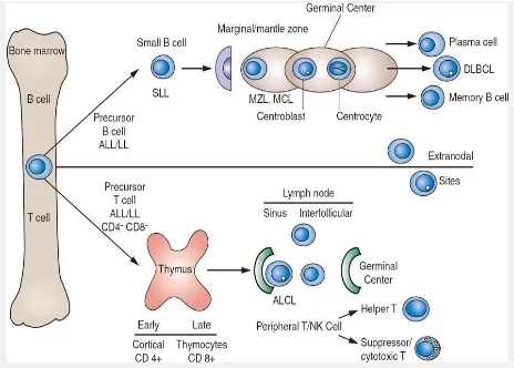
CELLS OF ORIGIN OF NON HODGKIN LYMPHOMA
34
35
36
The above illustration shows the different classes of lymphoma based on Lukes-Collin classification. The B cell lymphoma of small lymphocytes constitutes CLL/SLL, whereas the B-cell lymphoma of follicular centre cells with cloven nucleus (diffuse type) constitutes the centrocytic lymphoma. B-cell lymphoma of large follicular centre cells with non-cloven nucleus constitutes centroblastic lymphoma , whereas B-cell lymphoma of small follicular centre cells with non-cloven nucleus constitute lymphoblastic lymphoma.
IMMUNOHISTOCHEMISTRY
Immunohistochemistry is a technique which is used to detect the specific antigens over the tissues or cells based on the pattern of antigen-antibody reaction. Coons (81) in 1940 introduced immunoflorescence to localise corresponding antigen in frozen section. The enzymatic label horseradish peroxidase developed by Avrameas(82) and by Nakane and colleagues(83) provided the new path for the visualisation of the labelled antibody by light microscopy in the presence of a appropriate colorigenic substrate system.
37
main aim of performing IHC is to recognize microchemically the existence and distribution of substances which we have been made aware of macrochemically(84). Immunohistochemistry can provide a tissue based immunoassay with the reproducibility and quantitative characteristics of an ELISA test.
Demonstration of antigen in routinely processed formalin-fixed paraffin embedded tissues(85) was done for the first time in 1974 by Taylor and Burns in Oxford.Then as the immunohistochemistry evolved, its utilisation in diagnostic pathology steadily expanded such that more than one immunohistochemistry markers were used in routine surgical pathology especially with respect to tumour diagnosis and classification. The introduction of hybridoma technique(86) facilitated in the development of immunohistochemistry and the manufacture of many highly specific monoclonal antibodies.
The phenomenon of enzyme digestion was then introduced by Huang as the pretreatment to immunohistochemistry staining to unmask some antigens that had been altered by formalin fixation (87). The main disadvantage of using enzyme digestion was that it proved difficult to control the optimal digestion conditions for different tissue sections when stained with different antibodies. Hence, to overcome the difficulties encountered in enzyme digestion Shi and his associates in 1991(88, 89, 90, 91, 92,
93)
38
Fraenkel and co-workers (94, 95, 96). Various modifications of antigen retrieval has been described, among which majority of those used different buffer solution in the place of metal salt solution, which may have serious toxic effect(97, 98).
Antigen and antibody reaction
An antibody is a molecule that has the property of combining specifically with the antigen. Antigen-antibody recognition is based on three dimensional structure of the protein. Antibodies are immunoglobulin molecules containing pair of light chain and pair of heavy chain. An epitope is an antigenic determinant on which the antibody combines. For a protein, the term epitope corresponds to a cluster of aminoacid residues that binds specifically to the paratope of an antibody (99).
Antigen-antibody reaction depends on two factors that are related to antibody which includes sensitivity and specificity. Antibodies are primarily of two types monoclonal and polyclonal. Comparitive studies on sensitivity and specificity among monoclonal and polyclonal antibodies proves that polyclonal antibodies are more sensitive and less specific than monoclonal antibodies. The reason may be polyclonal antibody which is a composite of many antibodies may recognise several binding epitope whereas monoclonal antibody recognise single type of epitope.
39
have appeared on the market. Some of the rabbit derived antibodies offer advantage over murine clones for the detection of antigens by immunohistochemistry.
Enzyme labels in immunohistochemistry
Enzymes are the widely used labels used in immunohistochemistry. Incubation with the chromogen using proper histochemical method produces stable colour reaction end product that can be easily read by using light microscopy. Variety of enzymes and chromogens are available which produces many number of coloured end products. Horseradish peroxidase is the most commonly used enzyme label. When used in conjunction with most favoured chromogen like Diaminobenzidine tetrachloride yield insoluble, stable brown coloured end product. Horseradish peroxidase is most preferred as they are very small in size, and can be very easily obtained in pure form. It is very stable and does not undergo alteration during manufacture, storage and during application.
40
multilabelling technique. Rarely colloid metal label such as gold conjugates are used. When used alone it appears pink under microscope. A silver precipitation reaction can be used to amplify the visibility of gold conjugates. The use of radioisotopes as the tracer requires autoradiographic facilities.
Antigen retrieval
Antigen retrieval is the process of reversal of chemical reaction that has occurred between formalin and protein. Heat induced antigen retrieval technique is now widely used in pathology(100). It has certainly brought a very good improvement in the quality of immunohistochemistry. The optimal result of antigen retrieval in immunohistochemistry depends on heating temperature, period of heating and PH of antigen retrieval solution.
Various methods are used in the process of antigen retrieval which includes microwave antigen retrieval, pressure cooker antigen retrieval and retrieval using streamer, water-bath, autoclave and combined microwave and trypsin digestion antigen retrieval. The use of conventional heating at 1000c achieves results similar to those obtained by microwave. Distilled water could be used as the antigen retrieval solution with slight less effect
(84)
.
41
toxic citrate buffer. Cottoretti in 1993 argued that microwave antigen retrieval is an alternative to proteolytic enzymes. Various problems including uneven heating and hot spot production are the important problems with the use of microwave.
In 1994, Norton suggested the use of pressure cooker method for antigen retrieval. It is considered as one of the suitable method as it produces even heating and has no disadvantages which includes hot spot and cold spot production which often occur with the use of microwave. Streamer antigen retrieval is less efficient when compared to microwave and pressure cooker. Kawai in 1994 showed that water bath set at 900C is adequate for antigen retrieval. However, quality of antigen retrieval improves with increase in the temperature to 95-980C.
Evers and Nylings(101)in his study found that antigen retrieval depends on both pH and temperature. They concluded that it is not important what
kind of solution is used as long as the pH is appropriate. Chemical
composition and the molarity of the antigen retrieval solution may act as a cofactor for effectiveness of antigen retrieval and hence immunohistochemistry.
Non specific background staining
42
multiple unwanted antibodies. Blocking of endogenous enzymes activity is important. The degree of susceptibility of an enzyme to denaturation and inactivation varies from tissues to tissues. Any residual activity of endogenous enzymes must be abolished. Endogenous peroxidase activity is seen in neoplastic cells, erythrocytes, neutrophils, eosinophils, and hepatocytes. Usage of various substances for blocking endogenous peroxidase is suggested.
Some investigators thought that use of methanol-hydrogen peroxide may cause denaturation of antigen. Strans(102,103) advocated the use of phenylhydrazine by combination of phenyl hydrazine, nascent hydrogen peroxide and sodium azide. Mixture of hydrogen peroxide and sodium azide was found to be very effective(104). More recently cyclopropane hydrate was shown to inhibit endogenous activity without adverse effect(105). Endogenous alkaline phosphatase activity is usually blocked by addition of Levamisole to the substrate. Levamisole selectively inhibits certain types of alkaline phosphatase but not intestinal and placental. 20% glacial acetic acid is a better alternative to levamisole.
IMMUNOHISTOCHEMISTRY AND NHL
43
Much of the early work in diagnostic hematopathology required frozen section or acetone fixed frozen section as available antibodies failed to recognize the antigens fixed by cross linking fixatives. Partial enzyme digestion gives good result(108).
Understanding of the development, maturation and migration of lymphocytes provides rationale for immunophenotyping because stages in lymphocyte maturation are defined in part by the macromolecules they produce. Malignant cells expresses antigen that resembles the antigens of lymphocytes in different developmental stages. Lymphomas are thought of as being composed of cells arrested in development of certain stages(109). Hence the panel of markers directed against the malignant cells will be of more help.
44
totally specific and therefore needs careful selection of panel of markers to arrive at proper diagnosis.
LYMPHOMA DIAGNOSIS
The diagnosis of lymphoma is based on the World health organisation classification (110)(2008). Further classification is based on the stage of maturation and cell of origin. Other features which help in the diagnosis are anatomic architectural alteration in the lymphoid compartment, determination of cell size, nuclear character, and character of nucleoli.
B-cell markers
CD20 is the most common antibody used as the pan-B cell marker and is expressed by naive B cell until the final stages of maturation just prior to plasmacytic differentiation. Chronic lymphocytic lymphoma/small lymphocytic lymphoma rarely shows weak positivity(111) to CD20. In cases of prior rituximab therapy, CD79a should be added. Early stage including naive cells and other B-cell expresses Pax-5 but not the plasma cells(112). Few cases of precursor T-cell acute lymphoblastic lymphoma expresses CD79a and rare cases of AML and merkel cell carcinoma shows positive expression of Pax-5.
45
population, especially plasma cells. Immunohistochemistry although less sensitive in the detection of immunoglobulin expression, can be more valuable when used along with histopathology. Plasma cells and immunoblast shows strong expression of immunoglobulins which can be detected by paraffin immunoperoxidase method. Calorimetric insitu hybridisation is an alternate and most sensitive method used in the detection of immunoglobulin.
T-cell markers
CD3 is the most commonly used pan-T cell marker which detect the antigenic expression over the malignant and normal T-cell at the second stage of thymic differentiation and beyond. Sometimes there may be loss of pan-T cell marker over the malignant lymphoid cells as in case of anaplastic large cell lymphoma(113). T cells are divided into CD4 and CD8 subsets. Antibodies directed against these antigens are used as basic panels to detect abnormal distribution of lymphoid cells. CD5 is another pan-Tcell marker which is lineage non-specific and is not expressed on Natural killer cells. CD5 is present over small subset of B-cells especially among the cases of chronic lymphocytic lymphoma, mantle cell lymphoma.
Atypical cells and Lymphoma
46
carcinomas, Hodgkin lymphoma and anaplastic large cell lymphoma should be ruled out. This is done by using a panel of markers including CD45, CD15, CD30 and HMB45. The usage of antibody directed against ALK-1 may prove more valuable.
Other antibodies
Several other markers have found application in the diagnosis of Non-Hodgkin lymphoma. These include CD10, Bcl-6, cyclin D1, Bcl-2, CD56 and CD57. CD10 and Bcl-6 are the markers that show positive expression in normal and neoplastic follicular centres. CD10 also show positive expression on granulocytes, T-cells in angioimmunoblastic lymphadenopathy, precursor B-cells and T-cells, epithelial cell neoplasms like renal cell carcinomas, hepatocellular carcinomas and endometrial stromal cells. Bcl-6 is expressed in follicular centres and few T-cells. The presence of CD10 positive lymphocytes along with Bcl-6 positive cells outside the follicles strongly suggests neoplastic proliferation. But rare cases of follicular lymphoma which shows negative expression of CD10 have been recorded.
47
negative expression of Bcl-2 and there is strong expression of Bcl-2 in lymphoid cells of mantle zone and cells of primary follicle(115). The interpretation of Bcl-2 should be carried out in conjunction with CD3 staining for T-cells as T-cells normally show positive expression of Bcl-2. Most of the cases of marginal zone lymphoma shows Bcl-2 expression but reactive hyperplastic marginal zones of spleen and lymph nodes of abdomen also show Bcl-2 positivity. CyclinD1 is another immunohistochemical marker which has gained its importance in the diagnosis of Mantle cell lymphoma. Cyclin D1 is normally expressed in endothelium, nucleus of histiocytes, and fibroblast. It is usually expressed in the nucleus of lymphoid cells of mantle cell lymphoma. Cytoplasmic expression is considered non diagnostic. Although nuclear expression of CyclinD1 is almost diagnostic of mantle cell lymphoma many cases of CyclinD1 negative mantle cell lymphomas are on the records and even CyclinD1expression has been recorded in many cases of small lymphocytic lymphoma. Immunohistochemical markers like CD56 and CD57 play important role in the diagnosis of natural killer cell proliferation and neoplasms arising from natural killer cells.
PANEL OF MARKERS FOR NON HODGKIN LYMPHOMA
Ki-48
67/Mib1 antibodies. They found 100% positivity for CD20 in DLBCL and Burkitt lymphoma patients. Among DLBCL patients, 10.5% positivity was seen for bcl6, 22.2% positivity for MUM1/IRF4 and 66.7% for CD138. According to I.Satish Rao(1) distinction of monomorphic small B cell lymphomas are done by basic panel of markers such as CD5, 23, 10, cyclin D1 and bcl2, intermediate cells with diffuse pattern by markers such as CD20,10, Tdt, CD99 and Mib1 and large cells with diffuse pattern by CD20, CD79a, CD138 and EBER. Nirmala Ajit Jambhekar et al (117) suggests a panel of markers for Non Hodgkin lymphoma diagnosis which includes CD20 which is a B-cell marker showing membrane staining, CD3 is a T-cell marker, CD21/CD35 stains follicular dendritic cells and hence
are useful to demonstrate germinal centers within the lymphoid
proliferation, CD5 which detects B-cell CLL/SLL and mantle cell
lymphomas. CD23 shows positivity for B-CLL/SLL, whereas it is negative
in mantle cell lymphoma. Mantle cell lymphoma shows immunoreactivity to
Cyclin D1. Alk-1 is specific for anaplastic large cell lymphomas (ALCL)
which also shows CD30 positivity and focal EMA positivity. Bcl-2 is useful
in distinguishing reactive follicles (negative) from follicles of follicular
NHL (positive) whereas CD10 is positive in Burkitts lymphoma.
49
pan T cell marker (CD3). Hans et al (119) in their study on 152 cases of DLBCL demonstrated the expression of CD10 in 28% of cases, bcl-6 in 56%, MUM1 in 47%, bcl-2 in 50%, cyclin D2 in 13% and FOXP1 in 61% of cases. Ting Li et al (120) studied immunohistochemical features in 63 cases which resulted in expression of CD10 in 30% of cases, bcl6 in 35% and MUM1 in 51% of cases.
Strauchen JA and Mandeli JP (121) in their study of 345 cases of B cell NHL found Ig expression in 59% cases of large cell lymphoma and 100% of small cell lymphoma. Picker LJ et al (122) documented that immunoblastic variant especially those with plasmablastic differentiation showed cytoplasmic Ig expression. Piris M et al (123) through their study suggested that most of the anaplastic variant showed positive membrane staining for CD30. Fang JM et al (124) found that 20-30% of DLBCL showed CD10 positivity.
Follicular lymphoma cells show positive membrane staining for CD 19, 20, 22 & 79a with typical surface monoclonal Ig expression. Jaffe ES et al (29) through their study found out that follicular lymphoma cells do not express CD3, 5 and 23. Both follicular and inter follicular zones contain κ or λ clonally restricted B cell. Lai R et al (125)
50
Robert R. Lorsbach et al (127) through their study on 19 paediatric follicular lymphoma patients showed CD20 and bcl6 positivity in all cases and CD10 positivity in 80% of cases. Andrew J Davies et al (128) observed through their study that among the follicular lymphoma patients assessed for transformation to DLBCL, 89% showed germinal centre phenotype with CD10+, bcl6+, and MUM1- . Follicular lymphoma in addition to mimicking the growth pattern of reactive follicles, contain non neoplastic T cells and follicular dendritic reticulum cells. According to Said JW et al (129) follicular dendritic reticulum cells in follicular lymphoma and reactive follicular hyperplasia express CD21 and CD35 whereas follicular dendritic reticulum cells in follicular lymphoma but not in reactive follicular hyperplasia, lack the expression of fascin.
In mantle cell lymphoma, the neoplastic cells show membranous positive expression of CD19, 20, 22, 79a and CD5. Vasef MA (130) and Cheuk W (131) through their study showed that almost all the cases of mantle cell lymphoma showed strong nuclear expression for cyclin D1. In case of CLL/SLL, the cells are B cells with positivity for surface Ig light chain, IgM, IgD, pan B cell antigens, CD5 and 23 antigens.
51
CD2,3,CD5 and 7,43 and CD45RO but are negative for B cell antigens. Through their study Rudiger et al (134) showed that AITL express antigens like CD10, bcl6, CXCL13, SAP, programmed death-1 and activation induced cytidinedeaminase. In cases of anaplastic large cell lymphoma, ALK+ anaplastic large cell lymphoma by definition shows ALK positivity and CD30 expression in a distinctive pattern along with expression of T cell antigens.
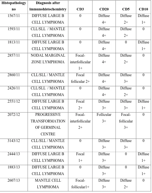
According to Afaf Abdel-Aziz, Abdel-Ghafar(135) the grading of immunohistochemical staining can be done using percentage of positive cells which include four grades, 0: no reaction, 1+: <5%, 2+: 5–9%, 3+: 10– 20% and 4+: ≥20%.
OTHER MODALITIES OF DIAGNOSIS OF LYMPHOMA
52
melanosomes(136), Ewing‘s sarcoma with abundant glycogen(137) and small cell carcinomas with neurosecretory granules(138).
Chronic lymphocytic lymphomas are characterised by increased synthesis of immunoglobulin light chain when compared to heavy chain. Ultrastructural examination in such cases reveals Ig light chain in both perinuclear space and rough endoplasmic reticulum whereas heavy chains are limited to rough endoplasmic reticulum.
Lymphoblastic lymphoma under electron microscope shows round cells with a high nuclear to cytoplasmic ratio with convoluted nucleus showing deep indentation and fine chromatin with peripheral nucleolus (139). The examination of angioimmunoblastic lymphoma shows nuclear indentation, speckled heterochromatin and prominent nucleolus (140).
53
Molecular diagnosis is considered as gold standard (141,142) in assessing monoclonality and chromosomal translocations. Unlike immunohistochemistry which detects proteins, molecular diagnosis detects DNA of genes encoding immunoglobulins and T cell receptor. Various techniques used in routine practice are conventional cytogenetics, southern blot technique, polymerase chain reaction and fluorescence insitu hybridisation. Polymerase chain reaction is a versatile technique used in the analysis of clonality, chromosomal translocation, genetic mutation, infectious agents and minimal residual disease. The main disadvantage is the occurrence of false positivity because of its high sensitivity.
Chromosomal translocation plays an important role in the occurrence of many lymphomas and hence identification of such translocation proves the diagnosis. This is carried out often using conventional cytogenetics or florescence insitu hybridisation technique. This can identify wide range of translocations which includes myc translocation in Burkitt‘s lymphoma(143),
54
CONDITIONS MIMICKING LYMPHOMA
Reactive lymphoid hyperplasia is defined as a benign reversible process characterised by proliferation of reactive lymphoid cells in various pattern secondary to antigenic stimulation. Clinically, this condition is manifested as enlarged lymph nodes in multiple sites including cervical nodes which are often affected in infectious mononucleosis, posterior cervical group as in the case of toxoplasmosis, axillary node in cat- scratch disease, parotid, submaxillary and epitrochlear group as in the case of HIV infection. When there is involvement of supraclavicular lymph nodes careful evaluation of the patient including age, sex, family history, symptoms and signs are performed. This is because these nodes are associated with either primary or metastatic malignancy in 25% of patients younger than 40 years and 90% of patients older than 40 years. (146)
55
Most of the hyperplastic nodes show nonspecific pattern. But characteristic morphological pattern can be seen in reactive follicular hyperplasia secondary to activated humoral immune response, diffuse paracortical hyperplasia as in the case of viral lymphadenitis and sinus histiocytosis. Histopathology of reactive follicular hyperplasia is characterised by numerous reactive secondary follicles of varying size and shape distributed in the cortex and medullary region, with well demarcated mantle cell zone. The germinal centre comprises mixture of small and large lymphoid cells.
The presence of numerous follicles throughout the cortical and medullary zones makes morphological distinction of exaggerated follicular hyperplasia from follicular lymphoma. However, according to Nathwani(150), numerous back to back follicles with even distribution of the cortex and medulla with scant inter-follicular area in addition with cytological features helps in the distinction. But in more difficult cases, immunohistochemistry may prove useful.
56
metastatic deposit from amelanotic melanoma. In difficult cases like this, immunohistochemistry solves the problem.
Atypical lymphoid hyperplasia is a group of disorder that is neither benign nor malignant which occurs in cases of primary immunodeficiency. This disorder occupies an intermediate position between reactive lymphoid hyperplasia and malignant lymphoma. The morphology is characterised by nodular or diffuse proliferation of lymphoid follicles admixed with many atypical large cells with large nucleus and prominent nucleoli. These atypical cells can be either singly scattered or in sheets, and when so can cause difficulties in differentiating it from malignant lymphomas and metastatic deposit. In such cases immunostaining to demonstrate monoclonal light chain (either κ or λ) is necessary.
57
p450. Gennis estimated that pseudolymphoma after phenytoin therapy occurs in the range of 1:1000. Anticonvulsant hypersensitivity syndrome (pseudolymphoma) occurs in association with haematological abnormalities such as eosinophilia, leucocytosis/ leucopenia and atypical lymphocytosis.
This is manifested as acute increase in the size of the lymph nodes with effaced nodal architecture but preservation of atleast portion of lymph node can be usually seen. Histopathologically, this is characetrised by paracortical expansion with rarely follicular hyperplasia. Paracortical expansion is characterised by proliferation of mixed inflammatory cell infiltrates composed of eosinophils, small lymphoid cells and large atypical looking immunoblast. Hence it should not be confused with malignant lymphomas including Hodgkin and Non-Hodgkin lymphomas especially T-cell lymphomas like anaplastic large cell lymphoma and angioimmunoblastic lymphomas. In case of difficulties immunohistochemistry, flow cytometry, PCR or molecular diagnostic approaches will be useful. The absence of monoclonal gene rearrangement favors drug associated lymphadenopathy over true malignant lymphomas.
58
DRUGS CAUSING LYMPHADENOPATHY Phenytoin
Carbamazepine Phenobarbitol Gabapentin Lamotrigine Penicillin Gentamicin Allopurinol Methyldopa Aspirin
Phenylbutazone
Primidone Methimazole Quinidine Tetracycline Ethosuximide Halothane Mexiletine Abacavir Ivermectin Gold
59
LYMPHNODE AND NECROSIS
Necrosis in the lymph node can be focal or diffuse replacing the nodal architecture. It can be of any type which includes coagulative, caseous and fibrinoid type. Caseous necrosis most commonly occurs in infections like tuberculosis in which the necrotic areas appear granular and eosiniphilic surrounded by epithiloid cell granulomas, lymphocytes and fibroblasts. In addition, the disease causing organisms can be demonstrated in such kind of lesions.
60
Lymph nodes are organs with rich vascularity and hence rarely infracted. However, cases of coagulative necrosis involving most of the nodal parenchyma, sparing only a narrow subcapsular rim have been recorded (156, 157). The causes include occlusive vascular thrombosis, thrombophlebitis, localised trauma secondary to fine needle aspiration and as a secondary complication to surgical procedure. Coagulative necrosis can occur secondary to malignant lymphomas. This may be due to expansion of lymphomas causing pressure effects over the vessels or due to direct invasion of the hilar vessels. Hence massive infarction should always be viewed with a suspicion of underlying malignancy. The demonstration of preserved antigen in the necrotic area through immune histochemistry is useful in such situations.
Castleman‘s disease is defined as the large benign asymptomatic lymphadenopathy usually involving mediastinal groups. The cause for Castleman lymphadenopathy is not known, however HHV-8 is considered to be a known etiological agent. Primarily two variants has been described namely hyaline vascular and plasma cell variant which can be unicentric or multicentric. The hyaline vascular variant is by far the most common variant
(158,159)
61
centre of the vessels. The expansion of mantle zone is observed with small lymphoid cells arranged concentrically in an onion-skin fashion. The interfollicular area shows mixed population of cells.
The plasma cell variant is characterised by sheets of plasmablasts and mature plasma cells expanding the interfollicular area. Immunohistochemical evaluation is needed when there is florid proliferation of these cells leading to confusion with lymphocytic lymphoma and plasma cell neoplasm.
TISSUE MICROARRAY
62
The origin of Tissue microarray technique is attributed to Dr. Hector Battifora‘s sausage block technique(164)
63
[image:78.595.134.487.202.485.2]64
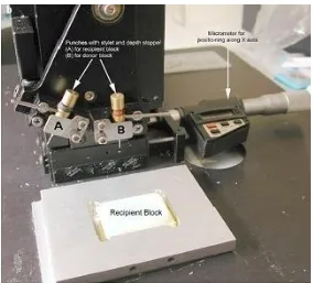
Figure 4(167): Beecher Instruments Tissue Arrayer
65
Materials for TMA
The source of the material for construction of Tissue microarray varies widely. Tissue microarrays are categorised based on material of origin. They are called simply as Tissue microarray when they are constructed from paraffin embedded materials. Tissue microarrays can also be constructed using resin as recipient block, if very thin sections are required. But the construction of resin Tissue microarray is very difficult when compared to paraffin Tissue microarray(168).Tissue microarrays can also be constructed using frozen sections in which case they are called as Cryoarrays(169)or can be constructed using paraffin embedded cell lines and by using cell blocks(170).
TMA categorisation
66
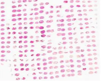
The common feature between all these devices is that they use hollow needles or punches and they adopt the technique of skin biopsy to extract tissue cores from a donor block and make a new paraffin recipient block. After cutting all the tissue cores appear as circular samples arranged in grid like fashion.
The initial step which is considered most important in construction of Tissue microarray is to clearly define the purpose of Tissue microarray construction and to decide the number and size of the tissue core which should be taken from the donor block. Then blocks and slides of the selected cases are carefully reviewed to mark the area of interest. The method of tissue sampling varies widely from tissue to tissue. If task is to compare the expression pattern of a marker from tumour centre and periphery, the cores from the particular location may be compatible. However the method of tissue sampling is entirely different when the task is to characterise the overall expression of the protein in a tumour. Targeted sampling technique is followed in the cases of comparing the expression patterns of tumour centre and periphery whereas Random sampling technique is best suited to study the overall expression patterns(172).
67
technique to overcome this problem. Although no standard and universally agreed sampling methods are in the records, it is intuitive that the more samples are taken it becomes the representative area for donor tissue. The concordance of Tissue microarray technique with full section eventually depends upon the number of cores obtained. Most of the studies seem to indicate that the results from triplicate Tissue microarray cores have upto 98% concordance with the results from full section(174). However Goethal through his study suggests that atleast four cores are needed to achieve greater accuracy, whereas others achieved 95% accuracy with only two cores (175).
It should be also noted that there are also technical reasons which increases number of cores taken from each tissue block. The reasons are tissue folding and complete loss of tissues during processing and section cutting. Total number of lost cases accounts for as high as 23% in the Tissue microarray construction study by using tissue cores from the cases of renal cell carcinoma(176). There are controversial data regarding the size of the cores that should be used in Tissue microarray technique and their influence over the technique.