0022-538X/10/$12.00 doi:10.1128/JVI.01625-10
Copyright © 2010, American Society for Microbiology. All Rights Reserved.
Pseudorevertants of a Semliki Forest Virus Fusion-Blocking Mutation
Reveal a Critical Interchain Interaction in the Core Trimer
䌤
Catherine Y. Liu,
1Christen Besanceney,
1† Yifan Song,
2‡ and Margaret Kielian
1*
Department of Cell Biology, Albert Einstein College of Medicine, Bronx, New York,1and Department of
Physics, City College of New York, New York, New York2
Received 3 August 2010/Accepted 31 August 2010
Semliki Forest virus (SFV) is an enveloped alphavirus that infects cells by a low-pH-triggered membrane fusion reaction mediated by the viral E1 protein. E1 inserts into target membranes and refolds to a hairpin-like homotrimer containing a central core trimer and an outer layer composed of domain III and the juxtamem-brane stem region. The key residues involved in mediating E1 trimerization are not well understood. We recently showed that aspartate 188 in the interface of the core trimer plays a critical role. Substitution with lysine (D188K) blocks formation of the core trimer and E1 trimerization and strongly inhibits virus fusion and infection. Here, we have isolated and characterized revertants that rescued the fusion and growth defects of D188K. These revertants included pseudorevertants containing acidic or polar neutral residues at E1 position 188 and a second-site revertant containing an E1 K176T mutation. Computational analysis using multicon-formation continuum electrostatics revealed an important interaction bridging D188 of one chain with K176 of the adjacent chain in the core trimer. E1 K176 is completely conserved among the alphaviruses, and mutations of K176 to threonine (K176T) or isoleucine (K176I) produced similar fusion phenotypes as D188 mutants. Together, our data support a model in which a ring of three salt bridges formed by D188 and K176 stabilize the core trimer, a key intermediate of the alphavirus fusion protein.
Enveloped viruses contain a phospholipid bilayer that sur-rounds and protects the viral genome until fusion of the virus and host membranes delivers the genome into the cytoplasm. Fusion is mediated by transmembrane fusion proteins in the virus envelope. Viruses have evolved specific mechanisms to trigger membrane fusion upon interaction with the host cell (15, 42). For example, the fusion protein of the human immu-nodeficiency virus is triggered by receptor and coreceptor bind-ing, while alphaviruses such as Semliki Forest virus (SFV) and flaviviruses such as dengue virus are triggered by exposure to acidic pH. The fusion trigger initiates the conversion of the fusion protein from the metastable prefusion state to the more energetically stable postfusion state (14, 15). The energy re-leased during the refolding of the membrane fusion protein drives the merger of the viral and host membranes.
Alphaviruses take advantage of the low-pH environment of the endocytic pathway to trigger membrane fusion during entry (37). E1 is the fusion protein and forms heterodimers with the E2 protein on the virus surface. These heterodimers are orga-nized into trimers (E2/E1)3to form the icosahedral glycopro-tein shell (21, 30, 43). Alphaviruses bind to cell surface recep-tors and are internalized by clathrin-mediated endocytosis and delivered to endosomes (16). Here, low pH induces E1/E2 heterodimer dissociation, E1 insertion into endosomal
mem-branes, and the refolding of E1 to the final postfusion homotri-mer conformation (16, 37). The resultant membrane fusion releases the viral RNA genome into the cytoplasm to initiate virus replication. During replication the envelope glycoteins are translated in the endoplasmic reticulum (ER), pro-cessed through the cellular secretory pathway, and delivered to the plasma membrane, where budding of virus particles occurs (20).
The alphavirus membrane fusion protein E1 and the flavi-virus membrane fusion protein E are structurally related. These proteins are often referred to as class II fusion proteins to distinguish them from the class I proteins (exemplified by influenza hemagglutinin [HA] and HIV gp41) and the class III proteins (exemplified by vesicular stomatitis virus G and bacu-lovirus gp64) (reviewed in references 15, 19, and 42). Class II fusion proteins such as the SFV E1 protein are composed almost exclusively of -sheets organized into three domains (DI to DIII) (22, 35). There is a central DI that connects to the elongated DII containing the hydrophobic fusion loop at the tip. The other side of DI connects to DIII, followed by the stem and transmembrane domain that anchors the protein to the viral membrane. Unlike the class I and class III proteins, the alphavirus and flavivirus fusion proteins are dimers in the pre-fusion state and homotrimers in the postpre-fusion state. During the prefusion to postfusion transition, DIII moves approxi-mately 37 Å toward the target membrane-inserted fusion loop. The resulting hairpin-like conformation brings the viral and host membranes together to mediate membrane fusion (4, 13, 31) (see Fig. 1 for the SFV E1 homotrimer structure).
Alphavirus membrane fusion is a necessary step for virus infection and occurs rapidly and efficiently with a threshold pH of⬃6.2 (reviewed in reference 16). Mutations that block tri-merization prevent virus fusion and infection (18, 29).
Simi-* Corresponding author. Mailing address: Department of Cell Biol-ogy, Albert Einstein College of Medicine, 1300 Morris Park Ave., Bronx, NY 10461. Phone: (718) 430-3638. Fax: (718) 430-8574. E-mail: [email protected].
† Present address: Department of Biochemistry, Albert Einstein College of Medicine, Bronx, NY.
‡ Present address: Department of Biochemistry, University of Wash-ington, Seattle, WA.
䌤Published ahead of print on 8 September 2010.
11624
on November 8, 2019 by guest
http://jvi.asm.org/
larly, chemical inhibition of trimerization inhibits fusion in a virus-liposome system (8). Fusion and infection are also spe-cifically inhibited by the addition of exogenous DIII, which binds a trimeric intermediate of E1 and prevents fold-back of endogenous DIII and formation of the final postfusion trimer (25).
Although formation of the E1 homotrimer is crucial to membrane fusion, little is known about the residues that reg-ulate the overall process and steps of trimerization. The dra-matic effects of local environment on the pKa of ionizable residues make it difficult to predict the key players that initiate and drive E1 refolding, despite the fact that it takes place in a physiological window between pH values of⬃5 and 7 (37). The postfusion structures of E1 and E show that DI and DII com-prise the central region of the trimer and that DIII and the stem pack against this core to form the outer layer of the trimer (4, 13, 31). It was recently shown that a truncated version of SFV E1 containing only DI and DII forms a stable core trimer with biochemical features similar to those of the full-length trimer (38). This result suggests that important interactions exist within the alphavirus core trimer.
Inspection of the E1 postfusion structure identified a con-served aspartate residue, D188, located in the central trimer interface. This residue was shown to play an important role in the initial events of trimerization (29). Mutation of D188 to lysine (D188K) blocks virus fusion and infection and prevents stable trimers from forming while having no effect on E2/E1 heterodimer dissociation or E1 membrane insertion. Here, we have selected and characterized viable revertants of the D188K mutant and used them to identify an important interaction of D188 with a lysine residue on the adjoining E1 chain. This ring of salt bridges acts to stabilize the E1 core trimer and helps to drive formation of an extended trimer intermediate.
(The data in this paper are from a thesis to be submitted by C. Y. Liu in partial fulfillment of the requirements for a Ph.D. in the Graduate Division of Medical Sciences, Albert Einstein College of Medicine, Yeshiva University, New York, NY.)
MATERIALS AND METHODS
Cells.BHK-21 cells were maintained at 37°C in BHK medium (Dulbecco’s modified Eagle’s medium containing 5% fetal calf serum, 10% tryptose
phos-phate broth, 100 U penicillin/ml, and 100g streptomycin/ml).
Construction of mutant SFV infectious clones.Mutagenesis was based on a pGEM5ZF-based plasmid, DG-1 (6), which contains an NsiI/SpeI fragment from the pSP6-SFV4 wild-type (wt) infectious clone (28). Site-directed mutagenesis
usingPfuTurbo DNA polymerase (Stratagene, Inc., La Jolla, CA) was used to
introduce mutations into DG-1 (E1 K176T, K176I, and D188I mutants) or into DG-1 containing the E1 D188K mutation (D188K/P14S and D188K/K176T double mutants) (29). NsiI/SpeI fragments containing the mutations were sub-cloned into the pSP6-SFV4 infectious clone (wt-ic) to generate D188K P14S-ic, D188K/K176T-ic, K176T-ic, K176I-ic, and D188I-ic. The NsiI/SpeI fragments were sequenced to confirm the presence of the expected E1 mutations and the
absence of other mutations. Viral RNAs were generated byin vitrotranscription
and electroporated into BHK cells for further analysis (28). All mutants were tested for cell surface expression of E1 and E2 by immunofluorescence with specific monoclonal antibodies (MAbs) (17).
Revertant selection.Independent revertants of the D188K mutant were iso-lated using two methods: (i) a liquid culture method and (ii) a direct plaque method.
(i) Liquid culture method.BHK cells were electroporated with D188K RNA
and plated on top of confluent, uninfected cells (ratio of⬃1:6). The cells were
cultured in individual plates at 28°C for 10 days, and viruses in the culture medium were plaque purified using an agarose overlay.
(ii) Direct plaque method.BHK cells were electroporated with D188K RNA and allowed to adhere for 3 h to confluent monolayers of BHK cells at ratios of 1:20 or 1:200. The cultures were then overlaid with agarose and incubated at 37°C for 5 days, and plaques were picked. For both methods, virus from isolated plaques was amplified by culture on fresh BHK cells for 2 days at 37°C or for 1 day at 37°C, followed by 3 days at 28°C. The viruses were pelleted, RNA was extracted, and the sequence of E1 was obtained by reverse transcription, PCR amplification, and automated sequencing (7).
Growth kinetics.Four representative revertants (one of each unique muta-tion) were chosen to characterize growth. BHK cells were infected with viruses at a multiplicity of 0.01 PFU/cell and incubated at 37°C. Media were collected at the indicated time points, and titers were determined by plaque assay or infec-tious center assay. Growth kinetics of the D188K/P14S, D188K/K176T, K176T, K176I, and D188I mutants were assessed by similar methods after either RNA electroporation into BHK cells or infection at low multiplicity, as indicated in the figure legends.
Fusion infection assay.To assess the pH dependence of fusion, viruses were bound to BHK cells on ice for 90 min with shaking, and unbound virus was removed by washing. Cells were then treated with media of the indicated pH for 1 min or 2.5 min at 37°C, washed, and cultured overnight at 28°C in medium containing 20 mM ammonium chloride to prevent secondary infection. Virus-infected cells were quantitated by immunofluorescence microscopy (29).
Cell-cell fusion assay.To assess the ability of E1 to induce cell-cell fusion, BHK cells were electroporated with viral RNA, mixed with unelectroporated
BHK cells (ratio⬃1:10), plated on 22-mm2
coverslips for 2 h at 37°C, and then cultured at 28°C overnight. The cells were then treated at 37°C with medium of the indicated pH and cultured for 3 h at 28°C to allow envelope protein expres-sion in the fused cells. Cells were stained with a polyclonal antibody to E1/E2, and the nuclei were stained with propidium iodide. The number of nuclei per E1/E2-expressing cell was evaluated using fluorescence microscopy. The fusion
index was calculated as follows: [1⫺(number of cells/number of nuclei)] (23).
Analysis with MCCE.Multiconformation continuum electrostatics (MCCE) combines continuum electrostatics and molecular mechanics to sample residue ionization states and side chain conformation as a function of pH (2, 10, 40). The structure of the SFV E1 trimer (Protein Data Bank [PDB] code 1RER) (13) was used as a starting point. All crystal waters and ions were removed. Ionization states of each ionizable residue were generated as conformers. In addition, heavy atom rotamers were generated for trimer interface residues. Rotamers were generated for all rotatable bonds in 30° increments for residues within 6Å of D188, D284, and K176. Lookup tables were calculated for electrostatic and nonelectrostatic conformer self- and conformer-conformer pairwise interactions. The electrostatic pairwise interactions and reaction field (solvation) energies were calculated with a finite-difference technique to solve the Poisson-Boltz-mann equation using the program DelPhi (3, 32, 34). The dielectric constant was 4 for protein and 80 for solvent, and the salt concentration was set to 0.15 M. The protein was given PARSE (parameters for solvation energy) charges and radii (39). The details of MCCE methodology can be found in Song et al. (40).
Monte Carlo sampling was used to generate a Boltzmann distribution of conformers at each defined pH. The occupancy of each conformer at each pH was generated, and information on the net charge of each ionizable residue
allowed nonlinear fitting of titration curves to generate the pKa. The most
occupied conformer at pH 6 was used to make the PDB file in Fig. 7.
RESULTS
Isolation of D188K revertants. The E1 D188K mutant is blocked in membrane fusion by a mechanism that prevents the initial formation of the core trimer (29). In order to identify important interactions of D188 in wt trimerization, we isolated viable revertants of D188K and characterized the changes in their E1 sequences. Revertant selections were performed by electroporating cells within vitrotranscribed D188K RNA and incubating the infected cells in the presence of nonelectropo-rated cells until the appearance of plaques or cytopathic ef-fects. Selections were performed at either 37°C, or at 28°C to reduce cell overgrowth, and required⬃5 days at 37°C or⬃10 days at 28°C. In total, 12 independent revertant viruses were recovered, resulting in the identification of six revertant geno-types (Table 1). The true revertant K188D was not isolated,
on November 8, 2019 by guest
http://jvi.asm.org/
presumably because this replacement would require two nu-cleotide changes, and thus all of the isolates are pseudorever-tants or second-site reverpseudorever-tants (referred to here generally as revertants).
Location of revertant amino acid changes.Almost all (11/ 12) of the viable viruses isolated were pseudorevertants at position 188 in E1. Single nucleotide changes converted lysine 188 to glutamic acid, glutamine, or asparagine, each of which was isolated independently two to five times (Table 1). While 10 of these 11 isolates contained only a single amino acid change at position 188, one isolate carried an additional amino acid change (K188Q and M280L). These same-site pseudo-revertants thus showed that rescue of the D188K phenotype strongly selects for a polar neutral or negatively charged resi-due at position 188 of the E1 protein.
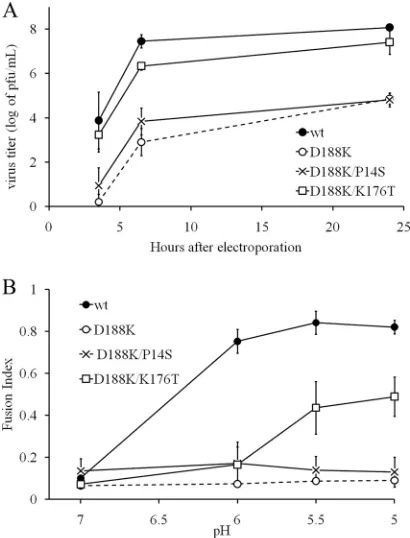
A single isolate retained the original K188 residue but ac-quired two additional mutations—P14S and K176T (Table 1). E1 P14 is located at the base of DI in the trimer, quite distant from the position of residue 188 on DII (Fig. 1A). It is mod-erately conserved among alphaviruses, with the majority of viruses having either a proline or serine at this position, sug-gesting that the P14S substitution in the revertant would not play an important functional role. In contrast, K176 is located in DII and positioned close to residue 188 in the central trimer interface (Fig. 1). K176, similar to D188, is completely con-served in all the reported alphavirus sequences. Thus, this revertant suggested that a K176T replacement could compen-sate for a lysine at position 188 in the core trimer.
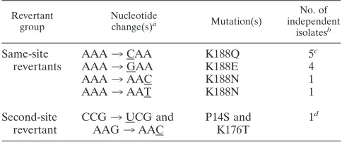
Growth properties of revertants.One example of each re-vertant containing a unique amino acid change was used for further characterization [D188(E/N/Q) and the second-site re-vertant D188K/K176T/P14S]. We first evaluated the growth properties of the revertants to assess the extent of reversal of the D188K fusion and infection block (Fig. 2). BHK cells were infected at low multiplicity, and the virus in the medium at the indicated time points was quantitated. D188K viruses exhibit virtually no progeny virus production by 24 h (29) (see also Fig. 5A). In contrast, all of the revertants showed efficient growth kinetics, with the growth of the D188E and D188Q rever-tants being similar to the kinetics of the wt. The D188N and D188K/K176T/P14S revertants had somewhat reduced growth kinetics, with final titers about 1.5 logs lower than the titer of the wt SFV.
Membrane fusion properties of revertants. While wt SFV fuses efficiently after treatment at low pH, the D188K mutant is fusion inactive at any pH from 4 to 11 (29). To evaluate the membrane fusion activity of the revertant viruses, serial dilu-tions of virus stocks were bound to BHK cells in the cold and treated with media of the indicated pH. The cells infected by virus fusion with the plasma membrane were quantitated by immunofluorescence (Fig. 3). wt SFV showed a fusion
[image:3.585.42.284.81.182.2]thresh-FIG. 1. Location of revertants in the E1 trimer. (A) The crystal structure of the postfusion E1* homotrimer (PDB entry 1RER) is shown with two chains in light gray and one chain colored as follows: DI in red, DII in yellow, DIII in blue, the fusion loop in green, the DI-DIII linker in black, and the N-terminal region of the stem in purple. The C-terminal stem connects to the transmembrane domain (neither of these is present in the crystal structure). The E1 residues discussed in this work are labeled and are represented as sticks high-lighted with colors for clarity. D188 on the g-h loop in DII is shown in cyan, K176 on the DII-strand f is in pink, and P14 on the DI-strand C0is in orange. (B) A view of the central trimer interface (fusion loops pointing toward the viewer) showing the positions of D188 and K176 in the crystal structure, with colors as in panel A but with oxygen shown in red and nitrogen in blue on the stick structures. A holmium atom (not shown) is coordinated by the three inwardly pointing D188 resi-dues. This figure was prepared using PyMol (9).
TABLE 1. Pseudorevertants of E1 D188K
Revertant group
Nucleotide
change(s)a Mutation(s)
No. of independent
isolatesb
Same-site revertants
AAA3CAA K188Q 5c
AAA3GAA K188E 4
AAA3AAC K188N 1
AAA3AAT K188N 1
Second-site revertant
CCG3UCG and AAG3AAC
P14S and K176T
1d
a
Nucleotide changes in the pseudorevertants are underlined.
b
Four 188Q revertants were isolated from 37°C cultures. All remaining re-vertants were isolated from 28°C cultures.
c
One also contains M280L.
d
Double mutation.
on November 8, 2019 by guest
http://jvi.asm.org/
[image:3.585.345.494.187.578.2]old of pH⬃6.0, maximal fusion at pH 5.75, and a decrease in fusion at lower pH due to the acid inactivation of the fusion protein. All of the revertants exhibited efficient fusion at their optimal pHs, with similar infectivities resulting from wt or revertant fusion with the plasma membrane (data not shown). However, all revertants showed an acid shift in the pH depen-dence of fusion, with maximal fusion occurring at pH values of
⬃5 to 5.5 (Fig. 3). All revertants showed acid inactivation at values below the optimum pH. The wild-type aspartic acid at position 188 thus appears optimal for fusion, but polar neutral residues (asparagine and glutamine substitutions) or negatively charged residues (glutamate) are fusion permissive. In addi-tion, D188K in combination with K176T permits low-pH-trig-gered membrane fusion, as discussed further below.
[image:4.585.58.265.66.233.2]Generation and characterization of D188I mutant. The same-site revertants included only polar residues even though a single nucleotide change could also have produced a muta-tion from lysine to isoleucine, a nonpolar residue of similar size as the parent aspartate residue. While our previous studies showed that a D188A mutation permits virus growth and mem-brane fusion, albeit with a more acidic pH threshold, the small size of the alanine side chain suggests that water molecules could be substituting for polar interactions in the core trimer. We therefore introduced the D188I mutation into the SFV infectious clone and characterized virus growth and fusion
FIG. 2. Growth kinetics of D188K revertants. BHK cells were in-fected with wt SFV or pseudorevertant viruses at a multiplicity of 0.01 infectious center/cell and incubated at 37°C. At the indicated time points the media were collected, and titers were determined. A rep-resentative example of two experiments is shown. Revertants are re-ferred to by the amino acid in the wt SFV sequence, its position, and the revertant amino acid replacement. Note that D188K is not shown here as its infectivity is greatly impaired (Fig. 5A).
FIG. 3. Virus-membrane fusion activity of D188K revertants. Wild-type and revertant viruses were prebound to BHK cells on ice, and virus fusion with the plasma membrane was triggered by treatment at the indicated pH for 1 min at 37°C. Cells were incubated overnight in the presence of ammonium chloride to prevent secondary infection, and infected cells were quantitated by immunofluorescence. Data are shown as the fraction of maximal fusion and represent the average of two experiments, with the bars indicating the range. Note that the parent D188K virus shows negligible membrane fusion activity (29).
on November 8, 2019 by guest
http://jvi.asm.org/
[image:4.585.112.474.373.671.2]properties. The mutant virus showed⬃1-log decrease in titer compared to the titer of the wt virus when cells were infected at low multiplicity (Fig. 4A) or by RNA electroporation (data not shown).
Fusion efficiency was first analyzed by testing cell-cell fusion. BHK cells were electroporated with wt or D188I RNA, incu-bated overnight to allow for abundant protein expression at the plasma membrane, and treated with low-pH medium to induce cell-cell fusion. wt E1 efficiently mediated cell-cell fusion with a threshold pH value of⬃6. At this pH⬃60% of the infected cells contained 2 to 11 nuclei/cell (Fig. 4B and data not shown). In contrast, the D188I mutant showed both a more acidic fusion threshold pH value of⬃5.5 and strongly reduced fusion efficiency. Minimal fusion was observed after treatment at pH 6, while even after treatment at pH 4,⬃60% of the infected cells showed no cell-cell fusion,⬃15% of the cells contained 2 nuclei/cell, and 25% of the cells contained 3 to 7 nuclei (Fig. 4B and data not shown).
Virus-membrane fusion was then tested by binding equiva-lent amounts of infectious D188I and wt SFV to BHK cells. Fusion was triggered by treatment with low-pH medium for 1 min and quantitated by determining the number of infected cells. Under these conditions, the fusion of D188I virus was decreased by⬃3 logs at pH 5 and⬃2 logs at pH 4 compared to fusion of wt SFV (Fig. 4C). Increasing the time of low-pH treatment to 2.5 min significantly increased D188I fusion al-though the D188I titers were still⬃2 logs lower than the titer
of wt SFV at pH 5 (Fig. 4D). Longer pH treatment did not significantly increase the amount of D188I fusion, and at all time points D188I had a significantly lower pH threshold (pH value of⬃4 to 5) than that of wt virus (pH value of⬃6.0) (data not shown). Thus, the kinetics of D188I membrane fusion were significantly slower than those of either the wt or revertant viruses. Together our data indicate that replacement of aspar-tate 188 with a similarly sized but nonpolar residue severely reduced the fusion activity of E1.
[image:5.585.109.474.65.323.2]Characterization of the second-site revertant.Our only sec-ond-site revertant was an E1 triple mutant containing the orig-inal D188K mutation plus P14S and K176T mutations (Table 1). Based on its position in DI (Fig. 1) and presence in alpha-viruses such as Venezuelan equine encephalitis virus, it ap-peared that the S14 residue was not responsible for the rescue of the D188K mutant. To directly determine the relevant mu-tation(s) in the revertant, we introduced mutations individually into the D188K infectious clone to generate D188K/K176T and D188K/P14S. Growth curves showed that, similar to the original revertant (Fig. 2), the D188K/K176T mutant repli-cated only about 1 log less efficiently than wt virus while growth of both D188K and the D188K/P14S double mutant was se-verely impaired (Fig. 5A). The E1 and E2 proteins of all four viruses were expressed at the plasma membrane in similar quantities (29; also data not shown), indicating that the defect in the D188K/P14S mutant was not due to defects in E1 folding or transport. Tests of cell-cell fusion activity showed that the
FIG. 4. Growth and membrane fusion properties of D188I virus. (A) Growth kinetics. The growth kinetics of wt and D188I virus were measured in BHK cells as described in the legend of Fig. 2. A representative example of two experiments is shown. (B) Cell-cell fusion activity of D188I. BHK cells were electroporated with wt or D188I RNA and incubated at 28°C overnight to permit abundant E1 and E2 expression at the cell surface. Fusion was induced by treatment at the indicated pH for 1 min at 37°C. Polykaryon formation was quantitated and expressed as a fusion index, as described in Materials and Methods. Data are the average and range of two experiments. (C and D) Virus-membrane fusion activity. Fusion activity of wt and D188I virus was measured as described in the legend of Fig. 3 using a treatment of 1 min (C) or 2.5 min (D) at the indicated pH. Equivalent titers of wt and mutant viruses were added to cells, but the fusion efficiency of D188I is significantly lower than that of wt virus. The average and range of two experiments are shown.
on November 8, 2019 by guest
http://jvi.asm.org/
D188K mutant was inactive in membrane fusion and that it was partially rescued by the K176T mutation while P14S had no effect (Fig. 5B). Thus, the K176T mutation was responsible for rescuing the fusion defect of D188K.
Generation of K176T and K176I mutants.The crystal struc-ture of the SFV E1 trimer shows that both D188 and K176 are located in the central trimer interface (Fig. 1). However, the native orientations of their side chains may be affected by the presence of holmium, which was added during crystallization to improve resolution and is coordinated by the three D188 residues of the trimer (13). Thus, holmium may have distorted the local bonding environment around D188, which our data indicate is an important region of protein-protein interaction in the trimer (29). The fusion defect of D188K was rescued by the K176T mutation, suggesting that K176 may also play an important role in trimer-ization and fusion. To test this, we generated SFV infectious clones containing a K176T mutation (the polar amino acid re-placement that rescued D188K) or a K176I mutation (a nonpolar residue that is closer in size to lysine).
Growth and fusion properties of K176T and K176I SFV.
Similar to the results with the D188(E/N/Q/I) mutations (Fig. 2 and 4), the K176T and K176I mutants were viable. When incubated at 37°C, K176T grew to similar titers in BHK cells as
the wt SFV (Fig. 6A). K176I exhibited a temperature-sensitive assembly defect at 37°C that was rescued at 28°C (data not shown). Growth curves performed at 28°C showed that growth of K176I was⬃2 to 3 logs lower than that of the wt SFV (Fig. 6B). Thus, similar to E1 D188, E1 K176 is not essential for virus replication.
The pH dependence of virus-membrane fusion was then determined for the K176T and K176I mutants. The wt virus showed maximal fusion at pH 6 (Fig. 6C and D). Both mutants had significantly more acidic fusion dependence: K176T showed maximal fusion at pH 5.5 (Fig. 6C) while K176I showed an even larger pH shift, with maximal fusion occurring at pH 5 (Fig. 6D).
Cell-cell fusion assays showed that the fusion efficiency of both mutants was reduced compared to that of the wt (Fig. 6E). Although the K176T threshold pH was⬃6.0, about 75% of the infected cells showed no fusion at pH 6 (data not shown). The fusion efficiency of K176I was markedly reduced even at pH 4.5. Over 80% of the K176I-infected cells showed no fusion after treatment from pH 5 to 6 (data not shown). Thus, in the context of the wt E1 sequence, threonine and isoleucine substitutions at K176 had strong effects on the pH dependence and efficiency of fusion. These data therefore sup-port a direct role for E1 K176 in fusion.
Computer modeling of D188 and K176.Together, our data showed that both D188 and K176 are important in regulat-ing the fusion step. Mutations at either of these positions produced similar phenotypes, including a more acidic pH dependence of fusion and decreased fusion efficiency. The ability of K176T to rescue the D188K mutant suggests a possible interaction between D188 and K176 in fusion. Be-cause the crystal structure of the trimer may not be an accurate representation of the bonding environment in this region, we used the modeling program multiconformation continuum electrostatics (MCCE) to capture the lowest en-ergy conformations of D188 and the residues surrounding it (2, 10, 40). The holmium ion was stripped from the crystal structure, and rotamers of D188 and the surrounding resi-dues were generated, keeping the alpha carbon backbone fixed as in the crystal structure. Protons were also added to the residues, and their free energy values were included in the energy lookup table, which allowed for the calculation of titration curves for each residue. The lowest energy, most occupied rotamer for each residue at a given pH was then selected and compiled in a PDB file (see Materials and Methods for details).
The results of these MCCE calculations demonstrated that D188 and K176 play an important role in stabilizing the trimer conformation. The calculated conformations of D188 and K176 indicate strong intermolecular salt bridges at the trimer interface, as shown in Fig. 7. D188 loses 7 kcal/mol of solvation energy due to its buried position in the interface, which would favor an increase in its pKa. Nonetheless, MCCE predicts that D188 is fully ionized in the calculated pH range (pH values of ⬃0 to 14) due to a number of intermolecular stabilizing interactions. In subunit A (D188a), interactions with the backbone of subunit C stabi-lize D188a by 2 kcal/mol, and interactions with the side
chains of subunit C stabilize by 7.5 kcal/mol. K176 of subunit C (K176c) interacts most strongly with D188a, with this FIG. 5. Definition of the second-site rescue mutation. To
deter-mine whether P14S or K176T rescued the D188K fusion defect, indi-vidual mutations were engineered into the infectious clone, and the viruses were characterized. (A) Growth kinetics. BHK cells were elec-troporated with the indicated viral RNAs and cultured for the indi-cated times at 37°C. The titer of progeny virus in the medium was determined. Data represent the averages and standard deviations of three independent experiments. (B) Cell-cell fusion activity. BHK cells were electroporated with the indicated viral RNAs, treated with pH medium for 1 min at 37°C, and assayed for low-pH-dependent cell-cell fusion activity as described in the legend of Fig. 4B. The average and range of two experiments are shown.
on November 8, 2019 by guest
http://jvi.asm.org/
[image:6.585.60.265.69.338.2]interaction stabilizing these two side chains by 4.5 kcal/mol. K123c, located below K176cand farther away from D188a, contributes about 2.5 kcal/mol to D188a stability. In
con-trast, residues from the same subunit (0.5 kcal/mol) and from subunit B (⬃0 kcal/mol) have minimal effects. As for D188, K176 is also buried in the trimer interface, losing 9 kcal/mol of solvation energy. However, K176 is similarly stabilized by intermolecular interactions, including D188 as well as several long-range interactions. Thus, both D188 and
K176 form strong interchain interactions, contributing to trimer stabilization.
DISCUSSION
[image:7.585.80.504.63.517.2]Effects of mutations in the core trimer interface.Our pre-vious work showed that the D188K mutant E1 protein inserts into target membranes but is unable to form the extended trimer intermediate (29). Formation of this core trimer is a
FIG. 6. Growth and membrane fusion properties of K176T and K176I viruses. (A) Growth kinetics of K176T. BHK cells were electro-porated with wt SFV or K176T mutant RNA and incubated at 37°C. At the indicated time points, the media were collected, and titers were determined by infectious center assay. (B) Growth kinetics of K176I. Growth kinetics were determined as described for panel A but using incubation at 28°C due to inefficient assembly of K176I virus particles at 37°C. Panels A and B each show a representative example of two experiments. (C and D) Virus-membrane fusion activity. Fusion of wt SFV, K176T, and K176I viruses with BHK cells was measured as described in the legend of Fig. 3. (E) Cell-cell fusion activity of K176T and K176I mutants. Cell-cell fusion of BHK cells infected with wt, K176T, and K176I RNAs was measured as described in the legend of Fig. 4B. Data in panels C to E are the average and range of two experiments.
on November 8, 2019 by guest
http://jvi.asm.org/
requisite step in E1 refolding to the final hairpin and is re-quired for virus fusion and infection. Most of the revertants we describe here contained replacements of K188 with either neu-tral polar residues (glutamine or asparagine) or a negatively charged residue (glutamic acid). These rescue mutations pro-duced viruses that grew quite efficiently but had more acidic pH thresholds for fusion, similar to the D188A mutation we had previously characterized (29). Replacing D188 with iso-leucine produced an even more acidic pH shift in fusion and decreased fusion efficiency and kinetics. This is in keeping with the large nonpolar isoleucine side chain inhibiting polar inter-actions that would be permitted by E, Q, and N substitutions or by water molecules that could enter the trimer interface due to the small size of the alanine side chain.
The second-site mutation K176T efficiently rescued the fu-sion block in D188K and provided key insights into the mech-anistic role of D188 in trimer formation. Computer modeling studies revealed a ring of interactions in the core trimer inter-face linking D188 of one chain with K176 of the neighboring chain.De novoreplacement of K176 with T or I produced a pH shift phenotype, and, similar to D188I, the K176I mutation had the strongest effect on pH dependence, fusion efficiency, and virus growth. The modeling and mutational studies strongly suggest that D188 and K176 form an interchain salt bridge that positions and stabilizes the trimer core.
The SFV E1 homotrimer is large (over 1,000 residues) and contains multiple contacts within the core trimer interface and between the core trimer and the DIII stem regions (13). None-theless, the revertants showed that relatively small changes in side chain length (e.g., D188E) or electrostatic potential (e.g., D188Q) in the trimer interface could produce marked changes in the pH dependence of fusion. The D188K mutation results
in six total positive charges in the core trimer interface, three from K188 and three from the adjacent K176. The resultant electrostatic repulsion appears sufficiently destabilizing to pre-vent compensation by other interactions since D188K rever-tants exclusively reflected substitutions at position 188 or 176. Together, these results highlight the importance of the core trimer interface and the D188-K176 interaction during forma-tion of the E1 homotrimer.
Computational analysis of titratable residues.Viruses that use a low-pH fusion trigger have developed mechanisms to respond to pH changes within the physiological range. Titrat-able residues can display a wide range of pKavalues depending on properties of the local environment, such as solvent acces-sibility or electrostatic potential (reviewed in references 37 and 41). For example, the aspartate carboxylate titrates at pH⬃4.4 in aqueous solution, while within a protein its pKacan range
from⬃2 to 10 (24). These properties can make it difficult to identify residues or interactions that are relevant to low-pH responses. Computer modeling programs are becoming in-creasingly more sophisticated in predicting the behavior of residues in their local environments. Because protonation of each residue is sampled and included in the energy lookup table, a titration curve for specific residues can be calculated, and the bonding configuration at a particular pH can be ana-lyzed.
In our work, the MCCE program was used to model the lowest energy bonding configuration of D188 in the trimer at pH 6.0. This analysis computationally removed the holmium that was added to improve resolution of the crystal structure and was coordinated by the three D188 residues in the trimer interface (13). D188 and its surrounding residues were then allowed to freely rotate and be titrated to generate the lowest energy; most occupied positions at pH 6.0. The results identi-fied a ring of interactions at the core trimer interface between D188 of one chain and K176 of the neighboring chain. The results of the computational analysis were corroborated by mutagenesis studies of K176.
Role of D188 and K176 in low-pH-triggered E1 refolding.
The pH dependence of alphavirus fusion is controlled by two major steps, the release of the E2/E1 heterodimer interaction and the subsequent refolding of E1 to the final trimeric hairpin (37). While the dissociation of the E2/E1 dimer is a critical early low-pH-dependent step, E1 also responds independently to low pH during trimerization. Our results indicate that al-though the revertants (e.g., D188Q and D188I) have signifi-cantly more efficient growth and fusion than D188K, their fusion requires a more acidic pH than that of the wt virus. Based on our prior results with D188A (29) and results pre-sented here, the more acidic pH threshold of fusion in the revertants is due to a more acidic pH threshold for formation of the E1 homotrimer rather than to effects on dissociation of the E2/E1 heterodimer.
[image:8.585.59.268.67.273.2]Many changes occur in the structure and bonding environ-ment of E1 as it transitions from its prefusion conformation through largely uncharacterized transient intermediates to the final stable homotrimer structure. In order for three E1 pro-teins to acquire the correct trimeric interactions, it seems likely that considerable sampling between charged pairs must occur. Sensitive assays show that alphavirus fusion occurs within sec-onds at low pH and physiological temperatures (5, 36), and
FIG. 7. Model of K176-D188 intersubunit interactions in the E1 homotrimer. The MCCE program was used to model the predominant conformation in the trimer interface at pH 6 (see Materials and Meth-ods). The figure depicts a section of the central trimer interface with the fusion loops pointing toward the viewer. Each chain in the trimer is indicated (chain A, B, or C), and residues K176 and D188 of each chain are indicated with the subscript denoting the chain. The dis-tances between each D188-K176 pair are indicated.
on November 8, 2019 by guest
http://jvi.asm.org/
thus the process of E1 trimerization is very rapid. MCCE analysis predicted that E1 residues D188 and K176 form a very strong bond that is stable even at pH 0. Do D188 and K176 play a direct role in the low-pH dependence of E1 refolding? Neither residue appears to have important contacts that would affect the stability of the prefusion conformation (35). It also does not seem likely that the D188-K176 salt bridge contrib-utes significantly to low-pH dependence, given the predicted stability of this bond across the pH range (pH 7 to 5) found in the endocytic pathway. Instead, we favor a model in which D188 and K176 make a key interaction during the initial for-mation of the core trimer, which helps to orient E1 molecules, constrain nonproductive E1 interactions, and enhance low-pH-dependent E1 interactions that then drive refolding to the postfusion trimer. As an example of one such low-pH-depen-dent interaction, a conserved histidine residue at position 3 on DI promotes the refolding of E1 to the final postfusion trimer (33). In the absence of the D188-K176 interaction, such as in the case of the same-site revertants, the efficiency of formation of such pH-dependent bonds is decreased, and lower pH or more time is required to promote trimerization. This model is supported by our results with D188I, whose fusion was pro-moted by either lower pH or longer incubation times.
Steps in the SFV E1 refolding reaction.Following release or rearrangement of the E2/E1 dimer, monomeric E1 inserts into the target membrane via the fusion loop (1, 11). Three E1 proteins then form the core trimer, a step that is promoted by the ring of D188-K176 interactions described here. Other in-teractions within the core trimer presumably also act to stabi-lize this intermediate. DIII and the juxtamembrane stem re-gion pack into the grooves formed by the core trimer (26, 27, 38), ending with the final postfusion E1 hairpin.
There is experimental information, albeit incomplete, on the low-pH dependence of these E1 refolding steps. Initial fusion loop-membrane insertion may not itself require low pH and may be reversible prior to trimer core formation (38). Muta-tion of H3 to alanine causes misregulaMuta-tion of the pH depen-dence of trimerization, perhaps reflecting key low-pH-depen-dent interactions of this ionizable residue (33). DIII and stem packing are critical for fusion (25), but this packing does not itself appear to be low pH dependent (38). While E1 trimers can interact cooperatively during membrane insertion (12, 38), the low-pH requirements of such intertrimer interactions and their role in fusion remain to be determined. A combination of computational, structural, and biochemical approaches should allow definition of the low-pH-dependent steps in E1 refolding and their functions in membrane fusion.
ACKNOWLEDGMENTS
We thank Mark Girvin of the Department of Biochemistry at Ein-stein for his helpful suggestions and hands-on assistance with the use of the MCCE program. We thank Marilyn Gunner of the Department of Physics of the City College of New York for helpful discussions and input on the MCCE program and its use. We thank all of the members of our lab for discussions and comments on the manuscript and Kartik Chandran and the members of his lab for their input. We thank Sonu Nanda for excellent technical assistance.
This work was supported by a grant to M.K. from the National Institute of Allergy And Infectious Diseases (R01-AI075647) and by Cancer Center Core Support Grant NIH/NCI P30-CA013330. C.Y.L. was supported in part through the Medical Scientist Training Program of the Albert Einstein College of Medicine (NIH T32 GM07288).
REFERENCES
1.Ahn, A., D. L. Gibbons, and M. Kielian.2002. The fusion peptide of Semliki
Forest virus associates with sterol-rich membrane domains. J. Virol. 76:
3267–3275.
2.Alexov, E. G., and M. R. Gunner.1997. Incorporating protein conforma-tional flexibility into the calculation of pH-dependent protein properties.
Biophys. J.72:2075–2093.
3.Bharadwaj, R., A. Windemuth, S. Sridharan, B. Honig, and A. Nicholls.
1995. The fast multipole boundary-element method for molecular
electro-statics: an optimal approach for large systems. J. Comput. Chem.16:898–
913.
4.Bressanelli, S., K. Stiasny, S. L. Allison, E. A. Stura, S. Duquerroy, J. Lescar, F. X. Heinz, and F. A. Rey.2004. Structure of a flavivirus envelope glyco-protein in its low-pH-induced membrane fusion conformation. EMBO J.
23:728–738.
5.Bron, R., J. M. Wahlberg, H. Garoff, and J. Wilschut.1993. Membrane fusion of Semliki Forest virus in a model system: correlation between fusion kinetics and structural changes in the envelope glycoprotein. EMBO J.
12:693–701.
6.Chanel-Vos, C., and M. Kielian.2004. A conserved histidine in the ij loop of the Semliki Forest virus E1 protein plays an important role in membrane
fusion. J. Virol.78:13543–13552.
7.Chanel-Vos, C., and M. Kielian.2006. Second-site revertants of a Semliki Forest virus fusion-block mutation reveal the dynamics of a class II
mem-brane fusion protein. J. Virol.80:6115–6122.
8.Corver, J., R. Bron, H. Snippe, C. Kraaijeveld, and J. Wilschut.1997. Membrane fusion activity of Semliki forest virus in a liposomal model
sys-tem: specific inhibition by Zn2⫹ions. Virology238:14–21.
9.DeLano, W. L.2002. The PyMOL user’s manual. DeLano Scientific, San Carlos, CA.
10.Georgescu, R. E., E. G. Alexov, and M. R. Gunner.2002. Combining con-formational flexibility and continuum electrostatics for calculating pK(a)s in
proteins. Biophys. J.83:1731–1748.
11.Gibbons, D. L., A. Ahn, M. Liao, L. Hammar, R. H. Cheng, and M. Kielian.
2004. Multistep regulation of membrane insertion of the fusion peptide of
Semliki Forest virus. J. Virol.78:3312–3318.
12.Gibbons, D. L., I. Erk, B. Reilly, J. Navaza, M. Kielian, F. A. Rey, and J. Lepault.2003. Visualization of the target-membrane-inserted fusion protein of Semliki Forest virus by combined electron microscopy and
crystallogra-phy. Cell114:573–583.
13.Gibbons, D. L., M.-C. Vaney, A. Roussel, A. Vigouroux, B. Reilly, J. Lepault, M. Kielian, and F. A. Rey.2004. Conformational change and protein-protein
interactions of the fusion protein of Semliki Forest virus. Nature427:320–
325.
14.Harrison, S. C.2005. Mechanism of membrane fusion by viral envelope
proteins. Adv. Virus Res.64:231–261.
15.Harrison, S. C.2008. Viral membrane fusion. Nat. Struct. Mol. Biol.15:690– 698.
16.Kielian, M., C. Chanel-Vos, and M. Liao.2010. Alphavirus entry and
mem-brane fusion. Viruses2:796–825.
17.Kielian, M., S. Jungerwirth, K. U. Sayad, and S. DeCandido.1990. Biosyn-thesis, maturation, and acid-activation of the Semliki Forest virus fusion
protein. J. Virol.64:4614–4624.
18.Kielian, M., M. R. Klimjack, S. Ghosh, and W. A. Duffus.1996. Mechanisms of mutations inhibiting fusion and infection by Semliki Forest virus. J. Cell
Biol.134:863–872.
19.Kielian, M., and F. A. Rey.2006. Virus membrane fusion proteins: more than
one way to make a hairpin. Nat. Rev. Microbiol.4:67–76.
20.Kuhn, R. J.2007.Togaviridae: the viruses and their replication, p. 1001–1022.
InD. M. Knipe, P. M. Howley, D. E. Griffin, R. A. Lamb, M. A. Martin, B.
Roizman, and S. E. Straus (ed.), Fields virology, 5th ed., vol. 1. Lippincott, Williams and Wilkins, Philadelphia, PA.
21.Kuhn, R. J., and M. G. Rossmann.2005. Structure and assembly of
icosa-hedral enveloped RNA viruses. Adv. Virus Res.64:263–284.
22.Lescar, J., A. Roussel, M. W. Wien, J. Navaza, S. D. Fuller, G. Wengler, and F. A. Rey.2001. The fusion glycoprotein shell of Semliki Forest virus: an icosahedral assembly primed for fusogenic activation at endosomal pH. Cell
105:137–148.
23.Levy-Mintz, P., and M. Kielian.1991. Mutagenesis of the putative fusion
domain of the Semliki Forest virus spike protein. J. Virol.65:4292–4300.
24.Li, H., A. D. Robertson, and J. H. Jensen.2005. Very fast empirical
predic-tion and rapredic-tionalizapredic-tion of protein pKa values. Proteins61:704–721.
25.Liao, M., and M. Kielian.2005. Domain III from class II fusion proteins functions as a dominant-negative inhibitor of virus-membrane fusion. J. Cell
Biol.171:111–120.
26.Liao, M., and M. Kielian.2006. Functions of the stem region of the Semliki
Forest virus fusion protein during virus fusion and assembly. J. Virol.80:
11362–11369.
27.Liao, M., and M. Kielian.2006. Site-directed antibodies against the stem region reveal low pH-induced conformational changes of the Semliki Forest
virus fusion protein. J. Virol.80:9599–9607.
on November 8, 2019 by guest
http://jvi.asm.org/
28.Liljestro¨m, P., S. Lusa, D. Huylebroeck, and H. Garoff.1991. In vitro mu-tagenesis of a full-length cDNA clone of Semliki Forest virus: the small 6,000-molecular-weight membrane protein modulates virus release. J. Virol.
65:4107–4113.
29.Liu, C. Y., and M. Kielian.2009. E1 mutants identify a critical region in the
trimer interface of the Semliki Forest virus fusion protein. J. Virol. 83:
11298–11306.
30.Mancini, E. J., M. Clarke, B. E. Gowen, T. Rutten, and S. D. Fuller.2000. Cryo-electron microscopy reveals the functional organization of an
enve-loped virus, Semliki forest virus. Mol. Cell5:255–266.
31.Modis, Y., S. Ogata, D. Clements, and S. C. Harrison.2004. Structure of the
dengue virus envelope protein after membrane fusion. Nature427:313–319.
32.Nicholls, A., K. A. Sharp, and B. Honig.1991. Protein folding and associa-tion: insights from the interfacial and thermodynamic properties of
hydro-carbons. Proteins11:281–296.
33.Qin, Z. L., Y. Zheng, and M. Kielian.2009. Role of conserved histidine residues in the low pH-dependence of the Semliki Forest virus fusion
pro-tein. J. Virol.83:4670–4677.
34.Rocchia, W., S. Sridharan, A. Nicholls, E. Alexov, A. Chiabrera, and B. Honig.2002. Rapid grid-based construction of the molecular surface and the use of induced surface charge to calculate reaction field energies: applica-tions to the molecular systems and geometric objects. J. Comput. Chem.
23:128–137.
35.Roussel, A., J. Lescar, M.-C. Vaney, G. Wengler, G. Wengler, and F. A. Rey.
2006. Structure and interactions at the viral surface of the envelope protein
E1 of Semliki Forest virus. Structure14:75–86.
36.Samsonov, A. V., P. K. Chatterjee, V. I. Razinkov, C. H. Eng, M. Kielian, and F. S. Cohen.2002. Effects of membrane potential and sphingolipid structures
on fusion of Semliki Forest virus. J. Virol.76:12691–12702.
37.Sanchez-San Martin, C., C. Y. Liu, and M. Kielian.2009. Dealing with low
pH: entry and exit of alphaviruses and flaviviruses. Trends Microbiol.17:
514–521.
38.Sanchez-San Martin, C., H. Sosa, and M. Kielian.2008. A stable prefusion intermediate of the alphavirus fusion protein reveals critical features of class
II membrane fusion. Cell Host Microbe4:600–608.
39.Sitkoff, D., K. A. Sharp, and B. Honig.1994. Accurate calculation of
hydra-tion free-energies using macroscopic solvent models. J. Phys. Chem.98:
1978–1988.
40.Song, Y., J. Mao, and M. R. Gunner.2009. MCCE2: improving protein pKa
calculations with extensive side chain rotamer sampling. J. Comput. Chem.
30:2231–2247.
41.Srivastava, J., D. L. Barber, and M. P. Jacobson.2007. Intracellular pH
sensors: design principles and functional significance. Physiology22:30–39.
42.White, J. M., S. E. Delos, M. Brecher, and K. Schornberg.2008. Structures and mechanisms of viral membrane fusion proteins: multiple variations on a
common theme. Crit. Rev. Biochem. Mol. Biol.43:189–219.
43.Zhang, W., S. Mukhopadhyay, S. V. Pletnev, T. S. Baker, R. J. Kuhn, and M. G. Rossmann.2002. Placement of the structural proteins in Sindbis virus.
J. Virol.76:11645–11658.