0022-538X/80/03-1026/08$02.00/0
Isolation and
Characterization of Recombinant DNA Clones of
Avian
Retroviruses:
Size
Heterogeneity and Instability of the
Direct Repeat
GRACEJU,LAWRENCE BOONE,ANDA. M.SKALKA*
DepartmentofCellBiology, RocheInstitute of Molecular Biology, Nutley, New Jersey 07110
Unintegrated proviral DNA of Schmidt-Ruppin B Rous sarcoma virus was
cloned in the bacteriophage A vector Charon 21A. A total of 12 independent
recombinantXSRBtdclones which were derived from the
transformation-defec-tive componentinthe viral preparationwereanalyzed with restriction
endonu-cleases and molecular hybridization techniques. Three classes of clones were
observed.Type Iclonescontained a5.0-megadaltoninsert of viral DNA, type II
clones contained phage withtwosizeclassesof inserts (5.0 and 5.2megadaltons),
andonetype IIIclone containedonly a5.2-megadaltoninsert. Thesmallerinsert
present in type II clones appeared to be derived by deletion of one copy of a
directlyrepeatedsequencewhichwaspresent in the larger insert. Mapping data
indicated that thedeletion includes all orpart of the terminalrepeat found in
lineardouble-stranded proviralDNA.Similarresults were obtained fromXRAV2
recombinant clones derived from Rous-associatedvirus type 2.Analysis of DNA
fromtype IIandtype IIIclones ofASRBtdandXRAV2revealed limited
hetero-geneityinthe size ofthedirectrepeat.
Replication of the retroviruses involves the
production of proviral DNA intermediates
be-fore integration into the host cell genome. At
least three types of intermediates have been
identifiedincells afterexogenousinfection:
lin-eardouble-stranded DNAandtwo sizes of
co-valently closed circular DNA (10). For the avian retroviruses, the linear double-stranded DNA
has been shown to contain a direct repeat of
approximately300basepairs (bp) (0.2
megadal-ton
[Md])
atitstermini.Thisduplication is alsopresent in the largerof the two circular
inter-mediates (16, 25), which may be formed from
the linearDNAbyintramolecularjoiningatthe
termini. Restrictionmappingdata indicatethat
thesmaller circular specieshasonlyonecopyof
the terminal sequence (25). The circular forms
arefoundexclusivelyinthenucleus(26),and,as
withbacteriophage lambda(9), acircular
inter-mediate of the provirus is thought to be the substrate for integrationinto the host genome (10, 13).Integrated provirushas also been found tocontain terminal redundancies (17, 24),
sug-gestingthattherepeatedsequencesareinvolved
intheintegrationreaction. Thelimitedquantity
of viral DNA found in infected cellshas made
analysis of these intermediates difficult.
How-ever,larger amountsofpurifiedDNAcan now
be obtainedbyusing recombinant DNA
meth-odology.
Unintegrated circular proviral DNA from
chicken embryo fibroblasts infected with
Schmidt-Ruppin B Rous sarcoma virus
(SR-RSV-B)wasisolated and cloned in the
bacterio-phageX vectorCharon21A(Ch21A)by usingan
enzymeknowntocuttheproviralDNA atonly
one site. Three types ofXSRBtd recombinant
DNA clones derived from the
transformation-defective (td) componentof the viruswere
iso-lated.OnetypecontainedaviralDNAinsert of
5.0Md,asecond containedalargerviralinsert
of 5.2 Md, and athird contained recombinant
molecules with either of thesetwosize classes of
inserts. Experimentsusing cloneswith twosize
classes indicated that the smaller insert could
arise by deletion of one copy of the repeated
sequence from thelarger insert during growth
within procaryotic host cells. Similar results
wereobtained with recombinant clonesprepared
fromunintegrated proviralDNAof
Rous-asso-ciated virustype2(RAV-2). Furthermore,in six
independent clones examined, the size of the
repeated sequences was found to be
heteroge-neous. The implications of these results for
mechanisms ofviralreplicationin vivoare
dis-cussed.
MATERIALS AND METHODS
Celis
and viruses. Chickenembryofibroblasts(gs-chf; SPAFAS, Norwich, Conn.) wereprepared and propagated as described previously (20). The SR-RSV-B usedin theseexperimentswasarecombinant
betweenNY68,atemperature-sensitivemutantof
SR-RSV-A (subgroup A), and RAV-2 (subgroup B). It 1026
on November 10, 2019 by guest
http://jvi.asm.org/
contained the temperature-sensitive mutation of NY68 in the transformation gene (src) and the sub-group B specificity of RAV-2 (18). The virus was obtainedas acloned stock from H.Hanafusa, Rocke-feller University. The A vector Ch21A has been de-scribedpreviously (4). Escherichia coli K-12 strains
employedweretheEK1 hosts ED8767(recArk-mk') (22) and C600 (recA-rk-mk), which was obtained fromR.Davis, Stanford University, and the EK2 host DP50supF(recA+).
DNAisolation. To prepareproviral DNA,25petri
dishes(150mm2) of chicken embryofibroblastswere infectedathighmultiplicity (multiplicity of infection, >1 focus-forming unit per cell). After48h, infected cellswerelysed with detergent, and high-molecular-weightnuclear DNAwaspelleted by the method of Hirt(15). The Hirtsupernatant fractionwastreated withproteinase K (50,Ig/ml) at37°C for 2.5 h, ex-tracted with phenol, and precipitated with ethanoL Theethanolprecipitatewas nextdissolved,extracted with chloroform-isoamyl alcohol (100:1), and again concentratedbyethanolprecipitation.The DNAwas then dissolved in TEN buffer (0.01 M Tris,pH 7.4,
0.001MEDTA,0.01MNaCl),treated withRNase A
(50,ug/ml)at37°Cfor1h, extracted with phenol and
chloroform-isoamyl alcohol,and then ethanol precip-itated. The precipitate wasdissolved and covalently closed circular viral DNAwasseparatedfrom relaxed circles and linear moleculesby isopycnic centrifuga-tion inacesium chloride-propidium diiodide density gradient (27).
Recombinant DNAtechniques. All cloning ex-perimentswereconducted under the P2-EK2 or P3-EK1 conditionsspecified by the December 1978 re-visedNational Institutes of Health guidelines.
A portion of the covalently closed proviral DNA
preparation of SR-RSV-B was digested with Sail, ligatedtoSalI-digested A Ch21A with T4 ligase (21),
encapsulatedby in vitro packaging techniques (3), and plated on the recA- strain ED8767. Approximately 36,000plaqueswerescreenedfor viral DNA inserts by using the method of Benton and Davis (2) anda3p labeled RAV-2 viral RNA probe. Positiveplaques were isolated andpurified twice before amplification and extraction ofDNAfor restriction endonuclease anal-ysis.
Nuclease digestion. Restriction endonucleases
AluI, SalI,HindIII,XhoI,XbaI,KpnI,andPvuIwere purchased from New England Biolabs, Beverly, Mass. EcoRIwaspurchased from Miles Laboratories,
Elk-hart, Ind.Digestionswereperformedin 25 or 50
pl
of theappropriate buffer.Gel electrophoresis, DNA transfer, and hy-bridization. Agarose gel electrophoresis and DNA transferwereperformedby the method ofSouthern
(28),aspreviously described (20). Hybridization pro-cedures havebeendescribed previously (20).For mo-lecular weight determinations, parallel lanes of the
gelscontainedHindIIIdigests of"C-labeledA DNA.
To determine the size of small DNA fragments
(<1,000 bp), digested DNA samples were analyzed by
electrophoresis invertical 6%polyacrylamide (acryl-amide/bisacrylamide ratio, 40:1) gels in TBE buffer (0.89 M Tris, pH 8.3,0.89 M boric acid, 0.01 M EDTA). Fragments of46X174 DNA digested withAluI were usedassizemarkers.
RESULTS
Molecular cloning of SR-RSV-B viral
DNA. Unintegrated covalently closed circular
proviralDNAwaspurifiedfrom chickenembryo fibroblasts infectedwithSR-RSV-Basdescribed above. The proviral DNA was digested with
restriction endonuclease Sall,whichwasfound tocleave the circular moleculesatasingle
loca-tion(Fig. 1). The cleaved DNAwasthenligated
totheAvectorCh21Aandpackagedintophage particles in vitro. Recombinant clones were
screened forvirus-specific inserts by using
32P-labeled RAV-2 viral RNA. SR-RSVand RAV-2
are approximately 80 to 90% homologous, as
determined by hybridization of SR-RSV 70S
RNA to complementary DNA prepared from
RAV-2 (14).
Cells infectedwith aviansarcomaviruses
con-tain nondefectiveand td DNAmolecules,which
have sizes of approximately 6.2 and 5 Md,
re-spectively (25). The td molecules are deletion mutantsderivedfromnondefective moleculesby loss of all or part of the viral src gene, which
codes for cellular transformation.Intheproviral DNApreparation used for cloning, td molecules represented approximately 50% of thecovalently closed circular molecules (data notshown). In
this report,recombinant clonescontaining only td moleculesarediscussed. Thecloning vehicle
Ch21A, in combination with the in vitro pack-aging procedure, selects for inserts witha
maxi-mum size of approximately 5.3 Md (8.2
kilo-bases) (lla). Because of its size limit, theuseof
Ch21Aallowedustoobtain clones withinserts
ofonly singletd molecules(Fig. 1and2). Restriction endonuclease analysis of XCh21A SR-RSV-B td (XSRBtd)
recombi-nant clones. A total of 12 independently de-rived clones containing viral DNA sequences
were isolated and plaque purified. DNA from
eachASRBtdclonewasdigested with Sall and
analyzed byagarose gel electrophoresis.
Diges-tion withSall generated thetwoarmsofCh21A
amI X Hindm XhoI SalI
I -I I
ll c
M
gag pol ."t
EcoRI EcORI Kpn I
JL Xbol Xbal
ECKO
RI
| CO RLPVUI PVUI
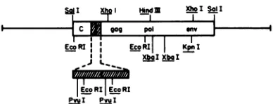
FIG. 1. Restrictionendonuclease map of ASRBtd
recombinant clones. Cleavage sites ofseven
restric-tionendonucleases inthelarger (5.2-Md) viral insert
of ASRBtdclonesareindicated. Theinsert isshown inorientationrwithadirectrepeat(shaded region). The relative orderof the viralgenesin the viral RNA
genome is: (5')gag, pol, env, C (3'). Ajagged line
indicatesthe siteofjoining of the repeatedsequences. VOL. 33,1980
on November 10, 2019 by guest
http://jvi.asm.org/
[image:2.504.252.446.508.582.2]C. 3 4 5 2 3 4 -, 6 7 8
EsI~A
de ,_
9 10 11 6 7 8 9 (
' #i#
. . _
v_mi,,
*4
B
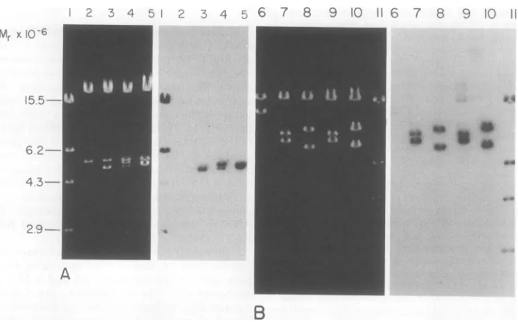
FIG. 2. SalI andHindIII digestionpatterns of ASRBtdclones. DNA from phage of XSRBtdcloneswas
extracted, digested, analyzed by electrophoresisin 0.7%agarose gels,and transferredtonitrocellulose filters
aspreviouslydescribed(20).Bandscontaining viralsequences weredetected after hybridization with
32P-labeled RAV-2 RNA (specific activity, -2x 10⁷ to 4 x 10⁷ Cerenkov cpm/lpg),prepared, and utilizedas
describedpreviously (20).Theleft panelsareethidiumbromide visualizations ofdigestedDNA from XSRBtd
clones,and therightpanelsareautoradiograms ofthegels after transfer andhybridization.(A)SalI digestion patterns. Lane1,marker"C-labeled DNAcleavedwithHindIII; lane2, Ch21A;lane3,XSRBtd-1; lane4, XSRBtd-2;lane5,ASRBtd-7.(B) HindIIIdigestionpatterns.Lane6,Ch21A; lane7,ASRBtd-1(orientation 1);
lane 8, XSRBtd-5 (orientation r); lane9, XSRBtd-2 (1); lane 10, XSRBtd-7(r); lane11,"C-labeled ADNA
cleaved withHindIII.
and the insertfragment(s) ineachrecombinant clone. Three types of clones could be distin-guished by differencesin theirdigestionpatterns
(Fig. 2A and Table 1). Digestion oftypeIclones (8of12) generatedaninsertfragment of 5.0 Md.
Type II clones (3 of 12) containedtwodifferent sizes of insert DNA(5.0 and5.2 Md). Onetype
IIIclone (XSRBtd-7) containedan insert of 5.2
Md.
Several XSRBtd cloneswere further charac-terizedby digestionwithanumber ofrestriction endonucleases. Figure 1 isa compositemap of
the larger viral insert in Ch21A, showing the
cleavage sites forsevenrestrictionenzymes. Al-though we observed differences in thenumber
and location ofsome ofthe HindIII and XbaI
sites, ourmapisingeneralagreement withthe
restriction maps reported for other avian sar-comavirus strains(16, 25).
With each of the clones, digestion with
HindIII,whichcleavesclosetothecenterofthe insertDNA(Fig.1)andoncewithin theAvector
leftarm,producedtwolarge fragments contain-ing virus-specificsequences.The sizes of the two
large fragments differed slightlyin the various
clones(Fig. 2B),asexpectediftheproviral DNA
TABLE 1. Recombinant DNAclonesof ASRBtd
Type XSRBtd Size of insert Orientation'
Tye clone (Md)a retain
I 1 5.0 1
3 5.0 l
4 5.0 l
5 5.0 r
6 5.0 r
8 5.0 r
11 5.0 r
12 5.0 r
II 2 5.0and5.2 1
9 5.0and5.2 r
10 5.0and5.2 r
III 7 5.2 r
aDeterminedby
SailI
digestion."DeterminedbyHindIIIdigestion.
was inserted in either one ofthe two possible
orientations. The orientationssuggested bythe
HindIIIdigestion patternswereverifiedby
anal-ysiswith other restriction endonucleases. Inserts
incorporatedsuch that the aviansarcomavirus
geneticmapis colinear withAlategenes
(tran-.1
Mr x 10-6
15.5-6
2-
43-
29-A
J. VIROL.
on November 10, 2019 by guest
http://jvi.asm.org/
[image:3.504.75.449.61.295.2] [image:3.504.269.460.408.574.2]1029
scribed tothe right) are designated as being in
the r orientation (Table 1). Inserts in the r
orientation yielded virus-specific fragments of 8.65 and 8.0 Md when digested with HindIII,
and inserts in the left (1) orientation produced
fragments of 9.2 and 7.45 Md after digestion with
HindIII (Fig.2B).ResultsofHindIIIdigestions
ofindividual clones also indicatedthat none of
the clones containedmultipletandem inserts.
Smallerinserts intype II clones arise by
deletionoflarger inserts. Because of the size
limitations of the Ch21A vector and the data
from restrictionanalysis, it was clear that each
recombinantphagechromosome contained only
oneproviral genome. However, two sizes of
in-serts weredetectedinDNAfromtype II clones. It seemed possible, therefore, that the 5.0-Md
insertsin type II clones might arise by one of
the two following mechanisms: (i) the original
plaque containedtwo ormore different
recom-binantphage particles;or(ii) deletionof part of
thelarger insertgeneratedthesmaller one
dur-ing passage and amplification of the original
plaque-purifiedclone.
Todistinguish between thesetwopossibilities,
one type II clone (XSRBtd-2) was replated at
low density, and phage from 16 independent
plaqueswereisolatedandamplified. SalI
restric-tion endonuclease analysis of DNA from the
subclones revealed that eightwere identical to
the original parental clone and also contained
phage with the same twosizes ofinserts. The
relative proportions of the larger and smaller
inserts were variable among these clones, as
shown by the four representative examples in
Fig. 3, lanes3 through6. The other eight
sub-clonescontainedonly the smaller 5.0-Md insert,
as shown by the four clones in Fig. 3, lanes 7
through10. None of the 16subclonescontained
onlythe 5.2-Md insert.
If the typeII cloneshad containedamixture
ofparticles, then thesubsequent plaque
purifi-cation should have produced clones that
con-tained either the larger or the smaller insert.
Since no subclones contained only the larger
insert and 50% of the subclonescontained two
sizes ofinserts, it seemed most likely that the
smallerinsert arosefromthelargeroneby
dele-tion.Thus, weconclude thateach type IIclone
probably originated from a recombinant
mole-cule which contained a single unique insertof
5.2 Md. A subsequent specific deletion of
ap-proximately0.2Mdduringpassage ofeachclone
generated themixture ofmolecules. The
varia-tion inthe relative proportion ofthe twosizes of
inserts in subclones ofXSRBtd-2 (Fig. 3)
indi-catedthatthedeletioneventprobably occurred
atadifferent time for eachsubclone. Although
the deletionof a0.2-Mdfragmentoccurred
fre-2 3 4 5 6 7 8 9 10
Mr X106
15.5-
6.2-
4.3-
2.9-FIG. 3. SailI digestion patterns of subclones of
ASRBtd-2. The type II clone ASRBtd-2wasplaque purifiedonstrain ED8767.Phage fromindependent plaques were isolated and amplified. DNAs from
these subcloneswereanalyzed bySalIdigestionand electrophoresis in 0.7% agarose gels. This figure shows results with the parent cloneASRBtd-2 and eight representative subclones.Fragments were vis-ualized after ethidium bromide staining. Lane 1, marker XDNA cleaved withHindIII; lane 2, parent
cloneXSRBtd-2; lane 3,XSRBtd-2.3;lane 4,
XSRBtd-2.6; lane 5,XSRBtd-2.7;lane6,ASRBtd-2.11; lane 7,
XSRBtd-2.4; lane 8, XSRBtd-2.14; lane 9, XSRBtd-2.15;lane 10, XSRBtd-2.16.
quently, no additional deletions were detected
inany of theXSRBtd clonesorXSRBtd-2
sub-clones.
All recombinant phage particles were
origi-nallygrown andamplified in ED8767, a
recA-host. To determine whether the Rec function
couldacceleratedeletionformation,typeI,type
II, and type III clones were also grown in a
secondrecA- mutant (derivedfrom C600) and
arecA+ (DP50supF;')host. Nosignificant
differ-enceswere observedintherestrictionpatterns
ofthe DNAs isolatedin any ofthese
prepara-tions (datanotshown).
Size heterogeneity of the direct repeat. The size difference between the larger and
smaller inserts (0.2Md)in type IIclones
corre-spondstothe size ofthedirectrepeat present in
vivo in the larger circular DNA species and absent in the smaller one (16, 25). The direct repeat contains a centrallylocated EcoRI site
(Fig. 1) (16, 25). Therefore, digestion with this
enzymeshouldproduceafragmentequalin size
to the repeat unit. To determine whether the
putative deletion from thelargerinsert in type
II clones involved loss of one copy of the
re-peatedsequence, DNAsfromtypeI,typeII,and
type IIIclonesweredigestedwith EcoRI
on November 10, 2019 by guest
http://jvi.asm.org/
[image:4.504.252.441.62.244.2]1030
tion endonucleaseandanalyzed by
electropho-resis in 6%polyacrylamide gels (Fig.4).
DNAs from type II and type III clones did
produce anEcoRI fragment of approximately
300 bp (0.2Md). DNAs fromtype I clonesand
from subclones of ASRBtd-2 (type II) which
containedonly the 5.0-Md insert didnotcontain
this EcoRI fragment. Similar results were
ob-tained whenPvuIwasused, which also cleaves
withinthe directrepeat(datanotshown).
Analysis of three ARAV2clonesprepared from
RAV-2proviral DNA (Boone andSkalka,
man-uscript in preparation) yielded analogous data.
Two type II clones, XRAV2-2 and XRAV2-12
(Fig. 4, lanes 9 and 10), produced small
frag-ments when digested with EcoRI. No small
EcoRI fragment was detected when a type I
clone, XRAV2-1(Fig.4,lane4),wasdigested.
Unexpectedly, the size of the small EcoRI
fragment in XSRBtd and XRAV2 clones was
heterogeneous. Digestion of six differenttypeII
and type III clones with EcoRI yielded frag-ments that ranged in size from 200 to 340 bp
(Table 2). This sizeheterogeneitywasprobably
notthe result ofinstability duringpassageof the
recombinant phage particles, because each of
the eight subclones of XSRBtd-2 which
con-tained thelarger insert produceda340-bpEcoRI
fragment identical in size tothat oftheparent
clone. Three discrete sizes of thisfragmentwere
observed in the six clones examined: 200, 250,
TYPE TYP
I I
J. VIROL.
and 340 bp.Therefore,the sizeheterogeneityof the smallEcoRI fragmentappears to be limited.
DISCUSSION
Usingunintegrated proviral DNAof
SR-RSV-B and the A vector Ch21A, we generated,
iso-lated, and characterized 12recombinant clones
ofASRBtd.Analysisofthe12clonesby
restric-tion endonuclease digestion revealed three
classes of recombinantclones, all derived from
the tdcomponentof theviral DNA. DNA from
type Iclonescontainedaviralinsert of5.0Md, type II clones contained phage DNAwithtwo
sizesof inserts (5.0 and5.2Md), and thetype III
clone containedphage DNA with only the larger
insert (5.2Md).Arestrictionendonucleasemap
of the SR-RSV-B td insert showed extensive
homology with the maps previously described
forproviral DNA ofthePrague (subgroupsA,
B, andC) and B77 strains of RSV (16,25).
Itwasof interesttodeterminetherelationship
between the 5.0-and 5.2-Mdinserts, aswellas
the origin of the three classes of clones.For the
analysis of the XSRBtd clones,twoassumptions
were made. First, each recombinant molecule
was assumed tocontain onlya single insertat
thetime ofconception and throughoutpassage.
Thepossibility oftandemincorporation oftwo
ormoreviral insertswasunlikely,because of the
size limit imposed by the use of Ch21A and
because of the results of restriction analysis.
E TYPE
BASE PAIRS
--358
-337 -276
!258,
254\247 \204 146
FIG. 4. Size ofrepeat unit in XSRBtd and XRAV2 clones. After digestion with EcoRI, DNAs from recombinantcloneswereanalyzed byelectrophoresisin6%polyacrylamide gels.Restrictionfragmentswere visualized by ethidium bromide staining. All XSRBtd clones andXRAV2-1 werederived by cleavage of proviral DNA withSalIbeforeinsertionintoCh21A. ARAV2-2andXRAV2-12werederived by cleavageof proviral DNA with HindIIIbeforeinsertion intoCh21A. Lanes1and12, marker cX174 DNA cleaved with
AluI;lane2,XSRBtd-1;lane3,ASRBtd-2.4; lane4,XRAV2-1;lane5,ASRBtd-2;lane6,XSRBtd-2.2;lane7, ASRBtd-9;lane8,ASRBtd-10;lane9,XRAV2-2;lane10,ARAV2-12;lane11, XSRBtd-7.
on November 10, 2019 by guest
http://jvi.asm.org/
[image:5.504.124.408.390.573.2]TABLE 2. Sizeheterogeneity oftherepeated
sequence
Size ofsmall Clone Type
~~~(<1,000-bp)
Clone Type EcoRI fragment
(bp)
XSRBtd-1 I NPa
XSRBtd-5 I NP
ASRBtd-8 I NP
XSRBtd-2 II 340
ASRBtd-9 II 250
ASRBtd-10 II 340
XSRBtd-7 III 200
XSRBtd-2.4b I NP
XSRBtd-2.10 I NP
XSRBtd-2.2 II 340
ASRBtd-2.3 II 340
XSRBtd-2.6 II 340
XSRBtd-2.7 II 340
XSRBtd-2.8 II 340
XSRBtd-2.9 II 340
XSRBtd-2.11 II 340
XSRBtd-2.13 II 340
ARAV2-1 I NP
XRAV2-2 II 340
XRAV2-12 II 250
aNP, Not present.
bClones
XSRBtd-2.2
toXSRBtd-2.13
aresubclones ofXSRBtd-2.Second, weassumed thatSR-RSV-Bis
homol-ogous to the other RSV strains in its EcoRI
digestion pattern. Therefore, thesmall
(<1,000-bp) fragment thatappeared after EcoRI diges-tionpresumably containedsequencesfrom the
directrepeat,andthe size of thisfragment
cor-respondedtothe size of therepeatunitin each
clone. Analogous results from PvuI digestion
supported this assumption.
Fromthe datain Fig.2andthe assumptions
made,weconclude thateachXSRBtdclone
orig-inated from arecombinant molecule that
con-tained a single insert of either 5.0 or 5.2 Md.
During plaque purification and amplification,
the type II clones with 5.2-Md inserts lost a
portion of the viral DNA and generated some
molecules with a 5.0-Md insert. This deletion doesnot seem to occurmore rapidly in recA+ hosts. Itisnotknownwhether the
480
general recombinationfunction(RED), which is carriedontheCh21Avector, plays a critical role in this
event.
The deletion apparently involved the loss of
all or part of one of the redundant sequences.
The DNA molecules with 5.2-Md inserts yielded
a small fragment upon digestion with EcoRI.
Thisfragment is the size of the terminal
redun-dancy of linear proviral DNA (-300 bp or0.2
Md).TypeIcloneswith 5.0-Md insertsdidnot
contain this EcoRIfragment. Although deletion
of this small repeat apparently occurred at a
highfrequency (in 50% of subclones of
XSRBtd-2), the 5.0-Md insertappeared tobe stable. No
otherdeletionsweredetected in either the Xor
the viral portion of the hybrid molecule. The
high frequency of deletion of the repeated
se-quence in the larger insert suggests that the
smaller circular DNA found in vivo may be
generated by the loss of this redundantsequence
from the larger circular species. Whether this
deletion is ofbiologicalsignificancein the viral
replicative cycle is unclear. Preliminary data
from DNA transfection experiments indicate
that the virus-specific inserts from both type I
andtypeIIclones havebiologicalactivity.
Stud-iesareunderwaytodetermine theefficiencyof
transfection andintegrationof viral DNA inserts
withand without therepeatedsequence.
The nature ofnucleotide sequences and the
mechanisms involved in the integration of
ret-rovirusesare notunderstood.Inprocaryotic
sys-tems,integrationoftranslocatable elements by
illegitimate recombination is associated with
di-rect orinvertedrepeatedDNAsequences.
Inser-tion elements and translocatable antibiotic
re-sistance elements contain inverted
complemen-tary repeats at their termini (6). In addition,
insertionandtranslocatable antibiotic resistance
elements and the bacteriophage Mu generate
smallduplicationsofhostsequencesatthe site
of insertion(1, 7, 12, 19).
Recently, translocatable elements in
eucar-yotic organismshave been described.These
ele-ments,which have been isolated fromyeast(8)
and fromDrosophila(23, 29),arestrikingly
sim-ilartothe retroviruses inseveral features. Like
the retroviruses, these elements contain direct
repeatsof0.25 to 0.5 kilobasesattheirtermini,
code foranabundantpolyadenylic
acid-contain-ing RNA, andcanintegrateatmanysitesin the
host genome. The presence of short repeated DNA sequences at the termini of procaryotic andeucaryotictranslocatable elementssuggests
that the direct repeats found in the proviral
DNAmay be essential forintegration and
pos-sibly translocation.
Deletionsarealsofrequently associated with
procaryotic translocatable elements.
Translocat-ableantibioticresistanceandinsertionelements
andbacteriophageMu generate
recA-independ-ent deletions ofnucleotide sequences (6). One
endpointof the observeddeletionsoccursat the
on November 10, 2019 by guest
http://jvi.asm.org/
1032
repeated terminus of the translocatable element.
However, cloned insertsofrepeated
nontranslo-catable eucaryotic DNA sequences have also
been observed to undergo deletion and other
rearrangementswhen propagated inrecA-
pro-caryotic hosts (5, 11, 30). Thus, it is not clear
that the instability of the direct repeat in the
ASRBtdclones isrelatedtoitspresumed role in
recombination andintegration into the host
ge-nome.
Analysis oftherepeatfragmentgenerated by
EcoRI digestion of DNA from XSRBtd and
ARAV2 clones revealed someheterogeneity in
the size of therepeatunit. Of six clones
exam-ined, each hadaredundancy ofacharacteristic
size, ranging from200to 340bp (Table2). This
size difference didnot seem to be dueto
insta-bility duringpassage,since subclones ofa
partic-ular clone (XSRBtd-2)hadaredundancy of the
same size as the parentclone. Because we
ob-served the same sizes of the redundant
se-quencesinXRAV2 clones and inXSRBtdclones,
it isunlikely that the size heterogeneity ofthe
direct repeat ispeculiar only to SR-RSV-B td
molecules.
Atleast three discrete sizes of therepeatunit
seem to bepresent inproviral DNA. The type
IIIcloneXSRBtd-7 hadalargerinsert thatwas
apparently stable and did not generate
mole-cules with 5.0-Md inserts. This clone also had
thesmallest EcoRIfragment(200bp). It is
pos-sible that a repeat unit of minimal size may
result inamorestable redundantsequence.
The size heterogeneity of the small EcoRI
fragment is of interest. It implies that the
se-quences ofthe terminal repeat in the proviral
DNA are variable. Thus, the termination
event(s) whichgeneratestheend of the terminal
redundancy and/or the recombination event
whichresults in covalent closuretoforn circular
molecules maynot besite specific. Analysisof
the small EcoRI fragment from several clones
should allow us to determine whether any
se-quencesare common atthe recombination
junc-tion.
LITERATURE CITED
1. Allet, B. 1979. Mu insertion duplicates a 5base pair
sequenceatthe host inserted site. Cell 16:123-129.
2. Benton,W.D.,and R. W. Davis. 1977.ScreeningXgt
recombinantclonesby hybridizationtosingle plaques insitu. Science1906:180-182.
3. Blattner, F. R., A. E. Blechl, K.
Denniston-Thomp-son,H.Faber,J. E.Richards,J.L.Slightom,P.W.
Tucker, and0.Smithies.1978.Cloninghuman fetal yglobin andmousea-type globin DNA: preparation andscreening of shotgun collections. Science 202:1279-1284.
4. Blattner, F.R.,D.0.Kiefer,D. D.Moore,J. R.de Wet, andB.G.Williams.1978.ApplicationforEK2 certification ofahost-vectorsystem forDNAcloning,
J. VIROL.
supplement9, Data on Charon 21A.
5. Brutlag, D., K. Fry, T. Nelson, and P. Hung. 1977. Synthesis of hybrid bacterialplasmidscontaining highly repeatedsatelliteDNA.Cell10:509-519.
6. Bukhari, A. I., J. A.Shapiro,andS. L. Adhya (ed.). 1977. DNAinsertion elements,plasmids,and episomes. ColdSpring Harbor Laboratory, Cold Spring Harbor, N. Y.
7. Calos,M. P.,L Johnsrud, and J. H. Miller. 1978. DNA
sequencesat the integration sites of the insertion ele-ment IS1.Cell 13:411-418.
8. Cameron, J.R., E. Y. Loh, and R. W. Davis. 1979. Evidence for transposition ofdispersed repetitive DNA families inyeast.Cell16:739-751.
9. Campbell,A.1971.Genetic structure, p. 13-44. In A. D.
Hershey(ed.), The bacteriophage lambda. Cold Spring Harbor Laboratory, Cold Spring Harbor, N. Y.
10. Coffin,J. M. 1979.Structure,replication,and
recombi-nation ofretrovirus genomes: someunifyinghypotheses. J. Gen. Virol. 42:1-26.
11. Cohen, A., D. Ram, H.0. Halvorson, and P. C.
Wen-sink.1978.Deletions within E.coliplasmids carrying
yeast rDNA. Gene3:135-147.
11a.de Wet, J.B., D. L. Daniels, J.L. Schroeder, B. G.
Williams,K.Denniston-Thompson,D. D. Moore,
andF. R. Blattner. 1980. Restriction maps for twenty-oneCharon vectorphages.J.Virol. 33:401-410. 12. Grindley, N. D. F. 1978. IS1 insertion generates
dupli-cation of a nine-basepair sequence at its target site. Cell 13:419426.
13. Guntaka, R. V., B. W. J.Mahy,J. M.Bishop, and H. E. Varmus. 1975.Ethidiumbromide inhibits appear-ance of closed circular viral DNA andintegration of virus-specific DNA in duckcells infected by avian sar-comavirus. Nature(London)253:507-511.
14. Hayward, W.S.,and H. Hanafusa.1973.Detection of avian tumor virus RNA inuninfected chickenembryo cells. J. Virol. 11:157-167.
15. Hirt, B. 1967.Selective extraction ofpolyoma DNA from infected mousecell cultures. J. Mol. Biol. 26:365-369. 16. Hsu, T. W., J. L.Sabran,G. E.Mark,R. V.Guntaka, andJ. M.Taylor.1978.Analysisofunintegratedavian RNA tumor virusdouble-strandedDNAintermediates. J.Virol. 28:810-818.
17. Hughes, S. H., P. R. Shank, D. H. Spector, H.-J.
Kung, J. M.Bishop,H. E.Varmus,P. K.Vogt,and M. L. Breitman. 1978. Proviruses of avian sarcoma virusareterminally redundant,co-extensivewith un-integrated linear DNA and integratedatmany sites. Cell 15:1397-1410.
18.Kawai,S.,and H. Hanafusa.1972.Genetic
recombina-tion with aviantumorvirus.Virology 49:37-44.
19.Kleckner,N.1979.DNA sequenceanalysisofTnlO
in-sertions:origin and role of9bpflanking repetitions
duringTnlOtranslocation. Cell 16:711-720.
20. McClements,W.,H.Hanafusa,S.Tilghman,and A.
Skalka. 1979. Structural studieson
oncornavirus-re-lated sequences in chickengenomic DNA: two-step
analysisof EcoRIandBgllrestrictiondigestsand
ten-tativemappingof aubiquitous endogenous provirus. Proc.Natl. Acad.Sci.U.S.A.76:2165-2169.
21. McClements,W.,and A. M. Skalka.1977.Analysisof
chicken ribosomal RNA genes and construction of lambda hybrids containing gene fragments. Science 196:195-197.
22. Murray, N. E., W.J.Brammer,and K.Murray.1977. Lambdoidphages thatsimplifytherecoveryof in vitro recombinants.Mol. Gen. Genet.150:53-61.
23. Potter, S.S., W.J.Brorein, Jr.,P.Dunsmuir,and G. M. Rubin.1979.Transpositionof elements ofthe412,
copiaand297dispersed repeatedgene familiesin
Dro-sophila.Cell 17:415-427.
24. Sabran, J.L.,T.W.Hsu,C.Yeater,A.Kaji,W.S.
on November 10, 2019 by guest
http://jvi.asm.org/
AVIAN
Mason, and J. M.Taylor. 1979. Analysis of integrated avian RNAtumorvirusDNA in transformed chicken, duck, and quailfibroblasts. J. Virol. 29:170-178. 25. Shank, P.R., S.H.Hughes,HF-J. Kung, J. E. Majors,
N.Quintrell, R. V.Guntaka,J.M.Bishop, andH.
E. Varmus. 1978.Mapping unintegrated aviansarcoma
virusDNA: termini oflinear DNA bear300nucleotides
presentonce ortwice intwospeciesof circular DNA. Cell 15:1383-1395.
26. Shank,P.R., andH. E.Varmus. 1978.Virus-specific
DNA inthe cytoplasm of aviansarcomavirus-infected cells isaprecursortocovalentlyclosed circular viral DNA in thenucleus. J.Virol. 25:104-114.
27. Sogo, J.M., M. Greenstein, and A. Skalka.1976.The
circle mode of replication of bacteriophage lambda: the role of covalently closed templates and the formation of mixedcatenated dimers. J.Mol. Biol. 103:537-562. 28.Southern, E. M. 1975. Detection ofspecificsequences
amongDNAfragments separated by gel
electrophore-sis. J. Mol.Biol. 98:503-517.
29.Strobel, E., P. Dumnmuir, and G. M. Rubin. 1979. Polymorphisms in the chromosomal locations of ele-mentsof the412, copia, and297dispersed repeated
genefamiliesinDrosophila. Cell 17:429-439. 30.Suzuki, Y., and Y. Ohshima 1977. Isolationand
char-acterization of thesilk fibroin genewith itsflanking
sequences.Cold Spring Harbor Symp. Quant.Biol. 42: 947-957.
VOL. 33,1980 1033
on November 10, 2019 by guest
http://jvi.asm.org/