0022-538X/81/010181-10$02.00/0
Isolation
of
Recombinant
DNA
Clones Carrying Complete
Integrated Proviruses of Moloney Murine
Leukemia Virus
LEE BACHELERt* AND HUNG FAN
The Tumor Virology Laboratory, San Diego, California 92138
EcoRI DNA fragments from a Moloney murine leukemia virus
(M-MuLV)-infected mouse fibroblast line (M-MuLV clone A9) were cloned in lambda phage
Charon4A cloning vector toderiveclones containing integrated M-MuLVproviral DNA. A 10- to 16-megadaltonclass ofEcoRI fragments was chosen for cloning, based on (i) its ability to induce XC-positive virus upon transfection of NIH/3T3 cells, and (ii) its content of a 0.8-megadalton viral KpnI fragment diagnostic for M-MuLV. Six recombinant DNA clones were isolated which contain a complete M-MuLV provirus, as judged by (i) restriction endonuclease mapping and (ii) the
fact that all ofthe clonesgave rise toXC-positive, NB-tropic virus upon DNA infection in NIH/3T3 cells. The sizes of the inserts were 12.0 (for three clones) or 12.5megadaltons (for three clones). Restriction mapping indicated that these six
clonesrepresentfive different M-MuLV proviral integrations into different
cel-lularDNA sites.
Moloney murine leukemia virus (M-MuLV),
likeother murine and avian retroviruses,
estab-lishesastable associationwith the host cell that
it infects. During infection, one or more DNA
copies ofthe viralRNAgenome are covalently
integrated into the chromosomal DNA of the hostcelltoform proviruses (26). In both avian
and murine systems, multiplesites for integra-tion of retrovirus DNA sequences into cellular DNA have been demonstrated by restriction enzyme mapping (1, 8, 14, 16, 24). Although it seems clear that integrationis not limited to a
singleor avery small number ofchromosomal locations,it is as yetunclearwhetherintegration
into cellular DNA sequences occurs totally at
random or whether there are
regional
and/orsequence
preferences
forM-MuLVintegration.
Althoughmultiple integrations
of M-MuLV andother retrovirusescanoccurinasingle cell,
notallsuchintegrationsare
functionally
equiv-alent. Keshet and Temin(3, 18) studied
integra-tions of reticuloendotheliosis virus and
spleen
necrosisvirusDNAs.They
demonstratedawide size rangeofprovirus-containing
restrictionen-zyme fragments in both acute and
chronically
infectedcells.Viralinfectivity,
asmeasuredinatransfection assay, however, resided in a
single
restrictionenzymefragmentsize(3,4).Jaenisch has derived a series of mouse strains whichgenetically transmit M-MuLV DNA sequences
insertedatdifferentchromosomallocations
(17,
17a). The patterns ofviral gene
expression,
astPresent address: Fels Research Institute,Temple Univer-sity SchoolofMedicine,Philadelphia,PA 19140.
wellas theeffects on mouse development, vary among the different proviral integrations and
providestrongevidencefor "positioneffects," or
regulation
ofthe expression ofthe inserted M-MuLV DNA.We (7)have indirectly studied the
transcrip-tionalactivity ofintegrated M-MuLV genomes in a numberof infected fibroblast linesby
mea-suring
the relative DNase I sensitivity of M-MuLV DNAsequences in isolated nuclei.Wein-traub and Groudine (29) have shown that the DNA ofactively transcribed genes is preferen-tially digested when nuclei or chromatin is treated with
pancreatic
DNase I. In cellswithseveral integrated copies of M-MuLV, only a
minority (as fewasonecopy)of the viralDNA sequences werefoundin apreferentiallyDNase
I-sensitive chromatin configuration. Most pro-viralcopies were resistant,suggestingthat many
oftheintegrated M-MuLV
copies
inthese cellswere notactively transcribed. Thus, fibroblasts, productivelyinfectedwithM-MuLV,apparently contain both transcribed and nontranscribed
copiesofproviralDNA.
Further studies of the fine structure and
expression of
integrated
M-MuLV genomes would be greatly facilitated by the availabilityofrecombinant DNA clones which contain
in-tegrated M-MuLV
proviral
DNA and thesur-rounding cellular DNA sequences. We report heretheisolationand
preliminary
characteriza-tion of a series of lambdaphage
recombinant DNA clones whichcontain suchintegrated
M-MuLVproviral
DNAs from an infected mouse fibroblast line.181
on November 10, 2019 by guest
http://jvi.asm.org/
All cells were maintained in Dulbecco-modified Ea-gle medium supplemented with 10% calf serum (GIBCO Laboratories, Grand Island, N.Y.). The deri-vation of M-MuLV clone no. 1 (10) and M-MuLV cloneA9(9) has beenpreviously described.
Isolationofhigh-molecular-weight DNA and unin-tegrated M-MuLV viralDNA, the synthesis of high-specific activity 32P-labeled M-MuLV representative complementary DNA (cDNA), and blot-transfer hy-bridization (23) have beenpreviously described (2, 11). Restriction enzyme digestions, gel electrophoresis inhorizontal agarose gels, and blot transferto nitro-cellulosewereperformedaspreviously described(2). For preparative isolation of DNA fragments, DNA samples wereelectrophoresed into thick (2-cm) 0.6% agarosegels, the DNAwasvisualized under UVlight after staining with ethidiumbromide, andappropriate regionsof thegelwereexcised. Thegelwasdissolved byheating in 10volumesof8MNaClO4at60°Cfor 1to 2 h and thenpassedover acolumnof hydroxy-apatite (BioRad), equilibratedin 0.05MPB (sodium phosphate buffer, pH 6.8) at60°C. The columnwas washed with 10 column volumes of1MNaCl04-0.05 M PB, and bound DNAwas eluted with 0.5MPB. The elutedDNA wasdialyzed against0.1 M ammo-niumacetateandconcentratedbylyophilization.
Ligation. A 1-pg amount ofgel-purified cellular DNAwasmixed with2jigofanequimolarmixtureof Charon4Alambdaphage left andrightendfragments
andlyophilized.TheDNAwasresuspendedin0.01M Tris(pH 7.4)-0.01MMgCl2-0.05MNaClat a
concen-tration of300,tg/ml and incubated at42°Cfor 1 h. Thesolutionwasadjustedto 0.4mMATPand1mM DTT, 2 U of T4 DNA ligase (Bethesda Research Laboratories,Bethesda,Md.)wasadded,and incuba-tionwascontinued for16to48hat12°C.
In vitropackaging. Products of theligation
re-actions were packaged into lambda phage particles
with purified lambdaproteinA, freeze-thaw lysates,
and sonicextractsof induced lambdalysogens bythe protocol of Blattner and co-workers (25; H. Faber, D. Kiefer and F.Blattner,personal communication, pro-tocolsdistributed with Charon vectors, 1978).Inone
600-pl
packagingreaction, 2.1,tg ofligatedDNAre-sulted in 2.2 x 10⁴recombinantphage. Ina second
600-,ul packagingreaction,7.2x105recombinantphage
resulted from thesame amountofinput DNA. The differences in efficiencies may have been dueto differ-entextentsofligation in thetwoDNAsamples.
Allpackaging experimentsandsubsequent biologi-calmanipulations wereperformed in acertified P-2 containmentlaboratory.
Screening ofrecombinant DNA clones. Prod-uctsof thein vitropackaging reactionswereadsorbed toEscherichia coliDP50supFandplatedonto 12 15-cmpetridishescontainingNZYDTagar (6). Nitrocel-lulose replicasofthepetri dishes were prepared by the method ofBenton and Davis(5)andhybridized with 32P-labeled cDNA. Dextransulfate (10%) wasadded tothehybridizationstoaccelerate theannealingrate (28).Afterautoradiographyof thenitrocellulose rep-licas,plaquesontheagarplateswhich showed hybrid-ization withthe M-MuLV cDNAprobe werepicked
tion andscreening until more than 90% of the plaques reacted positively withtheM-MuLV cDNA probe.A final plaque was then picked and used to preparea high-titerliquid lysate.
In oneexperiment, approximately 5x 10³ recombi-nant phage werescreened, and 12 plaques hybridized M-MuLV cDNA. Of these 12, 8 hybridized strongly and1wassubsequently foundtocontain a M-MuLV proviral integration. In a second experiment, approxi-mately 1.5 x 10⁴recombinant phage were screened, and30plaques hybridized M-MuLV cDNA, of which 11strongly hybridized. Six of the strongly hybridizing clones were further analyzed, and five contained M-MuLVprovirus whereas the other contained MuLV-related endogenous virus sequences.
Preparation of recombinant phage. Bulk prep-aration of recombinant phase was performed by the protocol of Blattner (Blattner et al., charon phage protocols, 1978). Briefly, high-titer lysates were used toinfect E. coli DP50 supF, and mass liquid cultures were grown.After lysis, the supernatant was clarified andphage wereprecipitated by addition of polyeth-ylene glycol. Precipitated phage were then banded throughastepCsCl gradient followed by banding to
equilibrium in CsCl. In cases where multiple phage bandsresulted (seeDiscussion), the individual bands wereharvestedseparately.
DNA infections.NIH/3T3cellswereinfected with 0.5to 2.0
iLg
ofpurifiedrecombinantphage DNA per 5-cmdish1day after seedingNIH/3T3cellsat 5x10⁵cells.Infectionswerecarriedoutbyamodification of thecalciumphosphateprecipitation technique of Gra-ham andvanderEb (15) byusing calf thymus DNA carrier anddimethylsulfoxidetreatment asdescribed by Lai and Verma (19). Cultures were assayed for virus-infected cells by UV-XC plaque assay (22) or passaged several timesto establishachronically in-fected culture.
RESULTS
The source of infected cell DNA for these
molecular cloning experimentswasthe cellline M-MuLVcloneA9. M-MuLVcloneA9cellsare
aclone ofNIH/3T3 fibroblasts productively
in-fected with M-MuLV, and we have previously
characterizedthemascontainingapproximately
4copies of M-MuLVDNA per haploid genome,
or 10 to 12 copies per cell (7, 9). EcoRI DNA fragments containing some ofthese integrated M-MuLV proviral DNAs (approximately 13.0, 12.0, 8.7, 5.4, and 4.6 megadaltons [Md]) have been identified in thesecells (1,2). In addition,
even though this cell line is productively in-fected, only a minority of the viral DNA se-quences exist in chromatin in a configuration which is preferentially sensitive to DNase I
digestion and therefore potentially
transcrip-tionallyactive (7).
We wishedtocloneM-MuLVproviruses from
an infectedmouse cell, the natural host for M-MuLV, and from a characterized cell clone to
on November 10, 2019 by guest
http://jvi.asm.org/
VOL. 37, 1981
facilitate further studies of the structure and function of individual M-MuLV integrations. A serious problem in attempting to screen for M-MuLV proviral DNAsequences in mouse cells
is the presence of an endogenous family of MuLV-related sequences which share partial se-quence homology with M-MuLV. Figure 1A
showsthe complex pattern of EcoRI fragments withnucleic acid sequence homology to a
rep-resentative M-MuLV cDNA probe (11) from uninfected mouse cells and M-MuLV clone A9 cells. Two EcoRIfragments present only in the
infected cell lineandabsent fromtheuninfected
cells canbeseenatapproximately 12 x106 to 13
x
10W
daltons, and they may contain integratedM-MuLV proviralDNA. That these new EcoRI
fragments actuallycontain integrated M-MuLV DNA sequences is shown by their enhanced hybridization to an M-MuLV cDNA specific for
M-MuLV DNA (and whichdoesnot recognize endogenous MuLV-related sequences in
unin-fectedcells) asshowninFig. 1B (2).
Our experimental design involved cloning of
size-selected EcoRI fragments from total
M-MuLV clone A9 DNA in the lambda phage
charon4Acloningvector (6). This strategy was
employed for several technical reasons. First,
EcoRI doesnotcleave M-MuLV DNA(12), so
EcoRI fragments frominfectedcellDNAwould
containcomplete copiesof MuLVproviralDNA
with adjacent5' and 3'cellular sequences.
Sec-ond, the Charon4Acloningvectorwas designed
forcloning of EcoRI fragments,and the sizes of
insertedDNAfragments thatcanbe
accommo-datedby thisvector(7.6to20.6kilobasepairs or 4.85 to 13.2 Md) include most of the EcoRI
fragments containing M-MuLV proviralDNA in M-MuLVcloneA9cells. Third, size selectionof
theEcoRI
fragments
wasdeemednecessarydueto the large number of EcoRI fragments
con-taining endogenous M-MuLV-relatedsequences present in both uninfected and infected cells. Generating lambdaphage clones from total
in-fected cellDNAwouldresult inalarge number of clonescontainingendogenous MuLV-related
sequences in additiontothesmaller number of desired M-MuLV
proviral
clones. Clones of theseendogenous virus-relatedsequenceswould also berecognized during
thescreening
proce-dures. Thus, many clones which hybridize M-MuLVcDNAmighthave to bescreenedbeforeone containing a genuine M-MuLV provirus mightbeobtained.Asshown in
Fig.
1A,certain sizes ofEcoRI fragmentsfrom M-MuLV clone A9cells containrelatively high
concentrations of M-MuLVproviral
DNA incomparison
toendogenous M-MuLV-relatedsequences, for in-stance, the 12 x 106_ to 13 x 106-dalton range.
M-MuLV PROVIRAL CLONES
183
Sc
Ii
12-13MdEC
5.4- I 12
2-l3
Md
d 5.4
Representative Specific
c DNA c DNA
FIG. 1. Identification of M-MuLV proviral
frag-ments. UninfectedNIH/3T3 andinfected M-MuLV clone A9cellDNAs (10pg per channel) were digested with EcoRI and analyzedby agarose gel electropho-resis and blot transfer to nitrocellulose filters. As a mobility marker, linearunintegrated M-MuLVDNA
(5.4Md) isolatedfrom freshly infectedcellswasalso included
("viral'".
Hybridization to two replicate blots wasperformed with either representative M-MuLV cDNA('representativecDNA"' orwith cDNAspecificfor M-MuL V whichwasgeneratedby
prean-nealing representativeM-MuL V cDNA withalarge excessof AKR MuLV 70S RNA ('specific cDNA'9. Autoradiograms ofthehybridized blotsareshown.
Therefore, appropriatesize selection of the
M-MuLV cloneA9EcoRI
fragments
beforecloning
couldprovide
an ixnportant enrichment ofM-MuLV
proviral
sequences overendogenous
MuLV-relatedsequences. For thesereasons. wechose toclone M-MuLV clone A9EcoRI
frag-mentsof12x 106to13x 106 daltons.
To prepare size-selectedfragments, 10mg of
M-MuLV cloneA9cellDNAwas
digested
withEcoRI and fractionated
by
electrophoresis
in0.6% agarose
gels.
DNA was extracted fromslices of the agarosegel bydissolvingthe
gel
inconcentrated
NaClO4
andbinding
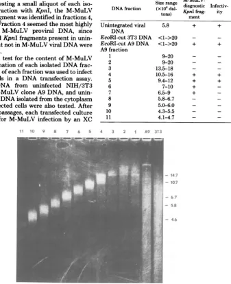
the DNAtohydroxyapatite. Figure 2 shows the size distri-bution of each of these isolated
fractions,
whicharealso listed in Table 1. Each size fraction of infected cell DNAwastested for its contentof
integrated M-MuLV sequences by two addi-tional criteria. First, a portion of each DNA fraction was tested for thepresence ofan
on November 10, 2019 by guest
http://jvi.asm.org/
[image:3.496.252.451.64.313.2]though not necessarily sequencecomposition,is
diagnostic of M-MuLV as opposedtoanyofthe
murineendogenous virus-related sequences (2). Figure 3 shows that this0.8 x 106-dalton
frag-ment present in unintegrated Moloney viral
DNA isalso detectedinthe totalcellular DNA of infected cloneA9.
Importantly,
novirus-re-lated fragment of similar size is liberated
by
KpnIdigestion
of uninfectedNIH/3T3
cellDNA. Bydigesting a smallaliquot of each
iso-lated DNA fraction with KpnI, the M-MuLV
diagnosticfragmentwasidentified in fractions 4, to 7
(Fig.
3). Fraction 4 seemed the mosthighlyenriched for M-MuLV proviral DNA, since MuLV-relatedKpnIfragmentspresent in unin-fected cellsbut not in M-MuLVviralDNAwere
largelyabsent.
In a second testfor thecontentof M-MuLV genetic informationof eachisolated DNA frac-tion,aportion ofeachfractionwasusedtoinfect NIH/3T3 cells in a DNA transfection assay.
EcoRI-cut DNA from uninfected NIH/3T3 cells, totalM-MuLV cloneA9DNA, and unin-tegrated viralDNAisolated fromthecytoplasm offreshly infected cells were also tested. After
one or more passages, eachtransfectedculture was assayed for M-MuLV infection by an XC
11 10 9 8 7 6 5
and total M-MuLV clone A9 DNA both
pro-duced XC-positive virus in the transfected
cul-tures, whereas uninfected NIH/3T3 cell DNA did not. Fractions 4 and 5 also induced
XC-positivevirus,confirmingtheir contentof
genet-TABLE 1. Characterization of size-selected EcoRI-cutDNAof M-MuLVinfectedcell line A9
Size range
M-MuLV-DNA fraction (X16dal- diagnostic Infectiv-tons) KpnI frag- ity
ment
Unintegrated viral 5.8 + +
DNA
EcoRI-cut3T3 DNA <1->20 - -EcoRI-cutA9DNA <1->20 + + A9fraction
1 9-20 -
-2 9-20 -
-3 13.5-18 -
-4 10.5-16 + +
5 9.4-12 + +
6 7-10 +
-7 6.5-9 +
-8 5.8-6.7 -
-9 5.0-6.0 -
-10 4.3-5.5 -
-11 4.1-4.7 -
-4 3 2 1 A9 3T3
- 14.7 - 10.7
- 6.7
- 5.
- 4.6
FIG. 2. Characterizationofsize-selectedDNAfractions. Aliquots of each size fraction(Ito11)ofpurified EcoRI-cut M-MuLVclone A9cellDNA wereelectrophoresedthrough a 0.6% agarose gel and visualized by staining with ethidium bromide. Alsoshown are total EcoRI digests of M-MuLV clone A9 cell DNA and uninfected3T3cellDNA. The positions of several restriction enzyme fragments of A DNA analyzed on the samegelareindicated,along with their sizes in Md. A DNA cut with EcoRI was mixed with the digested M-MuLVcloneA9DNAbeforepreparativefractionationtoprovide guides for slicing thegel.TheseA restriction enzymefragmentsarevisableinseveralisolated DNA fractions.
on November 10, 2019 by guest
http://jvi.asm.org/
[image:4.496.115.453.177.592.2]icallycompetent M-MuLV (Table 1). It is
inter-esting that fraction 7 may have contained M-MuLVproviral DNA by the KpnI cleavage test, but did not yieldinfectious virus upon
transfec-tion.
DNA fraction 4, with a sizerange of 10.5 x 10⁶
to 16.0 x
106
daltons, was cloned into the charon 4Acloningvector. The right and leftEcoRIendfragmentsof charon 4A,purifiedbypreparative agarose gel electrophoresis, were ligated with
fraction4DNA, and thehigh-molecular-weight
DNA was packaged into lambda particles in vitro by using the in vitro packaging system
described by Blattner(seeMaterials and
Meth-ods). In vitro-assembled phage particles were directly plated as primary plaques on E. coli DP50
supF
without initial amplification. Re-combinant phageplaqueswere screened for se-quencehomologytoM-MuLV byhybridization of an M-MuLV cDNA probe to nitrocelluloseE NC 1 2 3 4
filter replicas of the initial plates (5). Plaques
with strong sequence homology to M-MuLV werepicked, andplaqueswerepurified.
Recombinant DNA phage with strong se-quence homology to M-MuLV could represent clones of intact integrated M-MuLV proviruses, rearranged or otherwise aberrant integrations of
allorpartof theM-MuLVgenome, orclones of
endogenousMuLVrelatedsequences which
re-sideonEcoRIfragments of the size selected for cloning.To distinguishbetweenthese possibili-ties,DNA was purified from each recombinant
clone and further characterized with respect to its restriction enzyme cleavage pattern. Each DNAsamplewasdigested withKpnI to testfor the presence ofaM-MuLV-diagnostic0.8x 106-daltonfragment which is liberated from the 3'
region oftheviral genome. Figure 4 shows the
patterns of restriction enzyme fragments ob-tained from eight recombinant clones. Blot
5 6 7 8 9 10
0
j;' .
I
3.5
Md-2.5
-1.8
-0.8
-Eadogemeos
Fro o*nfs
- M-M.LV
[image:5.496.105.399.289.591.2]Specific
FIG. 3. KpnI cleavageofsize-selectedDNAs.Aliquotsofthesize-selected EcoRIfragments fromM-MuL V clone A9 cells were secondarily cleaved with KpnI and analyzed by electrophoresis and blot transfer
hybridizationbyusingarepresentativeM-MuL V cDNAprobe.Forcomparison,total cellularDNAfrom M-MuLVclone A9 anduninfected NIH/3T3cells, aswell asunintegratedM-MuLV linearDNA, was also digestedwithKpnIandanalyzed.Themobility ofthe M-MuLV-diagnostic0.8-MdKpnIfragment,aswellas anendogenous 2.5-MdKpnIMuL V-relatedfragmentpresent inuninfectedcells butnotcontainedingenuine
M-MuLVprovirus,isindicated.
37,
on November 10, 2019 by guest
http://jvi.asm.org/
cDNA is shown. Six of the eight clones tested contained theM-MuLVdiagnostic fragment of
0.8Md. Thissuggeststhatthey containatleast the3'portion ofanM-MuLVgenome,sincethis fragment is actually a doublet oftwointernal
viral DNA fragmentsliberated from the 3' end of the M-MuLV DNA sequences. These six clones also liberate a doublet of
M-MuLV-hy-bridizing KpnI fragments of1.8Md which arise from the 5' endofM-MuLV. This fragment is
not diagnostic ofM-MuLV, however, since the
vast majorityof theMuLV-related endogenous
sequencesalsocontain a1.8-Md KpnIfragment (Dolberg, Bacheler andFan, submitted for pub-lication).Indeed,clone27contains such a
virus-related 1.8-Mdfragment, butlacks the diagnos-tic 0.8-Md fragment. It is possible that clones which fail to release the M-MuLV-diagnostic 0.8-Md KpnI fragment carry M-MuLV
se-quences, either as
partial
integrations
orrear-rangedsequences,butsuchclones are not
can-didates for complete, potentially functional
M-MuLVintegrations. We conclude that clones48, 61, 63, 73, 75, and 76 are good candidates for
containing complete integrated M-MuLV
ge-M-MuLV-diagnostic
fragment
liberatedby
Bam digestion(1) whichcomigrated with the 1.9-MdBamfragmentfromunintegrated M-MuLV viral
DNAand whichhybridizedtoM-MuLVcDNA
(datanotshown).
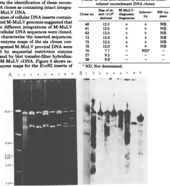
Figure5 showsadetermination of the sizes of
theinserted cellularDNA sequences.DNAfrom
each clonewas cutwithEcoRI whichseparates
the vector sequences from the inserted DNA and analyzed by gel electrophoresis. The six clones identified as containing M-MuLV-diag-nostic fragmentsfallinto twosizeclasses, having
inserts ofapproximately 12.0and 12.5Md. The sizesareverysimilartothe 12 and 13Md of the EcoRI fragments previously identified in total M-MuLV clone A9 DNA as containing
inte-gratedM-MuLV DNA sequences. The sizes of
the insertssuggestedthat atleasttwodifferent integrations of M-MuLV DNAwerecloned.
Thebiological activity of each ofthe sixclones
of integrated M-MuLV DNA sequences was
tested by using a portion of the phage DNA (after digestion withEcoRI) toinfectNIH/3T3 cells in atransfection assay. Each ofthese six
clones induced the fornation of XC-positive
2 so
1 46 *1 43 ,n Vs #
do
d ~~~*-J,
I
Al* %^ f
I
p
1.8 Md -
la
q_0.8 - _
*ipe
i
-_
_
-- M-MuLV
specific
FIG. 4. Test of recombinantclonesforM-MuL Vprovirus. DNAfromisolated recombinant clones or from linearunintegrated M-MuLV viral DNApurifiedfrom freshlyinfected cells('viral")was digested with KpnI andelectrophoresed througha 1% agarose gel. Hybridization to a blot transfer of this gel with M-MuLV-representative cDNA is shown. Themobility of the M-MuLV-diagnostic 0.8-Md KpnIfragmentis indicated. The fulldesignations of the clones are Ch4A-A9-4- followed by the clone number, butfor simplicity are referred to by the clone number.Inthefigure, Ch4A-A9-4-48 is indicatedas clone 48.
I*A
on November 10, 2019 by guest
http://jvi.asm.org/
[image:6.496.137.371.355.601.2]plaques in the infected cell cultures, whereas
other clones isolated in the same experiment,
which did notcontain M-MuLV diagnostic frag-ments, did not (clones 27 and 28) (Table 2). Supernatant virus from each
XC-positive-in-fectedculture was further characterized by
titra-tionon NIH/3T3 and BALB/c 3T3 cells. The XC-positive virus in each case titered with equal
efficiencyon bothcelltypes,indicatingthat the virusesliberated after cloned DNA infection had
theNB tropic host range ofM-MuLV itself. This NBtropic host range is also not characteristic of anyknownendogenous murine virus. This fur-ther supports theidentificationof these
recom-binant DNAclones ascontainingintact integra-tionsofM-MuLV DNA.
Thetwo sizes of cellular DNA inserts contain-ing integrated M-MuLV genomes suggested that at least twodifferent integrations of M-MuLV
DNA intocellularDNAsequences were cloned. Tofurther characterize the inserted sequences, restriction enzyme maps of the six clones
con-tainingintegrated M-MuLV proviral DNA were
constructed by sequential restriction enzyme
digestionsandby blot transfer-filter
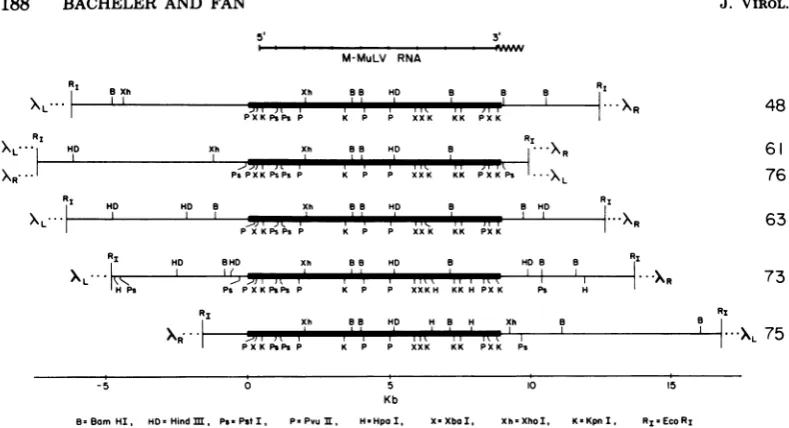
hybridiza-tionswith M-MuLV cDNA.Figure 6shows re-striction enzyme maps for the EcoRI inserts of
A 1 2 3 4 5 6 7 8 9
13.22-
4.54-3379
3.22-these sixclones. The mapshave been drawn to align the 5' end of the integrated viral DNA sequences and indicate that theintegratedviral DNAsequences are located in different positions in several of the clones. One 12.0-Md cellular DNAfragment was cloned twice, on clones 61
[image:7.496.96.444.206.585.2]and76, inoppositeorientations with respect to the Charon 4A cloning vector. Restriction en-zyme sites in the flanking cellular DNA se-quencesindicatethat clone 73contains a differ-entM-MuLVintegrationthan thatcontained in
TABLE 2. Characterization of isolated M-MuLV-relatedrecombinant DNA clones
Size of in- M-MuLV-
Infectiv-
NB tro-Clone no. sert(x106 diagnostic ity pismdaltons) fragments
48 12.5 + + NB
61 12.0 + + NB
63 12.5 + + NB
73 12.0 + + NB
75 12.5 + + NB
76 12.0 + + NB
70 7.7 - NDa
27 9.2 - -
-28 8.9 - -
-0ND, Not determined.
B
*
2 3 4 5 6 7 8 9
2.34-FIG. 5. Sizeofcloned inserts. DNAfromseveral recombinant DNA clones withstrongsequencehomology
toM-MuLVwas cutwithEcoRIandelectrophoresedthrougha0.6%agarosegel.The mobilitiesofmarker AEcoRI DNAfragmentsco-electrophoresedin thesamegelareindicated, alongwith their sizes (Md). (A) Ethidium bromidestainingofthegel;(B) hybridizationwith M-MuL V cDNAtoa blottransfer of thesame
gel.(Lane 1) MixtureofADNAcutwithHindIII,detectablebyethidium bromidestaining (A), and uncut M-MuLVunintegratedlinearDNA, detectedbyhybridizationwith M-MuLV cDNA(B);(lane2) Ch4A-A9-4-48; (lane 3)Ch4A-A9-4-61; (lane 4)Ch4A-A9-4-63; (lane 5) Ch4A-A9-4-73; (lane 6)Ch4A-A9-4-75; (lane 7) Ch4A-A9-4-76;(lane 8)Ch4A-A9-4-70; (lane 9)Ch4A-A9-4-27.
.'*.
0 .M 14 I
on November 10, 2019 by guest
http://jvi.asm.org/
M-MuLV RNA 8 Xh
I,
RXL
)L".I H
ID
PXKPsPs
Xh
Xh 8a HD 8 8 B R1
R
P K P P XXK KK PX K RI
Xll 8a HD BaS
I I" X
Ps K\ ,, P,,,\X Ps L
Ps PXK PsPs P K P P XXK KK PXK PS 'XL
R1
XL
---HD ND B Xh
PX K Ps Ps P
88 ND
K P .,
K P P XX K
8 8 HD RI
KK PXK
48
61
76
63
ND BHD Xh
Ps
X,iR
-5
v "IPPP
PS P X KPSPS P
0
88 NHD B ND B
''I I KKPXK
K P P XXKH KK H PXK Ps H
Xh 88 HD H B H Xh
5
Kb
10
..XR
a
...~~~~~~~~~~~~~~~~~~~~~~~~~~~~~~~~~~.
73
XL 75
s5
8-BamHI, HD-HindM, Ps.PstI, P-Pvu, H-HpaI, X.XboI, Xh.XhoI, K-KpnI, RI-EcoRI
FIG. 6. RestrictionmapsofM-MuLVproviralclones. Restrictionmapsforthe sixM-MuLVproviralclones areshown. For convenienceoforientation,coordinatesareshown with the M-MuLVproviralportions ofeach clonespanning from0to8.9 kilobasesonthemap, andtheyareshown in theconventional orientationwith respect to M-MuLV RNA. Adjacent cell sequences to the 5' side ofthe viral DNAs have negative map
coordinates, andadjacentcellsequencestothe 3' side havepositivecoordinatesgreaterthan 8.9 kilobases.
Themaps werederivedby cleavage ofrecombinantphageDNAs with various restrictionenzymes(singlyor incombinations) followed byagarosegel electrophoresis. The totalfragment patterns weredetermined by ethidium bromide staining, and those which contained viral sequences were identified by blot-transfer hybridizationwith M-MuLV cDNA. Theabbreviationsforthedifferentenzymesaregivenin thefigure;sites shown above the line havebeenmapped throughouttheinsert,whereas additional sitesmayexistforenzymes
shown below the line. The locationsoftheleft andrightarns ofthe Charon 4A cloning vectorarealso
indicated.
clones61and76,eventhough both integrations arecontained inEcoRI fragments ofverysimilar
size.Similarly, clones 48, 63, and 75 each
repre-sent different integrations of M-MuLV DNA
sequencesinto cellularDNA,eventhough each
provirus is contained in anEcoRI fragment of
12.5Md. In contrasttothediversity of restric-tionenzymesites in the cellular DNAsequences
flanking each integrated M-MuLVgenome,the viral DNAsequencesin eachcaseshow identical
restrictionmapswhich alsoagreewell with the
restrictionenzyme mapfor M-MuLVderived by
Gilboaetal. (13).
DISCUSSION
We have isolated six recombinant lambda phage DNA clones which contain both proviral M-MuLV DNAsequencesand the surrounding
cellular DNA sequences into which the viral
DNAwasinserted. Five different integrations of
M-MuLVarerepresentedontheseclones, even
though several of the EcoRI DNA fragments containing these integrationsareofverysimilar
size.We consider theseintegrations distinct
be-causeeach showsaunique distribution of
restric-tionenzymesites in the cellularDNAsequences
surrounding the proviral DNA sequences. The
number of different integrations of M-MuLV present in EcoRI fragments of 12.0 x 10⁶and 12.5 x 10⁶daltons wasinitiallysomewhat
sur-prising, since we had previously assumed that
the new EcoRI fragments of this size seen in
EcoRI digests of M-MuLV clone A9 cellular DNAwereeach the result of theinsertion ofa
single M-MuLV proviral DNA sequence. The
isolation of five different integrations is, how-ever, compatible with our previous estimates
that this cell line contains 10 to 12 integrated copies of M-MuLV (7). Furthermore, the fact thatthesameintegration sitewasindependently
isolated twice among six clones suggests that
most of the M-MuLV integrations in EcoRI fragments of this size have been cloned.
Although the cloning efforts with the M-MuLV clone A9 cell DNA successfully yielded the M-MuLV proviral clones described here,
effortstoperform similar experiments withtwo
other DNA fractions from other productively infected cells were unsuccessful. EcoRI
frag-mentsof6to10Md from thecellline M-MuLV
clone E7 (9) which carries integrated M-MuLV DNAsequences onEcoRIfragments of9.5, 8.6,
6.6, and 3.75 Md (2) were cloned, and 22,500
P,,PSPIP K KK, P
P X KPIPSP K P P XXK KK PX K Ps
-1 I I
RI
I
RI
X
L- -k
RI
B1
on November 10, 2019 by guest
http://jvi.asm.org/
[image:8.496.57.452.62.276.2]recombinant phage were screened. Ten phage
cloneswerefoundtohybridize M-MuLV cDNA and four strongly hybridized. However, none of the clones contained a M-MuLV provirus, al-though some contained endogenous
M-MuLV-related sequences. Similarly, EcoRI fragments
of6 to 10 Md fromM-MuLV clone 4Acells (9) were cloned, and 2.3 x
105
recombinant phage were screened; 34 phage clones hybridized withM-MuLV cDNA, and 15 strongly hybridized.
Again,none of theseclones contained a genuine
M-MuLV provirus. In the second experiment,
the 6- to 10-Md 4A EcoRI fragments were
di-gested with BamHIand found to yield a 1.9-Md
Bam fragmentdiagnostic of M-MuLV. In
addi-tion, this DNA gave rise to XC-positive virus upon DNAinfectionof NIH/3T3cells.Bythese
criteria,atleasttheM-MuLV clone 4AcellDNA
fractionshould havecontained intact M-MuLV
proviruses. Several possibilities might explain
the failureto obtainM-MuLV proviral clones in two cases. (i) Not enough recombinant phage may have been screened. This may have been the case forthe experimentswith theM-MuLV
cloneE7DNA. However, sufficient numbers of phage were screened for the M-MuLV clone 4A
trialsthat itcouldbecalculatedthat anysingle copy DNA sequence present should have been
cloned. (ii)Theparticular DNAfractionschosen may have containedrelatively higher concentra-tions of endogenous MuLV-related sequences. This wasprobably the case,sincethe 6- to
10-Mdrangeof EcoRI fragments from infected or
uninfectedmousecells containsmany more
frag-ments with sequence homology to M-MuLV
than the12- to 13-Mdrange. However,the fail-ure to isolate asingle M-MuLV proviral clone
contrastssharply with the relativeeaseof clon-ing from M-MuLV clone A9 cells. (iii) Some proviral integrations may not be clonable in bacterial systems. Evidence that certain
se-quences in the murine mammary tumor virus genomecannotbe cloned has been obtained
by
several research groups(G. Hager, personal
communication; H. E.Varmus, personal
com-munication), and it is
possible
that someM-MuLV
proviral integrations
may have similar properties.InthecaseofM-MuLV,
theputative
unclonable sequences would not be located in the viralsequencesthemselves(since
other M-MuLVprovirusesweresuccessfully cloned),
butrather inparticularadjacentcellular sequences. (iv) M-MuLV proviral
integrations
may have beencloned andsubsequently
lost.Instability
of recombinant phage clonescarryingproviral
se-quences has been observed for other murine retroviruses(20, 27).Retroviruses alsocarry
se-quencesresemblingclassical
procaryotic
andeu-caryotic transposable elements, which may
re-sult in sequence arrangement during growth
(22a). Indeed, we have observed that many of therecombinant phageclones isolated(carrying both genuine M-MuLV proviruses as well as endogenous MuLV-related sequences) display
instability during propagation. Growthof virus preparations isfrequentlyaccompanied by gen-eration ofdeleted forms of the originalphage, which can bedetected by the presence of phage
particles with lowerbuoyant density. We have found it necessary to routinely band all phage preparations inequilibrium CsCl gradientsand useonlythe phage of greatestdensity to mini-mize these problems.
Wehavetestedthebiologicalactivity of each M-MuLV-containing recombinant DNA clone by using the isolatedphage DNA toinfect
NIH/
3T3 cells. Eachofthe clones identifiedas
con-taining M-MuLV based on the presence of
di-agnostic restrictionenzymefragments alsogave rise to infectious NBtropic virus,confirmingthe
identification of these clones. The observation that DNAfromeachclonewasinfectious differs somewhat from several other studies in which
proviruses resultingfrom recent viral infection
(as opposedtogenetically transmitted
endoge-nousproviruses) were isolated as recombinant DNAclones.Lowyetal.(20) have isolatedthree
clonesofintegratedAKR viral DNA sequences
froma masscultureofinfectedfibroblasts.One
ofthese clones was noninfectious, whereas the
other twocloneswereinfectious, althoughwith
approximately 30-fold difference in specific
in-fectivity. O'Rear et al. (21) have cloned
inte-grated spleen necrosis virus DNA sequences from chronically and acutely infected chicken
cells. Both infectious and noninfectiousclones
were obtained. Although infectious clones
con-tainedinserts ofsmaller size thanthe
noninfec-tiousintegrationscloned,sizedidnotappearto
accountfor thedifferencein
biological activity.
Mullins et al. (J. I.Mullins,
M.Nicholson,
J.Casey,K.Burck and N.Davidson,
personal
com-munication) have cloned seven
integrations
offeline leukemia virus from an infected human
cell line and obtainedbothinfectiousand
non-infectious isolates. Nomajordifferences between the viral sequences ofinfectious and
noninfec-tious clones could be detected. It is unclear whether theinfectious natureof all ofourclones is merely chance or whether M-MuLV
provi-ruses are more often infectious than AKR, spleen necrosisvirus, or feline leukemia
provi-ruses.
The infectivity of all five of the M-MuLV proviruses cloned stands in apparentcontrastto
theDNase Iresistanceand,
therefore,
potential
on November 10, 2019 by guest
http://jvi.asm.org/
grated copies of M-MuLV found in the A9 cell line. Weare currently investigatingthe specific infectivity ofindividualcloned M-MuLV
provi-ruses and thetranscription activity of these
in-dividual integrationsof M-MuLV in the A9cell line.
ACKNOWLEDGMENTS
Thisworkwassupported byPublic HealthServicegrants
R01CA-22829(L.T.B.),R01CA-15747 (H.F.),contract
N01-CP-71008(H.F.),andcoregrantCA 14195,all fromthe
Na-tional Cancer Institute.
Wethank FrederickBlattnerand his associatesfor
provid-inguswith thecharon phagecloning kit,and Jeffrey Browne for adviceonin vitropackaging. Jennifer Price and Patricia
Kreikemeierprovided excellenttechnical assistance, and Mau-reenBrennantyped themanuscript. Wethankmenmbers of
theTumorVirologyLaboratoryforvaluable adviceand dis-cussions.
LITERATURE CITED
1. Bacheler, L. T., and H. Fan.1979.Multiple integration
sites for Moloney murine leukemia virus in productively
infectedmousefibroblasts. J. Virol. 30:657-667.
2. Bacheler, L., and H. Fan. 1980. Study of integrated
Moloney murine leukemia virus DNA using comple-mentaryDNA which does notrecognize endogenous relatedsequences.J. Virol. 33:1074-1082.
3. Battula, N., and H. M. Temin.1977.Infectious DNA of
spleen necrosis virus isintegratedatasingle site inthe
DNA of chronicallyinfected chicken fibroblasts.Proc. Natl.Acad. Sci. U.S.A. 74:281-285.
4. Battula,N., and H. M.Temin.1978.Sites of integration
ofinfectiousDNA of avianreticuloendotheliosisviruses indifferentavian cellular DNAs.Cell13:387-398. 5. Benton, W.D., and R. W.Davis.1977.Screening Xgt
recombinant clones byhybridizationtosingle plaques in situ.Science196:180-182.
6. Blattner, F. R., B. G. Williams, A. E. Bleckl, K.D. Thompson, H. E. Faber, C. A. Furlong, D. J. Grun-wald, D.0.Kiefr, D. D. Moore, J. W Schuman, G. L. Sheldon,and0.Smithies. 1977.Charon phages: safer derivatives ofbacteriophage lambda for DNA cloning.Science 196:161-169.
7.Breindl, M., L. Bacheler, H. Fan, and R. Jaenisch. 1980. Chromatin conformation ofintegratedMoloney
leukemiavirusDNAsequencesin tissues ofBALB/Mo
mice and virus-infectedcell lines. J. Virol.34:373-382.
8. Canaani, E., andS. A. Aaronson. 1979. Restriction
enzyme analysis ofmousecellulartypeC viralDNA:
Emergence of new viral sequences in spontaneous
AKR/J lymphomas. Proc. Natl. Acad. Sci. U.S.A.76: 1677-1681.
9. Fan, H., R. Jaenisch, andP. MacIsaac. 1978.
Low-multiplicity infection ofMoloney murine leukemiavirus
inmousecells:effectonnumberof viral DNA copies
and virus productionin producer cells. J. Virol.
28:802-809.
10. Fan, H., and M. Paskind. 1974. Measurement of the
sequencecomplexity of cloned Moloneymurine
leuke-mia virus 60 to 70S RNA: Evidence for a haploid
genome.J. Virol.14:421-429.
11. Fan, H.,and I. Verma. 1978. Size analysis and
relation-ship of murine leukemia virus-specific mRNA's:
evi-dence for transposition ofsequencesduring synthesis
andprocessing of subgenomic mRNA. J. Virol.
26:468-478.
12. Gianni, A. M., J. R. Hutton, D. Smotkin, andR. A.
Weinberg. 1976.Proviral DNAofMoloneyleukemia
virus: purificationand visualization. Science
191:569-571.
13. Gilboa, E., S. Goff, A.Shields, F. Yoshimura, S.
Mi-kbpterminallyredundantDNAcarryingtheinfectivity
ofMoloneymurineleukemia virus. Cell16:863-874. 14. Gilmer, T. M., and J. T. Parsons. 1979. Analysis of
cellularintegrationsitesinaviansarcomavirus-infected duck embryocells. J.Virol. 32:762-769.
15. Graham, F. I., andA. J. van derEb. 1973. A new techniquefortheassayofinfectivityof human adeno-virus 5DNA.Virology52:456-461.
16. Hughes,S.H., P. R.Shank,D. H.Spector,H.Kung, J. M.Bishop,andH. E.Varmus.1978. Provirusesof aviansarcomavirusesareterminallyredundant,
coex-tensivewithunintegratedlinearDNAandintegratedat
manysites.Cell 15:1397-1410.
17. Jaenisch, R. 1976.GermlineintegrationandMendelian transmissionof theexogenousMoloneyleukemiavirus. Proc.Natl.Acad. Sci.U.S.A.73:1260-1264.
17a.Jahner,D.,andR.Jaenisch. 1980.Integrationof
Mo-loneyleukemia virusintothegermlineof mice:
corre-lation betweensite ofintegration andvirusactivation. Nature(London)287:456-458.
18. Keshet, E.,and H.M.Temin.1978. Sites ofintegration
ofreticuloendotheliosis virus DNA in chicken DNA. Proc.Natl.Acad.Sci.U.S.A. 75:3372-3376.
19. Lai,M-H.T.,andI. M.Verma. 1980. Genome organi-zationofretroviruses.VII.Infectionbydoublestranded
DNA synthesized in vitrofrom Moloney murine
leu-kemia virusgeneratesavirusindistinguishablefromthe
original virus used in reverse transcription. Virology
100:194-198.
20. Lowy, D. R., E. Rands,S. K.Chattopadhyay, C.F. Garon, andG. L.Hager. 1980. Molecularcloning of infectiousintegratedmurineleukemiavirus DNA from
infectedmousecells.Proc.Natl.Acad. Sci. U.S.A.77:
614-618.
21. O'Rear, J.J., S. Mizutani, G.Hoffman, M. Fiandt,
and H. M.Temin. 1980.Infectiousand noninfectious recombinant clones ofthe provirus ofSNV differ in
cellular DNA and are apparently the same in viral
DNA.Cell 20:423-430.
22. Rowe, W. P.,W.E.Pugh,and J. W. Hartley. 1970.
Plaqueassay techniquesformurine leukemia viruses.
Virology42:1136-1139.
22a.Shoemaker,C.,S.Goff, E.Gilboa,M.Paskind,S.W.
Mitra,and D.Baltimore. 1980.Structureofacloned circularMoloneymurineleukemia virus DNAmolecule containinganinvertedsegment:implicationsfor
retro-virusintegration.Proc.Natl.Acad.Sci.U.S.A. 77:3932-3936.
23. Southern, E. M. 1975. Detectionofspecific sequences amongDNAfragmentsseparatedbygel electrophore-sis.J.Mol.Biol.98:503-517.
24. Steffen, D.,and R.A.Weinberg.1978.Theintegrated
genomeofmurineleukemiavirus.Cell 15:1003-1010.
25. Sternberg,N., D. Tiemeier,and I.Enquist. 1977. In vitro packaging ofaXDam vectorcontaining EcoRI
DNAfragments ofEscherichiacoliandphage P1.Gene
1:255-280.
26. Temin, H.M. 1970.Formation ofthe--provirus of RNA
sarcomaviruses,p.233-249.InR.D.Barryand B. W. J.Mahy(ed.),ThebiologyoflargeRNAviruses. Aca-demicPress,Inc.,NewYork.
27. VandeWoude,G. F.,M.Oskarsson,L. W.Enquist, S.Nomura, M.Sullivan,and P.J.Fischinger.1979. The cloningofintegrated Moloney sarcomaproviral
DNA sequencesinbacteriophagelambda.Proc. Natl. Acad.Sci.U.S.A.76:4464-4468.
28. Wahl,G.M.,M.Stern, andG. R.Stark.1979.Efficient transferoflargeDNAfragmentsfromagarosegelsto
diazobenzyloxymethyl-paper and rapid hybridization usingdextransulfate.Proc.Natl.Acad.Sci.U.S.A. 76:
3683-3687.
29. Weintraub, H., and M.Groudine.1976.Chromosomal subunitsinactivegeneshaveanalteredconformation.
Science193:848-856.