RESEARCH ARTICLE
EV71 virus-like particles produced
by co-expression of capsid proteins in yeast cells
elicit humoral protective response against EV71
lethal challenge
Xiaowen Wang
1, Xiangqian Xiao
1, Miao Zhao
1, Wei Liu
1, Lin Pang
2, Xin Sun
3, Shan Cen
4, Burton B. Yang
5,
Yuming Huang
2*, Wang Sheng
1*and Yi Zeng
1Abstract
Background: Enterovirus 71 (EV71) is the most common causative pathogens of hand, foot and mouth disease (HFMD) associated with severe neurological complications. There is a great need to develop prophylactic vaccine against EV71 infection.
Results: EV71 virus-like particle (VLP) was produced in yeast expression system by the co-expression of four EV71 structural proteins VP1–VP4. Immunization with the recombinant VLPs elicited potent anti-EV71 antibody responses in adult mice and anti-VLP sera were able to neutralize EV71 virus in vitro. Neonatal mice model demonstrated VLP immunization conferred protection to suckling mice against the lethal viral challenge.
Conclusions: Co-expression of four EV71 structural proteins VP1–VP4 in yeast expression systems is an effective method to produce EV71 VLPs. VLP-based vaccine shows great potential to prevent EV71 infection.
Keywords: Enterovirus 71, Vaccine, Yeast, Virus-like particle, Hand foot and mouth disease
© 2016 Wang et al. This article is distributed under the terms of the Creative Commons Attribution 4.0 International License (http://creativecommons.org/licenses/by/4.0/), which permits unrestricted use, distribution, and reproduction in any medium, provided you give appropriate credit to the original author(s) and the source, provide a link to the Creative Commons license, and indicate if changes were made. The Creative Commons Public Domain Dedication waiver (http://creativecommons.org/ publicdomain/zero/1.0/) applies to the data made available in this article, unless otherwise stated.
Background
Human enterovirus 71 is a non-enveloped RNA virus of the Picornaviridae family that was first reported in 1969. The virion is around 25–30 nm in diameter containing a single-stranded RNA viral genome of approximately 7500 nucleotides [1–3]. EV71 encoded a single large polypro-tein that is initially cleaved into P1, P2, and P3 regions. The P1 region is subsequently processed by protease 3CD to generate four capsid subunit proteins, VP1 to VP4. The viral genome is packaged in an icosahedral capsid which is composed of 60 copies of structural proteins. High-resolution structural analysis showed that VP1-3 form a
pseudo T = 3 icosahedral capsid that are located on the
surface of viral capsid [4]. VP4 is myristoylated at the N terminus and located inside virion [5, 6]. However, crys-tallographic analysis demonstrated that the structure of mature EV71 virion is similar to other enteroviruses [7].
EV71 has been identified as one of the major etiologi-cal agents of hand, foot and mouth disease (HFMD) [8,
9]. A number of HFMD epidemics caused by EV71 infec-tion occurred in the Asia–Pacific region and are asso-ciated with severe neurological complications such as aseptic meningitis, poliomyelitis-like paralysis and brain-stem encephalitis. The surveillance data from National Center for Disease Control and Prevention showed that more than 3 million HFMD cases and 1384 deaths were reported by the end of 2010 in mainland China [10–14]. HFMD is becoming the most common viral disease in these areas that seriously threat children health. How-ever, no appropriate vaccine is yet available to prevent EV71 infection.
Open Access
*Correspondence: [email protected]; [email protected] 1 College of Life Science and Bioengineering, Beijing University of Technology, 100 Pingleyuan, Chaoyang District, Beijing, People’s Republic of China
2 Department of Neurology, Beijing Ditan Hospital, Capital Medical University, Beijing, People’s Republic of China
based on viral VLPs have been successfully used in pre-vention against hepatitis B virus and human papillomavi-rus [21–23]. VLPs are empty non-infectious viral capsids that structurally mimic the conformation of native virion. High density of viral linear and conformational epitopes on the VLP surface may elicit strong immune responses [23]. In the present study, EV71 VLPs were successfully produced by co-expression of four structural viral pro-teins in yeast, which is safe, reliable and cost-effective platform for recombinant protein production.
Results
Generation and characterization of EV71 VLPs
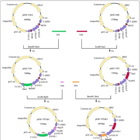
EV71 capsid is composed of 60 copies of each of the four viral structural proteins VP1, VP2, VP3 and VP4. In the pre-sent study, genes encoding VP1, VP2, VP3 and VP4 proteins of EV71 were inserted into vectors for co-expression of four viral proteins in yeast (Fig. 1). The expressions of viral pro-teins were investigated by SDS-PAGE and Western-blot. Viral proteins were purified using the method described previously with modification [3] and visualized using SDS-PAGE analysis. Three bands were observed which corre-sponded to VP1, VP2 and VP3 of EV71 virus, respectively, according to the molecular weight of each band (Fig. 2a). However, the band corresponding to EV71 VP2 protein was further proofed by using VP2-specific antibody MAB979 by Western-blot (Fig. 2b). To determine whether the co-expression of four viral structural proteins can generate EV71 VLPs, total proteins of yeast cells were extracted and loaded onto the sucrose and cesium chloride gradient to iso-late EV71 VLPs by ultracentrifugation. As shown in Fig. 2c, the formation of viral VLPs were observed in purified yeast lysates by transmission electron microscope and the diam-eters of particles were about 25–27 nm which appeared ico-sahedral. Our data indicate that co-expression of four viral structural proteins VP1, VP2, VP3 and VP4 in yeast cells can definitely lead to the formation of EV71 VLPs.
VLP immunization elicits neutralizing antibody responses against EV71
To investigate whether the EV71 VLPs were capable of inducing anti-EV71 antibody responses, female BALB/c
antibody response was detected in the yeast cell lysates-immunized group and the PBS group (Fig. 3). Our results demonstrated that EV71 VLPs, denatured VLPs and inac-tivated-EV71 virus induced similar levels of anti-EV71 antibody.
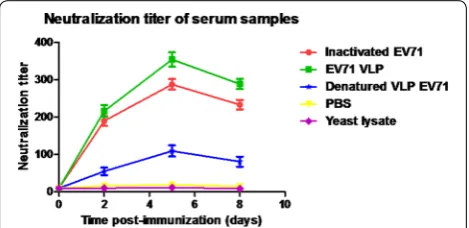
To evaluate whether the antibodies were able to neu-tralize EV71 virus, mouse serum samples were twofold serially diluted and mixed with infectious EV71 (100 TCID50) to test their ability to neutralize the live EV71 virus in vitro. EV71 Bj08 (genotype C4) and a variant of the prototype strain of EV71, BrCr-TR (genotype A) were used for in vitro neutralization assay. As shown in Fig. 4, remarkable increase in anti-EV71 neutralizing antibody titers was observed after the booster injection at 3 weeks post-injection. The EV71 VLPs elicited slightly higher neutralization titers against EV71 Bj08 compared to the heat-inactivated EV71. The denatured VLPs elic-ited lower neutralization titers than the purified VLPs, although they induced similar levels of IgG antibodies. These data are consistent with the previous report [3]. In vitro inhibition of EV71-mediated cytopathic effect (CPE) by anti-sera was illustrated by microscopic photo-graphs. Serum samples were diluted 256-fold for neutral-ization assay. Virus-mediated CPE was remarkably found in cells infected by either EV71 Bj08 or BrCr-TR (Fig. 5a, b) after 6 days of infection. In contrast, no obvious CPE was observed in the cells without viral infection (Fig. 5c, d). Virus-induced CPE could be significantly suppressed by the addition of anti-VLP sera (Fig. 5e, f) and anti-inac-tivated EV71 sera (Fig. 5g, h). Lower suppression of CPE was observed by using anti-denatured VLP sera (Fig. 5i, j), suggesting that antibody recognizing conformational viral epitopes are essential for VLP-induced immune pro-tection against EV71.
In vivo protection against lethal viral challenge in neonatal mouse model
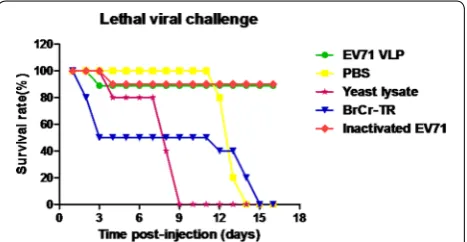
VLPs, inactivated-EV71 virus, yeast cell lysates and PBS. The mixture of EV71 virus and anti-sera was incubated overnight at 37 °C and used to inoculate intraperito-neally (i.p.) 1-day-old BALB/c suckling mice (n = 10 per group). Infection by BrCr-TR viral strain was used as positive control. The data showed that 90 % of mice treated with EV71 virus mixed with the EV71 VLPs- and
inactivated-EV71 virus-immune sera remained healthy and survived until the end of the experiments. In con-trast, mice inoculated with virus mixed with the yeast cell lysates and PBS-immune sera died at 16 days post-inoc-ulation, indicating that EV71 VLPs and inactivated EV71 virus conferred immune protection against EV71 lethal challenge in vivo (Fig. 6).
[image:3.595.56.541.88.576.2]Discussion
Vaccination is a commonly used and cost-effective method for infectious disease control. Various types of vaccine candidates against EV71 have been evaluated in animal model, including recombinant subunits [15, 24], synthetic peptide vaccines [18–20], live attenuated vac-cines [25, 26], VLP vaccines [3, 27, 28], DNA vaccines [29] and formalin-inactivated virion vaccines [30–33]. Recombinant subunit vaccines can be produced from different expression systems. It has been found that EV71 capsid proteins VP1, VP2 and VP3 are immuno-genic and capable of eliciting antibodies that recognize their corresponding viral proteins. Unlike VP2 and VP3, recombinant VP1 was capable of eliciting effective neu-tralizing antibody responses against EV71 [34]. Synthetic peptides containing neutralization epitopes are consid-ered as good vaccine candidates providing well-defined immunogens and safety advantages. Two linear neutral-izing epitopes SP70 and SP55 found in EV71 VP1, were capable of inducing neutralizing antibody responses in mice against EV71 virus by inhibiting viral infection at pre- or post-attachment steps [35]. Our previous study showed that immunization of N terminus of EV71 VP4 elicits cross-protective antibody responses [36]. Recently, a cross-neutralizing epitope was identified in EV71 VP2 [37]. However, neutralizing antibody responses induced by peptides are usually weak and require strong adju-vants. Live attenuated vaccines are widely used for viral disease prevention, however, the emergence of patho-genic viral revertants has raised vaccine safety concerns. Inactivated vaccine and VLP-based vaccine retain both
Fig. 2 Viral protein expression and electron microphotographs. a The expressions of EV71 structure proteins were visualized by SDS-PAGE. b The expression of EV71 VP2 was monitored using VP2-specific antibody MAB979 by Western blot. EV71 VLPs were isolated using sucrose and discon-tinuous cesium chloride (CsCl) gradient (1.4, 1.33, 1.29 and 1.25 g/ml) by ultracentrifugation. The formation of EV71 VLPs in yeast was monitored by electronic microscope. Size bar 50 nm
Fig. 3 Kinetics of antibody titer development in mice following immunization. The inactivated EV71 virus was coated on the plate surface to capture anti-EV71 antibody. The data were represented by the mean of reciprocal log2 endpoint titers ± standard deviation (SD)
[image:4.595.58.539.91.256.2] [image:4.595.58.290.323.442.2] [image:4.595.57.292.516.630.2]linear and conformational neutralizing epitopes respon-sible for eliciting potent neutralizing antibody responses against viral infection [38] and are thus widely accepted as effective vaccine candidates. VLP-based vaccine has a safety advantage over inactivated vaccine due to the lack of the viral genome.
Recombinant VLPs that mimic the conformation of authentic native viruses can be produced from variable expression systems such as insect cells, yeast and E. coli. EV71 VLPs have been successfully produced in vari-able expression systems via co-expression of P1 and 3CD viral proteins [28, 39]. 3CD is an EV71-encoded pro-tease responsible for the digestion of P1 polyprotein into structural subunit proteins, which form EV71 capsids via self-assembly.
Two types of EV71 viral particles (E- and F-particles) have previously been produced from Vero cells cultured in a serum-free condition. SDS-PAGE and Western blot analysis illustrated that the E-particle was an immature
particle containing incompletely cleaved VP0 protein, while in F-particle, VP0 protein was fully cleavaged into VP2 and VP4 by autocatalytic processing. In addi-tion, neutralization assay showed that the inactivated F-particle induced a more potent neutralizing antibody response against EV71 in the mouse model than the E-particle [40]. Thus, F-particle of EV71 is an ideal vac-cine candidate to prevent EV71 infection. In the present study, we report a technique for production of EV71 F-particles in yeast cells. TEM analysis showed that EV71 VLPs were successfully generated in yeast cells through the co-expression of four EV71 capside proteins (VP1 to VP4) without using 3CD protease. It has been reported that host cell apoptosis can be triggered by the coxsacki-evirus B3-encoded protease and HIV-encoded proteases induced cell death [41, 42]. Co-expression of P1 protein and 3CD can be used to produce EV71 VLP in different host cells. However, the effects of EV71-encoded pro-tease on host cell apoptosis or death still remains to be elucidated.
EV71 is phylogenetically classified into three distinct genotypes: A, B, and C based on the genetic variation of VP1 sequences. Genotypes B and C can be further divided into B1–B5 and C1–C5 subgenotypes, respec-tively [43, 44]. Recently, 3 new genotypes were reported, D, E and F subgenotypes [45, 46]. An ideal anti-EV71 vaccine should be able to provide broad protective activ-ity against a wide spectrum of EV71 strains. One sin-gle serotype was reported in EV71 when measured by hyperimmune animal sera. However, antigenic variations have been identified recently in post-infection human sera when measured cross-neutralizing antibody titers against different EV71 genotype strains. Antigenic vari-ation among different genogroups was reported [47]. Immunization of live attenuated EV71 vaccine (genotype
Fig. 5 In vitro neutralization assay. a Cells infected by EV71 Bj08 strain. b Cells infected by EV71 BrCr-TR strain. c, d Uninfected cells. e Cells infected by Bj08 strain reacted with anti-VLP serum. f Cells infected by BrCr-TR strain reacted with anti-VLP serum. g Cells infected by Bj08 reacted with anti-inactivated EV71 serum. h Cells infected by BrCr-TR strain reacted with anti-inactivated EV71 serum. i Cells infected by Bj08 reacted with anti-denatured VLP serum. j Cells infected by BrCr-TR strain reacted with anti-denatured VLP serum. Size bar 50 μm
[image:5.595.60.539.91.233.2] [image:5.595.58.292.302.423.2]as B4, B5, C2, C4 and C5 strains in health children and infants [48]. An potent cross-neutralizing antibody pro-tection against EV71 subgenotypes B1, B4, B5 and C4A were also observed in B4-based vaccine treatment [49]. Based on the antigenic heterogeneity among EV71 iso-lates and the efficacy of multivalent VLP vaccine dem-onstrated by the successful application of bivalent and tetravalent HPV vaccine, multivalent VLP-based vaccine has a great potential in the development of a safe and cost-effective anti-EV71 vaccine with broad cross-protection.
Conclusions
The co-expression of four EV71 capsid proteins in yeast resulted in effective production of EV71 VLPs, which elicited potent anti-EV71 antibody responses and pro-tected neonatal mice against lethal viral challenges. Mul-tivalent EV71 VLP-based vaccine has a great potential to be a safe and cost-effective vaccine candidate with broad cross-neutralizing activities against EV71 infection.
Methods
Construction of plasmids
The coding sequence of EV71 P1 protein of C4 sub-genotype was optimized according to the codon usage for S. cerevisiae. DNA fragment encoding P1 protein was then synthesized. VP1 gene was amplified using primers VP1-BamH1 (5′-AAGGATCCATGGGTGA CAGAGTCGCCGAT-3′) and VP1-XhoI (5′-CCGCTC GAGTCATAAAGTAGTGATGGCT-3′. The PCR prod-ucts were double-digested by XhoI and BamH1 and subsequently inserted into the vector pESC-URA (Inv-itrogen) to construct pESC-VP1. VP4 gene was ampli-fied using primers VP4-EcoR1 (5′-CCGGAATTCATGG GATCACAAGTTTCA-3′) and VP4-BglII (5′-GAAGA TCTCTTTAATGGGGCAGCCAT-3′). The PCR frag-ments were further inserted into the vector plasmid pESC-VP1 at EcoR1 and BglII sites to yield pESC-VP1&4. VP2 gene was amplified using primers VP2-EcoRI (5′-CGGAATTCATGTCTCCATCTGCCGAAGCA-3′) and VP2-SacI (5′-AACGAGCTCCTGAGTAACGGC TTGCCT-3′). The amplified PCR products were then
VP2&3 were further co-transformed into yeast of INVSc1 strain (Invitrogen, USA) by using Sc EasyComp transformation kit (Invitrogen, USA) according to the instructions of the manufacturer. The yeast transfor-mants were further selected onto a synthetic complete plates without uracil and histidine. Clonal isolates were then grown at 30 °C for 78 h in YPD containing 2 % galac-tose to an optical density. After centrifugation, the har-vesting cell pellets were broken with glass beads and cell lysates were analyzed for the expression of EV71 VP1.
Purification of VLPs
The yeast pellets were resuspended in sodium phos-phate buffer (pH 7.2) supplemented with 1×
SDS‑PAGE and western blotting
The purified VLP samples were denatured by boiling for 10 min and loaded onto SDS-PAGE (12 %) gel for elec-trophoresis. The recombinant proteins were detected by Western blotting using a monoclonal antibody against VP2 (MAB979, Millipore, USA). Briefly, the proteins were transferred onto PVDF membrane, which were blocked with 2 % (w/v) BSA in TBS solution for 1 h at room temperature, and further washed three times with TBS containing 0.05 % (v/v) Tween 20. The membrane was then incubated with primary anti-VP1 and anti-VP2 antibodies, respectively, for 1 h at 37 °C, and washed three times with TBS buffer. After incubation with the secondary goat rabbit and goat mouse anti-bodies conjugated with fluorescent dyes: IRDye 800 CW (KPL, USA) for 45 min, blotting images were acquired using the Odyssey infrared imaging system (Li-COR Bio-sciences, USA) and analyzed by the software provided by the manufacturer.
Electron microscopy
The formation of EV71VLPs was analyzed by electron microscopy as described previously [3]. Briefly, samples were adsorbed to carbon-coated copper grids and incu-bated for 1 min. The grids were then negatively stained for 45 s with 2 % phosphotungstic acid after washing twice with PBS and visualized using an electron micro-scope (H-7650, HITACHI, Japan).
Immunization of animals
Pathogen-free female BALB/c mice were purchased from Beijing HFK Bioscience Co. (Beijing, China). All animals were housed at pathogen-free conditions. Animal ments were performed according to the current experi-mental protocols involving animal study approved by the Institutional Animal Care and Use Committee of Peking University. For mice experiments, five female BALB/c mice (6–8 weeks) per group were immunized with 20 μg/ mouse of one of the following samples: purified VLPs, denatured VLPs, beta-propiolactone-inactivated EV71 virus (Bj08 strain), yeast cell lysate or PBS. The immuni-zation was boosted 3 weeks later with the same dosages. QuickAntibody™ from KBQ Biotechnology Co.
(Bei-jing, China) was used as an adjuvant. Control group was immunized with PBS plus adjuvant. The blood samples were collected at week 0, 2, 5, 8 and the sera were inacti-vated at 56 °C for 30 min and stored at −80 °C.
ELISA analysis
Inactivated EV71 virus were used as the coating antigen to titrate anti-EV71 IgG levels in the serum samples by sandwich enzyme-linked immunosorbent assay (ELISA)
as described previously [3]. Briefly, 96-well plates were coated with polyclonal anti-EV71 antibody overnight at 4 °C and blocked with the buffer containing 2 % (w/v) bovine serum albumin for 2 h at 37 °C. Inactivated EV71 virus was added to the well and incubated for 2 h after washing thrice with the buffer (0.05 % (v/v) Tween 20 in PBS). The sera were analyzed at twofold serial dilutions by starting at 1:100. The plates were incubated at 37 °C for 1 h and washed thrice with buffer. HRP conjugated goat anti-mouse IgG (CWBIO, China) was then added into each well in a 1:2000 dilution, and incubated at 37 °C for 1 h. The plates developed with TMB solution (Tiangen Biotech, China) in a dark room for 15 min after washing three times with buffer, and the reaction was stopped by adding 2 M H2SO4. The absorbance at 450 nm was evaluated using a microplatereader (Bio-Rad, USA).
In vitro neutralization assay
Neutralization assay was carried out as described previ-ously [36]. Briefly, EV71 BJ08 (genotype C4) and BrCr-TR (genotype A), were propagated in RD cells. The virus titers were determined in RD cells and expressed by the 50 % tissue culture infective dose (TCID50) accord-ing to the Reed-Muench method. To measure the neu-tralization titers, RD cells were cultured in the 96-well plates overnight until 60 % confluence. Serum samples were twofold serially diluted using Minimum Essential Medium (MEM, Gibco®) containing 2 % FBS and mixed with equal volume of EV71 (100 TCID50). After incuba-tion overnight at 37 °C, the mixture was used to infect RD cells. The highest serum dilution, which could fully protect infected cells from developing cytopathic effects, was considered as neutralization titer.
Mouse protection assay
The protective efficacy of the immunized sera was eval-uated by in vivo infection experiments. Briefly, 50 μl of sera from mice immunized with recombinant VLPs, inactivated EV71, yeast cell lysate or PBS were incubated with 10 LD50 of EV71 BrCr-TR at 37 °C for 2 h. Groups of 1-day-old BALB/c suckling mice (n = 10 per group) were inoculated intraperitoneally (i.p.) with the mixture of virus and sera. All mice were monitored daily for the appearance of death for up to 16 days after inoculation.
Authors’ contributions
31470076) and Jilin Program for Development of Science and Technology (No. 20106043, No. 2013C018).
Competing interests
The authors declare that they have no competing interests.
Received: 9 October 2014 Accepted: 30 November 2015
References
1. Schmidt NJ, Lennette EH, Ho HH. An apparently new enterovirus isolated from patients with disease of the central nervous system. J Infect Dis. 1974;129(3):304–9.
2. Brown BA, Pallansch MA. Complete nucleotide sequence of enterovirus 71 is distinct from poliovirus. Virus Res. 1995;39(2–3):195–205.
3. Chung YC, Ho MS, Wu JC, Chen WJ, Huang JH, Chou ST, Hu YC. Immunization with virus-like particles of enterovirus 71 elicits potent immune responses and protects mice against lethal challenge. Vaccine. 2008;26(15):1855–62. 4. Tuthill T, Groppelli E, Hogle J, Rowlands D: Picornaviruses. In: Johnson JE,
editor. Cell Entry by Non-Enveloped Viruses, vol. 343. Berlin Heidelberg: Springer; 2010. p. 43–89.
5. Chow M, Newman JFE, Filman D, Hogle JM, Rowlands DJ, Brown F. Myri-stylation of picornavirus capsid protein VP4 and its structural significance. Nature. 1987;327(6122):482–6.
6. Lewis JK, Bothner B, Smith TJ, Siuzdak G. Antiviral agent blocks breathing of the common cold virus. Proc Natl Acad Sci USA. 1998;95(12):6774–8. 7. Wang X, Peng W, Ren J, Hu Z, Xu J, Lou Z, Li X, Yin W, Shen X, Porta C, et al.
A sensor-adaptor mechanism for enterovirus uncoating from structures of EV71. Nat Struct Mol Biol. 2012;19(4):424–9.
8. McMinn PC. An overview of the evolution of enterovirus 71 and its clinical and public health significance. FEMS Microbiol Rev. 2002;26(1):91–107.
9. Suzuki Y, Taya K, Nakashima K, Ohyama T, Kobayashi JM, Ohkusa Y, Okabe N. Risk factors for severe hand foot and mouth disease. Pediatr Int. 2010;52(2):203–7.
10. Zeng M, El Khatib NF, Tu S, Ren P, Xu S, et al. Seroepidemiology of Entero-virus 71 infection prior to the 2011 season in children in Shanghai. J Clin Virol. 2012;53(4):285–9.
11. Wu Y, Yeo A, Phoon MC, Tan EL, Poh CL, Quak SH, Chow VTK. The larg-est outbreak of hand; foot and mouth disease in Singapore in 2008: the role of enterovirus 71 and coxsackievirus A strains. Int J Infect Dis. 2010;14(12):e1076–81.
12. Chen KT, Chang HL, Wang ST, Cheng YT, Yang JY. Epidemiologic features of hand-foot-mouth disease and herpangina caused by enterovirus 71 in Taiwan, 1998–2005. Pediatrics. 2007;120(2):e244–52.
13. Solomon T, Lewthwaite P, Perera D, Cardosa MJ, McMinn P, Ooi MH. Virol-ogy, epidemiolVirol-ogy, pathogenesis, and control of enterovirus 71. Lancet Infect Dis. 2010;10(11):778–90.
14. Chen SC, Chang HL, Yan TR, Cheng YT, Chen KT. An eight-year study of epidemiologic features of enterovirus 71 infection in taiwan. Am J Trop Med Hyg. 2007;77(1):188–91.
15. Wu CN, Lin YC, Fann C, Liao NS, Shih SR, Ho MS. Protection against lethal enterovirus 71 infection in newborn mice by passive
et al. Combined peptides of human enterovirus 71 protect against virus infection in mice. Vaccine. 2010;28(46):7444–51.
21. Kwag HL, Kim HJ, Chang DY. The production and immunogenicity of humanpapillomavirus type 58 virus-like particles produced in Saccharo-myces cerevisiae. J Microbiol. 2012;50(5):813–20.
22. Villa LL, Costa RL, Petta CA, Andrade RP, Ault KA, Giuliano AR, et al. Pro-phylactic quadrivalent human papillomavirus (types 6, 11, 16, and 18) L1 virus-like particle vaccine in young women: a randomised double-blindplacebo-controlled multicentre phase II efficacy trial. Lancet Oncol. 2005;6(5):271–8.
23. Ludwig C, Wagner R. Virus-like particles-universal molecular toolboxes. Curr Opin Biotech. 2007;18(6):537–45.
24. Wang M, Jiang S, Wang Y. Recombinant VP1 protein expressed in Pichia pastoris induces protective immune responses against EV71 in mice. Biocheml Biophys Res Commun. 2013;430(1):387–93.
25. Chiu CH, Chu C, He CC, Lin TY. Protection of neonatal mice from lethal enterovirus 71 infection by maternal immunization with attenuated
Salmonella enterica serovar Typhimurium expressing VP1 of enterovirus 71. Microbes Infect. 2006;8(7):1671–8.
26. Arita M, Nagata N, Iwata N, Ami Y, Suzaki Y, Mizuta K, Iwasaki T, Sata T, Wakita T, Shimizu H. An attenuated strain of enterovirus 71 belonging to genotype A showed a broad spectrum of antigenicity with attenuated neurovirulence in cynomolgus monkeys. J Virol. 2007;81(17):9386–95. 27. Chung CY, Chen CY, Lin SY, Chung YC, Chiu HY, Chi WK, Lin YL, Chiang BL, Chen WJ, Hu YC. Enterovirus 71 virus-like particle vaccine: improved production conditions for enhanced yield. Vaccine. 2010;28(43):6951–7. 28. Li HY, Han JF, Qin CF, Chen R. Virus-like particles for enterovirus 71
pro-duced from Saccharomyces cerevisiae potently elicits protective immune responses in mice. Vaccine. 2013;31(32):3281–7.
29. Tung WS, Bakar SA, Sekawi Z, Rosli R. DNA vaccine constructs against entero-virus 71 elicit immune response in mice. Genet Vaccines Ther. 2007;5:6. 30. Dong C, Wang J, Liu L, Zhao H, Shi H, Zhang Y, Jiang L, Li Q. Optimized
development of a candidate strain of inactivated EV71 vaccine and analysis of its immunogenicity in rhesus monkeys. Hum Vaccines. 2010;6(12):1028–37.
31. Liu L, Zhang Y, Wang J, Zhao H, Jiang L, Che Y, Shi H, Li R, Mo Z, Huang T, et al. Study of the integrated immune response induced by an inacti-vated EV71 vaccine. PLoS One. 2013;8(1):e54451.
32. Dong C, Liu L, Zhao H, Wang J, Liao Y, Zhang X, Na R, Liang Y, Wang L, Li Q. Immunoprotection elicited by an enterovirus type 71 experi-mental inactivated vaccine in mice and rhesus monkeys. Vaccine. 2011;29(37):6269–75.
33. Bek EJ, Hussain KM, Phuektes P, Kok CC, Gao Q, Cai F, Gao Z, McMinn PC. Formalin-inactivated vaccine provokes cross-protective immu-nity in a mouse model of human enterovirus 71 infection. Vaccine. 2011;29(29–30):4829–38.
34. Chou AH, Liu CC, Chang JY, Lien SP, Guo MS, Tasi HP, Hsiao KN, et al. Immunological evaluation and comparison of different EV71 vaccine candidates. Clin Dev Immunol. 2012;2012:831282.
35. Ye X, Ku Z, Liu Q, Wang X, Shi J, Zhang Y, Kong L, Cong Y, Huang Z. Chimeric virus-like particle vaccines displaying conserved enterovirus 71 epitopes elicit protective neutralizing antibodies in mice through divergent mechanisms. J Virol. 2014;88(1):72–81.
37. Xu L, He D, Li Z, Zheng J, Yang L, Yu M, Yu H, Chen Y, Que Y, Shih JW, et al. Protection against lethal enterovirus 71 challenge in mice by a recombi-nant vaccine candidate containing a broadly cross-neutralizing epitope within the VP2 EF loop. Theranostics. 2014;4(5):498–513.
38. Pushko P, Pumpens P, Grens E. Development of virus-like particle technol-ogy from small highly symmetric to large complex virus-like particle structures. Intervirology. 2013;56(3):141–65.
39. Chung YC, Huang JH, Lai CW, Sheng HC, Shih SR, Ho MS, Hu YC. Expres-sion, purification and characterization of enterovirus-71 virus-like parti-cles. World J Gastroenterol. 2006;12(6):921–7.
40. Liu CC, Guo MS, Lin FH, Hsiao KN, Chang KH, Chou AH, Wang YC, et al. Purification and characterization of enterovirus 71 viral particles produced from Vero cells grown in a serum-free microcarrier bioreactor system. PLoS One. 2011;6(5):e20005.
41. Zaragoza C, Saura M, Padalko EY, Lopez-Rivera E, Lizarbe TR, Lamas S, Lowenstein CJ. Viral protease cleavage of inhibitor of kappaBalpha trig-gers host cell apoptosis. Proc Natl Acad Sci USA. 2006;103(50):19051–6. 42. Blanco R, Carrasco L, Ventoso I. Cell killing by HIV-1 protease. J Biol Chem.
2003;278(2):1086–93.
43. Yoke-Fun C, AbuBakar S. Phylogenetic evidence for inter-typic recombi-nation in the emergence of human enterovirus 71 subgenotypes. BMC Microbiol. 2006;6:74.
44. Zhang Y, Tan X, Cui A, Mao N, Xu S, Zhu Z, Zhou J, Shi J, Zhao Y, Wang X, et al. Complete genome analysis of the C4 subgenotype strains of enterovirus 71: predominant recombination C4 viruses persistently circu-lating in China for 14 years. PLoS One. 2013;8(2):e56341.
45. Rao CD, Yergolkar P, Shankarappa KS. Antigenic diversity of enteroviruses associated with nonpolio acute flaccid paralysis, India, 2007–2009. Emerg Infect Dis. 2012;18:1833–40.
46. Bessaud M, Razafindratsimandresy R, Nougairède A, Joffret ML, Desh-pande JM, Dubot-Pérès A, et al. Molecular comparison and evolutionary analyses of VP1 nucleotide sequences of new African human enterovirus 71 isolates reveal a wide genetic diversity. PLoS One. 2014;9(3):e90624. 47. Huang SW, Hsu YW, Smith DJ, Kiang D, Tsai HP, et al. Reemergence of
enterovirus 71 in 2008 in Taiwan: dynamics of genetic and antigenic evolution from 1998 to 2008. J Clin Microbiol. 2009;47(11):3653–62. 48. Mao Q, Cheng T, Zhu F, Li J, Wang Y, Li Y, Gao F, Yang L, Yao X, Shao J, Xia
N, Liang Z, Wang J. The cross-neutralizing activity of enterovirus 71 sub-genotype C4 vaccines in healthy chinese infants and children. PLoS One. 2013;8(11):e79599.
49. Chou AH, Liu CC, Chang JY, Jiang R, Hsieh YC, Tsao A, Wu CL, Huang JL. Formalin-inactivated EV71 vaccine candidate induced cross-neutralizing antibody against subgenotypes B1, B4, B5 and C4A in adult volunteers. PLoS One. 2013;8(11):e79783.
• We accept pre-submission inquiries
• Our selector tool helps you to find the most relevant journal
• We provide round the clock customer support
• Convenient online submission
• Thorough peer review
• Inclusion in PubMed and all major indexing services
• Maximum visibility for your research
Submit your manuscript at www.biomedcentral.com/submit