A STUDY ON
VACHCHIRAROOPAM
Dissertation Submitted To
THE TAMIL NADU DR.M.G.R Medical University
Chennai – 32
In Partial fulfillment for The Award of Degree of
DOCTOR OF MEDICINE (SIDDHA)
(Branch – V, NOI NAADAL)
DEPARTMENT OF NOI NAADAL
Government Siddha Medical College
ACKNOWLEDGEMENT
I thank the Vice Chancellor, The Tamil Nadu DR. M. G. R. Medical
University, Chennai for giving me permission to do this dissertation work.
I express my thanks to Dr.M.Dinakaran, M.D.(S). Principal,
Government Siddha Medical College, Palayamkottai.
It is pleasure and deep sense of indebtedness to express my thanks to
Dr.R.Devarajan, M.D.(S)., Vice Principal, Head of the Noi Naadal Post
Graduate Department, Government Siddha Medical College, Palayamkottai
for his helpful suggestions and valuable criticism.
I express my gratitude to Dr.R.Thangamoney, M.D.(S)., Former
Asst. Lecturer, Post Graduate Noi Naadal Department.
I thank Dr.D.Rajaseker, M.D.(S)., Former Asst. Lecturer, Post
Graduate Noi Naadal Department for his valuable guidance.
I express my thanks to Dr.A.Vasuki Devi, M.D.(S)., Asst. Lecturer,
Post Graduate Noi Naadal Department.
I record my deep gratefulness to Dr.S.K.SASI, M.D.(S.)., Asst.
Lecturer, Post Graduate Noi Naadal Department for her meticulous guidance
at every stage in my dissertation work.
I express my thanks to Dr.S.Sundararajan, M.D.(S)., Former Asst.
I thank Dr. Paramasivam, M.D.(Patho).,M.D.(Forensic).,
Department of Pathology, Tirunelveli Medical College, Tirunelveli for his
valuable guidance.
I express my cordial thanks to Dr.K.Rajendra Retnam, MCh
(Neurology)., and Dr.V.T.Rajesh, M.D., (Paed)., P.G.I., and Dr.S.Raju
M.D. (Paed)., D.C.H., for permitting me to see and examine the cases.
I express my hearty special thanks to Prof.P.Arumugam, M.A.,
M.P.S., P.G.D.C.A., part time Professor of Bio statistics, Government
Siddha Medical College, Palayamkottai for giving guidance regarding
statistical analysis and interpretation for my dissertation work.
I would like thank Broad Band Net Café(BBNC), Palayamkottai for
their co-operation, and commitment for shape this work in an excellent
1
INTRODUCTION
Siddha system of medicine is indigenous medicine. This system has
been developed purely by the contribution of Siddhars on their own line of
thinking and achievements in the field of their research.
The word Siddha has derived from the word Siddhi which literally
means attaining perfection in life.
This system of medicine is founded by Siddhars on the basic
principles of nature and its elements after careful and thorough study of the
human systems. Siddha system has holistic view on patient.
Siddha is perhaps the earliest medical science that laid stress on
positive health, a harmonious blending of physical, mental, social, moral and
spiritual welfare of an individual.
This system has its own well developed chemistry and their tireless
striving in the direction of the development of alchemy, has resulted in the
genesis of thousands of mineral and metallic preparations.
Universe is made up of five elements (viz) Mann, Neer, Thee, Vayu
and Aakayam. Like Universe, Body is also made up of five elements. These
five elements are basic fundamentals of our body.
Seasonal variations and environmental pollutions tell on our body.
2
Mind at rest is a temple of joy - an emphatic yes. But it is rather
surprising that in the world of today, there seems to be no individual who is
blessed with peace of mind.
Leave alone human mental health, the person’s physical health is
completely shattered by his worry or anxiety or tension of however, one may
like to connote it. This deterioration in physical health is termed Disease.
Siddhars toiled to restore primarily his mental peace and thus to make
him healthy. For Mental health everyone should follow Attangha Yogam.
Attangha Yogam are mentioned in Tamil 3000 as follows,
‘,ak epakNk vz;zpyh Mjdk;
eaKW gpuhzh ahkk; gpuj; jpahfhuQ;
rakpF jhuiz jpahdQ; rkhjp
maKWk; ml;lhq;f khtJ khNk”
- jkpo; 3000.
According to Siddha Medicine, there is close and intimate connection
between the mind and the body. If any one of the mind or body alters from
its normal functions, other one will affect automatically.
So mind and body health are maintained in balanced level which is
very essential to protect our body and mind from disease. This is the
3
SIDDHA PHYSIOLOGY
SIDDHARS
A sect of people with tremendous power in themselves who were
called as Siddhars having perpetual power developed by their mental
concentration. They postulated a definite and distinct hypothetical theory for
the physiology, pathology and treatment of diseases.
SIDDHA LITERATURES
Tamil literatures are really a boon to our Siddha medicine, because
finished, on - going and forth coming researches are all based upon these
literatures.
Siddha literatures can give more information about multifaceted
siddha system of medicine. The importance of reading siddha literatures
have been revealed by the following poetry lines.
‘rPuhq;Fstp rpWGOitf; nfhz;lg;ghy;
NtwhnkhU $l;by; tpl;Nljd; - Ngwh
KUtjidj;jhd; Nehf;fTw;w rPHNghyf;
fUj E}yhyhFq; fw;W”
- fz;Zrhkpak;.
The transformation of wasp from the worm which was taken by
4
literatures and constant thinking about the same will certainly result in
beneficial effect.
Even though it is told commonly, it is very much need to Siddha
physicians indeed.
SIDDHA PHYSICIANS
Siddha physicians should have multifarious knowledge about
astrology, planetary positions and movements, numerology, scientific
respiratory practices, alchemy, saint Agathiyar’s medical texts, texts of
manthra, kanmakandam. It is given in the following lines from 18 Siddhars
Naadi Nool.
’Nrhjplk; gQ;r gl;rp
Jyq;fpa ruE}y; khHf;fk;
NfhjW tfhu tpj;ij
FUKdp XJ ghly;
jPjpyhf; ff;fp \q;fs;
nrg;gpa fd;k fhz;lk;
<njyhq; fw;W zHe;NjhH
,tHfNs itj;a uhthH”
- gjpndz; rpj;jH ehb E}y;.
Our body has three parts.
1. Subtle physical body (Nun udal)
2. Gross physical body (Paru udal)
5
1. Subtle physical body
It consists of Gnanenthriyam -5
Kanmenthrium -5
Anthakaranam -4
Uyirkkaal -1.
Thus totaling fifteen in the body.
2. Gross physical body
It built out of Pancha bootham.
3. Casual body
The detached nature of the above said two bodies constitute this type.
The subtle physical body is immediately behind the gross physical
body and is closely connected with it.
Man is a product of nature. Man harbours within himself Divine;
without the embraces the world. The soul is also a link between man’s past
life and future life.
Man develops three distinct personalities namely, the mind, the vital
or life force and the body.
Through the mind he thinks and wills; through the vital or life force he
executes his thought and will; through the physical body he expresses what
6
The mind and vital life force are hidden in the gross physical body and
evolve gradually. Death means the gross physical body without the subtle
physical body.
Vali, Azhal, Iyam are the three humors which are the life constituents
of the human body. But still, there’s predominant Vali, below the umbilicus,
predominant Azhal in the abdomen and thorax regions and predominant
Iyam in the head and neck region.
VALI
Vali is responsible for the Creation of our body.
Vali dwells in the following places; Umbilicus, Idagalai, Abaanan,
Faecal matters, Abdomen, Anus, Bones, Hip joints, Skin, Joints, Hair
follicles and Muscles.
KINDS OF VALI
It has got ten different forms and actions.
1. Uyirkkaal - It is essential for respiration.
2. Keelnokkukkaal - It contracts the anus. It is responsible for
excretion of urine and faeces. It helps to take
the essence of the digested food to the
different parts of the body.
3. Melnokkukkaal - It starts from the Udharaakkini and takes the
essence of food and stations it at appropriate
7
4. Paravukaal - It activates voluntary and involuntary
movements of the body and thus makes them
to extend or flex. It appreciates the sense of
touch.
5. Nadukkaal - This is responsible for the balance of the
other vayus. It equalizes the Arusuvai, Water
and Food. It helps in assimilation.
6. Vaanthikkaal - It is responsible for higher intellectual
functions. It causes closing and opening of
the eyelids and goose flesh.
7. Vizhikkaal - It causes winking of the eyelids, yawning,
closure of mouth and lacrimation. It helps to
visualize things.
8. Thummikkaal - It lies in the tongue and causes nasal and
salivary secretions. It is responsible for
sneezing, cough and hunger.
9. Kottavikkaal - Lethargy, tiredness and human passions are
attributed to this. It stays at the kutham
(anus) and kuyyam (penis or vagina)
10.Veengukkaal - It functions from the nose. It departs from
the body after third day of death by head
8
AZHAL
Azhal is responsible for the protection of our body.
Azhal sustains in the Pingalai, Praanan, Urinary bladder, and Heart.
KINDS OF AZHAL
It is of five types depending upon the locations and the functions.
1. Aakkanal - It lies between the stomach and the intestines. It
causes digestion.
2. Vanna yeri - It lies in the stomach and gives red colour to the
chyme and produces blood.
3. Aattralangi - It lies in the heart. It has intelligence, knowledge
and it finishes favourable works.
4. Nokkazhal - It lies in the eyes and causes the faculty of vision.
It helps of visualize things.
5. Olloliththee - It gives colour, complexion and brightness to the
skin.
IYAM
Iyam is responsible for the destruction of our body.
Samaanan, Suzhimunai, Head, Tongue, Uvula, Eyes, Nose, Bone
9
KINDS OF IYAM
1. Ali Iyam - It lies in the lungs and helps in respiration.
This is vital among all types of Iyam,
because it controls the other Iyam and
maintains equilibrium.
2. Neerppi Iyam - It lies in the stomach. It mixes the consumed
food and water. It promotes the digestive
process.
3. Suvaikaan Iyam - It lies in the tongue and helps to appreciate
the taste of the consuming food.
4. Niraivu Iyam - Sustaining in the head, this gives refrigerant
effect to cool the eyes and other sense
organs.
5. Ontri Iyam - Sustaining in the joints, this makes them
move freely and easily.
96 THATHUVAM
In Universe each and every atom consists of 96 thathuvam. 96
thathuvam control and act the Uyir in powerful manner.
Panchabootham - 5
1. Mann - All organisms and materials are formed and
10
2. Neer - It gives chillness, and softness. It unites all
things.
3. Thee - It gives heat, sharpness, dryness and
brightness. It makes all things colourfully.
4. Vayu - It gives tiredness to the body.
5. Vinn - It gives space to all other boothams.
Pori - 5
1. Ear
2. Skin
3. Eye
4. Tongue
5. Nose
Pulan - 5
1. Sound
2. Touch
3. Light
4. Taste
5. Smell
Kanmenthirium - 5
1. Mouth - Vinn
2. Leg - Vayu
3. Hand - Thee
4. Anus - Neer
11
Anthakaranam - 4
1. Manam - It thinks based upon delight and regret
2. Puththi - It analyses based upon Nal vinai and Thee vinai
3. Aganthai - Inspiration
4. Siddham - Determination and Achievement.
Arivu - 1 – Wisdom
Naadi - 10
1. Idakalai
2. Pinkalai
3. Suzhumunai
4. Siguvai
5. Purudan
6. Kanthari
7. Aththi
8. Alampudai
9. Sankini
10. Gugu
Vayu -10
1. Uyirkkaal
2. Keelnokkukkaal
3. Melnokkukkaal
4. Paravukaal
12
6. Vaanthikkaal
7. Vizhikkaal
8. Thummikkaal
9. Kottavikkaal
10. Veengukkaal
Aasayam - 5
1. Amarvasayam - Stomach
2. Pahirvasayam - Liver and intestines
3. Salavasayam - Urinary system
4. Malavasayam - Rectum and anus
5. Sukkilavasayam - Genital organs
Kosam - 5
1. Annamaya kosam - It consists of body with 7 Udal thathukkal.
2. Piranamaya kosam - Praanan + Kanmenthirium.
3. Manomaya kosam - Manam + Gnanenthirium.
4. Vingnanamaya kosam - Puththi + Gnanenthirium.
5. Aanandhamaya kosam - Praanan + Suzhuththi.
Aatharam - 6
1. Moolatharam - Between the anus and external genitalia
2. Swathitanam - It lies 2 inches above Moolatharam
3. Manipooragam - It lies 8 inches above Swathitanam
13
5. Vishuthi - It lies 10 inches above Anagatham
6. Aakkinai - It lies 12 inches above Vishuthi
Mandalam - 3
1. Thee mandalam - Moolatharam + Swathitanam
2. Gnayiru Mandalam - Manipooragam + Anagatham
3. Thingal Mandalam - Vishuthi + Aakkinai
Malam - 3
1. Aanavam
2. Maayai
3. Kaamiyam
Thodam - 3
1. Vadham - Derangement of Vayu
2. Pitham - Derangement of Thee
3. Kabam - Derangement of Neer
Edanai - 3
1. Porul patru
2. Puthalvar patru
3. Ulaga patru
Gunam - 3
1. Sathuva gunam
2. Rasatha gunam
14
Vinai - 2
1. Nal vinai
2. Thee vinai
Ragam - 8
1. Kaamam
2. Krotham
3. Lopam
4. Moham
5. Matham
6. Marcharyam
7. Idumbai
8. Agankaaram
Avaththai - 5
1. Ninaivu
2. Kanavu
3. Urakkam
4. Paerurakkam
5. Uyirppadakkam
‘Kg;gJ Kg;gJ Kg;gj;j WtUk;
nrg;G kjpYilf; NfhtpYs; tho;gtH nrg;GkjpYilf; Nfhtpy; rpije;jgpd; xg;gpyidtUk; XnlLj;jhNu”
15
96 thathuvam are omni present. Man having 96 thathuvam is like a
Divine in the temple.
If temple is collapsed, 96 thathuvam willn’t be stable there.
Udal Thathukkal - 7
Udal thathukkal control the normal functions of the body. Increasing
or decreasing of the Udal thathukkal can affect the body. So maintenance of
Udal thathukkal in their normal level are very important.
1. Saaram
2. Senneer
3. Oon
4. Kozhuppu
5. Enbu
6. Moolai
7. Sukkilam/Suronitham
Malam - 3
Malam means waste products of the body.
1. Motion
2. Urine
16
Udal Vanmai -3
1. Eyarkai vanmai - It is formed from Mukkunam naturally.
2. Kaala vanmai - It is formed by different age periods and
seasons.
3. Seyarkai vanmai - Body is protected in healthy level by diet,
good habits and medicine.
Vegangal - 14 (Natural Urges -14)
1. Vadham
2. Thummal
3. Siruneer
4. Malam
5. Kottavi
6. Pasi
7. Neer vetkai
8. Kasam
9. Elaippu
10. Nithirai
11. Vaanthi
12. Kanneer
13. Sukkilam/Suronitham
17
Udal Akkini - 4
1. Samaakkini
2. Mandhakkini
3. Deekshanakkini
4. Vishamaakkini
Suvai - 6
Suvai is appreciated by tongue. Each suvai consists of 2 bootham.
1. Inippu - Mann + Neer
2. Pulippu - Mann + Thee
3. Uppu - Neer + Thee
4. Kaippu - Vali + Vinn
5. Kaarppu - Vali + Thee
6. Thuvarppu - Mann + Vali
Seasons
Seasons are formed by rotation and revolution of earth.
Ancient Tamils had their own divisions of the year into different
seasons (Perumpozhudhu) and the day into parts ( Sirupozhudhu )
Division of the Year (Perumpozhudhu)
Revolution of the Earth causes seasonal changes.
The year is divided into six seasons consisting of two months each.
1. Kaar kaalam - Aavani, Purattasi
18
3. Munpani kaalam - Markazhi, Thai
4. Pinpanikaalam - Maasi, Panguni
5. Ilavenir kaalam - Chiththirai, Vaikasi
6. Muthuvenir kaalam - Aani, Aadi
Divisons of the Day ( Sirupozhudhu )
Rotation of the Earth causes day. The day is divided into six parts and
they are,
Vaikarai - Dawn
Kaalai - Morning
Yaerppaadu - Forenoon
Nannpagal - Noon
Maalai - Evening
Yaaman - Night
The Formation of Ayanam
The beginning of the year from the Tamil month Thai has two
divisions; so named as Ayanam; namely Uththarayanam and
Dhatshinayanam.
The Sun taking northward course is called Uththarayanam and the Sun
19
Urampokki Kaalam
In Uththarayanam, excessive heat and dryness are present. This can
decrease the strength of the entire living organism. This period is called as
Urampokki kaalam.
This consists of Pinpani kaalam, Ilavenir kaalam and Muthuvenir
kaalam.
Aakka Kaalam
In Dhatshinayanam, humidity is present due to rain. So all organisms
are having strength. This is called as Aakka kaalam.
20
SIDDHA PATHOLOGY
Pathology is the study of disease. The concept of disease is as old as
life. Since the beginning of mankind, there has been desire as well as need to
know more about the causes and mechanisms of disease.
The answers to these questions have evolved over the centuries - from
supernatural beliefs to the present state of our knowledge of modern
pathology.
DISEASE
Disease means any deviation or interruption from the normal functions
of body. Another interpretation about disease is what existing in the mind,
will experiencing in the body.
Siddha system approaches the disease by the basis of Vali, Azhal,
Iyam. In healthy individual the ratio of Vali, Azhal, Iyam is 1:1/2: 1/4. Any
imbalance in this ratio causes disease. The enjoyment of life is associated
with pleasure. Disease is opposed to this sense of pleasure.
Diseases are of two kinds,
1. Pertaining to the body
2. Pertaining to the mind.
The principle of Siddha system seems to answer the onset of the
21
CAUSES FOR DISEASE
Occurrence of disease in the body is due to,
1. Derangement of Uyir thathukkal
2. Alterations in Udal thathukkal
3. Seasonal variations
4. Changes in food habits
5. Constraint of 14 Natural urges.
1. DERANGEMENT OF UYIR THATHUKKAL
Uyir thathukkal are basic constituents of the body. Changes in Uyir
thathukkal are called Mukkutram which is assessed by Naadi.
Abnormal Functions of the Vali
1. Body ache and pain
2. Nervous debility
3. Tremor, Tremulousness
4. Dryness
5. Weight loss
6. Constipation, Concentrated urination
7. Weakness of functional organs and loss of functions
8. Goose flesh
9. Stiffness of upper and lower limbs
22
Abnormal Functions of the Azhal
1. Increased hunger
2. Increased thirst
3. Burning sensation in the body
4. Decreased sleep
5. Yellowish discolouration of the skin, eyes, urine and faeces.
Abnormal Functions of the Iyam
1. Reduced appetite
2. Increased salivation
3. Loss of Perseverance
4. Heaviness of the body
5. Chillness of the body
6. Pallor
7. Increased sleep
8. Flatulence
9. Cough
10. Weakness in all joints of the body
2. ALTERATIONS IN UDAL THATHUKKAL
The human body is made of seven basic physical constituents. These
constituents should be in harmony and normality. Any variation in them will
23
The Variations of the Udal Thathukkal
1. Saaram
Increased Saaram leads to diseases of increased Iyam like reduced
appetite.
Decreased Saaram leads to dryness of the skin, tiredness, and
diminished activity of the sense organs.
2. Senneer
Increased Senneer causes boils in different parts of the body,
reduced appetite, and reddish eye and skin.
Decreased Senneer leads to tiredness, nervous debility, pallor, and
desire to take sour and chill foods.
3. Oon
Oon in excess causes kandamaalai, kiranthi and hypermuscular in
the neck.
Decreased Oon leads to impairment of sense organs, joint pain and
jaw, thigh get shortened.
4. Kozhuppu
The signs of increased Kozhuppu are identical to that of Increased
Oon associated with tiredness.
Decreased Kozhuppu leads to pain in the hip region, and weight
24
5. Enbu
Excess of Enbu causes growth in the bones and teeth.
Decreased Enbu causes the pain in joints, loosening of teeth,
splitting and falling of hair and nails.
6. Moolai
Increased Moolai causes heaviness, swollen eyes, swollen
phalanges, diminution of urine, and non healing ulcers.
Decreased Moolai causes sunken eyes.
7. Sukkilam
Excess Sukkilam causes love and lust towards women, and urinary
calculi.
Decreased Sukkilam causes failure in reproduction, and pain in the
genitalia.
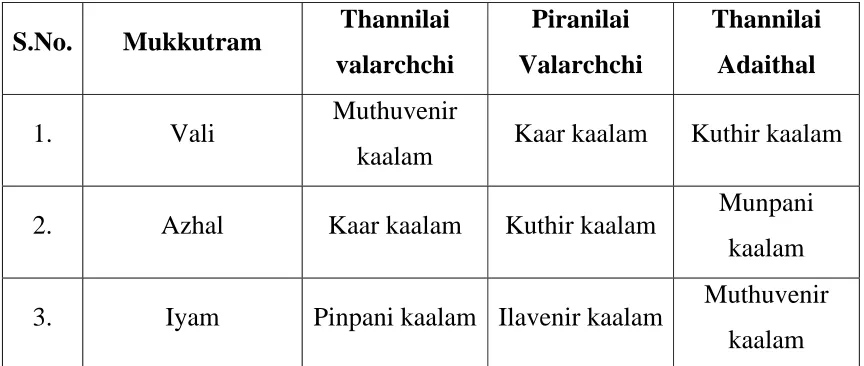
3. SEASONAL VARIATIONS
Seasonal variations which affect the normal constituents of the
body and these cause diseases.
Thannilai Valarchchi means Mukkutram are increasing from their
normal level.
Piranilai Valarchchi means increased Mukkutram spread in to other
places.
Thannilai Adaithal means Mukkutram are stable in their own
25
Table No : 1
Seasonal Variations of Uyir Thathukkal
S.No. Mukkutram Thannilai
valarchchi
Piranilai
Valarchchi
Thannilai
Adaithal
1. Vali Muthuvenir
kaalam Kaar kaalam Kuthir kaalam
2. Azhal Kaar kaalam Kuthir kaalam Munpani
kaalam
3. Iyam Pinpani kaalam Ilavenir kaalam Muthuvenir
kaalam
4. CHANGES IN FOOD HABITS
Food is the basic and essential requirement for keeping the body and
its parts, to grow well and do their work properly for a long time. In short,
the long life, mind, speech and actions of the body, depend upon one’s food.
According to tradition, consuming food twice a day is good. If he
takes food for more than three times a day, he will become sick and will be
inflicted with innumerable diseases.
When, one is in a state of distressed mind, anger or hunger, the heat of
the body will become more and at that state he should consume liquid food.
If he takes solid food under such conditions, it will cause improper
26
During eating, one should take the sweet taste at first then he takes
pungent, saline tastes with vegetables. Astringent taste, sour - curds and
pickles will be consumed at last. This method of taking the six tastes will
give the pleasure of eating.
Any alterations in taking six tastes will cause diseases.
The following foods are unsuitable and they may cause diseases of the
physical constituents, which lead to indigestion and diseases of the stomach.
1. The food that causes abominate feeling or flatulence.
2. The food that has been charred or scorched by fire.
3. Food is not cooked suitably or hard foods, over dried, very cold
food.
4. Foods cooked without properly removing the foreign bodies like
small stones, sand, grass or husk, worms, hair etc.,
5. Dried cooked rice or cooked rice kept in water for a long time.
6. Reheated the already prepared foods.
7. Food that prepared with a lesser quantity of vegetables.
8. Food that is very hot and contains more salt.
Food that consume at irregular timings or at irregular intervals
between the meals may cause severe diseases. And if one consumes the food
27
5. CONSTRAINT OF 14 NATURAL URGES
14 Natural urges are the indications of our body functions and these
should not be obstructed forcibly.
1. Abaanan (Flatus air)
If one resists the flatus air completely or partially, he will inflict with
the diseases of the chest, flatulence, constipation, and pricking sensation
through out the body.
2. Sneezing
Kirukaran vayu which lodges with nose and is responsible for
sneezing. Restriction of sneezing causes headache, and pain in the sense
organs.
3. Urine
If one does not pass urine regularly, it causes obstruction in the
urethral passage, ulceration in the urinary tract, joint pain and distension of
the lower abdomen.
4. Faeces
Abana vayu is responsible for defaecation. If it is obstructed, its
increased quantum pushes the stools. It also causes headache, pain in the
thigh, constipation, discomfort and inability.
5. Yawning
28
6.Hunger 7. Thirsty
If hunger and thirst are not quenched, these lead to impairment
of the functions of vital organs, tiredness and joint pain.
8. Cough 9. Rest (Tiredness)
If cough is controlled, it leads to violent cough, bad odour in the
breath, heart diseases, abdominal pain and tiredness.
If one does not take test properly, tiredness causes faintness and chills.
10. Sleep
If one does not sleep well daily, he may gets headache, redness of
eyes, impaired speech and hearing.
11.Vomiting
If vomiting is prevented forcibly, it leads to fever, itching, pallor, eye
diseases and cough.
12.Tears
Constraint of tears causes eye diseases, ulcer in the head, and heart
disease.
13. Semen
If semen is controlled, it leads to fever, diminution of urine, joint pain,
and chest pain.
14.Breathing
29
DIAGNOSTIC TOOLS IN SIDDHA SYSTEM
Siddha system has a unique diagnostic method to identify the diseases
and their causes.
Envagai Thervu
‘nka;f;Fwp epwk; njhdp tpopeh ,Ukyk;
iff;Fwp” - Njiuah;.
There are 8 tools of diagnosis.
1. Meikuri
2. Naa
3. Niram
4. Mozhi
5. Vizhi
6. Malam
7. Moothiram
8. Naadi
1. Mei kuri
By Meikuri, the following symptoms are observed. The temperature of
the skin (heat or cold), sweating, numbness, fissures, thickening of hairs, hair
30
2. Naa
Tongue colour (black, red, yellow, white), coating of tongue,
excessive salivation, dryness, ulcer, gums, teeth, taste, mouth deviation,
speech are noted.
3. Niram
Body colour (black, yellow, white, red, blue), flush, pallor, black
colour in eyes and teeth are observed.
4. Mozhi
Pitch of voice (high, low, normal), hoarseness of voice, fluency,
intelligence, articulation, character, breathlessness are observed.
5. Vizhi
Eyes easily reflect the pathological changes of the body. Size and
shape, colour (red, yellow, pallor, blue, muddy), lacrimation, dryness,
swelling of eyelids, ulceration, visual field, sharpness of vision, colour
vision, inflammation (ulcer in conjunctiva, cornea, pupil) are keenly
observed.
6. Malam
Colour (yellow, red, black, white, green), froth, solid or semisolid or
31
7. Moothiram
Urine is observed under 2 headings
1. Neer kuri
2. Nei kuri
7.1. Neer kuri
Collection of Urine for Testing
Before the collection of urine for testing, one should take supper
consisting of all the six tastes at the regular time based on one’s digestive
fire. After a sound overnight sleep, urine should be collected in a closed
glass ware and the test should be done before 90 minutes from dawn. This
rule is relaxable in severe cases.
General Features of Urine
Niram (Colour)
Manam (Odour )
Nurai (Froth )
Edai (Specific gravity )
Enjal (Quantity)
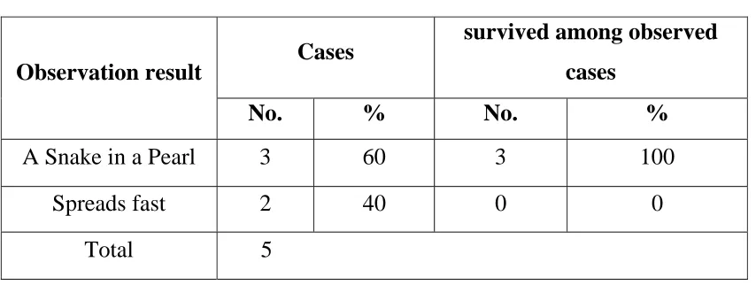
7.2 . Nei kuri
A drop of oil is dropped at the centre of oil bowl without any shaking.
It should be ensured that the direct sunlight does not fall on it, but bright
32
The changes of the oil drop in Urine suggest the diagnosis and condition of
the patient.
General Nature of Urine in Oil Examination
If the oil drop takes the shape of a shake, it indicates Vali disease. If it
spreads like a ring it indicates Azhal disease. If it stands like a pearl it
indicates Iya disease. If the oil drop sinks in the urine.
If all features of the three humors are seen together in the urine it
suggests derangement of all three humors. If the oil drop spreads fast, it will
indicate Asaathiayam.
8. Naadi
Naadi is very important tool. Diagnosis of the disease by assessing
Naadi gives a best way to treat the disease. Vali Naadi in felt in tip of Index
finger. Azhal Naadi is felt in tip of Middle finger. Iya Naadi is felt in tip of
Ring finger.
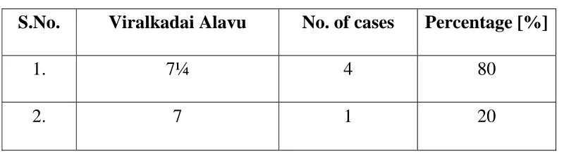
Manikkadai Nool
Manikkadai nool is another important diagnostic tool. It is a
measurement which is done 4 inches from the wrist by thread. The
measurement denotes the signs and symptoms of the disease. In severe
33
AIM AND OBJECTIVES
The importance of medical science is well appreciated when man
suffers by disease. The Siddha system of medicine gives both curative and
preventive aspects of the disease.
Diseases of the nervous system are fairly common in pediatrics than
adults. Neurological symptoms are also frequently encountered in a wide
variety of systemic illnesses.
Accurate early diagnosis in children is likely to be more rewarding
than in adults
Because the early detection of this disease is very much useful to
protect our future assets, i.e. children. This can give a healthy and victorious
India as we believe to reach.
The aim of this study is to evaluate the etiology and pathology of
Vachchiraroopam through 96 basic principles. This gives guide to diagnose
and treat the disease in successful manner.
The following objectives are carried to fulfill the aim.
To find out the etiology of this disease.
To establish the pathological view of this disease.
To keenly observe the changes of the Envagai Thervu for attain
diagnosis.
To collect Siddha literary evidences as well as the Modern theories.
34
ELUCIDATION ABOUT VACHCHIRAROOPAM
Vachchiraroopam is one of the Vatha diseases which is described in
Yugi Vaithiya Sinthamani in 302nd stanza. It is as follows.
tr;rpu&gk;
‘tpjpahd gplhpiaj;jhd; kpftp Oj;J kpff;fz;f sirtpy; yhkNy Nehf;fpj; jjpahd FNwhf;ifNghw; rj;jk; ngw;Wj; jiyNehT kpFjpaha; eLf;f Yw;W
Fjpahd euk;gpOj;Jj; jpkpUz; lhfpf;
nfhl;lhtp Nrhk;gyha;;f; $r;r Yz;lha; tjpahd kyryKQ; RWf;fp tPOk;
tr;rpu& ge;jd;dpd; tifap jhNk”.
- A+fp itj;jpa rpe;jhkzp tr;rpuk; - itukzp
&gk; - tbtk;
Vachchiraroopam denotes the stiffness of the body.
nghUs;:
gplhp - Occiput ( The back part of the head )
,Oj;jy; - Retraction ( The act of drawing back )
FNwhf;if Nghw; rj;jk; - Snoring like voice
jiyNehT - Headache
eLf;fy; - Tremulousness ( Shaking slightly )
euk;G - Muscles
35
Nrhk;gy; - Lethargy
$r;ry; - Making noise due to irritation
RWf;fp - Diminution
‘tpjpahd gplhpiaj;jhd; kpftp Oj;J”
Retraction of head is an extreme degree of cervical rigidity which
represents reflex protective spasm.
‘kpff;fz;f sirtpy; yhkNy Nehf;fpj;”
Fixed gaze which means look steadily in one direction. The patient
can’t see freely in all directions because the field of vision is contracted all
round the periphery.
‘jjpahd FNwhf;ifNghw; rj;jk; ngw;Wj;”
Snoring like voice which means Nasal twang.
Twang means the quality of speech sound which produced through
both the nose and the mouth.It is produced due to paralysis of the soft palate.
‘jiyNehT kpFjpaha; eLf;f Yw;W”
There will be severe headache and tremulousness.
‘Fjpahd euk;gpOj;Jj; jpkpUz; lhfpf;”
Muscle spasm and stiffness which is produced throughout the body
and it will make the appearance of the body rigid.
‘nfhl;lhtp Nrhk;gyha;;f; $r;r Yz;lha;”
The patient has symptoms such as yawning, lethargy and making
36
‘tjpahd kyryKQ; RWf;fp tPOk;”
Diminution of faeces and urine reflect the severity of the disease
becoming worst.
‘tr;rpu& ge;jd;dpd; tifap jhNk”
These are the features of the disease Vachchiraroopam.
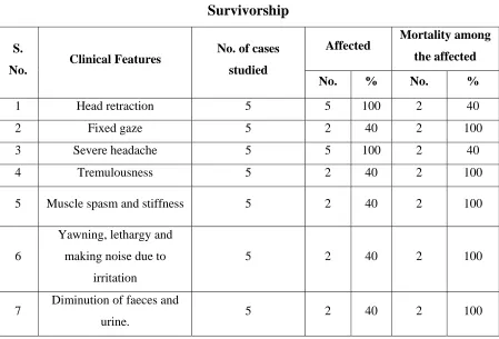
The Summary of Clinical Features of Vachchiraroopam are as follows.
In Vachchiraroopam, inflammation of the meninges causes head
retraction which is due to spasm of extensor muscles of the neck because of
irritation of nerve roots.
Fixed gaze is due to Papilloedema. Snoring like voice is the
manifestation of paralysis of muscles of soft palate.
Intense headache occurs due to stretching stimulus to the blood vessels
of the meninges.
Tremulousness occurs due to inflammatory toxic substances
circulating in blood which affects the hypothalamic temperature control
center.
Muscle spasm and stiffness occurs due to inflammation of spinal
meninges. Yawning, lethargy, making noise due to irritation are due to
37
PATHOLOGICAL VIEW OF DISSERTATION TOPIC IN
SIDDHA ASPECT
In Vachchiraroopam, both Uyir and Udal thathukkal are affected.
UYIR THATHUKKAL
INCREASED IYAM
Iyam is predominantly affected, because head is one of the dwelling
places of Iyam. It is revealed by the following poetry lines.
‘$wpNdhk; rpNyw;gdkJ rkhd thA
nfhOfpalh RopKidiag; gw;wp tpe;jpy;
rPwpNa rpurpyhf; fpidiar; Nrh;e;J
rpq;Fit mz;zhf;F epzkr;ir uj;jk;
kPwpNa epwq;Nfhzk; euk;ng Yk;gpy;
NktpaNjhh; %isngUq; Flypw; fz;zpy;
NjwpaNjhH nghUe;jplq;f nsy;yhQ; NrHe;J
rpNyl;LkkJ tPw;wpUf;Fe; jplq; fz;lhNa.”
- rjf ehb
1. Ali Iyam ( Avalambagam )
It is increased and exhibits the symptoms like dyspnea on exertion.
2. Neerppi Iyam ( Kilethagam )
38
3. Suvaikaan Iyam ( Pothagam )
It is increased and results sweet taste appreciated in tongue. It is
revealed by the following poetry lines.
‘ Nrj;Jk nkOe;jpUf;fpw; jpj;jpg;G ehtp NyWk ;”
- mfj;jpah; ehb.
4. Niraivu Iyam ( Tharpagam )
It is increased in this disease which is due to inflammatory changes in
the Cerebrospinal Fluid.
5. Ontri Iyam ( Santhigam )
It is increased and causes inability to flex the joints.
INCREASED VALI
1. Uyirkkaal ( Praanan )
It is increased and results dyspnea on exertion.
2. Keelnokkukkaal ( Abaanan )
In Vachchiraroopam, it is increased and exhibits the symptoms like
diminution of faeces and urine.
3. Melnokkukkaal ( Udhaanan )
It is increased and causes snoring like voice.
4. Paravukaal ( Viyaanan )
It is increased and exhibits the symptoms like pain andtenderness in
39
5. Nadukkaal ( Samaanan )
It is increased in this disease and results reduced appetite.
6. Vaanthikkaal (Naahan )
In Vachchiraroopam, it is increased and causes reduced intellectual
functions.
7. Vizhikkaal ( Koorman )
It is increased and it causes contraction of field of vision.
8. Thummikkaal ( Kirukaran )
It is increased and results coated tongue and reduced appetite.
9. Kottavikkaal ( Devathathan )
In Vachchiraroopam, it is increased and results lethargy and tiredness.
DECREASED AZHAL
1. Aakkanal ( Anar pitham )
It is decreased and causes reduced appetite.
2. Vannayeri ( Ranjaga pitham )
It is decreased and exhibits pallor ness due to anaemia.
3. Aattralangi ( Saathaka pitham )
It is decreased because of the ability to do works disturbed in
Vachchiraroopam.
4. Nokkazhal ( Aalosaka pitham )
In Vachchiraroopam, it is decreased and exhibits the symptoms like
40
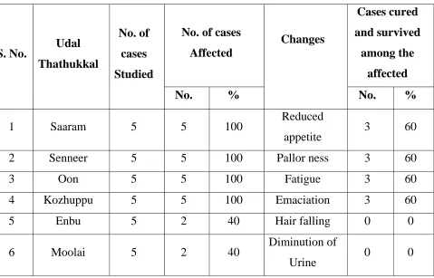
UDAL THATHUKKAL
In Vachchiraroopam, Udal thathukkal are deranged.
Saaram - Sluggishness
Senneer - Pallor ness
Oon - Fatigue
Kozhuppu - Emaciation
Enbu - Hair falling
Moolai - Stiffness of the body and Diminution of urine.
In Vachchiraroopam, primarily Iyam and secondarily Vali are
increased and Azhal is decreased. This Mukkutra nilaigal leads to
41
PATHOLOGICAL VIEW OF THE DISSERTATION TOPIC
IN MODERN ASPECTS
’tpjpahd gplhpiaj;jhd; kpftp Oj;J”
In Vachchiraroopam, Head retraction is the cardinal feature. Head
retraction is an extreme degree of cervical rigidity brought about by spasm of
extensor muscles of neck.
This is caused by irritation of the nerve roots during their passage
through the subarachnoid space which is infected.
’kpff;fz;f sirtpy; yhkNy Nehf;fpj;”
This symptom is the manifestation of increased C.S.F. pressure in the
Optic disc.
The normal pressure in the C.S.F. system when one is lying in a
horizontal position averages 130 mm of water (10 mmHg).
The C.S.F. pressure rises considerably when infection occurs in the
cranial vault.
In this condition, large numbers of white blood cells suddenly appear
in the C.S.F. and they can cause serious blockage of the small absorption
channels through the arachnoid villi. This also sometimes elevates the C.S.F.
pressure to 400 to 600 mm of water (about 4 times normal).
42
Anatomically the arachnoid mater and the pia mater extend forwards
into the orbit and cover the Optic nerve. So the subarachnoid space also
surrounds the optic nerve.
Elevation in Cerebrospinal fluid System Pressure
When the pressure rises in the Cerebrospinal fluid system due to
infection or inflammation, it also rises inside the optic nerve sheath, a few
millimeters behind the eye and then passes along with the optic nerve fibers
into the eye itself. Therefore,
1. High C.S.F. pressure pushes fluid first into the optic nerve sheath and
then along the spaces between the optic nerve fibers to the interior of the
43
2. The high pressure decreases outward fluid flow in the optic nerves,
causing accumulation of excess fluid in the optic disc at the center of the
retina; and
3. The pressure in the sheath also impedes flow of blood in the retinal vein,
thereby increasing the retinal capillary pressure throughout the eye, which
results in still more retinal edema.
The tissues of the optic disc become for more edematous than the
remainder of the retina and swells into the cavity of the eye. The swelling of
the disc can be observed with an Ophthalmoscope and is called as
Papilloedema
So the field of vision is contracted all round the periphery which is
44
’jjpahd FNwhf;if Nghw; rj;jk; ngw;W”
Snoring like voice which means Nasal twang.
It is produced due to Paralysis of the Soft Palate.
The muscles of the soft palate (except the tensor palati which is
supplied by the mandibular nerve) are supplied by the Pharyngeal plexus.
The fibres of this plexus arise from the upper part of the Inferior
ganglion of the Vagus, and contain chiefly the fibres of the cranial accessory
nerve.
The Inferior ganglion of the Vagus nerve which is cylindrical and lies
near the base of the skull. It is involved during the inflammation of
meninges. It causes paralysis of soft palate which produces nasal twang.
‘jiyNehT kpFjpaha;”
This may be the first symptom which calls the patient to physician.
Due to inflammation of the meninges, including the sensitive areas of
the dura, the sensitive areas around the venous sinuses and stretching
stimulus to the blood vessels of the meninges can cause headache.
The extreme headache refers over the entire head.
’eLf;f Yw;W”
Tremulousness occurs due to inflammatory toxic substances
circulating in blood which affects the hypothalamic temperature control
45
When the hypothalamic temperature control center is suddenly
changed from the normal level to higher as a result of tissue destruction due
to inflammation, the body temperature usually takes several hours to reach
the new temperature level.
Because the blood temperature is now less than the set point of the
hypothalamic temperature controller, the usual responses that cause
elevation of body temperature.
During this period, the person experiences tremulousness.
Tremulousness can continue until the body temperature reaches the
higher hypothalamic set point level.
‘Fjpahd euk;gpOj;Jj; jpkpUz; lhfpf;”
Due to inflammation of the Spinal meninges, the subarachnoid space
of the spinal cord is distended.
The spinal nerve roots are stretched. These cause muscle spasm and
stiffness.
‘nfhl;lhtp Nrhk;gyha;f; $r;r Yz;lha;”
During inflammatory process, many tissue products are released from
damaged tissue. These are histamine, bradykinin, serotonin, prostaglandins,
several different reaction products of the blood clotting system and multiple
46
Necrosin is released from damaged tissues at the site of infection,
which further damage the tissues locally and when it enters the general
circulation, it damages organs elsewhere.
Circulating toxic substances cause Delirium. This usually takes the
form of lethargy with disorientation and muddled thinking.
These may be associated with emotional disturbance such as anxiety,
irritability or depression.
There may be a high pitched Meningeal cry due to severe headache.
‘tjpahd kyryKQ; RWf;fp tPOk;”
Diminution of faeces and urine are due to Sacral radiculopathy.
Due to Sacral radiculopathy, the faeces arrives normally at the pelvic
colon, but their final evacuation is not adequately performed. Fragmentary
defaecation may takes place, i.e. Small amounts are passed, but much faeces
are left behind in the rectum to continue distending it.
Due to Sacral radiculopathy, micturition is incompletely performed;
i.e.large quantities of residual urine may be left in the bladder.
’tr;rpu ≥ jd;dpd; tifapjhNk”
47
MODERN ASPECTS
MENINGES
The Brain and Spinal cord are surrounded by the three membranous
coverings called the Meninges. The brain is very important but delicate
organ.
The Meninges are
1. Outer Duramater
2. Arachnoid mater
3. Inner Piamater.
The dura mater is also frequently called the pachymenix. The
arachnoid and pia mater are collectively called the leptomeninges.
Between the arachnoid mater and the pia mater there is the
subarachnoid space which contains Cerebrospinal fluid (C.S.F.). In relation
to the dura mater there are a series of venous sinuses which drain intracranial
structures including the brain.
DURA MATER
The dura mater is a thickest of the three meninges. It encloses the
cranial venous sinuses, and has a distinct blood supply and nerve supply.
The Cerebral Dura Mater
The dura mater is the outermost, thickest and toughest membrane
48
There are two layers of dura:
a. An outer or endosteal layer which serves as an internal periosteum or
endosteum or endocranium for the skull bones, and
b. An inner or meningeal layer which surrounds the brain.
The meningeal layer is continuous with the spinal dura mater.
The two layers are fused to each other at all places, except where the
cranial venous sinuses are enclosed between them.
The Endosteal Layer or Endocranium
The endocranium is continuous
a. with the periosteum lining the outside of the skull or pericranium
through the sutures and foramina, and
b. with the periosteal lining of the orbit through the superior orbital
fissure.
It provides sheaths for the cranial nerves. The sheaths fuse with the
epineurium outside the skull. Over the optic nerve, the dura mater forms a
sheath which becomes continuous with the sclera.
The Meningeal Layer
At places, the meningeal layer of dura mater is folded on itself to form
partitions. It divides the cranial cavity into compartments which lodges
49
The folds are the:
a. Falx cerebri
b. Tentorium cerebelli
c. Falx cerebelli
d. Diaphragma sellae.
Spinal Dura Mater
Spinal dura mater is a thick, tough, fibrous membrane which forms a
loose sheath around the spinal cord. It is continuous with the meningeal layer
of the cerebral dura mater.
The spinal dura extends from the foramen magnum to the lower
border of the second sacral vertebra; whereas the spinal cord ends at the
lower border of first lumbar vertebra. The dura gives tubular prolongations
to the dorsal and ventral nerve roots and to the spinal nerves as they pass
through the inter vertebral foramina.
Subdural Space
Subdural space is a capillary or potential space between the dura and
the arachnoid, containing a thin film of serous fluid. This space permits
movements of the dura over the archnoid.
ARACHNOID MATER
The arachnoid mater is a thin transparent membrane.
Cerebral Arachnoid Mater
50
Spinal Arachnoid Mater
It loosely invests the entire central nervous system. Inferiorly it
extends, like the dura, upto the lower border of the second sacral vertebra.
PIA MATER
This is a delicate membrane.
Cerebral Pia Mater
It closely invests the brain. It dips into sulci of the cerebrum.
The pia mater intracranially extends over the cranial nerves and fusing
with their epineurium. Blood vessels entering the cerebrum are covered by
the peri vascular sheath formed from pia mater.
The telachoroidea is an extension of pia mater into the ventricular
system. Telachoroidea is invaginated by the blood vessels to form the
choroid plexus. The choroid plexus secrete the Cerebrospinal Fluid.
Spinal Pia Mater
Spinal pia mater is thicker, firmer and less vascular than the cerebral
pia mater, but both are made up of two layers.
a. An outer epi – pia containing large vessels.
b. An inner pia – gia or pia intima which is in contact with nervous
tissue.
51
SUBARACHNOID SPACE
This is the space between the arachnoid and the pia mater. It surrounds
the brain and spinal cord like a water cushion.
The spinal subarachnoid space is wider than the space around the
brain.
It is widest below the lower end of the spinal cord where it encloses
the Cauda Equina. It is traversed by a network of arachnoid trabeculae which
give it a sponge like appearance.
It ends below at the lower border of the second sacral vertebra. The
subarachnoid space contains C.S.F. and large vessels of the brain. Cranial
nerves pass through the space.
The subarachnoid space in certain locations, they are dilated and
called cisterns.
The main cisterns are,
a. Cisterna pontis (Pontine cistern)
b. Interpeduncular cistern (Basal cistern)
c. Cerebello medullary cistern ( Cisterna magna )
The cisterna pontis lies anterior to Pons and Medulla.
Interpeduncular cistern lodges circle of Willis. This cistern is due to
the stretch of the arachnoid mater between the two cerebral peduncles.
Cerebello medullary cistern is situated posteriorly. The arachnoid
52
The Arachnoid villi especially well developed along the superior
sagital sinus. Collection of Arachnoid villi are called Arachnoid granulations
( Pacchionion bodies ).
Through the Arachnoid villi the cerebrospinal fluid is filtered into the
venous blood. There are fibrous sheath connecting arachnoid and pia mater.
Cranial nerves are crossing through the subarachnoid space.
CEREBROSPINAL FLUID (C.S.F.)
Cerebrospinal fluid is a clear and colourless watery liquid. It is a
modified tissue fluid. It is contained in the ventricular system of the brain
and in the subarachnoid space around the brain and spinal cord.
C.S.F. replaces lymph in the Central nervous system. The C.S.F.
provides a fluid cushion which protects the brain from injury. It probably
also helps to carry nutrition to the brain and to remove waste products.
SITE OF C.S.F. FORMATION
1. Choroid plexus of the lateral ventricle – 95%
2. Choroid plexus of the third and fourth ventricles
3. Perivascular spaces of the brain
53
CIRCULATION OF C.S.F.
Choroid plexus of lateral ventricle
↓
Foramen of Monro
↓
Third ventricle
↓
Sylvian duct
↓
Fourth ventricle
↓
Foramen of Luschka and Magendie
↓
Subarachnoid space
↓
Basal cisterns circulation over brain and spinal cord.
ABSORPTION OF C.S.F.
1. C.S.F. is absorbed chiefly through the arachnoid villi and granulations,
and is thus drained into the cranial venous sinuses.
2. It is also absorbed partly by the perineural lymphatics around the first,
second, seventh and eight cranial nerves.
54
FUNCTIONS OF C.S.F.
1. Nutrition
2. Excretion
3. Shock absorption
4. Regulation of intra cranial pressure.
NORMAL C.S.F.
Colour - Clear as water
Volume - 150ml (The whole volume
of C.S.F. replaced several times a day)
Rate of Formation - 0.3ml per minute
Specific gravity - 1.005
Reaction - Alkaline
pH - 7.33
Pressure - 60-150mm of C.S.F. in supine and
200-250mm of C.S.F. in sitting
position
Protein - 20 to 40 mg%
Glucose - 40 to 60 mg%
Chlorides - 720 to 750mg%
Cells - not more than 5 lymphocytes/cumm.
The C.S.F. secreted by ventricle dose not contains any cell. The
55
CHRONIC MENINGITIS
Meningitis refers to an inflammatory process of the leptomeninges and
cerebrospinal fluid within the subarachnoid space.
Infectious meningitis can be broadly classified as
1. Acute pyogenic meningitis (usuallybacterial)
2. Aseptic meningitis (usually viral)
3. Chronic meningitis (bacterial or fungal)
CHRONIC MENINGITIS
The condition is most commonly diagnosed when a characteristic
neurological syndrome exists for longer than 4 weeks and is associated with
a persistent inflammatory response in Cerebrospinal fluid (C.S.F.).
There are two principal types of chronic meningitis – one bacterial
(tuberculous meningitis) and the other fungal (cryptococcal meningitis).
Both types cause chronic granulomatous reaction and may produce
parenchymal lesions.
AETIOLOGY
Infection may reach the meninges by following routes:
1. Haematogenous route (most common) is used by pathogens that reach
the circulating blood during a systemic infection, by septic emboli
from suppurative lesions of lungs or infected valvular vegetations, or
56
2. Local extension can occur secondary to an established infection in an
airsinus (most of the the mastoid or frontal), an infected tooth, or a
surgical site in the cranium or spine causing osteomyelitis, bone
erosion, and propagation of the infection into the central nervous
system.
3. Direct implantations of organisms may result from penetrating
wounds, compound fractures of the skull, and neurosurgical
procedures (such as ventricular shunts or lumbar puncture), and rarely
from congenital neuro ectodermal defects.
4. Axonal transport - The peripheral nervous system can be a path of
infection into the central nervous system, as occurs with certain
viruses (eg: rabies, and herpes simplex).
Damage to nervous system may be the consequence of direct injury of
neurons or glia by the organisms, microbial toxins, the effects of the
inflammatory response, or immune mediated injury.
PATHO PHYSIOLOGY
C.S.F. produced by the choroid plexus of cerebral ventricles, exists
through narrow foramina into the subarachnoid space surrounding the brain
and spinal cord. It circulates around the base of the brain and over the
cerebral hemispheres and is resorbed by arachnoid villi projecting into the
57
C.S.F. flow provides a pathway for rapid spread of infections and
malignant processes over the brain, spinal cord and cranial and spinal nerve
roots. Spread from the subarachnoid space into brain parenchyma may occur
via the arachnoid cuffs that surround blood vessels that penetrate brain tissue
(Virchow – Robins’s space). Virchow – Robins’s space is potential spaces
surrounding blood vessels for a short distance as they enter the brain.
The cellular response of the nervous system to invading pathogens is
basically similar to inflammation elsewhere in the body.
The capsular K1 antigen seems to be of particular importance. It is
assumed that offending organisms lodge in the choroid plexus, producing
acute plexitis and subsequently spread into ventricular fluid to reach the
subarachnoid space.
Nociceptive fibers of the meninges are stimulated by the inflammatory
process. Purulent exudate appears as creamy streaks on sulcal cisterns and
forms a thick film around the spinal cord, especially over its dorsal aspect.
The ventricular fluid is turbid. The choroid plexus and the ependymal
lining are covered by pus.
During the first 2 to 3 days the exudates are composed entirely of
polymorphonuclear leukocytes, followed by lymphocytes and macrophages
58
All are largely confined to the subarachnoid space and extend only for
short distances into the Virchow – Robins’s spaces of the penetrating cortical
vessels and subependymal venules.
Cognitive and behavioral changes during the course of chronic
meningitis may also result from vascular damage, which may similarly
produce seizures, stroke or myelopathy.
Inflammatory deposits seeded via C.S.F. circulation are often
prominent around the brainstem and cranial nerves and along the
undersurface of the frontal and temporal lobes.
Such cases, termed basal meningitis, often present as multiple cranial
neuropathies with visual loss (Cranial Nerve II), facial weakness (C.N. VII),
hearing loss (C.N. VIII), diplopia (C.N. II, IV, VI), sensory or motor
abnormalities of the oropharynx (C.N. IX, X, XII), decreased olfaction
(C.N. I), or facial sensory loss and masseter weakness (C.N. V).
If the inflammation is not rapidly controlled, the exudate may become
basilar and fibrotic adhesions of the arachnoid, particularly in the basal
cisterns and along the sagital fissure, impede the freeflow of C.S.F. and
59
CAUSES OF CHRONIC MENINGITIS
Infectious Causes
1. Bacterial
a. Partially treated acute pyogenic infections
b. Mycobacterium tuberculosis
c. Syphilis (secondary, tertiary)
d. Parameningeal infections
2. Fungal
a. Cryptococcus neoformans
b. Candida albicans
c. Histoplasma capsulatum
3. Viral
a. Mumps
b. Echovirus
c. H.I.V.
d. Herpes simplex
4. Protozoal
a. Trypanosomiasis
60
Non Infectious Causes
1. Malignancy – Risk factors - metastatic carcinoma of breast, lung,
stomach or pancreas, melanoma, lymphoma, leukaemia, meningeal
sarcoma.
2. Primary inflammation C.N.S. sarcoidosis
3. Systemic lupus erythmatosis
4. Chemical irritation from craniopharyngioma or epidermoid.
TUBERCULOUS MENINGITIS
In C.N.S. Tuberculous meningitis is the most common form;
tuberculoma occurs less frequently and present as mass lesions.
PATHOGENESIS
Tuberculous meningitis is assumed to be caused by release of the
pathogen into the subarachnoid space from small tuberculomas, Rich’s foci.
These foci are difficult to identify, purportedly occurring anywhere in
the neuraxis, notably the cortex.
Tuberculous meningitis may also develop in the course of miliary
tuberculosis.
PATHOLOGY
The meninges over the surface of the brain and the spinal cord are
cloudy and thickened, but the process is usually most intense at the base of
61
A thick collar of fibrosis may form around the optic nerves, cerebral
peduncles, and basilar surface of the pons and midbrain.
The ventricles are moderately dilated and the ependymal lining is
covered with exudates or appears roughened (granular ependymitis). Minute
tubercles may be visible in the meninges, choroid plexus and cerebral
parenchyma.
On microscopic examination, the exudates in the thickened meninges
are composed chiefly of mononuclear cells, lymphocytes, plasma cells,
macrophages, and fibroblasts with occasional giant cells. The inflammatory
process may extend for a short distance into the cerebral substance where
microscopic granulomas may also be found.
Proliferative changes are frequently seen in the inflamed vessels of the
meninges, producing a panarteritis. These arteritic changes may lead to
thrombosis of the vessel and cerebral infarcts.
TUBERCULOMA
A tuberculous abscess in the brain is called tuberculoma which may be
associated with meningitis. When tuberculosis was rife, tuberculoma of the
brain were common, especially in children, and were found most often in the
cerebellum.
Tuberculous abscesses in the brain are often multiple. They may be
62
Microscopically, there is usually a central core of caseous necrosis
surrounded by typically tuberculous granulomatous reaction and
calcification may occur in inactive lesions.
CLINICAL FEATURES
Although tuberculous meningitis may occur at any age, it is most
common in child hood and early adult life.
The onset is usually subacute, with headache, vomiting, fever, bursts
of irritability, and nocturnal wakefulness as the most prominent symptoms.
The headache becomes progressively more severe. The pain often
causes the children to emit a peculiarly shrill cry (Meningeal cry) a high
pitched scream.
The patient becomes drowsy and at times delirious, but lucid intervals,
even up to a late stage of the illness, are characteristic.
Signs of Meningeal Irritation
Cervical Rigidity (Neck Stiffness)
It is observed by the observer placing his hand beneath the patient’s
occiput and endeavouring to flex the head so as to bring the chin towards the
chest.
There is resistance due to spasm of the extensor muscles of the neck,
63
Head Retraction
Head retraction is an extreme degree of cervical rigidity brought about
by spasm of extensor muscles.
Flexion of the neck causes a rise in the C.S.F. pressure in the cisterna
magna. When the meninges are inflamed this is painful; neck stiffness and
head retraction represent reflex protective spasm.
Kernig Sign
To elicit Kernig’s sign one knee is extended with the hip fully flexed;
when positive there is pain due to spasm of the hamstring muscles and
limitation of extension.
Brudzinski Sign
Brudzinski’s sign consists first of spontaneous flexion of the knees
and hips on attempted neck flexion and secondly spontaneous flexion of one
leg when the other is flexed passively.
These signs result from the presence of inflammatory exudates around
the roots in the lumbar theca.
Other Signs
The initial irritability is gradually replaced by apathy, confusion,
lethargy, and stupor.
Papilloedema, cranial nerve palsies, and focal neurological signs are
64
moderately elevated (100 to 1020F) in the early stage, rises to high levels
before death.
Involvement of cranial nerves may give ptosis, diplopia, facial
weakness, deafness, or dysphagia.
Retention of urine may be followed by incontinence of urine, less
often of faeces. Constipation is usual.
Injury may occur to motor and sensory nerve roots as they traverse the
subarachnoid space and penetrate the meninges.
These cases are present as multiple radiculopathies, with combination
of radicular pain, sensory loss, motor weakness, and sphincter dysfunction.
Meningeal inflammation can encircle the cord, resulting in myelopathy.
COMPLICATIONS
Acute Complications
1. Hydrocephalus
2. Cerebral infarction
3. Cranial nerve palsies
4. Convulsions
5. Fluid and electrolyte disturbances and
65
Chronic Complications
1. Obstructive hydrocephalus
2. Optic atrophy
3. Subdural effusions
4. Diffuse or localized spinal arachinoiditis
5. Spinal cord compression and
6. Development of tuberculoma in the brain and spinal cord.
DIAGNOSIS
C.S.F. Analysis
The diagnosis of tuberculous meningitis can be established by
recovery of the organisms from the C.S.F..
The C.S.F. findings are, Increased pressure, slightly cloudy or ground
glass appearance of the C.S.F. with formation of a clot on standing,
Moderate pleocytosis of 25 to 500 cells / cu mm with lymphocytes as the
predominating cell type, Increased protein content, Decreased sugar content,
a negative serological test for syphilis or streptococcal antigen and absence
of growth when the C.S.F. is inoculated on routine culture media.
COHEN’S LAW OF MENINGITIS (1929)
Substances which are more in C.S.F. than blood diminish in
meningitis and substances which are less in C.S.F. than blood increase in
meningitis (except sugar which is low in meningitis because it is used by the
66
Protein, Sugar, Cholesterol, Urea, Calcium and Phosphorus are
less in C.S.F. than blood.
Chlorides and Magnesium are more in C.S.F. than blood.
Antibodies, Enzymes, Penicillin and Streptomycin are never seen
in C.S.F. as they don’t cross the blood- brain barrier.
C.T. or M.R.I. Scan of the brain
C.T. or M.R.I. of the brain in Tuberculous meningitis, the exudates
which is frequently seen in the Basal cisterns, tends to be thick and adhesive.
Communicating hydrocephalus may result from obstruction at the
level of the basal cisterns. Vascular involvement of the arteries at the Base of
the brain or Sylvian fissures may result from vasculitis and surrounding
meningeal inflammation.
On Non contrast C.T. or M.R.I. studies, the Basal cisterns and Sylvian
fissures may be partially or completely obscured by the presence of purulent
exudates and inflammatory tissue, which appears to have a similar density or
signal intensity.
On Contrast - enhanced C.T. or M.R.I. studies, the involved cisterns
show intense enhancement.
M.R.I. studies are more sensitive than C.T. scans for detection of
67
On Contrast - enhanced C.T. scans, Tuberculomas may present a
pattern of Ring like enhancement or, less likely, areas of nodular
enhancement or irregular non homogeneous enhancement.
A central nidus of calcification with surrounding ring like
enhancement known as the Target sign is suggestive of Tuberculoma.
Other Diagnostic Aids
It includes a thorough search for a primary focus, including