0022-538X/97/$04.00
1
0
Copyright © 1997, American Society for Microbiology
Phosphorylation Sites in Polyomavirus Large T Antigen That
Regulate Its Function in Viral, but Not Cellular, DNA Synthesis
ALAKANANDA CHATTERJEE, BEVERLY J. BOCKUS,† OLE V. GJØRUP,‡
ANDBRIAN S. SCHAFFHAUSEN*
Department of Biochemistry, Sackler School of Graduate Biomedical Sciences, Tufts University
School of Medicine, Boston, Massachusetts 02111
Received 30 January 1997/Accepted 16 May 1997
Polyomavirus large T antigen (large T) is a highly phosphorylated protein that can be separated by
proteolysis into two domains that have independent function. A cluster of phosphorylation sites was found in
the protease-sensitive region connecting the N-terminal and C-terminal domains. Edman degradation of
32
P-labeled protein identified serines 267, 271, and 274 and threonine 278 as sites of phosphorylation. Analysis
of site-directed mutants confirmed directly that residues 271, 274, and 278 were phosphorylated. Threonine
278, shown here to be phosphorylated by cyclin/cyclin-dependent kinase activity, is required for viral DNA
replication in either the full-length large T or C-terminal domain context. The serine phosphorylations are
unimportant in the C-terminal domain context even though their mutations activates viral DNA replication in
full-length large T. This finding suggests that these sites may function in relating the two domains to each
other. Although the phosphorylation sites were involved in viral DNA replication, none was important for the
ability of large T to drive cellular DNA replication as measured by bromodeoxyuridine incorporation, and they
did not affect large T interactions with the Rb tumor suppressor family.
Large T antigen (large T) of murine polyomavirus (Py) is
multifunctional. Its most obvious role is to initiate viral DNA
replication (7). Large T also stimulates host cell DNA synthesis
(9, 35, 42) and transactivates a variety of cellular promoters
(17, 19, 20, 24, 34, 35). Such effects on the host cell are
re-flected in large T’s ability to immortalize primary cells (37) and
to inhibit differentiation of myoblasts (28). Both of these
ef-fects depend on interactions with members of the Rb tumor
suppressor family (8, 23, 27).
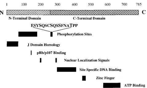
The functions of Py large T are assorted to different
struc-tural domains within its 785-amino-acid sequence (Fig. 1).
Pro-teolysis experiments point to a division between an N-terminal
domain (NT) and a C-terminal domain (CT) around residue
260 (14). NT is sufficient for immortalization by Py large T (14)
and for the stimulation of cell cycle progression in
serum-starved cells (9). It contains a binding motif for Rb family
proteins and a DnaJ domain that provides a binding site for
hsc70 (43a). CT is sufficient for viral DNA replication in
grow-ing cells (9) and contains the DNA bindgrow-ing domain, zinc fgrow-inger,
and ATP binding region. Py large T is localized to the nucleus
and contains two independent nuclear localization signals (16,
38).
It has long been known that Py large T is highly
phosphor-ylated (40). Most of the phosphorylation occurs after large T is
translocated to the nucleus (16). There is ample reason to
suspect that the phosphorylation may be functionally
signifi-cant. Both correlation and direct evidence suggest a role for
large T phosphorylation in DNA replication. A
slower-migrat-ing form resultslower-migrat-ing from phosphorylation (2) predominates
af-ter the initiation of viral DNA replication, while a fasaf-ter-mi-
faster-mi-grating form is seen early in infection. A number of mutants
defective in DNA replication also show altered patterns of
phosphorylation (2, 39). Wang and colleagues have shown that
phosphatase treatment of large T affects its activity in DNA
replication assays in vitro (49).
Although phosphorylation of large T has attracted
consid-erably attention, the sites have not been identified. Previous
work showed substantial phosphorylation in two regions, one
in NT and one more C terminal, near the junction of the two
domains (1). Work by Hassauer et al. (12) implicated residues
81 and 187 as sites of phosphorylation in NT. The work
pre-sented here identifies the more C-terminal phosphorylations
and tests their function. These phosphorylations appear
im-portant in large T function in initiating viral, but not cellular,
DNA replication.
MATERIALS AND METHODS
Cells, viruses, and plasmids. NIH 3T3 and 293 cells were maintained in
Dulbecco’s modified Eagle’s medium (DMEM; GIBCO) supplemented with 10% calf serum (Hyclone) (3T3 cells) or with 10% fetal calf serum (293 cells). Transfections were performed by the calcium phosphate precipitation method of Chen and Okayama (6). Cells were harvested after 48 h for transient assays. Stable 3T3 cell lines expressing wild-type and mutant large T’s were selected with geneticin as described previously (14).
Spodoptera frugiperdaSf9 cells were maintained at 27°C in Grace’s insect cell culture medium (GIBCO) supplemented with 10% fetal calf serum, Yeastolate, and lactalbumin hydrolysate. The Sf9 cells were infected with the recombinant baculoviruses and used 48 h after infection.
Baculovirus-infected cells expressing a glutathioneS-transferase (GST)–CT fusion was used for Edman degradation. Fusion to GST was done with pGEX 3X (Pharmacia) as a parent vector. CT of Py large T (residues 264 to 785) was cloned into theBamHI site of pGEX 3X as aBamHI/BclI fragment to generate pGEX 3X-CT. The baculovirus vector bGST-CT was constructed by PCR amplification using pGEX 3X-CT as the template, digesting the product withNheI/BamHI, and cloning it into the baculovirus transfer vector pVL1392 which has been digested withXbaI andBamHI. The resulting transfer vector was cotransfected with BaculoGold genomic DNA (Pharmingen) to yield the recombinant bacu-lovirus expressing GST-CT. The recombinant bacubacu-loviruses expressing p34cdc2
kinase and GST-cyclin B were a gift from H. Piwnica-Worms.
Wild-type pCMV-LT and pCMV-CT have been described previously (9). Site-directed mutagenesis to obtain mutants in the phosphorylation sites was carried out on pCMV-LT by PCR, using the site overlap extension technique. 59-GCGCGCGCTAGCTGATCATGGATAGAGTTCTGAGCAGAG-39 and
* Corresponding author. Mailing address: Department of
Biochem-istry, Tufts University School of Medicine, 136 Harrison Ave., Boston,
MA 02111. Phone: (617) 636-6876. Fax: (617) 636-6409. E-mail:
[email protected].
† Present address: Genzyme Corporation, Framingham, MA 01701.
‡ Present address: University of Copenhagen, 1353 Copenhagen K,
Denmark.
6472
on November 9, 2019 by guest
http://jvi.asm.org/
59-GGAAGCGCTAGCATCCGGGATCCGGGGGACCCTGATATGACGC GC-39 (LT6) were used as outside primers. Conversion of T278 to alanine, glutamate, and aspartate was done by using oligonucleotides 59-TCAATGCA GCGCCACCTAA-39, 59-TCAATGCAGAGCCACCTAA-39, and 59-TCAAT GCAGACCCACCTAA-39, respectively, and a common antisense primer, 59-AG GCATTTGAATTCGGGCCTGAAC-39. A triple mutant converting serines 267, 271, and 274 to alanine was made by PCR using a double mutant at serines 271 and 274 as the template, plus oligonucleotide 59-GAGAGCTACGCACAGA GCTGC-39and its complement. A quadruple mutant converting serines 267, 271, 274, and threonine 278 to alanine was made by PCR using the triple mutant as the template and oligonucleotide 59-TCAATGCAGCGCCACCTAA-39. The PCR products were digested withEspI andNsiI, and the fragment containing the mutations was recovered and cloned into pCMV-LT digested withEspI andNsiI. The N-terminal mutagenic oligonucleotide 59-GGGCTAGCGGATCCATCAT GGAGAGCTACGCACAGAGCTGCGCTCAGAGGCATTCAATGCAACG-39
was used to convert serines 267, 271, and 274 to alanines in CT, while the single mutant at threonine 278 was constructed by using pCMV-LT T278A as the template and the N-terminal oligonucleotide 59-CGATCGGCTAGCTGATCAT GGAGAGCTACTCACAGAGCTGCTCTCAG-39. LT6 was used as the C-ter-minal oligonucleotide.
pUC-ori contains the Py origin of replication from theBclI site at nucleotide 5024 to theSphI site at nucleotide 163, cloned into theEcoRI site of pUC12. Plasmid pA10-E2F-CAT (gift of A. Yee) consisted of the285 to230 adenovirus E2 promoter sequence and a minimal simian virus 40 (SV40) promoter fused to a chloramphenicolacetyltransferase (CAT) gene (26).
Cells, transfections, metabolic labeling, and protein analysis.Labeling,
ex-traction, and immunoprecipitation of Py large T have been previously described (1). Briefly, mammalian or insect cell proteins were labeled metabolically with carrier free [32P]orthophosphate (Dupont-NEN) in phosphate-free DMEM or
RPMI. For labeling insect cells, the pH of the phosphate-free medium was reduced to 6.1. Cells were labeled with 500 to 1,000mCi per ml in a 100-mm-diameter dish for 3 h. Large T was extracted in T extraction buffer (137 mM NaCl, 10 mM Tris-Cl [pH 9.0], 1 mM MgCl2, 1 mM CaCl2, 10% [vol/vol] glycerol,
1% [vol/vol] Nonidet P-40) supplemented with 20 mM NaF for 20 min on ice. Py large T was immunoprecipitated with a polyclonal antiserum and protein-A Sepharose (Pharmacia). After immunoprecipitation, the samples were subjected to chemical and enzymatic digestion and analyzed on discontinuous buffer so-dium dodecyl sulfate (SDS)-gels (22).
Procedures for the mapping of Py large T with hydroxylamine or Staphylococ-cus aureus V8 protease have been described previously (1). Hydroxylamine cleavage of Py large T was done by treating immunoprecipitates with 2 M hydroxylamine in 0.2 M K2CO3(pH 9.0) for 3 h at 45°C. The hydroxylamine
fragments of Py large T remain attached to the beads. After digestion, the beads were subjected to SDS-polyacrylamide gel electrophoresis (PAGE) on 7.5% polyacrylamide cylindrical gels. The cylindrical gel was then placed horizontally onto a 15% polyacrylamide gel, overlaid with 2 ml of enzyme solution (125 mM Tris-Cl [pH 6.8], 10 mM EDTA, 20% [vol/vol] glycerol, 0.015% [wt/vol] bromo-phenol blue, 100mg ofS. aureusV8 protease per ml), and electrophoresed at 50 V. The gels were dried, and the maps were visualized on Kodak XAR5 film or a PhosphorImager with ImageQuant software (Molecular Dynamics).
Western analysis was performed by SDS-PAGE followed by blotting onto nitrocellulose (46). The blot was then blocked in Tris-buffered saline–Tween (50 mM Tris-Cl [pH 7.5], 0.15 M NaCl, 0.05% [vol/vol] Tween 20) containing 5% (wt/vol) dried milk (Carnation) for 1 h at room temperature. The blot was then incubated with 1:50 anti-large T monoclonal antibody PN-116 followed by 1: 10,000 horseradish peroxidase-conjugated anti-mouse secondary antibody (Am-ersham). Protein was detected by enhanced chemiluminescence.
Edman degradation.Edman degradation was performed to localize
phos-phoamino acids in CT by the method of Sullivan and Wong (45). Briefly, labeled CT (released with factor Xa) was subjected to SDS-PAGE. The band corre-sponding to CT was excised, and CT was eluted in 0.1% SDS–100 mM sodium acetate (pH 8.5) by incubation for 16 h at 37°C. The eluate was dried, resus-pended in 30ml of 50% acetonitrile, and spotted onto a 1,4-phenylene diisothio-cyanate-Sequelon disc (Millipore). Covalent linkage was accomplished by adding
N-methylmorpholine for 5 min at 50°C. After 30 min at room temperature, the disc was washed extensively with water and then extracted five times with triflu-oroacetic acid (TFA) to remove unbound peptide. The disc was then extracted with methanol and subjected to Edman degradation. Edman degradation of the immobilized peptide was carried out by the following cycles of degradation: incubation with 0.5 ml of coupling reagent (methanol-water-triethylamine-phe-nylisothiocyanate [7:1:1:1, vol/vol]) for 10 min at 50°C (step 1), five washes with 1 ml of methanol (step 2), drying under vacuum for 5 min (step 3), incubation with 0.5 ml of TFA for 6 min at 50°C (step 4), extraction with 1 ml of TFA–42.5% phosphoric acid (9:1, vol/vol) (step 5), and finally Cerenkov counting of the disc and of washes from steps 4 and 5. At this stage, the disc was either stored in methanol at220°C or washed six times with 1 ml of methanol before the next cycle was started.
In vitro kinase reactions.p34cdc2and GST-cyclin B were produced in Sf9 cells.
The active kinase complex was precipitated on glutathione (GSH) beads and washed with cdc2 kinase buffer (20 mM Tris-Cl [pH 7.5], 10 mM MgCl2). The
kinase complex was then incubated with factor Xa-cleaved GST-CT along with 50mCi of [g-32
P]ATP in 200ml of kinase buffer for 15 min at room temperature. The reaction product was then subjected to SDS-PAGE and Edman degradation.
Replication assays.Replication assays were carried out as modifications of
published procedures (9, 36, 50). NIH 3T3 cells were plated at 43105
to 53105
cells per 60-mm-diameter dish; 12 to 24 h later, the cells were cotransfected with a large T or CT expression plasmid and pUCori with a total of 10mg of DNA per 60-mm-diameter dish. The total amount of transfected expression plasmid was normalized by the addition of pCMV vector without coding sequence. At 48 to 72 h after transfection, low-molecular-weight DNA was isolated by a modified Hirt procedure (13). After digestion withDpnI andHincII, the DNA obtained from 1/6 to 1/20 of a 60-mm-diameter dish was examined by Southern blotting. The DNA fragments were separated on a 1.2% agarose gel in 0.53 Tris-borate-EDTA buffer, transferred to a nylon membrane by capillary blotting overnight, and probed with a32
P-labeled random hexamer-primed 454-bpEcoRI restriction fragment from pUCori containing the entire Py origin. The washed blots were used to expose Molecular Dynamics PhosphorImager screens.
Measurement of cellular DNA synthesis.Cellular DNA synthesis was
deter-mined as described previously (9). NIH 3T3 cells were plated on coverslips and transfected at 15 to 25% confluence; then 200ml of calcium phosphate precip-itate (10mg of cytomegalovirus [CMV]-based pCMV-LT/ml) was added to 2 ml of medium in a 30-mm-diameter dish containing coverslips. The precipitate was left on the cells for 5 h, after which the cells were washed twice with phosphate-buffered saline and fed with DMEM containing 0.2% calf serum. Approximately 40 h after transfection, the cells were labeled with 100 mM bromodeoxyuridine (BrdU) for an additional 8 h. The cells were then fixed in 3.7% formaldehyde and permeabilized with methanol-acetone (3:7) for 10 min at220°C. Cells were simultaneously stained for Py large T with a polyclonal anti-T serum and for BrdU with monoclonal antibody BU-1 (Amersham). The cells were then stained with a mixture of rabbit fluorescein isothiocyanate-conjugated and anti-mouse tetramethylrhodamine isothiocyanate-conjugated secondary antibodies. Nuclear fluorescence was visualized with a Zeiss microscope.
CAT assays.NIH 3T3 cells in 100-mm-diameter dishes were transfected at 15
to 25% confluence with 5mg of expression plasmid pCMV-LT and 5mg of pA10-E2F-CAT. Cells were harvested 48 h posttransfection. CAT activity was measured by standard chromatographic techniques (10). Thin-layer plates were quantitated with ImageQuant software (Molecular Dynamics) to determine the percentage of acetylated [14
C]chloramphenicol.
RESULTS
Identification of phosphorylation sites in CT.
Previous work
had indicated that there was considerable phosphorylation
be-tween residues 257 and 285 of Py large T. This phosphorylation
was reflected in two V8 phosphopeptides, 5 and 7 (1).
Char-acterization of the large T variant LT 97 (24), which is deleted
of residues 271 to 280, narrowed the region of interest further.
Figure 2 shows partial V8 maps of wild-type and LT 97 large T
labeled in vivo with
32PO
4
. It is quite clear that V8 peptides 5
[image:2.612.59.298.69.212.2]and 7, the peptides from the 257-to-285 region, are absent.
Most of this region is contained in CT, starting at residue
264. CT is fully functional in DNA replication in growing cells
(9), so studies concentrated on these sequences. Baculovirus
vectors were used to obtain enough material for N-terminal
amino acid sequencing. The patterns of phosphorylation in
FIG. 1. Schematic diagram of Py large T indicating the structural domains and functional motifs. The amino acids in boldface indicate the sites of phos-phorylation identified in this study.
on November 9, 2019 by guest
http://jvi.asm.org/
insect cell systems for SV40 large T are qualitatively similar to
those in mammalian systems (15), and the same is true for Py
(not shown). Further, baculovirus-expressed Py large T is
func-tional in in vitro viral DNA replication (49). For ease of
puri-fication, a baculovirus-expressed GST-CT fusion including
amino acids 264 to 785 of Py large T was used. After in vivo
labeling of infected Sf9 cells with
32PO
4
, the fusion protein was
collected on GSH beads. CT was then released from the GST
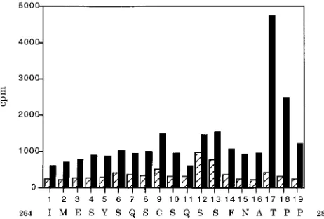
by cleavage with factor Xa. After separation by SDS-PAGE,
CT was eluted from the gel and subjected to manual Edman
degradation (45). Application of the manual degradation
pro-cedure resulted in four peaks of phosphate release, at turns 6,
10, 13, and 17 (Fig. 3). These peaks correspond to the serine
residues 267, 271, and 274 and threonine 278 in Py large T. The
threonine phosphorylation peak was the highest even though
its yield might have been expected to be lower since it came at
a later sequencing turn. While this finding suggests greater
phosphorylation at threonine 278 relative to the serines, it is
not possible to draw firm conclusions about relative
stoichiom-etry. The extent of labeling could depend on the rate of
turn-over at individual sites. In any case, quantitative differences
exist in SV40 large T phosphorylation in Sf9 cells compared to
monkey cells (15). This is likely to be true for Py as well.
Mutation of the phosphorylation sites.
To assess the
phos-phorylation sites identified in the baculovirus system, a series
of mutations were created both at individual sites and at sets of
sites (triple serine mutant S267A/S271A/S274A and quadruple
mutant S267A/S271A/S274A/T278A). The threonine at 278
was changed to alanine, asparagine, glutamate, and aspartate.
After transient transfection, both single- and multiple-site
mu-tants were generally expressed at levels similar to the wild-type
level as measured by Western blotting (not shown). The one
exception, single mutant S267A, was expressed at a level
five-fold lower than the wild-type level.
To test the effects of the mutations on phosphorylation,
large T labeled in vivo with
32PO
4
was analyzed first by partial
proteolysis with
S. aureus
V8 protease. V8 gives a series of
phosphopeptides (1), including peptides 5 and 7, that comes
from the more C-terminal cluster. No single mutation at 267,
271, 274, or 278 abolished labeling of these peptides. However,
a double mutant lacking S271 and S274 did not shown peptides
5 and 7 (not shown). This was unexpected in view of the
baculovirus results showing phosphorylation at 267 and 278
and suggested that the assay lacked sufficient resolution.
Therefore, double digestion of Py large T with hydroxylamine
and
S. aureus
V8 protease was carried out. Hydroxylamine
cleavage separates large T into two fragments (residues 1 to
210 and 211 to 785) (1). This makes it possible to examine the
C-terminal phosphorylations without the N-terminal
phos-phopeptide background (Fig. 4). The wild-type V8-NH
2OH
[image:3.612.136.217.67.187.2]phosphopeptide pattern of large T (Fig. 4A) shows that
pep-tides 5 and 7 arose from the C-terminal hydroxylamine
frag-ment. Mutation of threonine 278 left a pattern of V8 cleavage
that resembled the wild-type pattern qualitatively (Fig. 4C).
Mutation of the three serines (Fig. 4B) resulted in loss of
peptides 5 and 7, consistent with the result seen in the
one-dimensional V8 map. However, a new phosphopeptide with a
mobility similar to that of N-terminal V8 peptide 9 (Fig. 4B,
arrowhead) was now resolved. This new phosphopeptide
cor-responded to the phosphorylation at threonine 278, since it
disappeared when T278 was next mutated to alanine to give
FIG. 2.S. aureusV8 protease maps of Py large T. NIH 3T3 cell lines were labeled with32PO
4, and the mutant and wild-type Py large T’s were
[image:3.612.327.546.68.197.2]immuno-precipitated and electrophoresed on 7.5% polyacrylamide cylinders. The cylin-ders were then placed horizontally on top of a 10% acrylamide slab gel and electrophoresed in the presence of 200mg of V8 protease. WT refers to wild-type Py large T; LT 97 refers to mutant that is deleted of residues 271 to 280 of Py large T. The numbers and positions of the V8 phosphopeptides are shown on the left.
FIG. 3. Edman degradation of phosphorylated CT produced in Sf9 cells. The
32P released by each turn of Edman degradation was measured by Cerenkov
[image:3.612.57.287.508.671.2]counting. Each cycle of Edman degradation is shown, with the corresponding Py large T sequence indicated underneath. The first two amino acids indicated do not belong to the Py sequence but occur as a result of the in-frame GST fusion. The four sites of phosphorylation are indicated in boldface.
FIG. 4. Two-dimensional phosphopeptide mapping of large T phosphoryla-tion mutants. 293 cells were transfected with Py large T expression vectors and labeled with32
PO4. The mutant and wild-type Py large Ts were
immunoprecipi-tated, treated with hydroxylamine (HA), and electrophoresed on 7.5% polyacryl-amide cylinders in the direction indicated by the arrow labeled HA. The cylinders were then placed horizontally on top of 15% acrylamide slab gels and electro-phoresed in the presence of 200mg of V8 protease. (A) Wild-type Py large T; (B) triple mutant S267A,S271A,S274A; (C) single mutant T278A; (D) quadruple mutant S267A,S271A,S274A,T278A I, intact Py large T that was not cleaved by hydroxylamine; C, the C-terminal hydroxylamine fragment. The positions of V8 phosphopeptides 5 and 7 are indicated.
on November 9, 2019 by guest
http://jvi.asm.org/
the quadruple mutant (Fig. 4D). There are at least two
expla-nations for the difference in mobility of the 278-containing
peptide in the triple serine mutant and in the wild type. The
most likely possibility is that the mutations altered the site of
partial proteolysis, leading to a different fragment size.
An-other possibility is that the mobility of the phosphopeptide
depends on serine phosphorylation. Since the wild type does
not show peptides 5 and 7 as well as the new phosphopeptide,
phosphorylation would have to be functionally processive, with
phosphorylation of 278 leading to rapid phosphorylation on
serine.
Taken together, these results showed phosphorylation of
wild-type large T on threonine 278 and serines 271 and 274.
The phosphorylation of serine 267 could be neither directly
confirmed nor ruled out in this mapping.
[image:4.612.70.289.68.223.2]Mutant analysis demonstrates that T278 is critical for viral
DNA replication.
The next issue is the importance of the
phos-phorylation to large T function. Since there is already evidence
connecting large T phosphorylation to viral DNA replication
(2, 9, 39, 49), in vivo assays of Py DNA replication were
per-formed. Large T expression vectors with full-length or
C-ter-minal large T constructs were cotransfected with a plasmid
containing the Py origin of DNA replication. After 48 h, cells
were extracted and replication was measured by conversion of
input
Dpn
I-sensitive DNA to replicated
Dpn
I-resistant DNA.
Figure 5A shows that mutation of T278 to alanine (or
aspar-agine [not shown]) led to large T that was inactive in DNA
replication (compare lane 3 with lane 4). The same result was
obtained in a C-terminal domain construct (lane 6). By
con-trast, large T mutants lacking S267, S271, and S274 were
rep-lication competent as either full-length or CT constructs (Fig.
5A, lanes 2 and 5).
Since phosphorylation can sometimes be mimicked by
re-placement with a negative charge (43), T278D and T278E were
constructed. These two mutants were no more active than the
alanine mutant in DNA replication (Fig. 5B). All three
mu-tants were essentially inactive, although Western blotting (Fig.
5C) shows protein levels even higher than the wild-type level.
Replication assays were also performed after cotransfection
of wild-type and T278A large T (not shown). There was no
indication of a substantial dominant negative effect that might
have been expected if each subunit of the large T hexamer
needed to be wild type.
Threonine 278 is an in vitro site for cdc2 kinase.
Threonine
278 lies in the sequence (S/T)PX(R/K), which is a consensus
sequence for the cyclin/cyclin-dependent kinase (cdk) family of
kinases (18). Also, this sequence of Py large T (TPPKK) is
identical to the SV40 large T sequence starting at T124; T124
is an in vitro site for p34
cdc2(29). To test the Py 278 sequence,
CT was tested as a substrate for cyclin B and p34
cdc2. CT
generated from the baculovirus GST-CT construct was used in
an in vitro kinase experiment using baculovirus-expressed
GST-cyclin B and p34
cdc2. After labeling, CT was subjected to
manual Edman degradation. As a control for endogenous
phosphorylation, CT was phosphorylated with GSH beads
in-cubated with cyclin B alone (Fig. 6). While none of the serines
identified previously were phosphorylated by cyclin B and
p34
cdc2, T278 was highly phosphorylated. Thus, threonine 278
of Py large T is a site of phosphorylation by the cyclin/cdk
family of kinases.
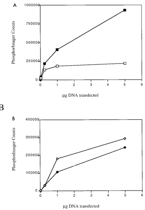
[image:4.612.318.549.70.226.2]Mutations of S267, S271, and S274 activate DNA
replica-tion.
As shown above, even the Py large T mutant at the three
serine phosphorylation sites is able to mediate viral DNA
rep-lication. In fact, from Fig. 5A, mutation of the serines
ap-peared to stimulate viral DNA replication. Therefore, we
com-pared the dose responses of the triple serine mutant and
wild-type Py large T (Fig. 7A). The results indicated that the triple
serine mutant could stimulate as much as fivefold with respect
to viral DNA replication. Mixtures of the triple serine mutant
and wild type gave the higher level of replication. These results
are in one sense different from the result for SV40. Mutation
of serines 120 and 123 results in loss of DNA replication (43).
However, in another sense they are similar. As for Py, removal
of phosphate from wild-type SV40 large T is activating (41, 47,
48).
FIG. 5.In vivoreplication assays of Py large T phosphorylation mutants. Southern blots show low-molecular-weight DNA from 3T3 cells transfected with Py origin plasmid (pUCori) and Py large T expression vectors. Extracted DNA was cut withHincII andDpnI. The blot was probed with32P-labeled Py origin
DNA (bp 5024 to 163). (A) Lane 1 is a control with pUCori and the CMV expression vector without Py large T coding sequences. All the other lanes are from cells transfected with pUCori plus S267A,S271A,S274A large T (lane 2), T278A large T (lane 3), wild-type large T (lane 4), S267A,S271A,S274A CT (lane 5), T278A CT (lane 6), and wild-type CT (lane 6). The positions of theDpn I-sensitive and -resistant bands are indicated. (B) In vivo replication assay of mutants at T278. Cells were transfected with pUCori plus the CMV expression vector without large T coding sequences (lanes 1 and 2), T278A large T (lanes 3 and 4), T278E large T (lanes 5 and 6), T278D large T (lanes 7 and 8), and wild-type T (lanes 9 and 10). (C) Western blot showing the amount of protein expressed in the replication assay in panel B. Cells were transfected with pUCori plus T278A large T (lanes 1 and 2), T278E large T (lanes 3 and 4), T278D large T (lanes 5 and 6), and wild-type large T (lanes 7 and 8).
FIG. 6. Edman degradation of CT produced in Sf9 cells and phosphorylated in vitro by GST-cyclin B and p34cdc2; hatched bars represent CT phosphorylated
by GST-cyclin B alone. Each cycle of Edman degradation is shown, with the corresponding Py large T sequence indicated underneath. The first two amino acids indicated do not belong to the Py sequence but occur as a result of the in-frame GST fusion. The four sites of phosphorylation identified in Fig. 2 are indicated in boldface.
on November 9, 2019 by guest
http://jvi.asm.org/
Interestingly, the same kind of dose-response experiment
done in the context of the serine phosphorylation mutants in
the C-terminal background showed no significant difference
between mutant and wild type (Fig. 7B). This result implies
that the role of these phosphorylations is connected to the
relationship between NT and CT.
Mutation of C-terminal serine or threonine
phosphoryla-tion sites does not seem to affect large T funcphosphoryla-tions related to
cellular DNA replication.
Large T not only is essential for viral
DNA replication but also stimulates cellular DNA replication
(9, 35, 42). Given the importance of T278 to viral DNA
repli-cation, it was of interest to determine what, if any, contribution
it makes to large T’s effect on cell cycle progression. BrdU
incorporation was used to measure cellular DNA replication.
3T3 cells were serum starved for 40 h after transfection and
then labeled with BrdU for 8 h. Double immunostaining was
used to score both expression of large T and DNA replication
in individual cells. Table 1 shows that the wild type, T278A, the
triple serine mutant, and a quadruple mutant lacking all four
sites were capable of efficiently promoting cellular DNA
rep-lication.
One important function of large T is to interfere with tumor
suppressors of the Rb family. As shown by the Rb
2large T, the
growth assay was done in cells where inactivation of Rb family
function is necessary for cell cycle progression. This suggests
that mutation of these phosphorylation sites did not
compro-mise large T’s ability to affect Rb family members. This
possi-bility was confirmed directly by using a CAT reporter assay
measuring E2F-mediated transcription. Wild-type large T
transactivates the A10 CAT construct in a manner dependent
on binding to Rb family members (reference 16 and Table 1).
As for cell cycle progression, mutation of the phosphorylation
sites had no effect on the ability of large T to transactivate.
DISCUSSION
This work characterizes the cluster of phosphorylation sites
which are found in the protease-sensitive region connecting
NT and CT of Py large T. Edman degradation of labeled
protein identified serines 267, 271, and 274 and threonine 278
as sites of phosphorylation. Labeling of mutant large T’s
con-firmed 271, 274, and 278 as sites of phosphorylation. The
threonine at 278 is required for viral DNA replication in either
the full-length or CT context, suggesting it is formally part of
CT. The serine phosphorylations are unimportant for viral
DNA replication in the CT context even though their mutation
activates full-length large T. This finding suggests that these
sites may regulate some aspect of domain-domain
communi-cation. None of these sites affect the ability of large T to drive
the cell cycle as measured by BrdU incorporation or the ability
to affect interactions with the Rb tumor suppressor family. This
finding is consistent with the view that these are largely
prop-erties of NT.
For both Py and SV40, the regions N terminal to their
homologous nuclear localization signals, residues 280 to 286 in
Py and 126 to 133 in SV40, are regions of high
phosphoryla-tion. The threonine at 278 that we have characterized aligns
with the threonine at 124 of SV40 precisely. The alignment of
the serines is less clear. In SV40, major sites are found at 120
and 123 (SQHS) compared to the Py sites 271 and 274 (SQSS),
which are further from the critical threonine. The
phosphory-lation of Py residue 267 occurs in a second SQS motif that is
not apparent in SV40. Interestingly, the E1 replication protein
of bovine papillomavirus is quite similar to those of both Py
and SV40, having a sequence (TPVK) analogous to threonine
278 and a proximal serine sequence (SQNS) that could be
aligned with either Py or SV40 large T.
[image:5.612.63.296.69.407.2]Edman degradation, peptide mapping of large Ts
phosphor-ylated in vivo, and analysis of mutants show that threonine 278
is a site of phosphorylation. Mutation at threonine 278
com-pletely abolished the ability of Py large T to mediate viral DNA
replication in both full-length and CT constructs. Substitutions
FIG. 7. Dose responses of viral DNA replication by the S267A,S271A,S274A mutant and the wild type. (A) In vivo replication assay done in the large T context. Solid squares represent the S267A,S271A,S274A mutant large T; open squares represent wild-type large T. (B) In vivo replication assay done in the CT context. Solid diamonds represent the S267A,S271A,S274A CT mutant; open diamonds represent wild-type CT. They axis shows replication measured in arbitrary counts assigned by PhosphorImager scans ofDpnI-resistant replicated DNA.
TABLE 1. Activities of phosphorylation site mutants
Construct
A10 E2F CAT assay (% conversion)a
S-phase induction
assay (% BrdU)b
pCMV
2
7
cpCMV-T278A-LT
45
60
pCMV-S267A,S271,S274A-LT
44
45
pCMV-S267A,S271,S274A,T278A-LT
46
51
pCMV-LT-Rb
20
5
pCMV-LT
44
52
aConversion of nonacetylated [14
C]chloramphenicol to acetylated forms.
bNumber of large T-positive cells that were also positive for BrdU. cThe BrdU background was measured by the number of BrdU-positive cells
per 100 cells.
on November 9, 2019 by guest
http://jvi.asm.org/
[image:5.612.316.556.83.182.2]of a negative charge at this site was ineffective in replacing this
function. These results support the view that phosphorylation
of threonine 278 is absolutely required for Py large T to
per-form its replication function. As with Py large T, mutation of
threonine 124 in SV40 large T abolishes viral DNA replication.
The DNA replication defect in the threonine 124 mutant has
been shown to lie in the unwinding of the viral origin (30, 32).
Recently it was shown that threonine 124 phosphorylation
en-hances the interaction of the minimal DNA binding domain
with the core origin of replication, possibly stimulating double
hexamer assembly and thus DNA unwinding (31). It is likely
that the DNA replication defect in T278 is similar to that of
T124 in SV40 large T. Since this phosphorylation is required in
the CT context, it is puzzling that the comparable
phosphory-lation site, in E1, which has been shown to be phosphorylated,
is not required for bovine papillomavirus replication (25).
Threonine 278 is a form of biosensor connecting large T
function in DNA replication to the state of the cell cycle. CT is
capable of efficient replication in growing cells but not in
rest-ing cells (9). It can be complemented by a variety of genes, such
as the NT, E1A, E7, or E2F1, that stimulate the cell cycle. This
complementation is accompanied by a shift in CT mobility that
suggests phosphorylation. Here we have shown that CT
con-taining threonine 278, but not serines 267, 271, and 274, is fully
capable of replication. Further, threonine 278 was identified as
a substrate for the cell cycle-regulated kinase p34
cdc2.
Threo-nine 124 in SV40 large T antigen (29) is also a substrate for the
cell cycle-regulated kinase p34
cdc2.
Mutation of serine phosphorylation sites at positions 267,
271, and 274 has a less dramatic effect on DNA replication
than the mutation at threonine 278. Further, these mutations
result in stimulation, not inhibition, of DNA replication. This
observation is likely related to the earlier result that treatment
of Py large T with calf intestinal alkaline phosphatase (CIAP)
at low levels resulted in a stimulation of viral DNA replication
in vitro (49). CIAP preferentially removes phosphates from
serines and leaves threonine phosphorylations intact. In that
work, Wang and coworkers implicated phosphorylations lying
between residues 89 and 110 and 133 and 167 in the
stimula-tion of viral DNA replicastimula-tion. It could well be that
phosphor-ylation in the N terminus as well as at 267, 271, and 274, can
also affect DNA replication. A stimulatory effect of
phospha-tase treatment was also seen in the case of SV40 large T, where
treatment with either CIAP or the catalytic subunit of protein
phosphatase 2A enhanced its ability to mediate SV40
origin-dependent replication in vitro (11, 21, 33, 41, 44). Subsequent
work has shown that phosphorylation on serines 120 and 123
by a casein kinase I isoform causes inhibition of unwinding and
DNA replication which can be reversed by protein
phospha-tase 2A (3–5). No molecular explanation for this inhibition is
currently available. Since the triple serine mutation had little
effect on DNA replication when placed in the context of CT, it
may be that phosphorylation regulates the connections
be-tween NT and CT.
The data presented here connect the phosphorylation of
large T at the N terminus of CT to viral DNA replication.
Experiments to examine the ability of large T to promote cell
cycle progression or to activate E2F sites suggest that these
sites are not important for such processes. This is reasonable in
view of the known ability of NT to act on the cell. NT is also
highly phosphorylated. It will be of considerable interest to
determine what contributions those phosphorylations have on
large T function.
ACKNOWLEDGMENTS
This work was supported by National Health Institutes of Health
grant CA34722.
We thank Helen Piwnica-Worms for the gift of baculoviruses
ex-pressing GST-cyclin B and p34
cdc2and Marcel Bastin for the gift of LT
97.
REFERENCES
1.Bockus, B. J., and B. Schaffhausen.1987. Localization of the
phosphoryla-tions of polyomavirus large T antigen. J. Virol.61:1155–1163.
2.Bockus, B. J., and B. Schaffhausen.1987. Phosphorylation of polyomavirus
large T antigen: effects of viral mutations and cell growth state. J. Virol.
61:1147–1154.
3.Cegielska, A., I. Moarefi, E. Fanning, and D. M. Virshup.1994. T-antigen
kinase inhibits simian virus 40 DNA replication by phosphorylation of intact T antigen on serines 120 and 123. J. Virol.68:269–275.
4.Cegielska, A., S. Shaffer, R. Derua, J. Goris, and D. Virshup.1994. Different
oligomeric forms of protein phosphatase 2A activate and inhibit simian virus 40 DNA replication. Mol. Cell. Biol.14:4616–4623.
5.Cegielska, A., and D. M. Virshup.1993. Control of simian virus 40 DNA
replication by the HeLa cell nuclear kinase casein kinase I. Mol. Cell. Biol.
13:1202–1211.
6.Chen, C. A., and H. Okayama.1987. High-efficiency transformation of
mam-malian cells by plasmid DNA. Mol. Cell. Biol.7:2745–2752.
7.Francke, B., and W. Eckhart.1973. Polyoma gene function required for viral
DNA synthesis. Virology55:127–135.
8.Freund, R., R. T. Bronson, and T. L. Benjamin.1992. Separation of
immor-talization from tumor induction with polyoma large T mutants that fail to bind the retinoblastoma gene product. Oncogene7:1979–1987.
9.Gjørup, O. V., P. E. Rose, P. S. Holman, B. J. Bockus, and B. S.
Schaff-hausen.1994. Protein domains connect cell cycle stimulation directly to
initiation of DNA replication. Proc. Natl. Acad. Sci. USA91:12125–12129.
10. Gorman, C., L. F. Moffat, and B. H. Howard.1982. Recombinant genomes
which express chloramphenicol acetyltransferase in mammalian cells. Mol. Cell. Biol.2:1044–1051.
11. Grasser, F. A., K. Mann, and G. Walter.1987. Removal of serine phosphates
from simian virus 40 large T antigen increases its ability to stimulate DNA replication in vitro but has no effect on ATPase and DNA binding. J. Virol.
61:3373–3380.
12. Hassauer, M., K. Scheidtmann, and G. Walter.1986. Mapping of
phosphor-ylation sites in polyomavirus large T antigen. J. Virol.58:805–816.
13. Hirt, B.1967. Selective extraction of polyoma DNA from infected mouse cell
cultures. J. Mol. Biol.26:365–369.
14. Holman, P. S., O. V. Gjørup, T. Davin, and B. S. Schaffhausen.1994.
Characterization of an immortalizing N-terminal domain of polyomavirus large T antigen. J. Virol.68:668–673.
15. Hoss, A., I. Moarefi, K. Scheidtmann, L. Cisek, J. Corden, I. Dornreiter, A.
Arthur, and E. Fanning.1990. Altered phosphorylation pattern of simian
virus 40 T antigens expressed in insect cells by using a baculovirus vector. J. Virol.64:4799–4807.
16. Howes, S. H., B. J. Bockus, and B. S. Schaffhausen.1996. Genetic analysis of
polyomavirus large T nuclear localization: nuclear localization is required for productive association with pRb family members. J. Virol.70:3581–3588.
17. Kellems, R., V. Morhenn, R. Pfendt, F. Alt, and R. Schimke.1979. Polyoma
virus and cAMP-mediated control of dihydrofolate reductase mRNA abun-dance in methotrexate-resistant mouse fibroblasts. J. Biol. Chem.254:309– 318.
18. Kennelly, P. J., and E. G. Krebs.1991. Consensus sequences as substrate
specificity determinants for protein kinases and protein phosphatases. J. Biol. Chem.266:15555–15558.
19. Kern, F., S. Pellegrini, A. Cowie, and C. Basilico.1986. Regulation of
polyomavirus late promoter activity by viral early proteins. J. Virol.60:275– 285.
20. Kingston, R. E., A. Cowie, R. I. Morimoto, and K. A. Gwinn.1986. Binding
of polyomavirus large T antigen to the humanhsp70promoter is not re-quired fortransactivation. Mol. Cell. Biol.6:3180–3190.
21. Klausing, K., K. H. Scheidtmann, E. A. Baumann, and R. Knippers.1988.
Effects of in vitro dephosphorylation of DNA binding and DNA helicase activities of simian virus 40 large T antigen. J. Virol.62:1258–1265.
22. Laemmli, U. K.1970. Cleavage of structural proteins during the assembly of
the head of bacteriophage T4. Nature227:680–685.
23. Larose, A., N. Dyson, M. Sullivan, E. Harlow, and M. Bastin.1991.
Poly-omavirus large T mutants affected in retinoblastoma protein binding are defective in immortalization. J. Virol.65:2308–2313.
24. Larose, A., L. St-Onge, and M. Bastin.1990. Mutations in polyomavirus
large T affecting immortalization of primary rat embryo fibroblasts. Virology
176:98–105.
25. Lentz, M. R., D. Pak, I. Mohr, and M. R. Botchan.1993. The E1 replication
protein of bovine papillomavirus type 1 contains an extended nuclear local-ization signal that includes a p34cdc2phosphorylation site. J. Virol.67:1414–
1423.
on November 9, 2019 by guest
http://jvi.asm.org/
26. Loeken, M. R., and J. Brady.1989. The adenovirus EIIA enhancer. Analysis of regulatory sequences and changes in binding activity of ATF and EIIF following adenovirus infection. J. Biol. Chem.264:6572–6579.
27. Maione, E., G. M. Fimia, P. Holman, B. S. Schaffhausen, and P. Amati.1994.
Retinoblastoma antioncogene is involved in the inhibition of myogenesis by polyomavirus large T antigen. Cell Growth Differ.5:231–237.
28. Maione, R., G. M. Fimia, and P. Amati.1992. Inhibition of in vitro myogenic
differentiation by a polyomavirus early function. Oncogene7:85–93.
29. McVey, D., L. Brizuela, I. Mohr, D. Marshak, Y. Gluzman, and D. Beach.
1989. Phosphorylation of large tumor antigen by cdc2 stimulates SV40 DNA replication. Nature341:503–507.
30. McVey, D., S. Ray, Y. Gluzman, L. Berger, A. G. Wildeman, D. R. Marshak,
and P. Tegtmeyer.1993. cdc2 phosphorylation of threonine 124 activates the
origin-unwinding functions of simian virus 40 T antigen. J. Virol.67:5206– 5215.
31. McVey, D., B. Woelker, and P. Tegtmeyer.1996. Mechanisms of simian virus
40 T-antigen activation by phosphorylation of threonine 124. J. Virol.70:
3887–3893.
32. Moarefi, I. F., D. Small, I. Gilbert, M. Hopfner, S. K. Randall, C. Schneider,
A. A. R. Russo, U. Ramsperger, A. K. Arthur, H. Stahl, T. J. Kelly, and E.
Fanning.1993. Mutation of the cyclin-dependent kinase phosphorylation site
in simian virus (SV40) large T antigen specifically blocks SV40 origin DNA unwinding. J. Virol.67:4992–5002.
33. Mohr, I. J., B. Stillman, and Y. Gluzman.1987. Regulation of SV40 DNA
replication by phosphorylation of T antigen. EMBO J.6:153–160.
34. Mudrak, I., E. Ogris, H. Rotheneder, and E. Wintersberger.1994.
Coordi-natedtransactivation of DNA synthesis- and precursor-producing enzymes by polyomavirus large T antigen through interaction with the retinoblastoma protein. Mol. Cell. Biol.14:1886–1892.
35. Ogris, E., I. Mudrak, and E. Wintersberger.1993. Distinct amounts of
polyomavirus large T antigen are required for different functions of the protein. Oncogene8:1277–1283.
36. Peden, K., J. Pipas, S. Pearson-White, and D. Nathans.1980. Isolation of
mutants of an animal virus in bacteria. Science209:1392–1396.
37. Rassoulzadegan, M., Z. Naghasfar, A. Cowie, A. Carr, M. Grisoni, R.
Ka-men, and F. Cuzin.1983. Expression of the large T protein of polyoma virus
promotes the establishment of culture of “normal” rodent fibroblast cell lines. Proc. Natl. Acad. Sci. USA80:4354–4358.
38. Richardson, W. D., B. L. Roberts, and A. E. Smith.1986. Nuclear location
signals in polyoma virus large-T. Cell44:77–85.
39. Rose, P. E., and B. S. Schaffhausen.1995. Zinc-binding and protein-protein
interactions mediated by the polyomavirus large T antigen zinc finger. J. Vi-rol.69:2842–2849.
40. Schaffhausen, B., J. Silver, and T. Benjamin.1978. Tumor antigen(s) in cells
productively infected by wild type polyoma virus and mutant NG18. Proc. Natl. Acad. Sci. USA75:79–83.
41. Scheidtmann, K. H., D. M. Virshup, and T. J. Kelly.1991. Protein
phospha-tase 2A dephosphorylates simian virus 40 large T antigen specifically at residues involved in regulation of DNA-binding activity. J. Virol.65:2098– 2101.
42. Schlegel, R., and T. L. Benjamin.1978. Cellular alterations dependent upon
the polyoma virus hr-t function: separation of mitogenic from transforming capacities. Cell14:587–599.
43. Schneider, J., and E. Fanning.1988. Mutations in the phosphorylation sites
of simian virus 40 (SV40) T antigen alter its origin DNA-binding specificity for sites I or II and affect SV40 DNA replication activity. J. Virol.62:1598– 1605.
43a.Sheng, Q., and B. Schaffhausen.Unpublished data.
44. Simmons, D., W. Chou, and K. Rodgers.1986. Phosphorylation
downregu-lates the DNA-binding activity of simian virus 40 T antigen. J. Virol.60:888– 894.
45. Sullivan, S., and T. W. Wong.1991. A manual sequencing method for
identification of phosphorylated amino acids in phosphopeptides. Anal. Bio-chem.197:65–68.
46. Towbin, H., T. Staehlin, and J. Gordon.1979. Electrophoretic transfer of
proteins from polyacrylamide gels to nitrocellulose sheets: procedures and some applications. Proc. Natl. Acad. Sci. USA76:4350–4354.
47. Virshup, D. M., M. G. Kauffman, and T. J. Kelly.1989. Activation of SV40
DNA replication in vitro by cellular protein phosphatase 2A. EMBO J.
8:3891–3898.
48. Virshup, D. M., and T. J. Kelly.1989. Purification of replication protein C,
a cellular protein involved in the initial stages of SV40 DNA replication in vitro. Proc. Natl. Acad. Sci. USA86:3584–3588.
49. Wang, E. H., S. Bhattacharyya, and C. Prives.1993. The replication
func-tions of polyomavirus large tumor antigen are regulated by phosphorylation. J. Virol.67:6788–6796.
50. Weichselbraun, I., G. Haider, and E. Wintersberger.1989. Optimal
replica-tion of plasmids carrying polyomavirus origin regions requires two high-affinity binding sites for large T antigen. J. Virol.63:961–964.