0022-538X/97/$04.0010
Copyrightq1997, American Society for Microbiology
Analysis of the 2-Kilobase Latency-Associated Transcript
Expressed in PC12 Cells Productively Infected with Herpes
Simplex Virus Type 1: Evidence for a Stable,
Nonlinear Structure
EYVIND RØDAHLANDLARS HAARR*
Centre for Research in Virology, University of Bergen, N-5020 Bergen, Norway
Received 22 July 1996/Accepted 29 October 1996
The major latency-associated transcript (LAT) expressed in PC12 cells productively infected with herpes simplex virus type 1 is a 2-kb, nonpolyadenylated RNA molecule that accumulates in the nuclei of infected cells. In actinomycin D-treated cells, the 2-kb LAT gene transcript has a half-life considerably greater than 12 h. After polyacrylamide gel electrophoresis, two species of the transcript were observed, a major species that was retarded in the gel and a minor species that migrated as a 1.96-kb RNA molecule. RNase H digestion after hybridization of the RNA with an oligonucleotide complementary to positions280 to2101 relative to the 3* end of the 2-kb LAT gene transcript changed the mobility of the retarded species into that of the rapidly migrating species. Our data indicate that the 2-kb LAT gene transcript expressed in productively infected PC12 cells is present in a stable, nonlinear form.
In neurons latently infected with herpes simplex virus type 1 (HSV-1), the only transcripts detected by Northern blot or in situ hybridization are the latency-associated transcripts (LATs) (9, 10, 19, 49, 50). The major LATs are 2, 1.5, and 1.45 kb long and constitute a family of colinear, nonpolyadenylated tran-scripts that accumulate in the nuclei of infected cells with sparing of the nucleoli (2). As many as 40,000 copies may be present in each latently infected cell (52). LAT gene transcrip-tion has been detected within an 8.3-kb unit of the joint region of the viral genome (Fig. 1) by in situ hybridization, and there is evidence that the major LATs are spliced derivatives of an 8.3-kb primary transcript (13, 17, 19, 54). Transcription from the LAT gene region has also been observed during productive infection, both in tissue culture cells and in neurons of exper-imentally infected animals (11, 37, 46). In productively infected cells, only a 2-kb major LAT gene transcript has been detected, suggesting that the splicing event generating the 1.5- and 1.45-kb LATs (47) is unique to latently infected neurons. Dur-ing the productive phase of infection, LAT gene transcripts are present predominantly in the nucleus but can also be detected in the cytoplasm (49). The function of LATs is not clear, but they may be important for induced reactivation of the virus from latently infected cells (4, 24, 28, 48).
Cell culture studies have indicated that the 2-kb LAT gene transcript is relatively stable (17). Increased stability of the transcript could also be the reason why it accumulates in la-tently infected neurons in vivo. Analysis of stable RNA mole-cules derived from splicing reactions has shown that they are either introns present as lariats (36) or circular exons that have been derived by a process termed missplicing (8, 34). Introns have also been shown to accumulate as lariats when the intron branch point has been mutated (25, 26) and in yeast strains lacking debranching activity (7). Apparently, when present as circles, the RNA molecules are less susceptible to degradation,
presumably because of the low level of endonucleolytic activity in the cell nucleus (36).
In this study, we have examined the stability and conforma-tion of the 2-kb LAT gene transcript expressed in PC12 cells productively infected with HSV-1. PC12 cells differentiate into cells resembling sympathetic neurons after treatment with nerve growth factor (NGF) (21) and have been used by several investigators to study the interaction between HSV-1 and neu-ronal cells in culture (3, 38, 39). They support high levels of LAT promoter activity compared with other cell lines (5, 29). (This work was presented in preliminary form at the 19th International Herpesvirus Workshop in Vancouver, British Columbia, Canada, in 1994.)
LAT gene transcripts expressed in productively infected PC12 cells. PC12 cells were grown in RPMI 1640 medium supplemented with 10% horse serum, 5% fetal calf serum, and 50 mg of gentamicin per liter. All cell culture reagents were from Gibco-Bethesda Research Laboratories. Differentiated cells were obtained after treatment with 2.5 S NGF (100 ng/ml) (Boehringer Mannheim GmbH) for 2 weeks. The cells were infected with HSV-1 strain KOS(M) (51) at a multiplicity of infection (MOI) of 1. Single-step growth curves confirmed that both differentiated and undifferentiated cells were permissive for HSV-1 infection (data not shown) (38). To examine the LAT gene transcripts expressed, total RNA was isolated from cells lysed in the presence of 5.5 M guanidinium isothiocyanate (Gibco-Bethesda Research Laboratories) by centrifugation through a cushion of cesium trifluoroacetate (Pharmacia-LKB). Highly purified RNA with no detectable DNA contam-ination is obtained by this procedure (31). A cloned HSV-1 fragment (ATD19) corresponding to genomic positions 119628
to 119975 (26) (Fig. 1) labelled with [a-32P]dCTP (Amersham)
(18) was used as a probe to detect LAT gene transcripts by Northern blot analysis (42). As in other productively infected cell lines (11, 46), the major LAT gene transcript detected in both differentiated and undifferentiated cells was a 2-kb RNA molecule (Fig. 2 and 3) that was not retained on an oligo(dT) column (Pharmacia-LKB) (data not shown). In addition, a series of transcripts ranging from less than 2 kb to more than
* Corresponding author. Mailing address: Centre for Research in Virology, University of Bergen, N-5020 Bergen, Norway. Phone: 47 55 58 45 07. Fax: 47 55 58 45 12.
1703
on November 9, 2019 by guest
http://jvi.asm.org/
8 kb hybridized with the LAT gene probe and were observed as a weak smear on the Northern blots. The 1.5- and 1.45-kb major LAT species were not seen. No LAT gene transcripts were detected in cells treated with phosphonoacetic acid (400
mM) prior to infection with strain KOS(M) (data not shown)
or in cells infected with IE175 (ICP4) deletion mutant KD6B11 (14, 44) (Fig. 2), indicating that viral replication was necessary for LAT gene transcript expression to occur. Similar kinetics of LAT gene expression have previously been ob-served in CV-1 cells (46). The intracellular distribution of the 2-kb LAT gene transcript was then examined by in situ hybrid-ization analysis (22) of NGF-treated PC12 cells maintained on plastic coverslips (Miles Laboratories) coated with collagen type I (Sigma). In this procedure, the tissue is extensively treated with DNase I, and it is generally accepted that the signals obtained are derived from hybridization with RNA (13, 22, 49, 50). With ATD19 labelled with tritiated nucleotides as a probe, the 2-kb LAT gene transcript was localized predom-inantly in the nuclei of cells infected with KOS(M) (Fig. 2). The distribution of the grains resembled that previously seen in productively infected neurons in vivo (49). It is not known, however, if the 2-kb LAT gene transcript expressed in tissue culture cells is identical to that expressed in neurons in vivo.
Since the level and length of the transcripts observed in undifferentiated cells were not significantly different from those observed in NGF-differentiated cells, undifferentiated cells were used throughout the rest of the study.
Decay of the 2-kb LAT gene transcript in actinomycin D-treated cells.In previous studies, Northern blot analysis of the 2-kb LAT gene transcript expressed in HeLa cells has sug-gested that the transcript is relatively stable (17). Its half-life, however, has not been estimated, and we therefore examined
decay curves of PC12 cells treated with actinomycin D (10
mg/ml). RNA was harvested no later than 24 h postinfection
(p.i.), since at that time point extensive cytopathic effects of the viral infection and the actinomycin D treatment were observed. The rate of degradation of the 2-kb LAT gene transcript was measured relative to that of 28S rRNA (the half-life of the most unstable 28S rRNA species is approximately 48 h [16]). It was also compared with mRNA from genes UL43 and UL44 {the probe used for Northern blot hybridization [the
SalI-EcoRI fragment of ATD54, genomic positions 94853 to 96763
(30)] did not distinguish between these genes [Fig. 1]}. In untreated cells, the level of the 2-kb LAT gene transcript increased from 12 to 24 h p.i., while in cells treated with actinomycin D no significant change was observed within the same period (Fig. 3). This indicates that the 2-kb LAT gene transcript was stable, with a half-life considerably longer than 12 h. In contrast, transcripts expressed from the UL43 to UL44 region decreased in cells treated with actinomycin D (Fig. 3) and their half-life was estimated by densitometric analysis to be approximately 3 h.
Actinomycin D inhibits RNA transcription by intercalating between the DNA bases (45). Some RNA transcripts have been shown to be stabilized by actinomycin D (32, 43). The mechanism is poorly understood, but in such cases actinomycin D is thought to inhibit the production of RNA-destabilizing factors. To test the possibility that actinomycin D stabilized the
2-kb LAT gene transcript, cells treated with a-amanitin (10
mg/ml) were examined. No decay of the 2-kb LAT gene
tran-script was detected over a 12-h observation period, confirming that the transcript is stable (data not shown). In other studies, evidence for a stable 2-kb LAT gene transcript has been ob-tained from experiments in which actinomycin D has not been
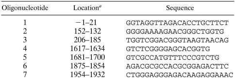
[image:2.612.319.552.67.281.2]FIG. 1. (A) Schematic representation of transcription detected within the LAT gene region of the HSV-1 genome. The 1.5- and 1.45-kb LATs are observed only in latently infected neurons. In addition to the transcripts indicated here, several minor, polyadenylated transcripts have been identified in productively infected cells (8). The positions of probe ATD19 and the SalI-EcoRI fragment (S-E) of ATD54 used in Northern blot and in situ hybridization analyses are indicated. A denotes ApaI sites flanking the 2-kb LAT gene. (B) Tentative structure of circular and lariat forms of the 2-kb LAT gene transcript with approximate positions of oligonucleotides used for RNase H digestion (oligonu-cleotide 6), primer extension analysis (oligonu(oligonu-cleotide 7), and RT-PCR (oligo-nucleotides 1 to 6) (numbered as in Table 1). The circular model shows a putative end-to-end ligation of the 59and 39ends of the transcript.
FIG. 2. Northern blot (A) and in situ hybridization (B and C) analyses of LATs expressed in NGF-differentiated PC12 cells infected with HSV-1 at 18 h p.i. (A) Total RNA (20mg in each lane) was separated by electrophoresis in an agarose gel containing 2.2 M formaldehyde, vacuum blotted onto a nylon mem-brane, and hybridized with32P-labelled, LAT-specific probe ATD19. Lanes: 1,
RNA from uninfected cells; 2, RNA from cells infected with HSV-1 strain KOS(M) (MOI of 1); 3, RNA from cells infected with IE175 deletion mutant KD6B11 (MOI of 10). The positions of 28S (4.7 kb) and 18S (1.9 kb) rRNAs are indicated on the left. For in situ hybridization analysis (B and C), ATD19 labelled with3H-labelled nucleotides was used as the probe. (B) Cells fixed 18 h
p.i. with KOS(M). (C) Uninfected cells. Magnification,3115.
on November 9, 2019 by guest
http://jvi.asm.org/
[image:2.612.59.294.76.284.2]present (17). Thus, it is not likely that the long half-life ob-served was an artifact caused by actinomycin D.
Polyacrylamide gel electrophoresis (PAGE) of the 2-kb LAT gene transcript.Stable RNA molecules derived from splicing reactions have been found to be present in a nonlinear form (8, 34, 36). To understand the basis for the stability of the 2-kb LAT gene transcript, it was therefore of interest to study its conformation. Several investigators have used PAGE to sepa-rate linear from nonlinear RNA molecules (15, 27, 35, 36, 40, 53). To avoid interference from the long LAT gene transcripts, we first separated total RNA by electrophoresis in agarose gels containing 2.2 M formaldehyde (42) and then excised the re-gion containing 18S rRNA (where the 2-kb LAT gene tran-script also is present) from the gel. RNA was eluted from the gel piece by centrifugation through siliconized glass wool in an Eppendorf centrifuge at 6,000 rpm for 10 min (23) and finally recovered by ethanol precipitation. Polyacrylamide gel electro-phoresis of RNA was performed in gels (15 by 35 cm) contain-ing 4% acrylamide, 7 M urea, and 90 mM Tris-borate buffer
with 2 mM EDTA (13TBE) at a constant current of 1 mA/cm
(36). The loading buffer used for PAGE was the same as that used for agarose gels (42). After electrophoretic transfer to nylon membranes (36) and subsequent Northern blot hybrid-ization using ATD19 as the probe, two distinct species of the 2-kb LAT gene transcript were observed: a minor band mi-grating as a 1.96-kb linear RNA molecule and a major band that was retarded in the gel (Fig. 4A). Extensive treatment of the RNA with proteinase K did not alter the mobility of the transcripts, indicating that the retarded form was not linked to a protein. We also examined the migration of an in vitro-transcribed 2.3-kb ApaI fragment that spans the 2-kb LAT
gene transcript (Fig. 1). The ApaI fragment was subcloned from the BamHI b fragment of the HSV-1 genome into plas-mid pSL301 (Invitrogen) and then transcribed with T7 RNA polymerase by using an in vitro transcription kit (Promega). As shown in Fig. 4B, this transcript migrated as expected for a linear molecule, indicating that under the conditions used terference from secondary structure caused by inter- or in-tramolecular hybridizations was minimal. We also considered the possibility that components used to isolate the 2-kb frac-tion of total RNA, or the procedure itself, could have altered the migration of the RNA molecules in a polyacrylamide gel. However, other RNA molecules, like 18S rRNA, migrated as expected (a strong 1.86-kb band; data not shown). Further-more, the retarded form of the 2-kb LAT gene transcript was also observed after PAGE of total RNA which had not been subjected to the isolation procedure (Fig. 4D), indicating that the retarded migration of the 2-kb LAT gene transcript was not a consequence of the purification process.
The experiments described above suggested that different forms of the transcript were present. Both circular and lariat RNA molecules, as well as large Y-shaped transcripts, are retarded in polyacrylamide gels (39, 53). Naturally occurring Y-shaped transcripts have been observed after trans splicing in trypanosomes (33). To distinguish between linear and nonlin-ear forms of the molecule, we used a strategy in which the RNA is hybridized with an oligonucleotide and treated with RNase H and the reaction products are then examined by PAGE and Northern blot analysis (36). The
[image:3.612.316.559.67.241.2]deoxyoligonucleo-tide used was a 22-mer, 59-AGA CGC GCC ACG CGG AGA
FIG. 3. Northern blot analysis showing the time course of viral RNA accu-mulation in PC12 cells infected with HSV-1 strain KOS(M) (MOI of 1). Acti-nomycin D was added 12 h p.i., and total RNA was isolated at various time points (0, 2, 6, or 12 h) thereafter. (A) The RNA molecules were separated by elec-trophoresis in a denaturing agarose gel and vacuum blotted onto a nylon mem-brane. Total RNAs from untreated (2) and actinomycin D-treated (1) cells were run in parallel lanes. The membrane was hybridized with 32P-labelled
[image:3.612.63.296.68.211.2]probes corresponding to genomic positions 119628 to 119975 (ATD19) for de-tection of LATs (top) and 94853 to 96763 (SalI-EcoRI fragment of ATD54) for detection of UL43-UL44 transcripts (middle), as well as with the 28S rRNA probe (1) (bottom). The membrane was stripped between individual hybridiza-tions in boiling 0.1% sodium dodecyl sulfate and then gently agitated while cooling for 7 min. (B) Graphic representation of the level of viral RNAs relative to the level of 28S rRNA estimated by densitometric analysis of the autoradio-grams. The autoradiograms were scanned with a Microtek Scan Maker IIXE (Microtek International), and relative band intensities were estimated with the National Institutes of Health Image 1.52 program on a Macintosh LC 475 personal computer (Apple Computer). The value obtained at 12 h p.i., when actinomycin D was added, was defined as 100%. Symbols:Ç, 2-kb LAT gene transcript without actinomycin D;å, 2-kb LAT gene transcript with actinomycin D;h, UL43-UL44 transcripts without actinomycin D;■, UL43-UL44 transcripts with actinomycin D.
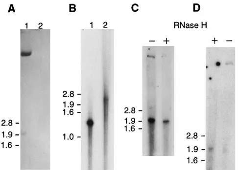
FIG. 4. PAGE and Northern blot analysis of the 2-kb LAT gene transcript. The upper 8 to 10 cm of the gel is shown. (A) Two-kilobase fraction of total RNA from PC12 cells infected with HSV-1 strain KOS(M) (lane 1) and from unin-fected PC12 cells (lane 2) separated by electrophoresis in a 4% denaturing polyacrylamide gel for 7 h. After electroblotting onto a nylon membrane, hy-bridization was performed with the32P-labelled ATD19 probe. Molecular weight
markers run in separate lanes were stained with methylene blue, and their sizes are indicated in kilobases on the left. (B) PAGE of RNA transcribed in vitro. Lanes: 1, 1.5-kb transcript synthesized as a control for the activity of the tran-scription kit; 2, 2.3-kb transcript expressed from the ApaI fragment spanning the 2-kb LAT gene region. (C and D) RNase H treatment. An oligonucleotide complementary to positions280 to2101 upstream of the 39end of the 2-kb LAT gene transcript was hybridized with either the 2-kb fraction (C) or total RNA (D) from PC12 cells infected with KOS(M). The samples were split into two parts, of which one was treated (1) with RNase H and the other was not (2). The reaction products were separated by PAGE and analyzed by Northern blot hybridization as described above. The increased amount of the 1.96-kb species present in the untreated sample in panel C is probably due to unspecific nicking of the RNA during the experimental procedure. The black dots in panel D are artifacts which are all located at the edges or outside of the lanes and do not
obscure potential RNA bands.
on November 9, 2019 by guest
http://jvi.asm.org/
CTT C-39(Oligos, etc.), which is complementary to positions
280 to2101 relative to the 39end of the 2-kb LAT [Fig. 1B
and Table 1, oligonucleotide 6]) (17). This oligonucleotide has previously been shown to work as a primer in reverse tran-scription of LATs from latently infected ganglia (47). If the retarded species had been a lariat, we would have expected this oligonucleotide to be located upstream of the branch point. As shown in Fig. 4C and D, after treatment with RNase H, the amount of the retarded 2-kb LAT species decreased signifi-cantly, while that of the 1.96-kb species increased. Densitomet-ric analysis of the bands in Fig. 4C and D indicated that more than 90% of the retarded form was converted to the 1.96-kb form by RNase H treatment. No additional bands were de-tected. If the retarded species had been a linear or Y-shaped molecule, two novel bands should have been observed. It is therefore likely that the retarded species is present either in a circular form or as a lariat.
In other studies, the 59end of the 2-kb LAT gene transcript
has been identified by primer extension analysis (52). This result argues against a circular structure, although the possi-bility that the extension products were obtained from a linear form of the transcript cannot be excluded.
We have tried several approaches to distinguish between circular and lariat forms of the retarded 2-kb LAT gene
tran-script. First, the specific 29-59 phosphodiester bond which
forms the branch point in a lariat can be cleaved by debranch-ing enzyme activity present in S100 extract from HeLa cells (12, 41). Although this extract efficiently cleaved the control
lariat IVS1 from humanb-globin, only a minimal effect on the
2-kb LAT gene transcript was observed (results not shown).
Second, primer extension analysis of the 39 end of the
tran-script was performed to identify a putative branch point at which the extension would be arrested. No specific extension products were generated from an oligonucleotide
complemen-tary to positions 21 to 223 relative to the 39 end of the
transcript (17) (Fig. 1B and Table 1, oligonucleotide 7). Third,
assuming that the 59and 39ends of the 2-kb LAT gene
tran-script were ligated to form a circular molecule, RT-PCR with primers on either side of the ligation would amplify a fragment spanning the joining point (6). No fragment was made from these primers (oligonucleotide 4 or 5 with oligonucleotide 2 or 3), while amplification products were generated from other sets located at different positions (oligonucleotide 4 or 5 with 6 and oligonucleotide 2 or 3 with 1) (Fig. 1B and Table 1).
The debranching experiment, primer extension analysis, and RT-PCR all produced negative results and should therefore be interpreted cautiously. In the first case, the result is consistent with the existence of either a circular molecule or a lariat that is resistant to HeLa debranching activity, as well as to similar activities present in the nuclei of neurons and PC12 cells. The primer extension analysis also did not distinguish between
cir-with the notion that the transcript has this form, we think that a lariat is the most likely structure of the transcript.
The origin of the minor 1.96-kb species observed after PAGE remains speculative. This species was also nonpolyade-nylated (data not shown) and could be derived from the non-linear 2-kb LAT by nicking during preparation. Alternatively, it could be the result of debranching of a lariat within the cell, or it may even represent a LAT species initiated or terminated at an alternative site (11, 20).
This work was supported by grants from the Norwegian Cancer Society. E.R. is a postdoctoral fellow of the Research Council of Norway.
We thank Farhad Sedarati for ICP4 deletion mutant KD6B11, Law-rence T. Feldman for plasmids pATD19 and pATD54, Valerie Preston for plasmid pGX159 (BamHI b fragment), and Bruce Howards for the PC12 cells. We are very grateful to Karin Wiebauer for performing the RT-PCR and for analysis of the HeLa cell S100 extract for debranch-ing activity.
ADDENDUM
After this report was submitted, evidence for a nonlinear form of the 2-kb LAT gene transcript was published by Wu and coworkers (52a).
REFERENCES
1. Aasland, R., L. A. Akslen, J. E. Varhaug, and J. R. Lillehaug. 1990. Expres-sion of the genes encoding transforming growth factor-aand its receptor in papillary carcinomas of the thyroid. Int. J. Cancer 46:382–387.
2. Arthur, J., S. Efstathiou, and A. Simmons. 1993. Intranuclear foci containing low-abundance herpes simplex virus latency-associated transcripts visualized by non-isotopic in situ hybridization. J. Gen. Virol. 74:1363–1370. 3. Block, T., S. Barney, J. Masonis, J. Maggioncalda, T. Valyi-Nagy, and N. W.
Fraser.1994. Long term herpes simplex virus type 1 infection of nerve growth factor-treated PC12 cells. J. Gen. Virol. 75:2481–2487.
4. Bloom, D. C., J. M. Hill, G. Devi-Rao, E. K. Wagner, L. T. Feldman, and J. G. Stevens.1996. A 348-base-pair region in the latency-associated transcript facilitates herpes simplex virus type 1 reactivation. J. Virol. 70:2449–2459. 5. Bratanich, A. C., and C. J. Jones. 1992. Localization of cis-acting sequences
in the latency-related promoter of bovine herpesvirus 1 which are regulated by neuronal cell type factors and immediate-early genes. J. Virol. 66:6099– 6106.
6. Capel, B., A. Swain, S. Nicolis, A. Hacker, M. Walter, P. Koopman, P. Goodfellow, and R. Lovell-Badge.1993. Circular transcripts of the testis-determining gene Sry in adult mouse testis. Cell 73:1019–1030.
7. Chapman, K. B., and J. D. Boeke. 1991. Isolation and characterization of the gene encoding yeast debranching enzyme. Cell 65:483–492.
8. Cocquerelle, C., B. Mascrez, D. He´tuin, and B. Bailleul. 1993. Mis-splicing yields circular RNA molecules. FASEB J. 7:155–160.
9. Croen, K. D., J. M. Ostrove, L. J. Dragovic, J. E. Smialek, and S. E. Straus. 1988. Latent herpes simplex virus in human trigeminal ganglia. Detection of an immediate-early gene “anti-sense” transcript by in situ hybridization. N. Engl. J. Med. 317:1427–1432.
10. Deatly, A. M., J. G. Spivack, E. Lavi, and N. W. Fraser. 1987. RNA from an immediate early region of the HSV-1 genome is present in the trigeminal ganglia of latently infected mice. Proc. Natl. Acad. Sci. USA 84:3204–3208. 11. Devi-Rao, G. B., S. A. Goodart, L. M. Hecht, R. Rochford, M. K. Rice, and E. K. Wagner.1991. Relationship between polyadenylated and nonpolyade-nylated herpes simplex virus type 1 latency-associated transcripts. J. Virol. 65:2179–2190.
on November 9, 2019 by guest
http://jvi.asm.org/
[image:4.612.57.299.90.175.2]12. Dignam, J. D., R. M. Lebovitz, and R. G. Roeder. 1983. Accurate transcrip-tion initiatranscrip-tion by RNA polymerase II in a soluble extract from isolated mammalian nuclei. Nucleic Acids Res. 11:1475–1489.
13. Dobson, A. T., F. Sederati, G. Devi-Rao, W. M. Flanagan, M. J. Farrell, J. G. Stevens, E. K. Wagner, and L. T. Feldman.1989. Identification of the latency-associated transcript promoter by expression of rabbit beta-globin mRNA in mouse sensory nerve ganglia latently infected with a recombinant herpes simplex virus. J. Virol. 63:3844–3851.
14. Dobson, A. T., T. P. Margolis, F. Sedarati, J. G. Stevens, and L. T. Feldman. 1990. A latent, non-pathogenic HSV-1-derived vector stably expressesb -ga-lactosidase in mouse neurons. Neuron 5:353–360.
15. Domdey, H., B. Apostol, R.-J. Lin, A. Newman, E. Brody, and J. Abelson. 1984. Lariat structures are in vivo intermediates in yeast pre-mRNA splicing. Cell 39:611–621.
16. Emerson, C. P. 1971. Regulation of the synthesis and the stability of ribo-somal RNA during contact inhibition of growth. Nature New Biol. 232:101– 106.
17. Farrell, M. J., A. T. Dobson, and L. T. Feldman. 1991. Herpes simplex virus latency-associated transcript is a stable intron. Proc. Natl. Acad. Sci. USA 88:790–794.
18. Feinberg, A. P., and B. Vogelstein. 1983. A technique for radiolabelling DNA restriction endonuclease fragments to high specific activity. Anal. Biochem. 132:266–267.
19. Fraser, N. W., T. M. Block, and J. G. Spivack. 1992. The latency associated transcripts of herpes simplex virus: RNA in search of function. Virology 191:1–8.
20. Goins, W. F., L. R. Sternberg, K. D. Croen, P. R. Krause, R. L. Hendricks, D. J. Fink, S. E. Straus, M. Levine, and J. C. Glorioso.1994. A novel latency-active promoter is contained within the herpes simplex virus type 1 ULflanking repeats. J. Virol. 68:2239–2252.
21. Greene, L. A., and A. S. Tischler. 1976. Establishment of a noradrenergic clonal line of rat adrenal pheochromocytoma cells which respond to nerve growth factor. Proc. Natl. Acad. Sci. USA 73:2424–2428.
22. Haase, A., M. Brahic, L. Stowring, and H. Blum. 1984. Detection of viral nucleic acids by in situ hybridization methods. Methods Virol. 7:189–226. 23. Heery, D. M., F. Gannon, and R. Powell. 1990. A simple method for
sub-cloning DNA fragments from gel slices. Trends Genet. 6:173.
24. Hill, J. M., F. Sedarati, R. T. Javier, E. K. Wagner, and J. G. Stevens. 1990. Herpes simplex virus latent phase transcription facilitates in vivo reactiva-tion. Virology 174:117–125.
25. Hornig, H., M. Aebi, and C. Weissman. 1986. Effect of mutations at the lariat branch acceptor site onb-globin pre-mRNA splicing in vitro. Nature 324: 589–591.
26. Jacquier, A., and M. Rosbash. 1986. RNA splicing and intron turnover are greatly diminished by a mutant yeast branch point. Proc. Natl. Acad. Sci. USA 83:5835–5839.
27. Kjems, J., and R. A. Garrett. 1988. Novel splicing mechanism for the ribo-somal RNA intron in the archaebacterium Desulfurococcus mobilis. Cell 54:693–703.
28. Leib, D. A., C. L. Bogard, M. Kosz-Vnenchak, K. A. Hicks, D. M. Coen, D. M. Knipe, and P. A. Schaffer.1989. A deletion mutant of the latency-associated transcript of herpes simplex virus type 1 reactivates from the latent state with reduced frequency. J. Virol. 63:2893–2900.
29. Leib, D. A., K. C. Nadeau, S. A. Rundle, and P. A. Schaeffer. 1991. The promoter of the latency-associated transcripts of herpes simplex virus type 1 contains a functional cAMP-responsive element: role of the latency-associ-ated transcripts and cAMP in reactivation of viral latency. Proc. Natl. Acad. Sci. USA 88:48–52.
30. McGeoch, D. J., M. A. Dalrymple, A. J. Davison, A. Dolan, M. C. Frame, D. McNab, L. J. Perry, J. E. Scott, and P. Taylor.1988. The complete DNA sequence of the long unique region in the genome of herpes simplex virus type 1. J. Gen. Virol. 69:1531–1574.
31. Mirkes, P. E. 1985. Simultaneous banding of rat embryo DNA, RNA, and protein in cesium trifluoroacetate gradients. Anal. Biochem. 148:376–383. 32. Mu¨llner, E. W., and L. C. Ku¨hn.1988. A stem-loop in the 39untranslated
region mediates iron-dependent regulation of transferrin receptor mRNA stability in the cytoplasm. Cell 53:815–825.
33. Murphy, W. J., K. P. Watkins, and N. Agabian. 1986. Identification of a novel Y-branch structure as an intermediate in trypanosome mRNA pro-cessing: evidence for trans splicing. Cell 47:517–525.
34. Nigro, J. M., K. R. Cho, E. R. Fearon, S. E. Kern, J. M. Ruppert, J. D. Oliner, K. W. Kinzler, and B. Vogelstein.1991. Scrambled exons. Cell 64:607–613. 35. Padgett, R. A., M. M. Konarska, P. J. Grabowski, S. F. Hardy, and P. A. Sharp.1984. Lariat RNA’s as intermediates and products in the splicing of messenger RNA precursors. Science 225:898–903.
36. Qian, L., M. N. Vu, M. Carter, and M. F. Wilkinson. 1992. A spliced intron accumulates as a lariat in the nucleus of T-cells. Nucleic Acids Res. 20:5345– 5350.
37. Rodahl, E., and J. G. Stevens. 1992. Differential accumulation of the herpes simplex virus type 1 latency associated transcripts in sensory and autonomic ganglia. Virology 189:385–388.
38. Rubenstein, R., and R. W. Price. 1983. Replication of thymidine kinase deficient herpes simplex virus type 1 in neuronal culture: infection of the PC12 cell. Arch. Virol. 78:49–64.
39. Rubenstein, R., and R. W. Price. 1984. Early inhibition of acetylcholinester-ase and choline acetyl transferacetylcholinester-ase activity in herpes simplex virus type 1 infection of PC12 cells. J. Neurochem. 42:142–150.
40. Ruskin, B., A. R. Krainer, T. Maniatis, and M. R. Green. 1984. Excision of an intact intron as a novel lariat structure during pre-mRNA splicing in vitro. Cell 38:317–331.
41. Ruskin, B., and M. R. Green. 1990. RNA lariat debranching enzyme as a tool for analyzing RNA structure. Methods Enzymol. 181:180–188.
42. Sambrook, J., E. F. Fritsch, and T. Maniatis. 1989. Molecular cloning: a laboratory manual, 2nd ed. Cold Spring Harbor Laboratory Press, Cold Spring Harbor, N.Y.
43. Schwartz, M. L., P. S. Schneidman, J. Bruce, and W. W. Schlaepfer. 1992. Actinomycin prevents destabilization of neurofilament mRNA in primary sensory neurons. J. Biol. Chem. 267:24596–24600.
44. Sedarati, F., T. P. Margolis, and J. G. Stevens. 1993. Latent infection can be established with drastically restricted transcription and replication of the HSV-1 genome. Virology 192:687–691.
45. Sobell, H. M., and S. C. Jain. 1972. Stereochemistry of actinomycin binding to DNA. J. Mol. Biol. 68:21–34.
46. Spivack, J. G., and N. W. Fraser. 1988. Expression of herpes simplex virus type 1 (HSV-1) latency-associated transcripts and transcripts affected by the deletion in avirulent mutant HFEM: evidence for a new class of HSV-1 genes. J. Virol. 62:3281–3287.
47. Spivack, J. G., G. M. Woods, and N. W. Fraser. 1991. Identification of a novel latency-specific splice donor signal within the herpes simplex virus type 1 2.0-kilobase latency-associated transcript (LAT): translation inhibition of LAT open reading frames by the intron within the 2.0-kilobase LAT. J. Virol. 65:6800–6810.
48. Steiner, I., J. G. Spivack, R. P. Lirette, S. M. Brown, A. R. MacLean, J. H. Subak-Sharpe, and N. W. Fraser.1989. Herpes simplex virus type 1 latency-associated transcripts are evidently not essential for latent infection. EMBO J. 8:505–511.
49. Stevens, J. G., E. K. Wagner, G. B. Devi-Rao, M. L. Cook, and L. T. Feldman. 1987. RNA complementary to a herpesvirusagene mRNA is prominent in latently infected neurons. Science 235:1056–1058.
50. Stevens, J. G., L. Haarr, D. Porter, M. L. Cook, and E. K. Wagner. 1988. Prominence of the herpes simplex virus latency-associated transcript in tri-geminal ganglia from seropositive humans. J. Infect. Dis. 158:117–123. 51. Thompson, R. L., M. L. Cook, G. B. Devi-Rao, E. K. Wagner, and J. G.
Stevens.1986. Functional and molecular analyses of the avirulent wild-type herpes simplex virus type 1 strain KOS. J. Virol. 58:203–211.
52. Wagner, E. K., G. Devi-Rao, L. T. Feldman, A. T. Dobson, Y.-F. Zhang, W. M. Flanagan, and J. G. Stevens.1988. Physical characterization of the herpes simplex virus latency-associated transcript in neurons. J. Virol. 62: 1194–1202.
52a.Wu, T.-T., Y.-H. Su, T. M. Block, and J. M. Taylor. 1996. Evidence that two latency-associated transcripts of herpes simplex virus type 1 are nonlinear. J. Virol. 70:5962–5967.
53. Zeitlin, S., and A. Efstratiadis. 1984. In vivo splicing products of the rabbit b-globin pre-mRNA. Cell 39:589–602.
54. Zwaagstra, J. C., H. Ghiasi, S. M. Slanina, A. M. Nesburn, S. C. Wheatley, K. Lillycrop, J. Wood, D. J. Latchman, K. Patel, and S. L. Wechsler.1990. Activity of the herpes simplex virus type 1 latency-associated transcript (LAT) promoter in neuron-derived cells: evidence for neuron specificity and for a large LAT transcript. J. Virol. 64:5019–5028.