A Dissertation on
“PREVALENCE OF HIV OCULAR MANIFESTATIONS IN RELATION
TO CD4 COUNT AT ART CENTRE, SMCH, CHENNAI"
Submitted to the
THE TAMILNADU DR. M. G. R. MEDICAL UNIVERSITY
In partial fulfilment of the requirements
For the award of degree of
M.S. (Branch – III)
OPHTHALMOLOGY
GOVERNMENT STANLEY MEDICAL COLLEGE & HOSPITAL
THE TAMILNADU DR. M. G. R. UNIVERSITY,
CHENNAI, TAMILNADU
CERTIFICATE
This is to certify that the study entitled “PREVALENCE OF HIV OCULAR
MANIFESTATIONS IN RELATION TO CD4 COUNT AT ART CENTRE,
SMCH, CHENNAI "is the result of original work carried out by Dr. Shubha.Raguram.K, under my supervision and guidance at STANLEY MEDICAL COLLEGE, CHENNAI. The thesis is submitted by the candidate in partial fulfilment of the requirements for the award of M.S Degree in Ophthalmology, course from May 2011 to April 2014 at Stanley Medical College, Chennai.
Prof. Dr. K.BASKER, M.S., D.O., Prof. Dr. K.BASKER, M.S., D.O.,
Head of the Department Professor and Head of the Department Department of Ophthalmology Department of Ophthalmology
Stanley Medical College and Hospital Stanley Medical College and Hospital,
Prof. Dr.S.GEETHA LAKSHMI, M.D, Ph.D
Dean
DECLARATION
I hereby declare that this dissertation entitled “PREVALENCE OF HIV
OCULAR MANIFESTATIONS IN RELATION TO CD4 COUNT AT ART
CENTRE, SMCH, CHENNAI" is a bonafide and genuine research work carried out by me under the guidance of Prof. Dr. K. Basker , M.S., D.O., Head Of The Department , Department Of Ophthlamology, Government Stanley Medical College and Hospital, Chennai- 600001.
Date: Signature
ACKNOWLEDGEMENT
I express my immense gratitude to The Dean, Prof. Dr.S.GEETHALAKSHMI, M.D.,PhD, Stanley Medical college for giving me the opportunity to work on this study.
I wish to express my heartfelt and profound indebtness to my teacher and guide
Dr K.BASKER, M.S.,D.O., Head Of The Department , Stanley Medical College, Chennai-1., for his invaluable feedback , suggestions and guidance till the completion of the study.
I take this opportunity to thank the ART Medical Officer, Dr. Jhansi,M.B.B.S., without whom this study would not have been possible.
I am grateful to Prof. Dr. K.Kanmani, M.S.,D.O., and Associate Prof. Dr. Thangarani.R. M.S., for the timely support and guidance.
My heartfelt thanks to my teachers and Assistant professors,Dr. T.R Anuradha,M.S , Dr P.Geetha M.S.,D.O., Dr.B. Meenakshi M.S., Dr.S.Venkatesh M.S., and Dr. A.Nandhini for their supervision and assistance during the progression of study.
My sincere thanks to all the staff at the ART centre, Stanley Medical College and Hospital, Chennai-1., for the help provided to carry out my study.
My thanks to all my collegues for their support.
No words can express my sense of gratitude and indebtness to my parents and my sister for their encouragement, motivation and patience which makes me feel blessed.
To all my friends, for being the catalyst to complete this thesis, which is greatly appreciated. Thank you so much.
I would like to thank all the people whose name I might have missed to mention. However their help is greatly appreciated and will not be forgotten.
Date: Signature
TOPIC:
PREVALENCE OF HIV OCULAR MANIFESTATION IN RELATION TO CD4 COUNT AT ART CENTRE, SMCH, CHENNAI..
KEYWORD: ocular HIV , HAART.
INTRODUCTION:
HIV;bane of new era has been a serious health issue since its discovery in the early 70’s. Although with the current fall in the incidence globally and India; as per UN 2012 reports ;India is still the third largest nation with HIV population.
HIV manifests indirectly and it does so by decreasing the immunity especially CD4 cells,thereby leading to secondary infections. It affects all systems alike ;eye and its adnexa is no exception.
HIV ocular complications include minor opportunistic infection,tumor, inflammation, vasculopathy and retinopathy. It is important that these manifestations are detected irrespective of symptoms or history and treated at the earliest possible.
AIM
To evaluate the prevalence of ocular manifestations in HIV seropositive patients, attending the ART centre, Stanley Medical College and Hospital. To evaluate how the manifestations correlate to the CD4 count.
To evaluate the impact of HAART on ocular HIV manifestations.
METHODS AND MATERIAL
STUDY DESIGN:
Cross sectional study was undertaken on 100 HIV seropositive patient attending the ART centre with known CD4 count was included in the study.The study period was from February 2013 till completion of hundred patients.
INCLUSION CRITERIA
HIV seropositve consented patients attending ART centre irrespective of ocular complaints, irrespective of treatment status.
Any age group
CD4 count obtained at the time of examination.
EXCLUSION CRITERIA
Severely ill patients.
CD4 count could not be obtained.
Pre-existing systemic illnesses.
The importance of ocular examination in them was thoroughly explained in patients own language.Consent in patients own language was obtained from all those who were willing to take part in study.Any photographic records of the lesions were taken only if the patient consented for the same.Upon completion of data collection, statistical analysis was applied.
STATISTICAL ANALYSIS:
ROUTINE OCULAR EXAMINATION WAS DONE AS FOLLOWS: Ocular history.
Ocular examination included:
Best corrected visual acuity. ( Snellen’s chart )
Slit lamp evaluation – Adnexa and Anterior segment
Dilated fundus evaluation (0.8 % tropicamide with 5% phenylephirine) – 90 D
and 20 D Neuro ophthalmological examination Orbit
Tonometry for IOP evaluation Schirmer’s for dry eye evaluation
Patient with suspected lesions were referred for complete systemic and laboratory work up.
Serological investigations included TORCH titre, PCR assay for HZV, VZ. Other investigations were done as per clinical indication included CT brain/
OBSERVATION AND DISCUSSION:
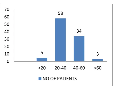
The mean age of patients in our study was 38.14 +/- 11.84 which fell in line with mean age of the comparative studies.Majority of the patient prevalence in our study fell under the reproductive age group of 20-40 years. This pattern was similarly noticed in Biswas et al , Gururaj et al and Lamichhane G et al study. However our prevalence rate of 58 % was closely comparable to the Gururaj et al study which had 54 %.Also comparable with the Gururaj et al study was the decrease in prevalence with increase in age.Of special mention is the similarity in the prevalence of patients in the age group < 20 years.
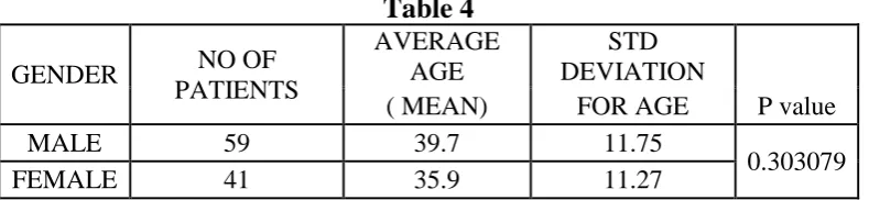
Our study shows a higher male prevalence in comparison to females. The age gender distribution in our study was not significant, p= 0.3. This was also the finding in Lamichhane G et all study.
Sexual route was the most common mode of disease transmission, and this was observed in study by Biswas et al and Gururaj et al. Gururaj et al had 1 homosexual route of exposure.P value of 0.1274 signifies that the route of exposure has no impact the prevalence of the ocular manifestations.Majority of the patients in our study were observed in clinical stage I and II. The findings were similar in the Gururaj et al and Amare et al study.
opportunistic infection in these stages. This was observed both in Gururaj et al and the Amare et al study as well.We observed an increased prevalence of ocular manifestations in the CD4 range of 200 – 500 cells/mm3. This was comparable to the Ethiopian study by Amare et al.P value in our study was 0.0235, which was significant. This significance was observed in all the above mentioned studies.Hence ocular manifestations is seen to increase significantly with decrease in CD4 count, especially in levels < 200 cells/mm3.
CONCLUSION
CONTENTS
PART ISL.NO TOPIC PAGE NO
01. INTRODUCTION 1
02. REVIEW OF LITERATURE 2
03. HISTORY 4
04. GLOBAL SCENARIO 5
05. HUMAN IMMUNODEFICIENCY VIRUS 6
06. CD4 AND HIV 11
07. HIV TRANSMISSION 13
08. PATHOGENESIS 15
09. HIV CASE DEFINITION AND CLASSIFICATION 17
10. LABORATORY DIAGNOSIS 19
11. ANTIRETROVIRAL THERAPY 20
12. OCULAR COMPLICATIONS OF ANTIRETROVIRAL THERAPY
21
13. HIV IN OPHTHALMOLOGY 22
14. DESCRIPTION OF HIV OCULAR LESIONS 24
15. TREATMENT OF HIV OCULAR LESIONS 30
PART II
16. AIM OF STUDY 35
17. MATERIALS AND METHODS 36
18. OBSERVATIONS 39
19. DISCUSSION 72
20. CONCLUSIONS 81
INTRODUCTION
HIV-AIDS was discovered in the early 1970’s. Amongst the various communicable diseases that have been well controlled with the advancing sciences, HIV- AIDS1 has been a major challenge. It poses a challenge not just to the doctors treating the disease but also to the patient subjected to societal stigma. AIDS with its multisystem manifestations calls for an increasing attention in understanding the disease and managing it and the associated opportunistic infections.
Eye with its adnexa is no exception to HIV infection. Ophthalmic2 presentations of HIV include microvasculopathy, neuro-ophthalmic disorders, neoplasms and opportunistic infections. Mainly seen and examined by the treating physician it becomes a necessity that all HIV infected patients undergo routine ophthalmic evaluations irrespective of symptoms for early identification of manifestations and prompt initiation of treatment to prevent visual loss.
REVIEW OF LITERATURE
1. Ocular manifestation of HIV/AIDS and correlation with CD4+ cells count
among adult HIV/AIDS patients in Jimma town, Ethiopia: a cross sectional study; Sisay Bekele, Yeshigeta Gelaw and Fasil Tessema.
Prevalence of HIV ocular manifestation was found to be lower than previous studies. They concluded this as probable result antiretroviral therapy. Low CD4 count is both risk factor and predictor for eye involvement in HIV.
2. Clinical study of ocular manifestations in HIV/AIDS and its correlation with CD4+T cells; Shivayogi Kusagur, Gururaj .K.J.
CD4+ cell counts and WHO clinical stage of HIV disease were concluded as important predictors of occurrence of ocular morbidity in HIV positive individuals.
4. Pattern of Ocular Manifestation of HIV/ AIDS among Patients on HAART in ART Clinic of Gondar University Hospital, Northwest Ethiopia; Bemnet Amare, Fisseha Admassu, Yared Assefa, Beyene Moges, Jemal Ali and Afework Kassu.
HISTORY
Theories of HIV origin and its epidemic have baffled many of us for ages. Simian immunodeficiency virus from monkeys3 spread4 into human community in various forms thereby mutating to Human immunodeficiency Virus (HIV). Homosexual5 practice along with social changes and urbanisation6 led into its amplification in human community.
The virus was identified and named7 lymphadenopathy associated virus in 1983 and renamed as Human lymphotrophic virus-III8 in 1984. In the year 1986 both names were discarded and the name Human immunodeficiency virus was put into use. On retrospective analyses of all the preserved samples (blood, lymph node) of suspected deaths (clusters of immunosuppressive episodes) the first known case was a documented HIV1 infection from a man in Congo in the year 1959. Second documentation was from a woman in Congo in 1960. Since then number of patients positive for HIV have increased progressively without any permanent cure and increasing opportunist infections.
GLOBAL SCENARIO
As on 2012, the estimated number of people living with HIV globally, is 35.3 (32.2–38.8) million. The number of new infection in 2001 was 3.4 million in comparison to 2.3 (1.9–2.7) million new HIV infections in 2012, showing a 33% decline in the number of new infections globally12. The AIDS related deaths have drastically declined from 2.3 million in 2005 to 1.6 million in 2012, a direct result of increasing care and antiretroviral treatment. Even India is still the third largest nation with HIV population.
In India the new infection number fell from 2.74 lakhs cases in 2000 to 1.16 lakhs in 2011(57%). An impact of the various interventions under National AIDS Control Programme. Of the four high prevalence States in India includes Andhra Pradesh, Karnataka, Maharashtra and Tamil Nadu and they account for 53% of all HIV infected population in the country.
HUMAN IMMUNODEFICIENCY VIRUS
MICROSCOPIC STRUCTURE OF HIV VIRION
Shape16: spherical enveloped virus Size: 90-120nm
Nucleocapsid: has an outer icosahedral shell and inner cone shaped core that which encloses ribonucleoproteins.
VIRAL GENES AND ANTIGENS:
STRUCTURAL GENES:
These are the gag, pol, env; the products of which are the viral antigens17.
1. gag: precursor proteins that determine the core and the shell of the virus.(p15, p18, and p24).
p24 helps in disease detection and indications re-exacerbation.
2. env: determines synthesis of envelope glycoprotein gp120 and gp 41. gp120- forms surface spikes17.
gp 41 -forms the trans membrane anchoring protein18.
3. pol :codes for polymerase reverse transcriptase ,protease and endonuclease
NON-STRUCTURAL AND REGULATORY GENE:
1. tat (trans activating gene): enhances expression of all viral genes.
2. nef (negative factor gene) : down regulates viral replication.
3. rev (regulator of virus gene ): enhances expression of structural proteins.
4. vif (viral infectivity factor gene): influencing infectivity of viral particles.
6. vpr stimulating promoter region of the virus.
7. LTR (long terminal repeat)19 sequences, one at either end, containing sequences giving promoter, enchancer and integration signals.
ANTIGENIC VARIATIONS:
HIV has been characterized into HIV-1 and HIV-220.
HIV1: responsible for majority of infection. It is more virulent of the two. Three main are Group M (majority of cases), Group O and Group N.
HIV2: largely confined to West Africa because of its low virulence.
VIRUS STABILITY:
CD4 AND HIV
CD is an abbreviation of the cluster of differentiation /cluster of designation23. They are a defined sub classification of cellular surface receptors (> 250 in number), also called epitope; which identify cell type and stage of differentiation. These receptors coat the surfaces of the B and T lymphocytes and are recognized by antibodies. Of all the epitopes classified CD4 receptor present on the T lymphocytes has been in limelight for its highest association with HIV pathogenesis.
CD4 is a membrane glycoprotein present on precursor T cells and also on a few mature T cells. Being a helper cell, when activated in presence of an infecting agent (antigen) triggers a cascading event thereby activating the co immune cells especially the killer T cells (CD 8) thereby eliminating the antigen24.
CD4+ T cell count in HIV infected individuals categorises the systemic/ opportunistic manifestations. CD4 cell counting includes various techniques of which flow cytometry26 is essentially used. CD4 values ranging from 500-1200 cells/mm3 is considered normal range for HIV infected person. A fall below 500cells/mm3 warrants frequent monitoring, and a fall below 350cells/mm3 necessitates start of antiretroviral therapy.
However a child is born with a very high level of CD4 (several thousands) and this cannot be used in monitoring disease. Therefore CD4 percentage in relation to other immune cells is used27.
CD4% of 12-15% = <200 cells/mm3,
CD4% of 29% = > 500 cells/mm3,
CD4% of about 40% = HIV negative34.
For treatment purposes in HIV infected child World Health Organisation recommends the following28:
AGE Infants and
children <24 months of age
≥24 months of age to 59 months of age
Five years of age or
Older
%CD4+ Absolute
lymphopenia
≤25 NA
Absolute CD4 count
Absolute lymphopenia
HIV TRANSMISSION
TYPES OF EXPOSURE CHANCE OF INFECTION
PER EXPOSURE Sexual intercourse: anal , vaginal , oral 0.1 – 1.0 %
Blood and blood products, blood transfusion
> 90% Tissue and organ donation: semen ,
cornea, bone marrow, kidney etc
50 -90% Injections and injuries: injection with
unsterile syringes, drug addicts shared needles, needle stick injury , surgical wounds
0.5 -1.0%
Mother to baby :trans-placental , at birth , after birth breast milk
30%
SEXUAL TRANSMISSION:
Predominant mode of transmission. Risk of transmission increases with multiple sexual partners29. People indulged in riskful sexual practices. These people and their partners should be counselled regarding safer sex methods.
BLOOD AND BLOOD PRODUCTS:
ORGAN TRANSPLANTATION:
Organs are now screened for HIV and risk of transmission has been drastically reduced. This is applicable for semen, cornea, bone marrow, kidney and other organs.
INJECTIONS:
Relevant in intravenous drug abusers who share needles31. Also in medical and health professional the danger of needle stick injury persists to a great extent, though the chance of infection is much less.
MOTHER TO CHILD:
PATHOGENESIS
HIV CASE DEFINITION AND CLASSIFICATION
PROVISIONAL DIAGNOSIS FOR AIDS, WHO: (1986)36
Presence of at least two major signs along with one minor sign, from those listed below
MAJOR SIGN
Weight lose greater than 10% of body weight
Fever for more than one month, intermittent or continuous
Chronic diarrhoea for more than one month, intermittent or continuous MINOR SIGN
Persistent cough for more than one month General skin manifestation
Recurrent herpes zoster Oropharyngeal candidiasis
Chronic progressive and disseminated herpes simplex infection Generalised lymphadenopathy
CDC CLASSIFICATION SYSTEM FOR HIV DISEASE: (1986) Group I: acute infection
Group II: asymptomatic infection
Group III: persistent generalised lymphadenopathy Group IV: other diseases
Subgroup A constitutional disease Subgroup B neurologic disease
Subgroup C secondary infectious disease Subgroup D secondary cancers
Revised CDC classification of 1995
Stage 1(HIV
infection)
CD 4 t lymphocyte count greater than or equal to 500 cells/Ml
Stage 2(HIV
infection)
CD4 T lymphocyte count of 200-499 cells/uL Stage 3 (AIDS) CD4 T lymphocyte count of less than 200
cells/uL
WHO STAGING:
HIV-ASSOCIATED IMMUNODEFICIENCY
AGE RELATED CD4 VALUES IN % <11
months
12-35 months
36-59 months
>5 years None or not significant >35 >30 >25 >500
Mild 30-35 25-30 20-25 350-499
Advanced 25-29 20-24 15-19 200-349
Severe <25 <20 <15 <200 or
LABORATORY DIAGNOSIS
Test includes for immunodeficiency as well as specific test for HIV. Specific test for HIV37
1. Antigen detection: p24 is the earliest virus marker to occur and detected by ELISA.
2. Polymerase chain reaction: sensitive and specific test. DNA PCR and RNA PCR are the tests performed. The tests are complex and costly and indicated only when other tests do not give definitive results. 3. Virus isolation: From peripheral lymphocytes. Patient’s lymphocytes
co cultivated with uninfected lymphocytes in presence of interleukin -249.
4. Antibody detection: two type of tests- screening and confirmatory ELISA: screening test. Simple, inexpensive but false positives are common.
WESTERN BLOT: it’s a gold standard confirmatory test.
Immunological tests:
1. Total leucocyte and lymphocyte count 2. CD4 cell count
3. Platelet count
ANTIRETROVIRAL THERAPY
Standard antiretroviral therapy (ART)38 consists of the combination of at least three antiretroviral (ARV) drugs to maximally suppress the HIV virus and stop the progression of HIV disease.
Various antiretroviral drugs approved for therapeutic use in India:
NRTI NNRTI Protease
inhibitor Entry inhibitors Integrase inhbitors Zidovudine Stavudine Lamivudine Abacavir Didanosine Tenofovir Dideoxycytidine Nevirapine Efavirenz Etravirenz Delaverdine Saquinavir Indinavir Ritonavir Nelfinavir Lopinavir Atazanavir Darunavir Fos-Amprenavir Tipranavir Enfuvitride Maraviroc Raltegravir
NRTI : nucleoside reverse transcriptase inhibitor; NNRTI: non-nucleoside reverse transcriptase inhibitor
Highly active antiretroviral therapy (HAART) is the combination of two or more groups of drugs that acts at different level of viral replication thereby increasing patient compliance and also decreasing chances of drug resistance. HAART is initiated when the CD4 count <350/uL, the combination depending on patient health status and other co morbidities.
OCULAR COMPLICATIONS OF ANTIRETROVIRAL THERAPY
Given below are the common drug used in our set up and its side effects;NRTI ADVERSE EFFECT
Zidovudine Amblyopia, macular edema,
photophobia
Stavudine -
Lamivudine Red itchy eye
Abacavir Red itchy eye
Tenofovir -
didanosine Retinal changes, optic neuritis
NNRTI ADVERSE EFFECT
Nevirapine Red itchy eye
PROTEASE INHIBITOR ADVERSE EFFECT
Saquinavir blepharitis, dry eye syndrome, eye irritation, xerophthalmia, cytomegalovirus retinitis Indinavir Ocular pain , orbital edema Ritonavir Iritis , visual field defects, abnormal
EOG
HIV IN OPHTHALMOLOGY
CORRELATION OF CD4 T LYMPHOCYTE COUNT AND OCULAR LESION39:
Stage Seroconversion63 Early infection
Intermediate infection
Late infection
CD4+ 1000 500-1000 200-500 <200
External eye
Inflamed
conjunctiva, Dry eye
Allergic conjunctivitis
Dry eye, Blepharitis, Bacterial andfollicular conjunctivitis, Kaposi’s sarcoma, Molluscum contagiosum, Opportunistic infections and tumours affecting all ocular structures Anterior segment Reiters syndrome Intermediate uveitis Herpes simplex, Herpes zoster Posterior segment HIV retinopathy Retinal vasculitis HIV retinopathy, Tuberous uveitis Neuro ophthalmic
Headache Retro orbital pain
Optic neuropathy
SITE WISE OCULAR LESION IN AIDS:
SITE LESIONS
ADNEXA Herpes zoster ophthalmicus
Kaposi sarcoma
Molluscum contagiosum Eyelid infections
Allergic or infective conjunctivitis ANTERIOR SEGMENT Dry eye
Infective keratitis Uveitis
Microvasculopathy POSTERIOR SEGMENT HIV retinopathy
CMV retinitis
Progressive outer retinal necrosis Acute retinal necrosis
Fungal endophthalmitis Toxoplasma retinochoroiditis Ocular syphilis
Mycobacterium infection
ORBIT Orbital cellulitis
Burkitts lymphoma NEURO OPHTHAL Cranial nerve palsies
DESCRIPTION OF HIV OCULAR LESIONS
EYELID INFECTIONS
Blepharitis and meibominitis are common adnexal presentations and require prompt treatment which includes lid hygiene, hot fomentation and support antibiotics.
MOLLUSCUM CONTAGIOSUM
HIV infected patients have an extensive, coalescing lesions and these recur on treatment. Treatment includes cautery- electrical or chemical, surgical excision.
KAPOSI SARCOMA (eyelid and conjunctiva)
A vascular lesion, reddish purple in colour arising from the lid and or conjunctiva. Usually said to be associated with herpes virus 8 infections. Kaposi sarcoma in HIV has not been reported in Indian context.
CONJUNCTIVAL MICROVASCULOPATHY
AIDS virus is said to directly attack the vascular wall leading to its dilation/ segmentations, and cock-screwing.
HERPES ZOSTER OPHTHALMICUS
INFECTIVE KERATITIS
HIV individuals are easily susceptible to corneal infection from bacterial, viral, fungal and protozoal.
UVEITIS
Unexplained uveitis in an individual should prompt investigations for HIV. Uveitis in HIV-infected patient should prompt a search for an underlying infection, including tuberculosis, syphilis, histoplasmosis, coccidioidomycosis, and toxoplasmosis.
HIV RETINOPATHY
It is a non-infectious microvascular disorder of the retina in HIV infection. With cotton wool spots, microaneurysms retinal haemorrhages and capillary non perfusion this almost mimics diabetic retina. Therefore it becomes a necessity to rule out HIV in unexplained vascular occlusive diseases.
Hemorrhages are seen less commonly seen than cotton-wool spots.They may involve both the nerve fiber layer and the deeper retina and may appear as flame-shaped, dot, or blot hemorrhages.
CYTOMEGALOVIRUS RETINITIS
Is the most common opportunistic infection in HIV. Cytomegalovirus is a DNA herpes group virus occurring in advanced HIV disease. This DNA virus invades the retinal cells causing retinal necrosis. Manifests either in indolent or fulminant form. The indolent variety has granular white retinal deposits, has minimal or no vasculitis and no retinal hemorrhage. The fulminant type presents with retinal deposits, vasculitis and haemorrhages. Both varieties begin in the periphery and spread to centre along the vascular arcade leading to triangular / wedge zones. Vitritis if at all will be minimal. Frosted branch angiitis in another mode of presentation. With eventual loss of retinal pigment epithelium the underlying choroidal vasculature becomes prominent.
Treatment includes both systemic and intravitreal injections of gancyclovir. Other drugs include valcyclovir, foscarnet. Administer till lesion heals. Retinal detachment can be a major challenge while treating CMV retinitis.
ACUTE RETINAL NECROSIS
is common with complications like retinal detachment and proliferative vitreoretinopathy. Ganciclovir /foscarnet, with adjunctive high-dose intravenous acyclovir (15 mg per kilogram every 8 hours)is the treatment of choice.
TOXOPLASMA RETINOCHOROIDITIS
Protozoan parasite which exists in cyst and trophozoites form. Though the infection occurs in early in extremely immunocompromised person, the ocular manifestation occurs usually on immune recovery on administration of HAART. This is because the immune mediated reaction is directed towards the cyst which is severe. In HIV, toxoplasmosis is characterised by multifocal retinochoroidal fluffy lesions involving entire retina with severe vitritis. Degree of haemorrhage is less in comparison to CMV retinitis. Fluorescein angiography differentiates toxoplasmosis from CMV retinitis by active leakages.
Serological testing for toxoplasma in HIV is unreliable. They either show low IgG titres or are unable to distinguish old from new lesions. Negative IgG titre however makes it unlikely for toxoplasma infection to be present.
PNEUMOCYSTIS CHOROIDITIS:
Pneumocystis jiroveci (PCP), is a fungus showing protozoal characteristics. It presents as multiple yellow white choroidal lesions, generally round and of variable size. It eventually leads to choroiadal necrosis. The earliest evidence of PCP retinal infection was seen in 1982, where the histopathological report showed evidence of PCP in ganglion and plexiform layer of retina. In 1987 histopathologic examination revealed areas of choroidal thickening and exudate which harbored the characteristic cysts of P jiroveci in HIV patients. Treatment will be same as for respiratory PCP infection.
SYPHILIS
OPTIC DISC
Optic disc involvement includes papilledema as a result of intracranial space occupying opportunistic lesions, optic neuritis or neuropathy.
TREATMENT OF OCULAR HIV MANIFESTATIONS
LID INFECTIONS:
Lid hygiene is of utmost importance. Antibiotics ointment and oral antibiotics are necessary. Also strict adherence to antiretrpviral therapy helps to maintain good immunity.
MOLLUSCUM CONTAGIOSUM:
Strict ART follow up. Topical trichloroacetic acid can be applied. Surgically curettage,excision or cryotheraphy can be done.
CONJUNCTIVITIS:
Infective conjunctivitis needs to be proved on grams stain and culture, and treated accordingly.
If allergic the drug causing should be replaced and symptomatic topical medication like lubricating drops applied.
CONJUNCTIVAL MICROVASCULOPATHY:
HERPES ZOSTER OPHTHALMICUS:
Acyclovir 10 milligram per kilogram of body weight administered IV every 8 hours for 1 week. Maintenance dose of oral acivir 800mg, 3-5 times a day indefinite.
On follow up patient had to be treated for post herpetic neuralgia if sny.
Alternatives include valacyclovir.
VIRAL KERATITIS:
If the cause is varicella zoster, oral acivir 800mg 5 times a day is given. For resistant cases IV foscarnet has been useful. Topical trifluridine 1 percent is also seen to be helpful in controlling varicella keratitis.
If the cause is herpes zoster; same treatment as for varicella. Topical application of antiviral medication is most indicated in herpes keratitis as it tends to recur more frequently in HIV AIDS patients.
FUNGAL AND BACTERIAL KERATITIS:
UVEITIS:
Usually drug induced, treatment consists of topical steroids tapered over weeks till resolution.
GLAUCOMA:
Usually of the inflammatory component, the underlying cause has to be treated first and also topical anti glaucoma medication have to be administered taking care to avoid prostaglandin analogue.
HIV RETINOPATHY:
Immune status needs to be evaluated and patient followed up every 3 months for dilated fundus examination.
CMV RETINITIS:
Ganciclovir 5mg/kg IV, 12th hourly for 3 weeks. Followed by maintenance doses of 5mg/kg/day is given twice weekly until lesion is inactive.
Immune status has to be improved with strict adherence to HAART. If the patient has never been on HAART prior to the occurrence, regime can be postponed to prevent immune recovery uveitis.
Patient needs to be educated about the recurrence and must be advised for a serial follow for dilated fundus examination.
TOXOPLASMOSIS:
Trimethoprim/ sulfamethoxozole 400mg administered per oral twice a day for 4 to 6 weeks.
Patients with very low immunity have to be given at least one drug life time or till immune status improves.
ACUTE RETINAL NECROSIS/ PROGRESSIVE OUTER RETINAL NECROSIS:
Intravenous 15mg/kg acyclovir is given 8th hourly. Foscarnet or Valcyclovir could be prescribed alternatively. Patient needs to be monitored for drug induced complications. Usually progresses rapidly.
MULTIPLE CRANIAL NERVE PALSY:
OPTIC NEURITIS:
Underlying cause has to be found and treated.
OCULAR TUBERCULOSIS:
AIM
To evaluate the prevalence of ocular manifestations in HIV seropositive patients, attending the ART centre, Stanley Medical College and Hospital.
To evaluate how the manifestations correlate to the CD4 count.
METHODS AND MATERIAL
STUDY DESIGN:
Cross sectional study was undertaken on 100 HIV seropositive patient attending the ART centre with known CD4 count was included in the study.
The study period was from February 2013 till completion of hundred patients.
INCLUSION CRITERIA
HIV seropositve consented patients attending ART centre irrespective of ocular complaints, irrespective of treatment status.
Any age group
CD4 count obtained at the time of examination.
EXCLUSION CRITERIA
Severely ill patients.
CD4 count could not be obtained.
The importance of ocular examination in them was thoroughly explained in patients own language.
Consent in patients own language was obtained from all those who were willing to take part in study.
Any photographic records of the lesions were taken only if the patient consented for the same.
Upon completion of data collection, statistical analysis was applied.
STATISTICAL ANALYSIS:
ROUTINE OCULAR EXAMINATION WAS DONE AS FOLLOWS:
Ocular history.
Ocular examination included:
Best corrected visual acuity. ( Snellen’s chart )
Slit lamp evaluation – Adnexa and Anterior segment
Dilated fundus evaluation(0.8 % tropicamide + 5% phenylephirine)90 D
and 20 D
Neuro ophthalmological examination
Orbit
Tonometry for IOP evaluation
Schirmer’s for dry eye evaluation
Patient with suspected lesions were referred for complete systemic and laboratory work up.
Serological investigations included TORCH titre, PCR assay for HZV, VZ.
OBSERVATIONS
This study includes 100 consented HIV seropositive patients, irrespective of age or gender, provided CD4 count was available at the time of ocular examination.
[image:56.595.199.425.387.557.2]AGE WISE DISTRIBUTION: N = 100
Table 1
AGE IN YEARS NO OF PATIENTS
<20 5
20-40 58
40-60 34
>60 3
The overall average age of the study population was 38.14 years.
The reproductive age group of twenty to forty years formed the major part of our study, with 58 patients falling under this group.
5
58
34
3 0
10 20 30 40 50 60 70
<20 20-40 40-60 >60
Of these fifty-eight patients, forty-three fall under age group of thirty-one to forty years.
GENDER DISTRIBUTION: N= 100
[image:58.595.182.442.310.474.2]With no attempt to maintain an equal count of the gender population, we observed 59% males and 41% females.
Table 2
GENDER NO OF PATIENTS
MALE 59
FEMALE 41
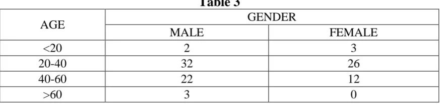
[image:58.595.94.533.572.678.2]AGE DISTRIBUTION OF GENDER WAS AS OBSERVED BELOW:
Table 3
AGE GENDER
MALE FEMALE
<20 2 3
20-40 32 26
40-60 22 12
>60 3 0
The age of the male patients ranged from thirteen to seventy-seven years. Whereas age of female patients ranged from four to fifty-seven years.
59
41
0 20 40 60 80
MALE FEMALE
The mean age for the fifty nine male patients is 39.7 years. And for the forty-one female patients is 35.9 years.
Table 4
GENDER NO OF
PATIENTS
AVERAGE AGE
STD
DEVIATION ( MEAN) FOR AGE P value
MALE 59 39.7 11.75
0.303079
FEMALE 41 35.9 11.27
Upon statistically validating the age – gender distribution using chi square test, we arrived upon a p value of 0.303079.
OCCUPATION: N = 100
Table 5
OCCUPATION NO OF PATIENTS
SKILLED 17
UNSKILLED 79
UNEMPLOYED 4
TOTAL 100
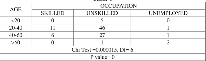
[image:60.595.95.531.545.674.2]AGE WISE OCCUPATION DISTRIBUTION:
Table 6
AGE OCCUPATION
SKILLED UNSKILLED UNEMPLOYED
<20 0 5 0
20-40 11 46 1
40-60 6 27 1
>60 0 1 2
Chi Test =0.000015, Df= 6 P value= 0
21
75
4 0
20 40 60 80
SKILLED UNSKILLED UNEMPLOYEED
79 Unskilled workers formed a major part of our study, with 46 of them falling under the reproductive age group. Majority of these unskilled workers were drivers.
The 5 children were included under the unskilled group.
The 11 skilled workers, including businessmen also were of the reproductive age group.
MARITAL STATUS: N= 100
Table 7
MARITAL STATUS NO OF PATIENTS
SINGLE 8
MARRIED 90
WIDOWED 2
TOTAL 100
90 out of the 100 patients in our study were married and contracted disease either from multiple sexual partners or from the exposed spouse.
Interestingly 1 widow was diagnosed with the disease following death of the male spouse from undiagnosed illness.
The single group of 8 people included 5 children and 3 unmarried adult males.
8
90
2 0
20 40 60 80 100
SINGLE MARRIED WIDOWED
MODE OF DISEASE EXPOSURE: N = 100
Table 8
MODE OF DISEASE EXPOSURE NO OF PATIENTS
SEXUAL 91
BLOOD TRANSFUSION 2
INJECTIONS 2
MTC 5
TOTAL 100
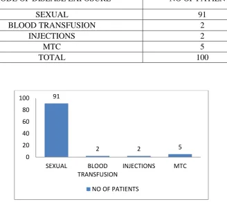
We observed that Sexual exposure was the most common form of disease transmission, with all 91 patients having had heterosexual exposure.
5 children had vertical transmission of the disease from the exposed mother (mother to child transmission/ MTC).
4 patients amongst the reproductive age group had non sexual exposure. Two of which had history of blood transfusion in the recent past, and 2 had high risk IV drug abuse behaviour with needle sharing.
91
2 2 5
0 20 40 60 80 100
SEXUAL BLOOD
TRANSFUSION
INJECTIONS MTC
WHO CLINICAL STAGE: N = 100
Table 9
WHO CLINICAL STAGE NO OF PATIENTS
I 73
II 7
III 10
IV 10
[image:64.595.94.533.492.629.2]TOTAL 100
Table 10
WHO CLINICAL STAGE OCULAR LESION TOTAL
PRESENT ABSENT
I 54 19 73
II 7 0 7
III 8 2 10
IV 9 1 10
TOTAL 78 22 100
P Value = 0.003
WHO clinical stage I consisted of 73 patients, indicating that the patients were asymptomatic or had acute flu like symptoms on disease detection.
73
7 10 10
0 20 40 60 80
I II III IV
The 20 patients from clinical stage III and IV were all diagnosed with HIV following systemic illnesses that should be presumed to be due to immunocompromised state.
One patient had esophageal candidiasis, four patients had recurrent viral infections, two had protozoal infections, two had intra cranial space occupying lesions (One expired during follow up, one diagnosed with tumefactive demyelination due to HIV infection), four patients had tuberculosis and the remaining seven patients had HIV wasting syndrome.
Nine out of these twenty patients had HIV associated severe ocular manifestations. Interestingly one patient, a child of four years of age, in clinical stage IV (extra pulmonary TB) of age did not exhibit any ocular lesion and her CD4 count was 2149.
CD4 COUNT DISTRIBUTION: N = 100
In our study we had CD4 count ranging from 28 – 2149 cells/mm3.
The average CD4 count therefore was 503.57 cells /mm3.
Table 11
CD4 COUNT NO OF PATIENTS
<200 11
201 – 500 45
>500 44
Table 12
CD4 COUNT AGE DISTRIBUTION TOTAL
<20 20-40 41-60 >60
<200 0 7 3 1 11
200-500 3 29 12 1 45
>500 2 22 19 1 44
TOTAL 5 58 34 3 100
Eleven patients were severely immunocompromised with CD4 count of < 200 cells/mm3.
Note that none of the children observed in our study were severely immunocompromised. All five children maintained CD4 count > 200.
11
45 44
0 10 20 30 40 50
<200 201 - 500 >500
DURATION OF THE HIV DISEASE: N = 100
Table 13
DURATION SINCE DISEASE
DETECTION (MONTHS) NO OF PATIENTS
<12 13
13-24 45
25-60 36
>60 6
TOTAL 100
We observed patients with disease duration ranging from as early as 2 months up to 9 years.
The average duration of disease being 4.03 years.
13
45
36
6
0 10 20 30 40 50
<12 13-24 25-60 >60
HAART REGIME: N = 100
Table 14
PATIENTS ON HAART NUMBER
YES 85
NO 15
HIV seropositive patients detected with CD4 count of < 350 /mm3 at any point; at or later date to disease detection were started on HAART regime as per WHO recommendations.
Fifteen patients were on observation with half yearly CD4 re-evaluation and treated symptomatically.
Eighty-five patients were on HAART regime, which was initiated as per WHO recommendation, and as per patient’s systemic associated conditions and response to drugs.
85
15
0 10 20 30 40 50 60 70 80 90
YES NO
DURATION SINCE HAART TREATMENT: N = 85
Table 15
DURATION SINCE STARTING HAART (MONTHS) NO OF PATIENTS
<12 15
13-24 17
25-60 47
>60 6
TOTAL 85
Table 16
DURATION SINCE STARTING HAART
OCULAR LESION
TOTAL
PRESENT ABSENT
<12 12 3 15
13-24 12 5 17
25-60 38 9 47
>60 5 1 6
TOTAL 67 18 85
Chi Test = 0.827356 , Df =3 P value= 0.2399
Sixty-seven out of the eighty-five patients on HAART manifested ocular lesions in various forms.
15 17
47
6
<12 13-24 25-60 >60
[image:69.595.93.530.520.666.2]TB STATUS: N = 100
[image:71.595.105.523.102.426.2]Table 17
SYSTEMIC TB STATUS NO OF PATIENTS
CURRENT 6
PAST 25
NIL 69
TOTAL 100
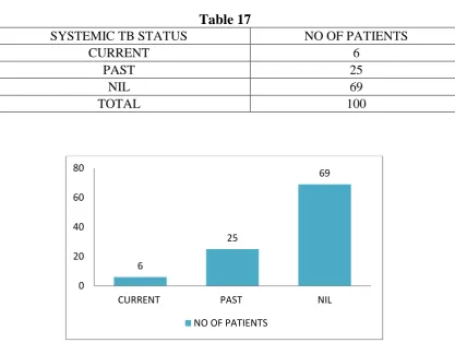
Six out of hundred patients were recently diagnosed and currently under treatment for tuberculosis. Two of these were children of age less than fourteen.
Twenty-five patients were treated for tuberculosis in the past, in the time frame of HIV detection- duration, of which one was a child.
6
25
69
0 20 40 60 80
CURRENT PAST NIL
OCULAR SYMPTOMS: N = 100
Table 18
OCULAR SYMPTOMS NO OF PATIENTS
PRESENT 30
ABSENT 70
[image:72.595.102.533.134.189.2]OCULAR FINDINGS: N = 100
Table 19
OCULAR FINDINGS NO OF PATIENTS
PRESENT 78
ABSENT 22
Seventy-eight out of the hundred patients that we examined had findings from the adnexa up to the optic nerve. The average CD4 count in these 78 patients was noted to be 468.16.
These findings were either a manifestations of viral load, or of low immunity, and/ or of opportunistic infections.
OCULAR SYMPTOMS VERSUS OCULAR FINDINGS:
Table 20
OCULAR FINDINGS OCULAR SYMPTOMS TOTAL
PRESENT ABSENT
PRESENT 29 49 78
ABSENT 1 21 22
TOTAL 30 70 100
P value = 0.003178
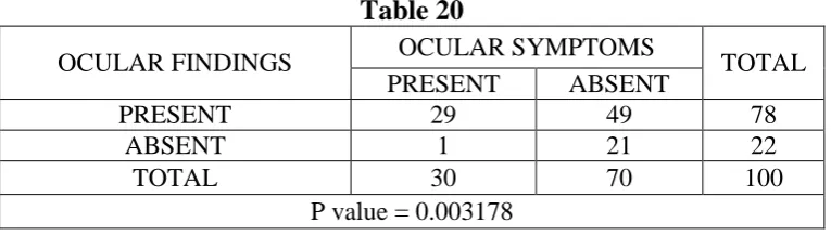
Thirty HIV seropositive patients had ocular symptoms. Of these twenty-nine patients almost all had severe blinding opportunistic manifestations. The remaining one patient had refractive error.
VISION DISTRIBUTION:
[image:74.595.102.524.137.243.2]The severity of manifestations on visual acuity was as observed below;
Table 21
VISION STATUS RIGHT EYE LEFT EYE BOTH EYES
BLIND 3 2 1
<3/60 1 3 2
3/60- 6/60 1 0 0
6/60- 6/18 2 2 2
>6/18 93 94 87
Five out of hundred patients had ocular manifestations leading on to bilateral impairment in vision. Of which one patient was completely blind.
Nine out of hundred patients had unilateral vision impairing manifestations.
The patient with bilateral blindness was diagnosed with severe toxoplasma retinochoroiditis.
OCULAR FINDINGS IN OUR STUDY:
[image:75.595.111.514.272.755.2] Seventy eight out of hundred patients had HIV related ocular manifestation. Majority of the patients in our study had multiple manifestations at the time of examination; blepharitis and conjunctival microvasculopathy were commonly found and accounting for the overlap in the total number of manifestations.
Table 22
OCULAR FINDINGS NUMBER OF PATIENTS WITH FINDINGS
PERCENTAGE ADNEXA
BLEPHARITIS 29 24.79
MOLLUSCUM CONTAGIOSUM 2 1.71
STYE 2 1.71
HERPES ZOSTER 1 0.85
ALLERGIC / INFECTIVE
CONJUNCTIVITIS 6
5.13
CONJ. MICROVASULOPATHY 53 45.3
ANTERIOR SEGMENT
COMPLICATED CATARACT 1 0.85
HERPES ZOSTER
OPHTHALMICUS 1
0.85
VIRAL KERATITIS 1 0.85
ANTERIOR UVEITIS 1 0.85
NON HEALING FUNGAL ULCER 1 0.85
INFLAMMATORY GLAUCOMA 1 0.85
POSTERIOR SEGMENT
HIV RETINOPATHY 4 3.42
CMV RETINITIS BURNT OUT 2 1.71
ARN 1 0.85
TOXOPLASMOSIS 2 1.71
RETINAL VASCULITIS 1 0.85
INTERMEDIATE UVEITIS 1 0.85
RETINITIS 1 0.85
NEURO OPHTHAL
MULTIPLE CRANIAL NERVE
PALSY 1
0.85
OPTIC NEURITIS 1 0.85
OPTIC ATROPHY 4 3.42
OCULAR FINDING AND AGE DISTRIBUTION: N = 78
Table 23
AGE IN YEARS NO OF PATIENTS WITH FINDINGS
<20 3
20-40 44
41-60 29
>60 2
TOTAL 78
75.85 % of the ocular manifestation was seen in the age group of twenty to forty years (58 people).
However when checked for its significance (by chi square test), the age wise distribution of ocular lesion proved to be statistically insignificant.
3
44
29
2
78
0 20 40 60 80 100
<20 20-40 41-60 >60 TOTAL
GENDER WISE DISTRIBUTION OF OCULAR FINDINGS: N = 78
[image:77.595.109.535.127.435.2]Table 24
GENDER NO OF PATIENTS AVERAGE CD4 COUNT
MALE 44 432.32
FEMALE 34 606.09
Forty-four out of these seventy-eight patients with HIV ocular manifestations were male when comparison to thirty-four females; who had disease manifestations.
The average CD4 count in the forty-four male patients with ocular manifestations was found to be 432.32.
The average CD4 count of 606.09 was observed in thirty-four female patients with ocular manifestations.
44
34
0 10 20 30 40 50
MALE FEMALE
OCULAR FINDING AND ROUTE OF EXPOSURE: N= 78
Table 25
MODE OF DISEASE EXPOSURE NO OF PATIENTS AVERAGE CD4 COUNT
SEXUAL 73 602.8
BLOOD TRANSFUSION 1 780
INJECTIONS 2 261
MTC 2 371.5
This data shows the prevalence of manifestations for various routes of transmission.
Injection/ IV drug abuse had 100% manifestations (though a non-blinding manifestation) when compared to blood transfusion which exhibited 50% prevalence of manifestation. However this data is insignificant in terms of sample size.
73
1 2 2
0 10 20 30 40 50 60 70 80
SEXUAL BLOOD
TRANSFUSION
INJECTIONS MTC
Ocular manifestations were more seen in patients with sexual mode of transmission.
Seventy-three of ninety-one (80%) were sexually exposed patients who exhibited ocular manifestations.
Most of the ocular blinding manifestations were observed to occur in patients exposed to HIV sexually, and this specific group had CD4 ranging from 28- 1916 cell/mm3. And these in itself are manifestations of other associated sexually transmitted diseases.
The 2 patients, who contracted disease by the IV route, had CD4 counts of < 350 cells/mm3. 1 out of the 2 patients, who had been seen after a year showed just a minimal improvement in immunity. This probably indicates the high viral load transmission via injections/IV drug abuse. However none of the two had opportunistic manifestations.
OCULAR FINDINGS AND WHO CLINICAL STAGING: N = 78
Table 26
WHO CLINICAL STAGE NO OF PATIENTS
I 54
II 7
III 8
IV 9
TOTAL 78
Fifty-three out of the seventy-eight patients with ocular manifestation were observed under the clinical stage I of WHO classification. All patients in our study group with HIV retinopathy fell under this clinical staging of HIV. One patient with intermediate uveitis was under WHO clinical stage 1.
Clinical stage II we observed seven people. Of these two patients had molluscum contagiosum, two had herpes zoster ophthalmicus, one had
54
7 8 9
0 10 20 30 40 50 60
I II III IV
allergic conjunctivitis, one had non healing fungal corneal ulcer and one had microvasculopathy.
Seventeen patients from WHO clinical stage III and IV categorised under ocular manifestations had vision impairing opportunistic diseases. These included viral keratitis, CMV retinitis , toxoplasma retinochoroiditis, acute retinal necrosis, multiple cranial nerve palsy, optic neuritis etc.
OCULAR FINDING AND CD4 COUNT: N =78
Table 27
CD4 COUNT OCULAR LESION % OF OCULAR LESION
PRESENT ABSENT
<200 10 1 90.90
200-500 34 11 75.55
>500 34 10 77.27
The prevalence of ocular manifestations is noted to increase with decrease in CD4 count which can be observed from the table above.
On the contrary, with the increase in CD4 count the prevalence of lesions were found to be decreasing which can be observed from the table above.
10
34 34
0 10 20 30 40
<200 201 - 500 >500
Table 28
CD4 COUNT GENDER TOTAL
MALE FEMALE
<200 8 2 10
200-500 21 13 34
>500 14 20 34
TOTAL 43 35 78
From the table above, we observed that the prevalence of male patient with ocular lesions is 80 % with the CD4 count < 200 cells/mm3.
OCULAR FINDINGS AND HAART REGIME DURATION: N= 78 Table 29 DURATION OF HAART TREATMENT ( MONTHS) NUMBER OF OCULAR LESIONS
MEAN AND STD. DEV CD4 COUNTS %
INCREASE IN CD4 COUNT BASE CD4 STD DEV PRESENT CD4 STD DEV.
<12 12 315.33 194.64 314.66 217.31 -0.21
13-24 12 324.58 271.2 461.76 272.21 29.71
25-60 38 319.52 271.2 473.45 272.21 32.51
[image:84.595.99.534.164.283.2]>60 5 425.16 270.2 766.16 273.04 44.51
Table 30
DURATION SINCE HAART TREATMENT
OCULAR LESION
TOTAL
PRESENT % ABSENT %
<12 12 80 3 20 15
12-24 12 70.58 5 29.41 17
25-60 38 80.85 9 19.14 47
>60 5 83.33 1 16.66 6
When comparing the baseline CD4 count at the start of HAART to the CD4 count at the time of examination, we observed a significant increase in CD4 count, suggesting the effectiveness of HAART.
CORRELATING THE FINDINGS WITH CD4 COUNTS: N =156 EYES
Table 31
CD4 COUNT DISTRIBUTION FOR N = 156
ADNEXA <200 200-500 >500
BLEPHARITIS 18 22 16
MOLLUSCUM CONTAGIOSUM 0 4 0
STYE 0 2 1
HERPES ZOSTER 0 1 0
ALLERGIC / INFECTIVE
CONJUNCTIVITIS 2 8 2
CONJ. MICROVASCULOPATHY 19 34 52
ANTERIOR SEGMENT
COMPLICATED CATARACT 0 2 2
HERPES ZOSTER
OPHTHALMICUS 0 0 1
VIRAL KERATITIS 0 1 0
ANTERIOR UVEITIS 0 0 2
NON HEALING FUNGAL
CORNEAL ULCER 0 1 0
INFLAMMATORY GLAUCOMA 0 0 2
POSTERIOR SEGMENT
HIV RETINOPATHY 0 6 1
CMV RETINITIS BURNT OUT 0 0 3
ARN 2 0 0
TOXOPLASMOSIS 0 2 2
RETINAL VASCULITIS 0 0 2
INTERMEDIATE UVEITIS 0 0 2
RETINITIS 2 0 0
NEURO OPHTHAL
MULTIPLE CRANIAL NERVE
PALSY 1 0 0
OPTIC NEURITIS 1 0 0
OPTIC ATROPHY 2 0 6
From the above tabular column the following observations are made:
Blepharitis was present in patients irrespective of the CD4 counts.
CONJUNCTIVAL MICROVASCULOPATHY
Stye and molluscum contagiosum manifested in CD4 cell range of 200- 500 cells.
Allergic conjunctivitis was observed in all ranges of CD4 counts.
Herpes zoster infection was seen to occur in CD4 of > 200 - < 1000 cells/mm3.
Complicated cataract occurred respectively in patients with anterior uveitis and toxoplasmosis induced inflammatory anterior reaction in the respective CD4 cell ranges.
Keratitis, both viral and fungal manifested in the range of 200 – 500 cells/mm3. Viral keratitis healed with patient’s strict adherence to treatment.
The fungal ulcer was non-healing and referred to higher centre for surgical management.
HEALED HERPES ZOSTER
FIELD DEFECTS IN PATIENT WITH TUMEFACTIVE
DEMYELINATION OF BRAIN
The two patients we examined to have CMV retinitis, both females; had burnt out retina. And both these patients had their CD4 > 500 cells.
This indicated a probable infection with CMV when severely immunocompromised, and healing of lesion with adequate treatment.
One patient presented to us with Acute Retinal Necrosis with a CD4 count of 28cells/mm3. The patient expired during follow up.
The intermediate uveitis was of unknown eitiology. All necessary investigation for TORCH and PCR for herpes and varicella came negative. Patient was not on any uveitis inducing drugs.
This probably was a case of HIV induced uveitis.
The patient with features of retinal vasculitis and no systemic manifestation of tuberculosis (CD4 of 1043 cells/mm3) failed to review with reports and therefore the laboratory diagnosis was incomplete.
Retinitis was seen in one patient involving both eyes with CD4 count of < 200 cells / mm3. Patient was positive for toxoplasma gondii, herpes simples virus and cytomegalovirus.
However the exact nature of the lesions remained unknown as the patient expired during follow up.
One patient presented himself with atypical optic neuritis associated with hemiparesis and fever. Thorough systemic evaluation revealed multiple space occupying lesions in the brain. Patient’s CD4 count was found to be 100 cells/ mm. After battery of tests it was diagnosed to be HIV white matter disease, progressive in nature.
The exact diagnosis was Tumefactive demyelination disease.
DISCUSSION
SOCIODEMOGRAPHIC STATISTICS
AGE WISE STATISTICS:
Table 32
AGE BISWAS
ET AL
GURURAJ ET AL
LAMICHHANE G ET AL
PRESENT STUDY
N 100 100 117 100
<20 7% 7% NA 5%
20 -40 74.60% 54% 78.60% 58%
41-60
19% 36% NA 34%
>60 3% NA 3%
MEAN
AGE 34.16 36.4 30.14+/- 11.82 38.14+/- 11.84
The mean age of patients in our study was 38.14 +/- 11.84 which fell in line with mean age of the comparative studies.
Majority of the patient prevalence in our study fell under the reproductive age group of 20-40 years. This pattern was similarly noticed in Biswas et al , Gururaj et al and Lamichhane G et al study. However our prevalence rate of 58 % was closely comparable to the Gururaj et al study which had 54 %. Also comparable with the Gururaj et al study was the decrease in prevalence with increase in age.
GENDER STATISTICS:
Table 33
GENDER
BISWAS ET AL
GURURAJ ET AL
LAMICHHANE G ET AL
AMARE ET AL
PRESENT STUDY
N 70 100 117 126 100
MALE 74% 46% 64.90% 34.12% 59%
FEMALE 21.42% 54% 35.10% 65.87% 41%
Our study shows a higher male prevalence in comparison to females. This finding was similarly noted in Biswas et al and Lamichhane G et al study.
MODE OF TRANSMISSION DISTRIBUTION:
Table 34
MODE OF TRANSMISSION
BISWAS ET AL
GURURAJ ET AL
PRESENT STUDY
N=70 N=100 N=100
SEXUAL 60% 81% 91%
BLOOD
TRANSFUSION 18.50% 1% 2%
INJECTIONS 7.10% NA 2%
MTC 7.10% 7% 5%
P= 0.1274
Sexual route was the most common mode of disease transmission, and this was observed in study by Biswas et al and Gururaj et al. Gururaj et al had 1 homosexual route of exposure.
In our study heterosexual route was the only sexual route of transmission and this finding was consistent with the Biswas et al study.
All 5 children in our study acquired infection from vertical transmission.
Two males from reproductive age acquired infection by sharing of needle.
WHO CLINICAL PROFILE:
Table 35
WHO CLINICAL STAGING
GURURAJ ET AL
AMARE ET AL
PRESENT STUDY
N 100 126 100
I
62%
63.50% 73%
II 13% 7%
II
38%
17.50% 10%
IV 6.35% 10%
P <0.05 <0.01 0.003
Majority of the patients in our study were observed in clinical stage I and II. The findings were similar in the Gururaj et al and Amare et al study.
It was observed in our study that all patients those who fell under the Clinical category III /IV had ocular HIV manifestation, and mostly of the opportunistic type. This was equally observed in the studies to which we compared to in the above table.
CD4 COUNT AND EYE INVOLVEMENT:
Table 36
CD4 COUNTS
GURURAJ ET AL
PATHAI ET AL
AMARE ET AL
PRESENT STUDY
N 46 26 27 78
<200 92.90% 76.92% 25.90% 15%
200-500
7.10% 23.10% 48.10% 50%
>500 25.90% 34%
P 0.001 <0.01 <0.01 0.0235
We observed an increased prevalence of ocular manifestations in the CD4 range of 200 – 500 cells/mm3. This was comparable to the Ethiopian study by Amare et al.
However the prevalence of ocular manifestations in the range < 200 cells/mm3 fell drastically in comparison Gururaj et al and Pathai et al, and significantly with Amare et al.
P value in our study was 0.0235, which was significant. This significance was observed in all the above mentioned studies.
OCULAR MANIFESTATIONS
[image:101.595.94.528.156.531.2]ANTERIOR SEGMENT FINDINGS:
Table 37 OCULAR FINDINGS GURURAJ ET AL (DAVANGER E 2013) LAMICHHA NE G ET AL
(NEPAL 2010) AMARE ET AL(NW ETHIOPIA 2011) BEKELE ET AL (ETHIOPA 2013 ) PRESENT STUDY (2013 )
N 100(%) 117(%) 126(%) 348(%) 100(%)
BLEPHARITIS 1 4(2.5) 3(2.38) 11(3.2) 29 MOLLUSCUM
CONTAGIOSUM 1 0 3(2.38) 9(2.6) 2
STYE 3 0 0 0 2
HERPES ZOSTER 42 4(4.27) 3(2.38) 3(0.8) 2 KAPOSI SARCOMA
OF ADNEXA 0 0 1(0.7) 2(0.5) 0
CONJUNCTIVAL
SCC/ LYMPHOMA 0 0 1(0.7) 8(2.3) 0
SCLERITIS 1 0 0 0 0
CONJUNCTIVITIS 0 3(1.7) 0 0 6
CONJUNCTIVAL MICROVASCULOPA THY
1 0 0 8(2.3) 78
DRY EYE 3 0 1(0.7) 40(11.5) 0
KERATITIS(BACTE RIAL/VIRAL/FUNG AL)
34 0 1(0.7) 2(0.5) 2
ANTERIOR UVEITIS 21 0 5(3.9) 1(0.2) 2 COMPLICATED
CATARACT NA 0 0 0 2
INFLAMMATORY
GLAUCOMA NA 0 0 0 1
Blepharitis and conjunctival microvasculopathy was the commonest anterior segment findings observed in this study. The prevalence of both of which were significantly higher in comparison to the studies mentioned above.
findings were present in the African studies. And this has been implicated due to the homosexual practises.
[image:102.595.111.516.286.453.2] Comparing to the two Indian studies, we see differences in most of the anterior segment presentations. This can be attributed to the geographical change in location of the study.
Table 37
OCULAR FINDINGS (N=100) BISWAS ET AL (2008)
PRESENT STUDY
N 100 100
BLEPHARITIS 1 28
MOLLUSCUM 2 2
HERPES ZOSTER OPHTHALMICUS 6 2
CONJUNCTIVAL
MICROVASCULOPATHY NA 78
ANTERIOR UVEITIS 4 2
KAPOSI SARCOMA 1 0
On comparing our study with Biswas et al, we found a similar prevalence of blepharitis and conjunctival microvasculopathy. Biswas et al mentions in text both the lesion to be the most common anterior segment manifestation. The one important patient mentioned above was of non-healing staphylococcal blepharitis.
POSTERIOR SEGMENT: Table 38 OCULAR FINDINGS BISWAS ET AL (CHENN AI 2013) GURURAJ ET AL (DAVANGE RE 2013) LAMICHHA NE G ET AL
(NEPAL 2010 ) AMARE ET AL (NORTHWE ST ETHIOPIA 2011) BEKELE ET AL (ETHIOP IA 2013) PRESE NT STUDY (2013)
N 1000(%) 100 117(%) 126 348 100
HIV
RETINOPATHY 43(4.3) 12 19(16.23) 9 2(0.6) 4 CMV RETINITIS 248(24.8) 7 5(4.23) 0 0 2 TOXOPLASMOSi
S 28(2.8) 1 2(1.7) 7 1(0.3) 2
ACUTE RETINAL NECROSIS
11(1.1) 1 0 12 0 1
INTERMEDIATE
UVEITIS 18(1.8) 0 0 0 0 1
OCULAR TB 26(2.6) 4 1(0.9) 0 0 0
OCULAR
SYPHILIS 0 0 0 0 0 0
OTHER RETINAL DISEASES
NA 3 3(2.6) 0 1 2
CRVO 3(0.3) 0 0 0 0 0
ENDOPHTHALM
ITIS 7(0.7) 0 0 0 0 0
The prevalence of HIV retinopathy, toxoplasmosis acute retinal necrosis and uveitis in our study was comparable to the prevalence in Biswas et al study.
The same finding was not so with CMV retinitis. This limitation was observed probably due to the sample size in our study.