THE FIRST AND SECOND NEURAL PROJECTIONS OF
THE INSECT EYE
Ian A. Meinertzhagen
A Thesis Submitted for the Degree of PhD
at the
University of St Andrews
1971
Full metadata for this item is available in
St Andrews Research Repository
at:
http://research-repository.st-andrews.ac.uk/
Please use this identifier to cite or link to this item:
http://hdl.handle.net/10023/14865
THE FIRST AND SECOND NEURAL PROJECTIONS OF THE INSECT EYE
by
Ian A* Meinertzhagen The Catty Marine Laboratory
and
Department of Natural History University of St # Andrews
1971
ProQuest Number: 10166620
All rights reserved
INFORMATION TO ALL USERS
The quality of this reproduction is dependent upon the quality of the copy submitted.
In the unlikely event that the author did not send a com plete manuscript and there are missing pages, these will be noted. Also, if material had to be removed,
a note will indicate the deletion.
uest
ProQuest 10166620
Published by ProQuest LLO (2017). Copyright of the Dissertation is held by the Author.
All rights reserved.
This work is protected against unauthorized copying under Title 17, United States C ode Microform Edition © ProQuest LLO.
ProQuest LLO.
789 East Eisenhower Parkway P.Q. Box 1346
SUPERVISOR’S CERTIFICATE
I certify that Ian Meinertzhagen has fulfilled the conditions laid down under Ordinance No. 16 of the
University Court, St. Andrews, and is accordingly qualified to submit this thesis for the Degree of Doctor of Philosophy
DECLARATION
I declare that the work reported in this thesis is my own and has not previously been submitted for any other degree.
CURRICULUM VITAE
I graduated in Physiology from the University of Aberdeen in 1966. During the years 1966 to 1969 I was in
receipt of a St. Andrews Studentship awarded by the
University. The work reported in the thesis was commenced early in 1968 and was continued in the Department of
XXI
ACKNOWLEDGEMENTS
It is a privilege to acknowledge my supervisor Professor G. A . Horridge P.R.S. for the opportunity to
work in his laboratory, for his enthusiasm, his understand ing of nervous systems and his imperturbability in the
face of scepticism*
I also owe a great deal to the staff and students of the Gatty Marine Laboratory and of the Department of
Neurobiology, Australian National University for innumerable discussions and helpful advice. In particular my thanks are due to A* C* loannides Esq. for useful discussions on compound eye physiology and to P. M. J. Shelton Esq. for valued advice on anatomical methods. My thanks are also due to Dr. Jennifer Altmann for her criticism of the manuscript,
IV
CONTENTS
ACKNOWLEDG EMENTS iii
CONTENTS iv
INDEX OF FIGURES ix
INDEX OF TABLES Xll
SUMMARY
INTRODUCTION
A*. The general .features of insect retinae and optic
lobes 3
The retina 7
The first projection and lamina 9
The second projection and medulla 12
B* The insects studied and the reasons for their
selection 13
C. The methods available for the analysis of
connectivity patterns in insect optic lobe 15
Light microscopical•methods 16
1* Hodological methods 16
2* Serial sectioning methods 20
V
Methods combining electron and light microscopy 26
D» The retina and optic lobes of the insects studied 28
The fly 28
1, The retina of the fly 28
2, The neurones of the fly lamina 30
3, The lamina-medulla projection of fly 34
The water-bug and Notonecta 37
Ic. The retinae 37
2. The optic lobes 38
The locust 39
1, The retina and lamina of the locust 39
The honey-bee 40
1. The retina of the bee 40
2* The neurones of the bee lamina 42
3, The lamina-medulla projection 45
Pieris 45
1, The retina of Pieris 45
2* The neurones of the lamina of Pieris 46
3. The lamina-medulla projection of Pieris 48
The skipper 48
1, The retina of the skipper 48
E* The physiological and behavioural analysis of
compound eye function 49
Evidence from the fly 50
vx
Evidence from the locust 53
Evidence from the skipper 55
Evidence from Pieris 56
Evidence from water-bug and Notonecta 56
MATERIALS AND METHODS
A* Animals . 58
B » Fixation ' 59
Co Dissection 60
The fly 60
The locust 60
The bee and dragonfly 61
The water-bug and Notonecta 61
Pieris 61
D. Microtomy 63
E. Microscopy, photomicrography and tracing
techniques 64
F* Electron microscopy • 66
G* Reconstruction of axon pathways 66
RESULTS
Ao The fly Calliphora 69
The short retinula axons 73
The long retinula axons 79
v i l
The composition of the lamina cartridges 85
The neurones ox the lamina 87
The lamina-medulla projection 90
Bo The water-bug Lethocerus 101
C. Notonecta : the backsvvimmer 106
Do The locust 108
Bo The drone bee Apis 115
The retina-lamina projection 119
The lamina 122
The lamina-medulla projection 131
F. Pieris: the small white butterfly 139
Go The skipper Trapezites 150
The retina-lamina projection 150
The lamina-medulla projection 155
Ho The dragonfly Aeschna 157
DISCUSSION
Ao The projection of the retina upon the medulla 160
The retina-lamina projection 161
1, The retinula cell composition of the
ommatidium 165
2, The long visual fibres 167
3, The short retinula axons 173
The lamina-medulla projection 175
Bo Functional consequences of the projection
V l l l
Visxial information available at the medulla
cartridges 182
The retinula cell input to the cartridges of
the water-bug lamina 190
The significance df the open-rhabdomere retina 193
The significance of neurone-glia synapses in
the fly lamina 195
Lateral interaction and movement perception 197
1. Behavioural performance of movement
perception systems in arthropods 198
2. Models derived from behavioural
performance 200
3» Components of the models and their
possible anatomical substrate 202
C . The probleBi of connectivity 206
Do Some developmental aspects of connectivity in
the optic lobe 210
The study of errors in the fly first projection 210
1, The occurnsnce of errors 211
2. Variability in the composition of
cartridges 214
3* The origin of the equator in the fly eye 215 The development of optic lobes and the
significance of chiasmata 218
E* The prospects for future work 224
I X
INDEX OF FIGURES
Figure
1 * Apis HLS of retina and optic lobe 4
2* Diagram illustrating the three main planes
of section of a compound eye 6
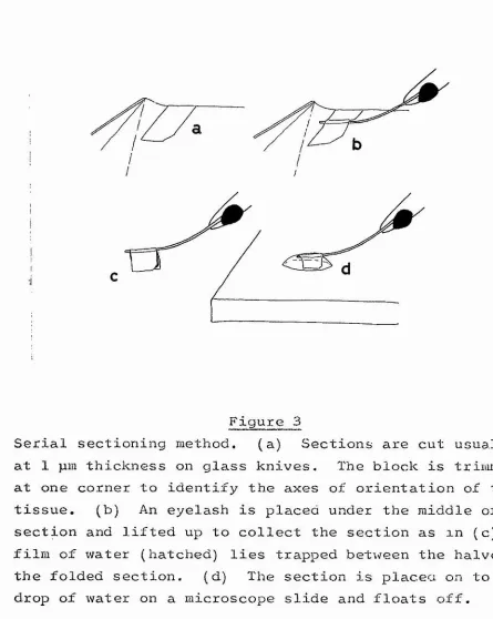
3* Serial sectioning method 62
4, Cc stygia wholemount of cornea 67
5* C. vomitoria plan of retina 68
6. C, vomitoria projection of individual
ommatidia 70
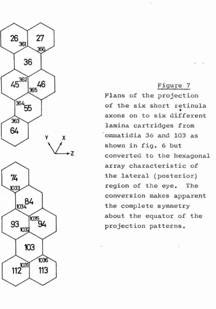
7 » Projection plans converted to an hexagonal
array facing 71
8* vomitoria plan of lamina facing 74
9o. Projection patterns of axons that terminate in
error 76
10* Block diagram of lamina facing 78
11* C* vomitoria micrograph of lamina 78
12* C* stygia HLS of first projection and
lamina facing 79
1 3 o Reconstruction of axon lattice 80
1 4 o C . vomitoria pull-out series of micrographs 81 15* C . vomitoria organization of lamina cartridges 84 16* Co stygia cartridge array at the edge of the
lamina 86
17, 18, C * vicina whole field of lamina-medulla
projection 88, facing 89
19, 20, C, vicina one row of lamina-medulla
X
2 1 o Reconstruction oi chiasmal pathways 97
22* C» vicina tangential process of medulla 98
2 30. Benacus random sections through retina and
optic lobe 102
24, Lethocerus and Notonecta 104
25* Notonecta lamina-medulla projection 105
26. Locusta retina, Feulgen stain 107
27. Schistocerca random sections through retina
and optic lobe 109
28* Schistocerca whole field of retina-lamina
projection 110
29. Schistocerca retina-lamina projection of
individual ommatidia 112
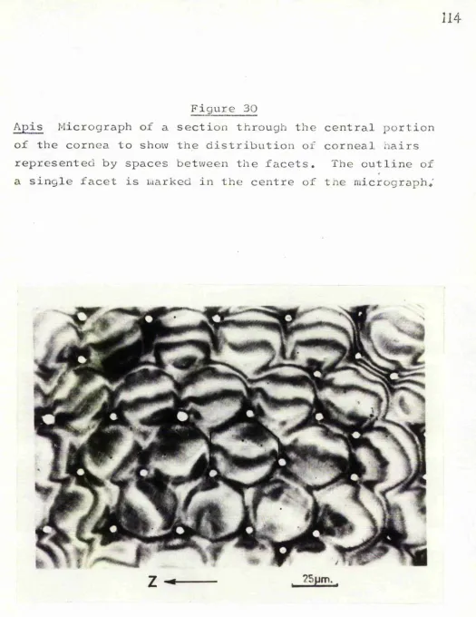
3 0 o Apis section through cornea * 114
31* Apis micrographs of retina 116
32* Apis plan of retina 117
33* Apis plan of lamina 118
.34* Apis micrograph of lamina 120
35* Apis retina-lamina projection of individual
ommatidia 121
36* Apis whole field of lamina cartridges 123
37* Apis the axons of individual lamina cartridges 125
38* Diagrams of the axons of a lamina cartridge 126 39* Reconstruction of the first projection and
lamina 128
40, 41 Apis whole field of lamina-medulla
projection 129, 130
43* Apis the medulla 133 44*. Apis lamina-medulla projection of individual
ommatidia 135
45* Pieris the arrangement of retinula cells 137
46* Pieris electron micrographs of retina 138
4 7 *r Pieris plan of retina 140
48, 49 * Pieris whole field of retina-lamina
projection 142, 143
50* Pieris plan of lamina 145
51* Pieris retina-lamina projection of individual
ommatidia 146
52* Trapezites random electron micrographs of
retina and optic lobe 148
53* Trapezites whole field of retina-lamina
projection 149
54* Trapezites retina-lamina projection of
individual ommatidia 151
55* Trapezites the projection of cartridges
through the lamina 153
56*. Trapezites the lamina-medulla projection 154
57* Trapezites tangential processes of the medulla 156
XXI
INDEX OF TABLES
Table
1. The first and second neural projections of
insect eyes 27
2* The seventeen short retinula axons that
terminate in incorrect cartridges 76
3* The long visual fibres and their cells of
origin 166
4o The short retinula axons and their cells of
SUmiARY
1, The patterns of projection of some of the perpendicular
neurones between the retina and medulla of the optic lobes of various insects have been studied. Axon paths have been studied from consecutive semi-thin plastic sections cut transversely and stained with toluidine blue. The termination positions and the paths of axons are both highly ordered and predictable,
2, In all insects with fused-rhabdomere eyes the axons of one ommatidium project to one cartridge of the lamina and
the array of cartridges duplicates the array of ommatidia. In insects with open-rhabdomere eyes visual information is distributed amongst a number of lamina cartridges so that
each cartridge receives information originating from one visual axis.
3, In both open- and fused-rhabdomere types the cartridge, array of the lamina is exactly duplicated in the medulla but by the intervention of the chiasma is reversed about
4. Most of the retinula axons from one ommatidium terminate in the lamina but usually one pair passes directly to the medulla. These are from the central
retinula cells (open-rhabdomere eyes) or from the small retinula cells (apposition type fused-rhabdomere eyes). Retinal responses are known mainly only for the short
retinula axons so that visual information delivered to the medulla cartridge is still largely unresolved.
5. The lamina neuropile probably contains the elements responsible for the lateral correlation between parallel
receptor inputs which is necessary for movement perception, but units with long lasting responses which could act as
the delay circuit of movement perception are unknown.
6. The occurrence of errors in termination of the first projection of the optic lobe of the fly, which are reported
for the first time in this w o r k , provide no direct clues to the developmental processes by which such a morphologic ally complex system arises. Nevertheless errors may arise within a sequence of growth processes which are fundamentally quite simple and not obvious from knowledge of the
INTRODUCTION
A* The general features of insect retinae and optic lobes The insect visual system consists of the external
compound eyes and the internal optic lobes. The eye consists of the cornea and associated dioptric apparatus which overlie
a layer of photoreceptors. These photoreceptors are collect ively called the retina and they generate visual responses that are processed in the optic lobe. The optic lobe is
composed of three main regions which, from distal to proximal, are the lamina, medulla and lobula. In both Diptera and
Lepidoptera the most central region, the lobula, is sub divided into two regions, the lobula and lobula plate
(Bullock and Horridge, 1965), Each region is composed of a band of neuropile surrounded by the cell bodies of that neuro pile. The retina and lamina are connected by the axons of
the first projection, while the lamina, medulla and lobula are connected in sequence by two chiasmata. The plane of d e
cussation of these is horizontal in the lamina-medulla chiasBia and vertical in the medulla-lobula chiasma. The main burden of this work has been to establish some of the neural
Figure 1
Apis Horizontal longitudinal section of retina and optic lobe to illustrate the general features of the insect compound eye. From the periphery to the centre are seen the retina, with the ommatidia obliquely sectioned,
basement membrane, retinula axon bundles, lamina, horizontal chiasma and medulla. Both lamina and medulla neuropile
Basement membrane
Retinula axon
Lamina monopolar cell body
Upper & middle lamina zones i
[ A
Lower lamina j ^ ^ "
zone 1 z*
'ê
> Retina
> 1st. projection
> Lamina
I L-M Chiasma
Medulla monopolar cell body
primarily in the light of their functional significance,
A concept of general interest that has been emphasized by the work is the high level of predictability and order of
the connections within optic lobe* This emphasis raises some challenging developmental questions concerning the generation of such a precise organization. The classic study of Cajal and Sanchez (1915) did much to reveal the organization of the optic lobe into many pathways in parallel, each highly ordered. These features make the tissue favourable for
tracing connections between neurones. Together with the con temporary trend towards the analysis of neuronal networks these factors have resulted in the renewed impetus with which the morphology of insect optic lobe has been studied in recent years, notably in the work of Trujillo-Cenoz, Braitenberg and Strausfeld on the fly.
An obvious feature of the eye and optic lobe is their organization into perpendicular and tangential components (fig. 1). The retina is composed entirely of a perpendicular array of cylindrical ommatidia while the lamina and medulla are divisible perpendicularly into arrays of cartridges which
i
Figure 2
Diagram illustrating the three main planes of section ( 1 - 3 ) of one quadrant of the insect compound eye. 1 = horizontal (antero-posterior); 2 = vertical (dorso-vent ral); 3 = tangential. Drawn from a photograph of the
retina or neuropile that contains their cell bodies; they convey information centripetally towards a more central neuropile or centrifugally away from it. Tangential cells have processes that spread across the visual field and collect visual information from many perpendicular cells in parallel which is then conveyed perpendicularly. Amacrine cells are
intrinsic to one neuropile only and have both perpendicular and tangential processes but do not convey .information between ganglia.
This work is concerned almost entirely with the per pendicular cells between the retina and medulla analysed from
series of tangential sections.
The three main planes of section are illustrated by Strausfeld and Blest (1970) , and given by them the names in common usage viz* tangential,, vertical (i.e. dor so-vent ral) and horizontal (i.e. antero-posterior) (see fig. 2),
The retina
Light reaches the photoreceptive rhabdome through the dioptric apparatus which is composed of the cornea, overlying the whole eye^ and the cone and ancillary structures of each
ommatidium. The surface of the cornea is an array of facets the arrangement of which varies between species. Commonly
X and y either at 30*^ to the horizontal e.g. Drosophila or at 60^ to the horizontal e.g* bee, locust and white butterfly Pieris. The flies Musca and Calliphora have a corneal array
that has both types of pattern (in the anterior and lateral regions of the eye respectively) separated by a mid-lateral
transitional zone of a rhomboidal array (Braitenberg, 1967,
1970) * The axes x,, y and z are useful because they also define
the array of ommatidia underlying the corneal facets*
Each ommatidium in tangential section has the appearance of a rosette or retinula composed of approximately eight
retinula cells each bearing many microvilli containing visual pigment along the length of its inner margin* Categories
distinguished within the functional morphology of different insect retinae include the arrangement of rhabdomeres and the pattern, arrangement and number of retinula cells within
ommatidia (Table 1),
The relationship between the distal end of the rhabdomeres and the central end of the cone has been the basis for the
longstanding and possibly erroneous distinction between retinae of different insect groups into apposition and
superposition types, first proposed by Exner (1891). Superposition eyes have a refractile crystalline thread
interposed between cone and rhabdome whereas in apposition
eyes the rhabdome abutts onto the cone.
be fused (the rhabdomeres are united into a single axial rod: locust, bee etc.) or open (the rhabdomeres are separated from each other: f l y , v/ater-bug) .
The arrangement of retinula cells within an ommatidium also varies; usually the cells are of two sizes, the larger long retinula cells (often six) and the small basal or central
retinula cells (one or two). The ommatidial complement of retinula cells is eight for f l y , locust,, dragonfly,; water-bug and the backswimraer Notonecta ; and nine for drone b e e , the small white butterfly Pieris and the skipper Trapezites.
The long retinula cells may contribute rhabdomeric microvilli for the entire length of their inner margin or the rhabdome may be divided along its length so that the rhabdomere one cell is in front of that of another along the optical path, in which case the retina is said to be tiered.
The ommatidium is limited proximally by a basement membrane pierced at intervals by bundles of retinula axons which proceed centrally to the lamina.
The first projection and lamina
ID
established, it is the long retinula cells that have short
retinula axons. This situation is known for fly, bee and Pieris and is probably true for water-bug too ; it is perhaps a general principle for all eyes. The existing terminology will however be retained to avoid further confusion.
The ommatidial bundles of retinula axons travel proximally, passing in turn the basement membrane, the fenestration zone containing parallel rows of horizontal tracheae,, the cell
body layer which is composed of columns of cell bodies mainly of lamina monopolar neurones and finally reaching the
neuropile of the external plexiform layer. Commonly retinula axon bundles twist during their passage to the lamina
although the rotational sequence of axons within the bundle remains constant, at least for the short retinula axons. This sequence is the relative positions of the axons estab
lished at the base of the ommatidium*
In the external plexiform layer of the lamina the short retinula axons terminate within the unit structure of the lamina neuropile, the cartridge, where they make synaptic
contact with various lamina neurones. The long retinula axons go straight through the lamina without synaptic contact, at
least in fly and worker bee* The distribution of the retinula axon terminals from one ommatidium amongst the cartridges
of the lamina is of two types : either each terminal is
11
also water-bug) or the terminals end in a single cartridge (as in all fused-rhabdomere eyes so far examined)*
The neurone types of the lamina are known from Golgi and methylene blue studies on various insects (notably Cajal and Sânchez, 1915 on various species, Zawarzin, 1913 on the dragonfly Aes c h n a , Strausfeld and Blest, 1970 on some
Lepidoptera and Strausfeld, 1970 on some Diptera), The
perpendicular neurones comprise monopolar neurones with cell bodies overlying the lamina neuropile and centrifugal neurones with cell bodies to one side of the chiasma* Centrifugal
neurones are usually T-shaped with a termination in the
medulla and a second termination in the lamina of which one type is the basket ending» The term centrifugal, now in common usage,will be used throughout this v/ork. It is used entirely as a morphological description and should not be
taken to imply polarity of conduction* The monopolar neurones which were further classified by Cajal and Sanchez (1915) as
large (having lateral dendrites for a large vertical depth of the lamina) and small (all other forms with lateral dendrites localized to only one or a few positions in the
lamina) have been reclassified by Strausfeld and Blest (1970) into large (those with overlapping fields of lateral dendrites spreading through more than one cartridge) and small (those
12
-they conflict ; in this work both schemes are used after the two original accounts and acknowledgement is made to whichever account is used. Tangential neurones are known from several species, one type with a cell body above the lamina (fly), another with a Cell body at the anterior edge of the lamina (Pieris) , a third with a cell body beneath the medulla
(Aeschna) but the cell bodies of other tangential neurones in these insects are not known nor are they known for other
insects, Amacrine neurones have cell bodies proximal to the lajJiina and peripherally directed processes (fly and Aeschna) .
The organization of the different lamina neurones and in coming retinula axons within the cartridge is variable. The cartridges may be separate (fly, Pieris) or indistinct
(locust, Trapezites, bee, water-bug, and Notonecta) depending largely on::the extent of lateral arborization of the
component dendrites. In the fly most synaptic connections of the lamina occur between inwardly directed dendrites.
The long retinula axons may occupy a central position in the cartridge (locust, bee, P i e r i s , Trapezites and probably
water-bug) or a satellite position to the cartridge (fly)»
The second projection and medulla
Axons from horizontal rows of lamina cartridges enter
the first optic chiasma (which will be called here more simply the chiasma) and become disposed in horizontal strata within
13
becomes inverted about a vertical plane. Axons from near the anterior or posterior margins of the lamina move more
obliquely than those at the centre and the axons are segre gated into alternate horizontal rows » Consequently
tangential sections have a striated appearance with axons cut longitudinally (those undergoing large horizontal
displacements) and axons cut transversely (those making only a small lateral shift) occupying alternate layers. The
uncrossed projection of some lamina neurones upon the medulla has also been depicted by Cajal and sânchez (1915) but
apparently does not apply to the perpendicular neurones with which this work is primarily concerned,
Chiasma axons again group into bundles at the central end of the chiasma, pass through the ganglion cell body layer of the medulla and enter the neuropile of that region. They pass through columns of medulla cell bodies in the medulla ganglion cell layer and each bundle eventually forms a
single cartridge of the medulla. Each cartridge also receives the axons from a number of monopolar neurones from the columns of medulla cell bodies. No medulla neurones have been studied in this present work,
14
behaviour and physiology. The results have been supplemented by studies on other insects with large axons that are easy to trace.
Since the early anatomical work on compound eye some insect species have been preferred,, notably fly, bee and locust while other whole insect groups have been relatively neglected. Bee and fly have attracted most attention
because of their interesting behavioural patterns whilst locust has been particularly favourable for
electrophysio-logical analysis of its optic lobes. Of the less well-studied species the water-bug Lethocerus is potentially interesting because it is a second example of an open-rhabdoraere eye
(but with an apparently more complex organization of the
lamina than that of the fly), Two lepidopterans have also been studied in this work,, members of two specialised
superfamilies the Papilionoidae and Hesperoidae respectively ; they are the white butterfly, Pieris and the skipper Trapezites Though lepidopteran vision has been little studied physio
logically and behaviourally these insects represent an important group with a variety of retinal organizations
including both superposition (e.g. the skipper) and apposition types (e.g, Pieris) . An important recent anatomical account
of pieris and the hawkmoth Sphinx (Strausfeld and Blest, 1970) was the immediate stimulus for the inclusion of Pieris in
15
divided lobulae, an arrangement not found in any other group.
In the next sections are given the anatomical methods of study currently available while in the last two sections
follow for each species firstly, a more detailed account of the present state of knowledge of anatomy of the compound eye and secondly, a brief reviev/ of the physiological and behavioural evidence of compound eye function already available.
C, The methods available for the analysis of connectivity patterns in insect optic lobe
Available methods may be divided into those utilizing light microscopy, those utilizing electron microscopy and those which rely upon a combination of both levels of
resolution. Each method has its own advantages but none is wholly adequate, and only by a correlation between methods will a complete analysis of connectivity be possible* However a problem is that individual workers specializing in different techniques prefer particular animals e.g. flies arc
impregnated well by the Golgi method; diverging pathways are shown well in reduced silver preparations; small animals
have smaller volumes of neuropile to reconstruct by the
laborious methods of electron microscopy; only some animals
16
use of different methods results in diversification of the morphological descriptions as well as consolidation» In general dipteran flies have received most attention and are most favourable for analysis by both anatomists and
physiologists alike.
Light microscopy provides gross details within a large volume of tissue of cell types, axon pathways, termination positions e tc., but the resolution of electron microscopy is needed for identification of synaptic contacts between cells. Electron microscopy however can only be applied to reconstruct small volumes of tissue.
The small size of the optic lobe and its arrangement into many parallel pathways have facilitated the analysis of a single neuronal circuit because the important features are emphasized by their repetition in numerous essentially identical circuits. The favourability of the tissue is
indicated by the definitive studies that have appeared recent ly (Trujillo-Cenoz and Melamed, 1963; Trujillo-Cenoz, 1965; Trujillo-Cenoz and Melamed, 1966; Braitenberg, 1967; Melamed and Trujillo-Cenoz, 1968; Trujillo-Cenoz, 1969; Boschek, 1970; Horridge and Meinertzhagen, 1970 a / b; Strausfeld and Blest, 1970; Strausfeld, 1970; Strausfeld and Braiten berg , 1970; Trujillo-Cenoz, 1970; Varela, 1970),
Light microscopical methods 1. Hodological methods
17
insect optic lobe rest is provided by the so-called
hodological methods (i.e. Golgi impregnation and methylene blue staining), in which the entire extent of only a few
neurones in either whole mounts or thick slabs of tissue are selectively stained. These methods provide information on neuronal profiles, perikarya positions etc. and from them the classification of morphological cell types. Completely Impregnated, the individual processes of the dendritic
arborization of a particular neurone are visible with light microscopy; the lower limit of the diameters of the finest
processes found in the neuropiles of the optic lobes and of many other insect ganglia range between 0.1 and 0.15
(Trujillo-Cenoz, 1959; Trujillo-Cenoz and Melamed, 1962;
Smith and Treherne, 1963; Smith, 1965; Trujillo-Cenoz, 1965; Steiger, 1967; Trujillo-Cenoz and Melamed, 1970), Such
fine processes however are only just visible with the largest numerical aperture objectives, which are those least suitable for photomicrography because of their shallow depth of focus.
Selectivity is the essential quality of these methods which gives them their great clarity. However selectivity also imposes limitations. Two such important limitations are that no reliable information on the relative numbers of differ ent cell types is provided and that little information is
18
are inherent in the methods; the former, that some neurones are consistently left unstained in every preparation, the latter, that the technique may produce artifactual staining either of non-neuronal structures or of part of a neurone only. These limitations have always to be borne in mind when assessing results obtained by these methods (Blest, 1966), though the general agreement between the accounts from
methylene blue preparations by Zawarzin (1913) and from Golgi preparations by Zawarzin (1913) and Cajal and Sanchez (1915)
is good evidence of the relative reliability of both methods. In the case of Golgi impregnations these limitations have
been fully discussed by Strausfeld and Blest (1970) for the
optic lobes of Lepidoptera and Diptera. Strausfeld (1970) and Strausfeld and Blest (1970) found every cell type
previously described by the Spanish authors which is evidence against random artifactual impregnation. In addition they found nevf types not described by Cajal or described only
from partially impregnated preparations. In his work Straus feld used a greater variety of impregnations from a larger number of animals than Cajal and paid particular attention
to the reconstruction of wide-field elements stained in their entirety only infrequently. Lastly, no major disagreement
19
reliable than the Golgi impregnations of Cajal and Strausfeld) Even so it is still not clear to what extent the large number of cell types and subtypes which may be compiled with
relative ease using the Golgi method are a meaningful
classification in terms of the connectivity of the neurones. A recent modification of a reduced silver staining method
(Strausfeld,. personal communication) introduces a degree of selectivity into the impregnation and permits the staining of entire single neurone classes preferentially, thus
allov/ing a degree of correlation between Golgi and reduced silver methods not previously attainable. The results are not yet available.
A third method of great potential value for the
-Ing
selective stain of entire neurones arises with the recent demonstration by Lund and Collett (1968) that various of the
reduced silver techniques for demonstrating neuronal
degeneration in vertebrates work satisfactorily in insect optic lobe. The profile of the entire neurone is visible by light microscopy while the terminal processes of degener ating neurones are visible by electron microscopy of the same section. The method has the limitation that discrete areas
of degeneration have to be induced. Consequently not only has the insect species to be selected for postoperative
20
experimental lesion except for those neurones where the soma is isolated from its neuropile e.g. for neurones v/ith somata in the contralateral optic lobe.
Light microscopical methods 2. Serial sectioning methods
Serial sections examined by light microscopy provide in general the intermediate between Golgi impregnation and electron microscopy; they allow the description of the pro jection of axonal pathways between neuropile layers of neurone types known from Golgi impregnations, but without the final resolution of synaptic contact. These projections are espec ially difficult to study because they consist of relatively long axons (say 100pm -1mm) of fine calibre (say 0.25pm~5jim) . The methods are most useful only when the staining of all
the elements of a projection is possible. Two types of serial sectioning method have been used; the reduced silver staining methods of e.g. Bodian, 1936;, Holmes, 1943 and its modific ations by Blest, 1961; Rowell, 1963; etc, and the following of axon pathways through serial 1 /L"sections of plastic
embedded material stained non-selectively by toluidine blue (Horridge and Meinertzhagen, 1970 a, b ) .
Reduced silver staining methods have proved
particularly successful for the study of anatomical pathways at single axon level in a few favourable cases where the axons move horizontally in a section and are separate from their
21
first projection of the fly optic lobe because the pattern of divergence is the characteristic feature of this system.
By identifying individual retinula axons within the separate ommatidial bundles (from a correlation v/ith their cyclic
order at the point of origin near the basement membrane) their pathways over the top of the lamina could be followed
individually because they were cut longitudinally in that region. Similarly Strausfeld and Braitenberg (1970) were able to use reduced silver staining methods to study the ex tremely fine lateral prolongations from the L4 neurones at the base of the fly lamina after identifying the profile of the L4 axis fibre from its size and position within the cartridge* The specificity of reduced silver staining for neurones makes axons easy to visualixe against a clear
background so that the connections of very fine processes may be studied. However, the confusion resulting from the staining of a tract of axons viewed in a 10pm section is
so great that it is difficult to identify individual axons even of 2pm diameter. Thus reduced silver methods are unsatisfactory for the study of projections where bundles of retinula axons unite into large axon tracts as they do in all insects examined except the f l y .
22
microscopy 1 p m plastic transverse sections cut consecutively. The number of sections is thus not too prohibitive although still very large, whereas the thinness of the section allows near maximal resolution available with light microscopy.
This method is much more laborious than either of the silver methods and has much in common with the methods of serial sectioning electron microscopy though lacking their extreme resolution. The poorer resolution of light microscopy is
of course compensated for by the larger field and most import ant the greater depth that can be studied. In common with
ail the light microscopic methods statements can only be made of neuronal proximity and not of synaptic contact which must be inferred from other electron microscopical studies or from
physiology. Synaptic connections between adjacent neurones can be excluded for many classes of fibres if the fine pro cesses of the neurones are known. These are best described
from silver preparations since they are not so easily seen in toluidine blue stained sections. Although light microscopy
of 1 p m sections allows maximal resolution and eliminates
the confusion resulting from the superimposition of the images of many parallel axons stained in a thick section, the
visibility of individual, fine axonal processes travelling horizontally within a section is much greater in such a thick section either impregnated by the Golgi method or stained
23
reasons; firstly, it is easier to visualize a process
stained as a black thread against a clear ground than as a hollow profile of the same diameter in a thin section,
and secondly, because any one thick section contains a larger portion of the process.
Electron microscopical methods
The remaining methods, those of electron microscopy,
provide the definitive morphological evidence of connectivity and refer usually to connections within a neuropile (as
opposed to the preceding methods which are concerned mainly with connections between neuropiles).
The great increase in resolution available with electron microscopy, which is necessary for the identif ication of synaptic contacts between neurones, results in a very great increase in the limitations and difficulties of this technique compared with those of light microscopy.
Only small volumes of tissue may be sampled because of the thinness of section required so that neuronal processes can rarely be traced from their cell of origin. Consequently cells of origin must be inferred from previous light micro scopical work by the position and orientation of their axons at the region where these enter the neuropile. This has been possible in the case of the ordered projections of perpendicular elements in the fly lamina (Trujillo-Cenoz,
24
(Varela, 1970) and will probably also be possible for the medulla cartridges in some insects. Ultrastructural clues
are also available in some cases by which the cell type of ^ neuronal processes are recognizable without needing to
trace the neurone through long distances, but it is doubtful
if all cell types can be so labelled and impossible to identify each representative of that type uniquely (e.g. between fly long retinula axon terminals 7 and 8 in the medulla),
Similarly the pattern of dendritic arborization can be used to identify a neurone in a reconstruction from serial
electron microscopy by correlation with the same pattern seen by light microscopy from Golgi impregnations. A
recent development (Shelton, Horridge and Meinertzhagen in press) in which thicker sections are cut for viewing with electron microscope accelerating voltages of 200kV may
prove a partial answer to the problem of cell identification since larger volumes of tissue may be sampled and neural
processes followed to their cell of origin, but there is still an upper limit to section thickness, imposed by the confusion
in the micrograph where the collective images of many processes are superimposed. Synaptic contacts are still conspicuous
25
and Sandri (1968) (Lamparter e t ^ a l . , 1969).
In g e n e r a l a r t h r o p o d central synapses have all the
features of chemical junctions in vertebrate nervous tissues, with well defined presynaptic ribbons and two sizes of vesicle
(Trujillo-Cenoz, 1965; Hamori and Horridge, 1966,a;
Steiger, 1967; Smith, 1967 ; Lamparter et al-, 1969 ; etc») although Varela (1970) saw no conspicuous presynaptic ribbons in bee lamina. The occurrence of specialized synaptic
regions between two neurones can, according to current ideas, be taken as evidence of synaptic interaction and has been used, in the fly (Trujillo-Cenoz, 1967; Boschek, 1970) to
infer the polarity of transmission.
Most authors are at pains to qualify conclusions based on such analyses (for the most recent of many reviews see Szentagothai, 1970; Akert and Sandri, 1970), In view of the polarized nature of chemical transmission it seems just ified to accept the asymmetrical organization of
morphological synapses as an indication of polarity until proved wrong more times than is acceptable. One anomaly to
have emerged is the active site specializations identified
between tall epithelial glial cells of the fly lamina and either a photoreceptor terminal or a centrifugal process as the presynaptic element (Boschek, 1970)* This finding
presumably requires a modification of our ideas of the
26
of an active site as a region of rapid chemical trans mission (see Discussion p. 195),
Methods combining electron and light microscopy
A variety of combined methods may be devised in an effort to complement the resolution of electron microscopy with the section size and thickness available with light
microscopy. Only one such method has so far been used (Trujillo-Cenoz and Melamed, 1970) the EM/Golgi combined method (Blackstadt, 1965; Stell, 1967; Hillman, 1969). This method allov/s the examination by electron microscopy of the distribution of the processes of an impregnated
neurone identified by light microscopy. Because the ultra structure of the impregnated processes is not visible,
synapses in which they are the pre-fibre cannot be identified, but by comparison between electron micrographs of Golgi
silhouettes and limited serial electron microscopic
reconstructions, Trujillo-Cenoz and Melamed (1970) were able to study the synaptology of the lamina basket endings of one of the medulla centrifugal neurones of the fly.
Table 1
Retina Type Open (0)
Fused (F) Genera Order Retinula Cell Number
0 Calliphora. Mus ça Lucilia
(Drosophila)
Diptera 8 (6 + 2)
(Dietrich 1909 etc.)
0 Benacus
Lethocerus Hemiptera 8(Walcott, in press)(6 + 2) '
0 Notonecta Hemiptera 8 (6 + 2)
(Horridge, 1968)
F Schistocerca Orthoptera 8 (6 + 2) :
(Horridge and Barnard, 1965 Horridge, 1966) *
F Apis (drone) Hyîiienoptera 9 (6 4- 3)
(Perrelet, 1970)
Cells 1 and 4 larger ,
F Pieris Lepidoptera 9 (4 4- 4 4’ 1 )
Cells 8 and 6 larger (Nowiko f f, 1931)
F Trapezites Lepidoptera 9 (7 4- 2)
F Libellula Odonata 8 (4 4-4)
(Horridge, 1968; for Sympetrium)
8 (5 4- 2 4- 1)
Retinula axon projection
» 2 central cells have long retinula axons to medulla
6 short retinula axons terminate in six different cartridges (Musca, Braitenberg, 1967; Lucilia, Trujillo-Cenoz and Melamed, 1966; Calliphora, Horridge and Meinertzhagen, 1970).
Retinula axons diverge on entry into lamina. Exact pattern of connections unknown.
not known. Long visual fibres seen in Golgi preparations (Horridge, unpublished).
8 axons go to one lamina cartridge,
2 basal cells and one other have, small axons,
axons go to one lamina cartridge. Cells 1-6 have short retinula axons
cells of 7, 8 , 9 have long retinula axons to medulla.
9 axons go to one lamina cartridge. Cells 1-4, 6 and 8 have short retinula axons. Cells 9, 7 and 5 probably have long retinula axons. Strausfeld (1970) incorrectly describes some axons diverging
9 axons go to one lamina cartridge;8 and 9 extremely fine, have central position and possibly are long retinula axons,
8 axons to one lamina cartridge one axon extremely fine. Zawarzin (1913) describes only short
retinula axons for Aeschna.
Kno\-m perpendicular neurons of lamina cartridge ; v{
é
.! leaving :
li
6 short retinula terminals from different ' ommatidia
2 long retinula axons
2 large monopolars 2 small monopolars
2 centrifugal from medulla ^2 long retinula
(Boschek, 1970; Musca)
I
I
f. 7.7' entering ; 8 axons from an unkno^m number of ommatidia
leaving : at least six axons
#
I
'entering‘ leaving : not knownat least six axons including two long retinula axons
8 axons from one ommatidium
unkno^'Ai
leaving : at least six axons of unknoim, identity 7 /entering ;
J/;f emaindkir
7 entering
-, / ' ■'
) leaving :
r6 short retinula terminals U fine axons of cells 7-9
"2 long retinula axons (from cells 7-9) 3 monopolars probably 2 large, 1 small 1 large axon, probably a fourth
monopolar
1 fine axon, probably a centrifugal
i
I
16!entering:
1 leaving :
V entering : / leaving :
{
6 short retinula axons and 3 axons of cells 7-92 long retinula axons probably and at least 4 more including one with a peripheral position in the cartridge
9 retinula axons
at least 6 axons of unknown identity
I
i
(!Lamina-meclulla projection
Àt least eight axons of one cartridge traverse the chiasma, of which six go between a single lamina and a single medulla cartridge. These are; 2 long retinula, monopolars , L2 and L3 and one other probably centrifugal.
hot knoT/n
At least six axons go between a single lamina and a single medulla cartridge.
f---:---ot known
t least seven axons go between a single lamina and single medulla cartridge. These are: 2 long retinula, 3 monopolar and 2 other (one of which probably is centrifugal).
;
Possibly permutation of lateral relations between long retinula and lamina monopolar neurones
(Strausfeld, 1970). Otherwise not known.
{ |:At least six axons go between a single lamina and a single medulla cartridge.
i l l
21
' ' •;
U':'
7 :
:'V ,
I
..
6:61
# 1
m '•/« >?/
;
I;
r
m./'
1/
28
D* The retina and optic lobes of the insects studied A summary is given of the current state of knowledge
about the arrangement and organization of the retinae and optic lobes of the insect species that have been studied in this work* The account is condensed in table 1* Information is original where no reference is given.
The fly. 1. The retina of the fly
The ommatidium of the fly retina has an open-rhabdomere arrangement of eight retinula cells. Photoreceptive
rhabdoraeres are borme on the inner margin of six separate retinula cells (1-6), while the seventh rhabdome is composed of two rhabdomeres, those of the pair of central cells 7 and 8 (Dietrich, 1909; Trujillo-Cen6z, 1968). In transverse
section the seven structures form a characteristic radially asymmetrical pattern best seen just beneath the dioptric apparatus. This pattern, first described by Dietrich (1909), is seen in a wide variety of dipterans of the sub-order
Brachycera (the pattern in various nematoceran dipterans on
the other hand is symmetrical). The retinal array of these rhabdomere patterns has a sharp discontinuity about a
horizontal equator so that the pattern of an ommatidium of the ventral half is the mirror image of that in the dorsal (Dietrich, 1909). The retinula cells which were numbered
1-8 by Dietrich have opposite rotational sequences in the
29
cells 7 and 8 in the ring of photoreceptor cells is as follows:,
cell 7 (the superior central cell of Melamed and Trujillo-Cenoz ; 1968) is situated between cells 1 and 6 and contributes
the distal portion of the central rhabdome; cell 8 (the inferior central cell)is situated between cells 1 and 2 and contributes only 75 pm of the central rhabdome. The rhabdomere of cell 8 has microvilli arranged orthogonally to those of
the superior rhabdomere overlying it and throughout the basal 75 pm in which it is situated, cell 7 is represented by an axon (Trujillo-Cenoz and Melamed, 1966; Melamed and Trujillo-Cenoz, 1968).
The optical consequence of the distribution of the intraomraatidial rhabdomeres at the bottom of the pseudocone
is that each rhabdomere accepts light from a different
portion of the visual field to its intraomraatidial neighbours (Autrum and Wiedemann, 1962; Kuiper, 1962;. Wiedemann, 1965) the exact portion being determined by the lateral position
of the rhabdomere (Kirschfeld, 1967). The spatial relation ships between the rhabdomere optical axes within ommatidia and the angles between neighbouring ommatidia is such that seven rhabdoraeres (each of a different cell number, one within each of seven neighbouring ommatidia) have the same optical axis (Kirschfeld, 1967). This pattern is the
30
described the patterns of facet rows of the cornea in terms of two sets of concentric arcs,the centres of which are located in the dorsal and ventral halves of the retina. The patterns of intersection of these arcs can be used to predict, with respect to the external coordinates of the animal, the exact arrangement of the rhabdoraeres of any ommatidium and the exact arrangement within any group of ommatidia of those rhabdoraeres with coincident optical axes. 2, The neurones of the fly lamina
In this work results from various genera of brachyceran flies will be used interchangeably except where specific di f ferences are known to exist* Cajal and Sanchez (1915) used
Calliphora vomitoria and the horse-fly Tab anus bovinus
Kirschfeld, Braitenberg, Boschek and the Tubingen group use the house-fly Musca domestica; Trujillo-Cenoz uses blowflies mainly Lucilia spp, but also Calliphora. and the flesh fly Sarcophaga; Strausfeld (1970) used Calliphora vomitoria and C. vicina (= erythrocephala) , and the hover flies Eristalis tenax, Syrphus elegans and S, nitidicollis while Horridge and Meinertzhagen (1970 a) used Calliphora vomitoria.
Six short retinula axons from six different ommatidia converge upon a single cartridge of the lamina (Trujillo-Cenoz and Melamed, 1966; Braitenberg, 1967; Horridge and Meinertzhagen, 1970 a) where they have simple cylindrical
31
They encompass and make synaptic connection with the axons of two monopolar neurones (Trujillo-Cenoz and Melamed, 1963), which were labelled LI and L2 by Braitenberg (1967) but
which are called g (=L1) and G (=L2) in this account. These two neurones are uniquely identifiable by subtle differences in their position and morpho3-ogy. Two additional smaller monopolar neurones L3 and L4 with cell bodies situated
immediately above those of LI and L2 have axons that proceed as a pair down the posterior-equatorial quadrant of the
periphery of the cartridge, usually between retinula terminals 5 and 6 (Braitenberg, 1967; Boschek, 1970)» The axon of
L3 is larger than that of L4 and receives synaptic input from at least one retinula terminal, but the axon of a fifth monopolar neurone described by Trujillo-Cenoz and Melamed (1970) for Lucilia (and also for Calliphora and Sarcophaga) is not
known. A variety of types of monopolar neurones is known
from Golgi impregnations ( Cajal and Sanchez, 1915;, Strausf e l d ,
1970) but in Calliphora the lateral extents of their dendrites are never more than one cartridge width (Strausfeld, 1970),
The correlation between the Golgi silhouettes of Strausfeld (1970) and the classification derived from reduced silver
methods (Braitenberg, 19 67 ; Strausfeld and Braitenberg, 1970)
based on cell body position and axonal location within the cartridge, is not yet available (see Discussion, p.179 ).
3Z
neighbouring cartridges (Strausfeld and Braitenberg, 1970),
and probably these connections are presynaptic to either or both through an unidentified fibre fragment (Boschek, 1970). Both L3 and L4 receive synaptic inputs in Musca from the two members (a and p) of the remaining class of perpendicular
neurone, the centrifugal neurones of the medulla (Boschek, 1970). Three types of centrifugal neurones are known from Golgi impregnations of several species (Cajal and Sanchez, 1915; Strausfeld, 1970)* One of these has a basket ending
which surrounds a single lamina cartridge and in L u cilia,
Calliphora and S a rcophaga the processes of this ending are said to contain synaptic ribbons at sites presynaptic to retinula terminals (Trujillo-Cenoz, 1965; Trujillo-Cenoz and Melamed, 1970)* In contrast, in Musca it is the two centrifugal neurones which are said to be postsynaptic to the retinula axon terminals (Boschek,, 1970). In addition,,
The two centrifugal neurones make synaptic contacts with them selves and v/ith additional unidentified fibre fragments
(Boschek, 1970) but the lamina ending of the second (or
possibly third) centrifugal neurone(s) is not yet identifiable with types known from Golgi impregnations, nor is it known
if the same types are found in all lamina cartridges of one species. The remaining known synaptic connections, in
33
between the cartridges throughout the lamina. These cells are post synaptic to the short retinula cells and to one of
the centrifugal neurones of the cartridge (Boschek, 1970) but have not been described as presynaptic to any element.
Identification of synaptic polarity in all cases is based upon the location of synaptic ribbons in presumed presynaptic neurones, with the exception of the main retinula terminal-monopolar synapses which are known from electrophysiological
records of the responses of dye-injected postsynaptic cells (Autrum, Zettler and Jarvilehto, 1970; loannides, personal communication). The long retinula axons are unique among the perpendicular neurones in having no synaptic contacts in the lamina (Trujillo-Cenoz, 1965; Boschek, 1970).
In addition to the perpendicular neurones of the lamina, representatives of tangential and amacrine cells are also
known to occur in Golgi impregnated preparations. Three types of tangential neurone with aperiodic distributions are
described by Strausfeld (1970), one type with a large cell
body beneath the lamina; of the remaining pair one (Lam :tan2) has not been seen in Calliphora. Amacrine cells of one type only are known and are described both by Cajal and Sanchez
(1915) and Strausfeld (1970); they have a periodic distribut ion with a small cell body immediately beneath each cartridge. Their synaptic connections are not known but their lateral
34
compass of the immediately adjacent cartridges. Absence of further information is an important omission in our knowledge of the functional connections of the cartridge. 3. The 1amina-medul1a projection of fly
Each lamina cartridge is represented by a bundle of axons
in the chiasma that contains at least eight axons visible by electron microscopy (Boschek, 1970). These are: two large
monopolar LI and L2, two small monopolar L3 and L 4 , two long retinula and two centrifugal. Five of these were seen and traced through the medulla by Horridge and Meinertzhagen (1970 a, b) using light microscopical methods. The final number of axons is probably larger than eight since Trujillo-Cenoz and Melamed (1970) report a fifth monopolar neurone,
and Strausfeld (personal communication) has counted at least ten and up to twelve axons in each chiasma bundle, although at least one of the twelve is of an aperiodic tangential neurone.
Some (and probably all) axons of these bundles project homotopically upon the medulla after crossing in the chiasma
(Strausfeld, 1970; Horridge and Meinertzhagen, 1970 a , b ) ,
In the medulla, axons from the chiasma form cartridges but the number of cartridges is not clear * Thus, Strausfeld
(1970) describes from reduced silver stained sections two