JOURNAL OFVIROLOGY,Apr. 1993,p. 2396-2401 0022-538X/93/042396-06$02.00/0
Copyright © 1993,American Society forMicrobiology
Truncation of the
Carboxy-Terminal 28 Amino Acids of
Glycoprotein
B
Specified by Herpes Simplex Virus Type
1
Mutant
ambl511-7 Causes Extensive Cell Fusiont
ABOLGHASEMBAGHIAN, LI HUANG, SUSAN NEWMAN, SUKHANYA JAYACHANDRA,
ANDKONSTANTIN G. KOUSOULAS*
Department of VeterinaryMicrobiology and Parasitology, School of Veterinary Medicine, Louisiana StateUniversity, BatonRouge, Louisiana 70803-8416
Received 3November 1992/Accepted 12 January 1993
Three amber mutations were introduced proximal to the syn3 locus of the herpes simplex virus type 1 glycoprotein B (gB)genespecifying gB derivatives lacking the carboxy-terminal28, 49,or64 aminoacids. A
complementation system that utilized gBs expressed in COS cells to complement gB-null virus KAT was
established. The 49- or 64-amino-acid-truncated gBs failed to complement gB-null virus KAT, while the 28-amino-acid-truncated gB complemented KAT efficiently. Mutant herpes simplex virus type 1 KOS (amblS11-7) specifying the 28-amino-acid-truncated gB fused Vero cells extensively.
Herpes simplex virustype 1(HSV-1) glycoprotein B (gB) is virallyencoded and embedded in the viral envelope and in cell membranes of infected cells. The gB gene was
se-quenced,andtheprimaryandsecondarystructuresof thegB protein were predicted (3, 17). gB is essential for the
productionofinfectious virusparticles (6, 16). Analyses of several mutant viruses showed that gB is essential in the penetration of virions into host cells and virus-induced membranefusion ofadjacent cells (3, 6, 9, 13, 15, 16, 21-23). A mutation ingB causing extensive syncytium(syn) forma-tion wasmapped to thecarboxy-terminal portion ofgB (2, 13)whichwaspredictedtolie inthecytoplasm (8, 17). This mutation (syn3) is most probably an Arg-to-His change at amino acid position857 in the publishedHSV-1(KOS)
pro-tein sequence (2), which is amino acid position 828 in the
HSV-1(KOS)andHSV-1(F) matureproteins aftercleavage of thesignal peptide (17). This amino acidchangeis located near the carboxy terminus of a predicted 31-amino-acid a-helical peptide segment (Fig. 1B). A second site in the cytoplasmic portion of gB that when altered causes cell fusion was identified between residues 816 and 817 in the HSV-1(KOS) protein (4),whichareresidues 786(glutamine) and 787(leucine)intheHSV-1(KOS)and HSV-1(F)mature proteins (17). An elegant complementation system which utilizesgB-nullviruses andtransfectedgBgeneswasusedto
map functional domains ofgB (4-6). The carboxy-terminal 41-amino-acid segment of HSV-1 gB was shown to be nonessential for production of infectious virus particles, glycoprotein maturation, andvirus-induced cell fusion (11). Wereporthere thedevelopment ofasystemwhich utilizes simian virus 40-based expression vectors to complement gB-nullvirusesandtocreaterecombinant viruses specifying altered gBs. We used this system to delineate carboxy-terminaldomains ofgB involvedinthe production of infec-tious virus andcell fusion.
Construction of gB genes that specify carboxy-terminal truncations. The syn3 mutation is located at the end of a
* Correspondingauthor.
tThis is publication GL 112 ofthe Department ofVeterinary Microbiology and Parasitology GeneProbes and Expression Sys-temsLaboratory"GeneLab."
predicted 31-amino-acid a helix(Fig. 1B). To examine the
roleof thispredicteda-helical domain ofgBin virus-induced cell fusion and virus growth, we constructed three amber
mutationsingBgenes.PlasmidpRB2080containsa3,353-bp
HSV-1(F)DNA including the entire codingregionofgB,150 bp from the 5'-flanking sequence, and 493 bp from the 3'-flanking sequence (13). A SalI-BamHI DNA fragment
frompRB2080containing thegBsequencewascloned intoa
pUC19 plasmid (24)thatwaspreviouslymodifiedto carrya
BglII site within its polylinker, creating plasmid p2080. Subsequently, gB was excised from p2080 with BglII and BamHI and cloned into theBglIIsite ofP90123, placingthe 5' end of the gB gene proximal to the adenovirus late promoterin P90123 (26)asexplainedforasimilarconstruct previously (18).Plasmidconstructsp1511, p1528,andp1513
were constructed by insertion of the synthetic oligonucle-otidelinkerSpeI* (CTAGACTAGTCTAG)that carries stop codons in all three reading frames and the recognition
sequencefor restrictionenzymeSpeIintouniquerestriction
sites BstEII (p1511), NcoI (p1528), and NheI (p1513) of p2080. Plasmid p1537wascreated byan in-frame fusion of clones pRB2024 and pRB2107 (13) resulting in an internal truncationfrom amino acids 130 to697. These modifiedgB
genes were then cloned into P90123 to produce plasmids
P9-1511, P9-1528, P9-1513, and P9-1537as explainedabove forp2080.The DNAsequence ofeachconstructcontaining the site of the SpeI* linker insertion was determined by polymerase chain reaction(PCR)-assistedDNAsequencing
as described previously (12). Briefly, a 297-bp DNA
frag-ment ofgB was amplified by PCR using primers gBl
(5'-CTAACCACCAAGGAGCTCAAGAA-3') and gB2 (5'-AG GTCGTCCTCGTCGGCGTCA-3'). PCRs were performed with2.5 U of TaqDNA polymerase (GIBCO-BRL, Gaith-ersburg, Md.)in 100-,ul reaction mixtures containing 0.01% bovineserum albumin and 0.1% Tween 20 in 20 mM Tris-HCl (pH 8.3), 25 mM KCl, 2 mM MgCl2, 200 ,uM (each) deoxynucleoside triphosphates, and eachprimer at0.2 ,uM. Thesampleswereoverlaid with mineraloil andsubjectedto
22cyclesof 2sat98°C, 1 minat66°C,and 2minat72°Cin aprogrammableDNAthermalcycler(Eppendorf,Fremont, Calif.). Final extension was at 72°C for 5 min. Single-stranded DNA of the amplified gB DNA fragment was
2396
Vol. 67, No. 4
on November 9, 2019 by guest
http://jvi.asm.org/
VOL. 67,1993
Nhe I 804
804Asp Phe Asp Glu Ala Lys Leu Ala Glu Ala Arg Glu Met Me Arg
HSV-1(F)gB GAC
1fT
GAC GAG GCC AAG CTA GCC GAG GCC CGG GAG ATG ATA CGGCTA GAC
TAG
TCT AG Leu Asp STOPNcoI
Tyr Met Ala Leu Val Ser Ala Met Glu Arg Thr Glu His Lys Ala TACATGGCC CTG GTG TCT GCC ATG GAG CGC ACG GAA CAC AAG GCC
P9 1528 _ ATC TAG P9-1528
Ilie
STOPBstEII
Lys Lys Lys Gly Thr Ser Ala Leu Leu Ser Ala Lys Val Thr Asp AAGAAG AAG GGC ACG AGC GCG CTG CTC AGC GCC AAG GTC ACC GAC
TAG ACT P9-1511 and amb1511
-_.-
STOPCARBOXY-TERMINUS
OF gB
K
N
1513
1528(SpeI*tNheI) (SpeI* atNcoI)
)
/
/
~~~~~~~Syn
3
A L R R M I S m ambBl
RRVT K ~~~~~~ (SpeI*
atBstE
II)H17a
5i
B
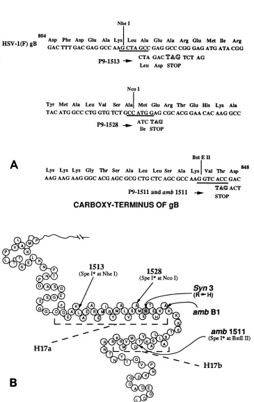
FIG. 1. (A)DNAandpredicted aminoacid sequences ofaportionoftheHSV-1(F) gBgene,ambl511-7,andplasmidsP9-1511,P9-1513, and P9-1528from amino acids804to848(17). Thecorrespondingportion of thepublishedHSV-1(KOS) gBsequenceis from amino acids 834 to878(3). The SpeI* insertionsinamb1511-7, P9-1511,P9-1513,andP9-1528areshown underneaththegBsequence, and thein-framestop codonsareindicated. (B)Aschematicrepresentationof thepredictedsecondarystructureofthecytoplasmicportionofglycoproteinB is shownasdescribedpreviously(17).ThegBtruncations inamblandambl511-7mutantviruses, P9-1513,P9-1528,and thesyn3mutationare indicatedbyarrows.The relevantpredictedahelicesaredenotedasH17aandH17b andmarkedbydashed lines.
NOTES 2397
on November 9, 2019 by guest
http://jvi.asm.org/
[image:2.612.129.494.72.650.2]2398 NOTES
1
2
-3
4
5.
94k-
67k-43k*
...,,,.,,..,.j.s,,U,.,,,,^,i...i's;
...;*-;..-
t."'i:'','','?*,;
,.,, .. ,,., . '. .,,s
.: ':
.. '.
*:
?...;...:
... .:30k-
::
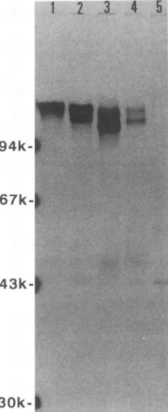
FIG. 2. COS cellsweretransfected withplasmids P9-2080, P9-1511, P9-1528, P9-1513, and P9-1537 and labeled with 35S-methi-onine from 40 to 46 h posttransfection. COS cell lysates were immunoprecipitatedwith anti-gBMAbHSB1.Immunoprecipitates wereelectrophoresedinanSDS-10%polyacrylamide geland visu-alizedby autoradiography.Lanes: 1,P9-2080gB; 2,P9-1511gB; 3, P9-1528gB; 4,P9-1513gB; 5,P9-1537gB.Molecularmassstandards werephosphorylaseb (94 kDa), bovine serum albumin (67 kDa), ovalbumin (43 kDa), and carbonic anhydrase(30 kDa). The posi-tionsof the molecularmassstandardsweremarked with radioactive inkafter thegelwasstained with Coomassie blue.
produced byPCRusing 10 ,ulof theamplified gBDNA and primer gB2 at0.2 ,uM. PCR parameterswere25 cyclesof 3 s at 98°C, 1 min at 66°C, and 1.5 min at72°C with a final incubation at72°Cfor5min.The best resultswereobtained
by using5to10pmolofprimer gBlperreaction fordideoxy DNAsequencingreactions (20).
ThegBDNAsequence specifyingthe45-amino-acid
[image:3.612.347.529.78.233.2]pep-tide segment from amino acids 804to 848of the HSV-1(F) proteinthatcontains theSpeI* linkerinsertionsisshownin
Fig. 1A.The predicted secondarystructureof the carboxy-terminalportionofgB showingthe locations of the trunca-tions specified by P9-1511, P9-1513, andP9-1528 aswell as thelocations ofother relevant mutations is shown inFig. 1B. P9-1511, P9-1513, and P9-1528 are predicted to specify carboxy-terminal truncations of28,49, and64 amino acids, respectively. In P9-1511, the insertion of the SpeI* linker produced an in-frame stop codon. Insertion of the SpeI* linker in P9-1513 added two amino acids (Leu and Asp) before encountering a stop codon. Similarly, an additional Ilewasaddedin P9-1528(Fig. 1A).
Expression of gB genes in COS cells. COS cells were transfected with gBgene constructs cloned into P90123 by the calcium phosphate method (7, 10). Transfected cells
298-FIG. 3. Photograph of an ethidium bromide-stained agarose gel in whicheachPCRDNAfragment obtained with primersgBland gB2wasdigestedwithSpeI andelectrophoretically separated.Lane 1, kphageDNA 1-kbpladder(BRL); lanes 2, 3,4, 5, and 6, PCR DNAfragments restrictedwithSpeIfromKOSandambl511-4, -7, -9, and-19viralDNAs,respectively.
werelabeled with
35S-methionine
from 40 to 46 h posttrans-fection. Proteinswereextracted from transfected cells with 1% Nonidet P-40 and 0.5% sodium deoxycholate in extrac-tion buffer (50 mM Tris buffer, 50 mM NaCl, 100 ,ug of phenylmethylsulfonyl fluoride per ml, 1 ,ug ofaprotininper ml, pH7.5)andimmunoprecipitatedasdescribedpreviously (18). Samples wereelectrophoresed insodiumdodecyl sul-fate (SDS)-polyacrylamide gels(14,
19). Monoclonal anti-body (MAb) HSB1 was produced in this laboratory by following standard procedures(1).
HSB1 precipitated gB from cellular extracts ofHSV-1(KOS)-infected
Vero cells and COS cells inamanner similartothat of MAb H126(18,
19). A comparison of the reactivities of MAbs HSB1 and H126 is shown inFig.5. Becauseof theavailabilityoflarger
amounts of MAb HSB1, it was used throughout these experiments. Radioimmunoprecipitates ofgB expressed in COScellsareshown inFig. 2. P9-2080specified gB migrat-ingwith an apparent molecular mass ofapproximately 115 kDa, determined by comparison with the molecular mass
standards described in the legend to Fig. 2. P9-1511, P9-1528, and P9-1513 expressed two forms ofgB. The faster-migrating gB form produced by each gB construct repre-sented mostprobably the high-mannose precursor form of gB,since itcomigratedwithgB radiolabeledduringa30-min labeling pulse (not shown). As expected, gB was not de-tectedin P9-1537-transfected COS cells.
Isolation of virus recombinants. To isolate viruses that specify the truncated gB derivatives, COS cells were cotransfected with intact plasmids 1511, 1513, P9-1528, and P9-2080 (15 ,ug each per
25-cm2
flask) andintact wild-type HSV-1(KOS) viral DNA (1 ,ug per flask). Viral DNAwaspreparedbycentrifugationofinfected-cell lysates in NaI density gradients (25). Progeny viruses were har-vested 72 h postinfection (hpi) and used to infect confluent monolayers of Vero cells. Individual, well-separated viral plaqueswereexaminedby phase-contrast microscopy at 24 to48 hpi.With the exceptionofvirus stocks from cotrans-fections with P9-1511, all other cotransfections produced virus plaques that were indistinguishable from wild-type KOS plaques. Viruses obtained from cotransfections of wild-type KOS viral DNA with P9-1511 reproducibly re-J. VIROL.on November 9, 2019 by guest
http://jvi.asm.org/
[image:3.612.102.223.80.376.2]NOTES 2399
FIG. 4. Phase-contrast photomicrographs (magnification, x125) of KOS (A), tsB5 (B) and amblS11-7 (C) virus-infected Vero cells observedat24hpi.
VOL. 67,1993
on November 9, 2019 by guest
http://jvi.asm.org/
[image:4.612.148.477.83.697.2]2400 NOTES
sulted in approximately 5% syncytial plaques inVero cells butnotin HEp-2 cells (datanot shown).
Several syncytial plaques from cotransfections of intact KOS viral DNA and P9-1511 were isolated and plaque
purified three times. Viral DNAs from each virus plaque weresubjectedtoPCR analysis using primersgBl and gB2, and PCR productsweredigested with restriction
endonucle-aseSpeL. Theresultant DNA fragmentswerevisualizedafter
electrophoresis in a 1.5%agarosegel followed by ethidium bromide staining (Fig. 3). Digestion of the 297-bp DNA fragments obtained from syncytial amb4, amb7, amb9, and ambl9mutants withSpeI resulted infaster-migrating DNA species, indicating that these DNAfragments contained an
SpeI site. In contrast, digestion with SpeI of the 297-bp fragment derived from KOS viral DNA did not result in faster-migrating DNA species (Fig. 3). The presence of the
SpeI* linker was confirmed by DNA sequencing of the PCR-amplified 297-bp DNA fragments. No other mutation wasfound within thePCR-amplified 297-bpDNAfragments. AgB DNAsegmentcontaining the SpeI* linkerfrommutant
ambl511-7 virus is shown in Fig. 1A. Additional DNA sequencing of approximately the terminal one-third of the entire gB gene of the ambl511-7 virus revealed no other
mutations.
Cell fusion caused by ambl511-7.Confluent monolayers of Vero cellswereinfectedwiththeKOS, tsB5, andambl511-7 strainsatamultiplicity of infection of 5, and the cell cultures were examined by phase-contrast microscopy and
photo-graphed at 24 hpi. The ambl511-7 strain fused Vero cells extensively (Fig. 4) but failed to fuse HEp-2 cells (not shown).
Synthesis ofgB specified by mutantamblS11-7. Vero cell monolayers were infected with HSV-1(KOS), the tsB5
strain, ortheambl511-7 strainat amultiplicityof infection of5and labeled with 50,Ciof35S-methionine perml from6 to 24hpi. Infected cellswere harvested byscrapingwith a rubberpolicemanandwerewashedtwotimesin phosphate-buffered saline. Immunoprecipitates from KOS, tsB5, and amblS11-7 strain-infected cellularextractswith MAb HSB1 or H126 revealed the presence of gB migrating with an
apparentmolecularmassofapproximately115 kDa(Fig. 5). Inaddition, twogB-related peptidesof 49 and 44 kDawere
precipitated. Cell surface expression of gB was tested by immunofluorescence assay and fluorescence-activated cell sorting analysis. amblS11-7 strain-infected cells expressed gBthatwastransportedtothe cell surface inamountssimilar to theamount ofgBsynthesized inKOS-infected cells (not shown).
ComplementationofgB-nullvirusKAT byCOScells trans-fected withgBgenes.gB-nullvirus KATreplicatesin D6cells but not Vero cells. To ascertain whether gB constructs cloned into expressionvector P90123 under the control of the adenovirus latepromoterwereabletocomplement KAT, COS cellsweretransfectedwiththegBconstructs and at 16 h posttransfection were infected with gB-null virus KAT. Infected cellswerecollectedat 24hpi,and viral stockswere
preparedand their titersweredeterminedonboth Vero and D6 cells. The results of these experiments are shown in
Table 1.IncorporationofafunctionalgBmolecule(provided by the transfected gB gene) into the KAT virion envelope resulted in infectious virus particles that successfully
in-fected D6 and Vero cells (complementation). Viral plaques formed on D6 cells because these cells provided gB for
subsequent rounds of infectious virus particle production requiredforplaque formation. Viruses that formedplaques
on Vero cells probably represent KAT rescued virus in
M
1
2
3
94k--67k-e
4
5
6
*MO--W Vag
4-,
A-, vo
43k-
__
[image:5.612.355.514.79.354.2]30k-b
FIG. 5. Verocells were infected with KOS, tsB5, andambl511-7
virus strains andlabeled with35S-methioninefrom 6 to 24 hpi. Cell lysates were reacted with anti-gB MAb HSB1 or H126, and the precipitates were electrophoretically separated in an SDS-10%
polyacrylamide gel. Lanes: M, molecular mass standards as de-scribedin thelegendtoFig.2; 1and2,HSV-1(KOS);3and 4, tsB5 virus; 5 and 6, amb1511-7 virus. Odd-numbered lanes, reactions withMAbH126;even-numberedlanes, reactions with MAb HSB1.
which the truncation in the gB gene of KAT virus was repaired through recombination with the transfected gB gene. P9-1537 served as the negative control for these experiments. Thisplasmid carriesalarge deletion in the gB gene andcannotcomplementorrescuethe KATvirus. Virus stocks prepared from COS cells transfected with P9-1537 andsuperinfectedwith virus KATdidnotproduceanyviral plaques on either D6 or Vero cells. Virus stocks from cultures transfected with P9-2080 and later superinfected with virus KATproduced5.2x
103
virusplaques on D6 cells and4 plaquesonVerocells,indicating
thatgBspecified
by P9-2080 complemented virus KAT. The small number of viral plaquesproduced on Verocells probablyrepresented KATrescued viruses. Plasmid P9-1511 complemented KAT virusasefficientlyasP9-2080. In contrast,plasmids P9-1513,TABLE 1. Complementation ofHSV-1(KAT) byCOS cells transfected withgBgenes
Transfected Growth(PFU/ml)ain:
plasmid D6cells Vero cells
P9-2080 5.2 x 10³ 4
P9-1511 2.4x 10³ 8(6syn+ + 2syn)
aCOScellsweretransfectedwithgBgenes andinfectedwithmutantKAT
16hlater.Virus stockswerepreparedat24hpi.Therewas nogrowthin either
D6 or Verocells transfected withP9-1513,P9-1528,orP9-1537.
J.VIROL.
on November 9, 2019 by guest
http://jvi.asm.org/
NOTES 2401 P9-1528, and P9-1537 failed to complement KAT (Table 1).
Virus stocks from P9-1511 transfections produced syn and syn+ wild-type virus plaques on Vero cells. These viral plaques were collected, and viral DNA was extracted and tested for the presenceof an SpeI* linker by restriction with SpeI and DNA sequencing as explained earlier. Viral DNA from syn viral isolates contained an SpeI* linker site within the PCR-amplified gB DNA fragments. Viral DNAs from syn+ viruses did not contain SpeI* linkers.
Conclusions. Transient expression vectors such as P90123 used inthis study replicate to high numbers in COS cells, allowing the expression and characterization of modified gB proteins in the absence of any other viral proteins. We show here for the first time that modified gB genes cloned into P90123can be used to efficientlycomplement gB-null virus KAT and toisolate recombinant viruses.
It was reported that mutant ambl virus specifying a 41-amino-acid-truncated gB derivative replicated normally in Vero cells, yielding titers similar to those yielded by the wild-typevirusstrain HSV-1(KOS) (11). Similarly, we found that the truncation of 28 amino acids of gB specified by mutant amblS11-7 did notaffect theability of this virus to replicate in cell culture. The 41-amino-acid gB truncation specified bythe ambl virus leaves the a-helical domain H17a intact (Fig. 1B). We found that truncated gBderivatives of 49 and 64 amino acids failed to complement gB-null virus KAT, and recombinant virusesspecifyingthese truncations couldnotbe isolated. These truncationsdisrupttheax-helical domain H17a (Fig. 1B). Therefore, domain H17a may be importantfor virus replication.
The ambl virus did not fuse Vero cells, although it produced smaller plaques (11). We show here that the 28-amino-acid gB truncationspecified by amblS11-7 caused extensive cell fusion. This truncation eliminates half of a
predicted14-amino-acid at helix(H17b)locatedimmediately after the31-amino-acidahelixH17a(Fig. 1B)which may be directly involved in the production of cell fusion. Alterna-tively,wefavorthehypothesisthat the28-amino-acid trun-cation induces certainconformational changes in the cyto-plasmic portionofgBthatresultin cell fusion. Thisimplies that the conformation of the
cytoplasmic portion
ofgB
may beimportantinvirus-induced cell fusion.This researchwassupported bygrantA127886from theNational InstituteofAllergy and InfectiousDiseasestoK.G.K.
REFERENCES
1. Baghian,A.,L.Shaffer,andJ. Storz. 1990.Antibodyresponse to epitopes of chlamydial major outer membrane proteins on
infectiouselementary bodies and ofthe reducedpolyacrylamide gelelectrophoresis-separatedform. Infect.Immun.58:1379-1383. 2. Bzik, D. J., B. A. Fox, N. DeLuca, and S. Person. 1984. Nucleotide sequence ofa region ofthe herpes simplexvirus type-1 gBglycoprotein gene: mutations affecting rateof virus entryandcellfusion.Virology137:185-190.
3. Bzik, D. J., B. A. Fox, N. DeLuca, and S. Person. 1984. Nucleotide sequencespecifying the glycoproteingene, gB, of herpessimplexvirus type 1.Virology133:301-314.
4. Cai, W.,B.Gu,and S.Person.1988. Role ofglycoproteinB of herpes simplexvirus type 1 in viral entry and cell fusion. J. Virol. 62:2596-2604.
5. Cai, W., S. Person,C. DebRoy, and B.Gu. 1988. Functional regionsandstructuralfeaturesof thegBglycoproteinofherpes
simplexvirus type 1:ananalysisof linkerinsertionmutants.J. Mol. Biol.201:575-588.
6. Cai, W.,S.Person,S. C.Warner,J.Zhou,and N. A. DeLuca. 1987. Linker-insertion nonsense and restriction-site deletion mutations ofthegBglycoproteingeneofherpes simplexvirus
type 1. J.Virol. 61:714-721.
7. Chen, C.,and H.Okayama. 1987. High-efficiency transforma-tion of mammalian cells by plasmid DNA. Mol. Cell. Biol. 7:2745-2752.
8. Claesson-Welsh, L., and P. G. Spear. 1987. Amino-terminal sequence,synthesis, and membrane insertionofglycoproteinB ofherpes simplexvirus type 1. J.Virol.61:1-7.
9. DeLuca, N. A., S. Person, D. J. Bzik, and W. Snipes. 1984. Genome locations of temperature-sensitive mutants in glyco-protein gB of herpes simplex virustype1.Virology137:382-389. 10. Graham,F.L.,and A.J.Van der Eb.1973.Anewtecnnique for
theassayofinfectivityofhumanadenovirus 5 DNA. Virology 52:456-467.
11. Huff, V.,W. Cai, J. C. Glorioso, and M. Levine. 1988. The carboxy-terminal41amino acids ofherpes simplexvirus type 1 glycoprotein B are not essential for production of infectious virusparticles.J.Virol. 62:4403-4406.
12. Kaltenboeck, B., J.W.Spatafora,X.M.Zhang,K.G. Kousou-las, M. Blackwell, andJ. Storz. 1992. Efficient production of single-stranded DNAas long as 2kb for sequencing of PCR-amplifiedDNA.BioTechniques12:164-171.
13. Kousoulas,K. G., P. E. Pellett, L. Pereira, and B. Roizman. 1984. Mutationsaffectingconformationorsequenceof neutral-izing epitopesidentifiedby reactivityof viableplaquessegregate from syn and ts domains of HSV-1 (F) gB gene. Virology 135:379-394.
14. Laemmli,U. K.1970.Cleavageof structuralproteins duringthe assemblyof the head ofbacteriophage T4. Nature (London) 227:680-685.
15. Little, S. P., J. T. Jofre, R.J. Courtney, and P. A. Schaffer. 1981. Avirion-associatedglycoproteinessentialfor infectivity ofherpes simplexvirus type 1.Virology115:149-160. 16. Manservigi, R., P.G. Spear,andA. Buchan. 1977.Cellfusion
inducedby herpessimplexvirusispromotedandsuppressed by differentviralglycoproteins.Proc.Natl. Acad. Sci. USA 74:3913-3917.
17. Pellett, P. E., K. G. Kousoulas, L. Pereira, and B. Roizman. 1985.Anatomy of theherpes simplexvirus1strainF glycopro-tein B gene:primarysequenceandpredictedproteinstructure
ofthe wild type andof monoclonalantibody-resistantmutants.
J. Virol. 53:243-253.
18. Pereira, L., M.Ali, K.G. Kousoulas, B. Huo, and T. Banks. 1989. Domain structure of herpes simplex 1 glycoprotein B: neutralizing epitopesmapinregionsof continuous and discon-tinuousresidues.Virology172:11-24.
19. Pereira, L.,T.Klassen,andJ.R.Baringer.1980.Type-common andtype-specificmonoclonal antibodiestoherpessimplexvirus type 1.Infect. Immun. 29:724-732.
20. Sanger, F.,S.Nicklen,and A. R. Coulson. 1977.DNA sequenc-ing with chain-terminating inhibitors. Proc. Natl. Acad. Sci. USA 74:5463-5467.
21. Sarmiento, M., M. Haffey, and P. G. Spear. 1979. Membrane proteins specified by herpes simplex viruses. III. Role of glycoproteinVP7(B2)in virioninfectivity.J.Virol. 29:1149-1158. 22. Spear, P. G. 1984. Glycoproteins specified by herpessimplex viruses,p. 315-316. In B. Roizman(ed.), The herpesviruses, vol. 3.PlenumPublishing Corp.,New York.
23. Spear,P.G. 1985. Virus-inducedcellfusion,p.3-32. In A. E. Sowers(ed.),Cellfusion,vol. 1. Plenum
Publishing Corp.,
New York.24. Vieira, J., and J. Messing. 1982. The pUC
plasmids,
anM13mp7-derivedsystem for insertionmutagenesisand sequenc-ingwithsyntheticuniversalprimers.Gene19:259-268. 25. Walboomers,J.M., andJ.TerSchagget. 1976. Anewmethod
for the isolation ofherpessimplexvirustype2DNA.
Virology
74:256-258.
26. Wong,G.,G.Witek,J.S.Temple,P. A.Wilkens,K.M.Leary, A. C.Luxenburg,D.P.Jones, S. S.Brown,E. L.Kay, R. M. Orr,E.C.Shoemaker,C.Golde,D.W.Kaufman,R.J.Hewick, E.A.Wang,andS.C.ClarL1985. Human GM-CSF:molecular cloning of the complementary DNA and