CopyrightC) 1988,AmericanSociety forMicrobiology
Isolation and Characterization of a Hepatitis B
Virus
Endemic in
Herons
ROLF SPRENGEL,1* ERHARD F. KALETA,2 ANDHANS WILL'
Max-Planck-Institutfur Biochemie, 8033Martinsried,
Munich,'
and InstitutfurGeflugelkrankheiten,
University of Giessen, 6300 Giessen,2 Federal Republic ofGermany
Received18April 1988/Accepted 28 June 1988
Anewhepadnavirus (designated heronhepatitis B virus[HHBV]) hasbeenisolated;this virus is endemicin
grey herons (Ardea cinerea)inGermany and closely relatedtoduckhepatitisBvirus(DHBV)by morphology of viral particles andsizeofthegenomeandofthemajorviralenvelopeandcoreproteins. Despiteitsstriking similarities to DHBV, HHBV cannotbetransmitted to ducksby infectionorbytransfection with cloned viral DNA.Aftertheviral genomewascloned andsequenced,acomparativesequenceanalysisrevealedanidentical
genome organization of HHBV and DHBV (pre-C/C-, pre-S/S-, and pol-ORFs). An open reading frame, designatedX in mammalianhepadnaviruses, is notpresentinDHBV. DHBV and HHBVdifferby21.6%base
exchanges, and thustheyarelessclosely related than thetwoknown rodenthepatitisB viruses(16.4%). The
nucleocapsid protein and the 17-kilodalton envelope protein sequences of DHBV and HHBV are well conserved. Incontrast, thepre-S part of the 34-kilodalton envelope proteinwhich isbelieved to mediate virus
attachment to the cell is highly divergent (<50% homology). The availability oftwo closely related avian
hepadnaviruses will now allow us to test recombinant viruses in vivo and in vitro for host specificity-determining sequences.
Human hepatitis B virus is the prototype member ofthe hepadnavirusfamily(10).Other members have been isolated
fromeastern woodchucks(woodchuck hepatitis virus [40]),
ground squirrels (ground squirrel hepatitis virus [18]), and
Pekin ducks (duck hepatitis B virus [DHBV] [20]). Related viruses seem to exist in tree squirrels andpossibly inother
animalsbuthavenotbeencharacterizedindetail(6,12).The narrow host range and the difficulties in establishing viral
infection in cultured cells have forced the use of animal
systems in hepadnavirus research. DHBV-infected ducks represent the most convenient animal system which has been used successfully for elucidating many aspectsofthe molecularbiology ofhepadnaviruses (for reviews,see
refer-ences8and 37 and F. Schodel,R. Sprengel,T. Weimer,D. Fernholz, R. Schneider,and H. Will, Adv. Viral Oncol., in press).
Hepadnavirusreplication involvesreversetranscription of
an RNA pregenome which leads to a heterogeneous popu-lation of DNA and DNA-RNA replicative intermediates in theliver (19, 39). In the virion, the viral genome is a partially
single-stranded circular DNA molecule with none of the DNA strands covalently closed (13). To the 5' ends of the DNAplus and minus strands, an oligoribonucleotide and a
protein, respectively, are covalently linked; these linkages
serve asprimers for DNA synthesis (9, 14, 15, 23, 31, 44). In vitro, the virion-encapsidated genome can be converted into
a double-stranded DNA molecule by use of the virion-encapsidated polymerase (13).
DHBV is the smallest hepadnavirus (3.0 kilobase pairs
[kb]), with a simple genome organization of three overlap-ping open reading frames (ORFs) designated pre-C/C-, pre-S/S-, and pol-ORF (16, 35, 37). The majornucleocapsid protein (duck hepatitis B core antigen [DHBcAg]) (34) and soluble derivatives thereof (duck hepatitis B e antigen
[DHBeAg])of unknown function are encoded by thepre-C/
*Correspondingauthor.
C-ORF (4, 30). This is similar for mammalian
hepadnavi-rusesexceptfor the nucleocapsid andeantigens, whichare
larger in DHBV. The pre-S/S-ORF encodes two major
envelope proteins of 17 (duck hepatitis B surface antigen [DHBsAg]) and 36 kilodaltons (kDa) (pre-S) (17, 27, 29), whereas three envelope proteins (HBsAg,pre-Si, and
pre-S2)areexpressed fromthe corresponding ORF of
mamma-lianhepadnaviruses (11). Thepre-Sproteins arebelievedto
mediate specific binding ofthe virus to a cellular receptor (25, 27). The longpol-ORF overlapswith the pre-C/C-and
pre-S/S-ORF and most likely encodes the viral reverse
transcriptase (38, 41, 42, 45) and probably an RNase H
activity (F. Schodel, T. Weimer, H. Will, and R. Sprengel, AIDS Res. Hum. Retroviruses, in press). Three major transcripts, 1.7, 2.1, and 3.4 kb in length, are used to
produce these proteins (1), whereas only two major
tran-scripts are consistently found for mammalian
hepadnavi-ruses(2, 3, 5, 22, 44). AfourthORF,designated X, is present in all mammalian viruses but not in DHBV (16, 35). Since DHBVis theonly avian hepadnavirus identifiedsofar, itis
notknown whether the lack ofanX-ORF is characteristic for avian hepadnaviruses or whetherthis iscorrelated with the lowornonexisting pathogenicity of DHBV.
Being interested in defining features that differentiate
mammalian and avianhepadnaviruses and in searching for a convenient in vitro and in vivo system in which host range determinants andvirus receptors can bestudied, westarted
a systematic search for DHBV-related viruses. Here we describe the isolation and characterization of a new avian hepadnavirus closely related to DHBV which is endemic in grey herons(Ardea cinerea) and cannot infect Pekin ducks.
MATERIALS AND METHODS
Enzymes and chemicals. Restriction endonucleases were
purchased from New England BioLabs and Boehringer Mannheim Biochemicals. Radiochemicals
[a-35S]dATP,
[o-3832
on November 10, 2019 by guest
http://jvi.asm.org/
32P]dXTPs,
and1251I-protein
A were purchased from Amer-shamBuchler
Braunschweig.Liver and sera. Liver and serum samples of herons were kindly provided by colleagues from the
Veterinarmedizi-nische Hochschule, Hannover, Federal Republic of Ger-many. One-day-old Pekin ducklings were purchased from commercial suppliers. Sera from Pekin ducks were obtained by cardiac puncture or bleeding of the jugular vein. Sera were assayed for the presence ofDHBV-related DNA by dot blot hybridization with
[ca-32P]dCTP-labeled
DHBV DNA prepared from plasmid pDHBV16-t-27 by nick translation (34). Virus-negative ducks were infected with DHBV by injection of cloned virus stocks or by transfection of cloned viral DNA (34, 36).Immunoblotting. Protein extracts were prepared by ho-mogenization of liver tissue in PBS-NP buffer (10 mM phosphate [pH 7.5], 140 mM NaCl, 0.1% Nonidet P-40), separated by sodium dodecyl sulfate-polyacrylamide gel electrophoresis (SDS-PAGE), and electrophoretically trans-ferred onto nitrocellulose filters. The filters were saturated with bovine serum albumin (1.5% in phosphate-buffered saline [PBS]) and incubated with antiserum (dilution 1:1,500) in 1.5% bovine serum albumin-PBS overnight. The antise-rum used was raised in rabbits against DHBcAg expressed in Escherichia coli (unpublished data). After being washed intensively with PBS-NP, the filter was incubated for 3 h with 2.5
,uCi
of125I-labeled
protein A in 1.5% bovine serum albumin-PBS, washed several times in PBS-NP and H20,dried, and exposed to an X-ray film.
Isolation and cloning ofHHBV DNA. For partial purifica-tion of heron hepatitis B virus (HHBV) particles, 2 ml of five viremic sera were pooled and pelleted for 30 min at 12,000 rpm in a Sorval SS34 rotor to remove aggregated pro-teins and other debris. The supernatant was passed through a sterile filter (Milex-GS;
0.22-pum
pore size; Millipore Corp.), and virus particles of the flowthrough were pelleted in aSW40
Ti rotor for 2 h at 35,000 rpm. The sediment was suspended in 50pul
of 20 mM Tris hydrochloride (pH7.5)-20
mM EDTA, and 5RI
of the virus pellet was sub-jected to SDS-PAGE for analysis of viral proteins or used for an endogenous polymerase reaction as described previously (34). For the isolation of viral DNA, 20[l1
of protease K (5 mg of protease K per ml, 0.2% SDS, 100 mM NaCl) was added to 20pI
of the virus pellet and incubated for 4 h at37°C.
The sample was deproteinized by phenol-chloro-form extraction, and viral DNA was precipitated by the addition of 2.5 volumes of ethanol. For molecular cloning, the viral DNA was digested with restriction enzyme KpnI and inserted into bacteriophage vectors M13mpl8 andM13mpl9
linearized by KpnI. For transfection studies, a plasmid carrying almost two HHBV genomes in a head-to-tail orientation (pUHHBV4-26T) was obtained after inser-tion of two subgenomic HHBV DNA fragments from phagemp18HHBV-4
(XbaIlKpnI,
2,914 base pairs [bp];KpnIlPstI,2,945 bp) into the
XbaI/PstI-linearized
plasmid pUC19. Alternatively, transfection was performed with a monomer of the HHBV genome in linear (released byKpnI digestion) or recircularized (linear genome with KpnI ends ligated at low DNA concentration) form.DNA sequence analysis. The nucleotide sequencing reac-tions were carried out by using the dideoxy-chain termina-tion method with
[ot-35S]dATP
(21, 28). Sequencing kits were obtained from New England BioLabs. The completenucle-otide sequences of both viral DNA strands were obtained by using DHBV-specific oligonucleotides and single-stranded
.t
FIG. 1. Electron micrograph ofnegative-stained particles from
ultracentrifuge deposits of HHBV- (a) and DHBV- (b) positivesera.
phage
M13mpl8/19
DNAs containing subgenomic HHBV DNA inserts.Comparative sequence analysis. For sequenceanalysisand
alignments, the programs Seqed, Lineup, Gap, Bestfit, and
Gapshow (version 5, 1987) of the University of Wisconsin Genetics Computer Group (UWGCG) computer software were used.
RESULTS
Identification of DHBV-related DNA and protein in sera and liver tissue of herons. To searchfor DHBV-related viruses in
different avian species, 54 sera from herons were
investi-gated by DNA spot test hybridization, using an
a-32P-labeled, cloned DHBV DNA (pDHBV16-t-27; 34) as a
probe. Strong signals were observed with eight sera, and intermediate and weak signals were obtained with three sera (data not shown). With the cloned DHBV as a quantitative
standard, theintensity of the signals corresponded to a viral titer ofapproximately 1010 to 1011 genome equivalents per
ml.
Thesedata suggestthat a DHBV-related virus is endemic in the herons of the area tested.To get further information on this virus, viral particles
presentin seraof infected herons and DHBV-infectedducks were pelleted and analyzed morphologically by electron microscopy(Fig. 1). Essentially two types of particles were
observed which closely resemble, in size (40to 60nm) and morphology, complete and empty viral particles ofDHBV. As for DHBV, the putative empty HHBV viral particles
(homogeneously staining) were in great excess compared to
virions (densely staining core).
When the proteins of the virus pellets of HHBV- and DHBV-positive sera were analyzed by SDS-PAGE (Fig.
2A), virus-specific 17- and 36-kDa proteins (17, 27, 29)
corresponding to the major envelopeproteinswere identified in both typesof sera but not in virus-free samples. Thus, the numbers and sizes of the envelope proteins of both viruses seem to be very similar, if not identical. In the same virus pellets, neithera DHBV nor aputativeHHBV nucleocapsid
protein could be identified by Coomassie brillant blue stain-ing, consistent with the electron microscopy data which showed a high prevalence of empty viral particles in sera.
bt, .If
b
t 1;on November 10, 2019 by guest
http://jvi.asm.org/
[image:2.612.321.557.74.271.2]B)
d 1 2 3 4 5 61
2 3
4 5 6 7 8
kb
9268
-46
-C
30
-2.4-^h
-2.4-
_
4m
-l[in
1 4..
FIG. 2. Analysis by SDS-PAGE of envelopeandcoreproteinsof HHBVand DHBV. (A) Coomassiebrilliant bluestainingofproteins from ultracentrifuge deposits obtained from HHBV- (lane 2)and DHBV-(lane4)containing and virus-free (lanes1and3)heron and ducksera.Theposition of the pre-Sand small Senvelope proteins
areindicated.(B)Immunoblotanalysisof HHBV and DHBVcore
proteins performed with an anti-DHBV core antiserum (34) and
1251I-labeledproteinA.Proteins ofanHHBV-infectedanduninfected
heron liverwereseparated and probed in lanes 1and 2. Coreprotein
analysiswasperformedwithultracentrifuge depositsobtained from
serafrom ducks(lanes3and 4) and herons(lanes5 and6). Lanes 3
and 5 are from infected animals, and lanes 4 and 6 are from
uninfectedanimals.
With an anti-DHBc antiserum, immunoblotting was per-formed with the same virus pellets and with liver protein
extracts to visualize nucleocapsid proteins (Fig. 2B). This
method revealedaspecificimmunereactionwithaproteinof approximately 32 kDa. Thisprotein comigrates with
DHB-cAgof DHBVvirions andcore particlesof DHBV-infected
liver (34) and therefore might correspond to the HHBV
nucleocapsid protein. Immunoblotanalysis ofproteins ofa collection of heron livers revealed the 32-kDa viral core protein in 29 of 63 samples (data not shown), which is
consistent with the high frequency ofHHBV infection in
herons asdemonstrated byDNA dotspot hybridization.
Theanalysis of the viral DNA in liver tissue ofinfected
herons by Southern blotting showed a pattern strongly
reminescent ofreplicative intermediates ofDHBV(datanot
shown). ThissuggeststhatHHBVreplication,likethatofall
hepadnaviruses, involves reverse transcription of a
prege-nome.
To test whether the HHBV genome is partially single stranded and can be repaired by a virion-encapsidated polymerase, an endogeneous polymerase assay was per-formed. By using this assay, the HHBVgenome could be radioactively labeled. Size fractionation of the DNA on
agarose gels revealedtwobands corresponding totheopen
circular and linearforms of the viral genome (Fig. 3). The labeled DNA comigrates with the corresponding genomic DNA of DHBV, which indicates that DHBV and HHBV have similar, ifnot identical, genome sizes. However, the
restrictionpattern obtained with HHBV (Fig. 3)was
drasti-callydifferentfrom that obtained withorpredicted for other
DHBV isolates (16, 35, 37) and thus predicted a major sequence divergenceof HHBV andDHBV.
Infectivity of HHBV in Pekinducks. Toexamine whether HHBV caninfectPekin ducks, 14Pekinducks, 1to 3 days
old, were inoculated intrahepatically with six different HHBV-positive seraand4animalswereinjected withapool
1.2-FIG. 3. Restrictionenzymeanalysisof HHBVgenomesobtained fromultracentrifuge depositsofapoolofviremic heron sera.The viralgenomes werelabeled with[a-32P]dCTPbyusingan endoge-nouspolymeraseassay(34). AfterproteinaseKdigestion,theviral DNAwasextracted, digestedwith restrictionenzymes(lanes1to7,
uncut, EcoRl, PstI, BamHI, BgIII, HindIII, and KpnI, respec-tively), andanalyzed byagarose gelelectrophoresis and autoradi-ography. Lane 8, DHBV DNA labeled and analyzed (uncut) as
described above. Minor bands around 1.2 kbprobablyderive froma
minor fraction of linearized viralgenomes (34). OC, Opencircular genomic DNA; lin, lineargenomicDNA.
of five sera. At four weeks after injection, none of the animals became viremic as demonstrated by DNA dot blot
analysis ofserum samples (data not shown). In the same experiment,four control animals injectedwith
DHBV-posi-tive duck serum became DHBV positive (datanot shown).
When liver tissue wasanalyzed for theexpression of viral nucleocapsid proteins by immunoblotting or for replicative
intermediates by Southern blotting, serafrom the four
pos-itive DHBV-injected ducks showed characteristic patterns of DHBcAg expression and viral replication, whereas all HHBV-infected animals were negative (data not shown).
The immunoblot and the Southern blots were sensitive enoughtodetecteven10to50 timesless-efficientreplicative
intermediate and core-protein synthesis in HHBV-infected
ducks than in DHBV-infected ducks. It is therefore likely
that HHBV cannot infect Pekin ducks. To confirm this
interpretation further, 1-day-old ducksweretransfected with amixtureof monomericlinear andcovalently closed circular (16 ducks) ordimeric plasmid-integrated (12 ducks) cloned HHBV DNA (see below). As in the infection experiment, none of the animals transfected with HHBV developed viremia whereas cloned DHBV DNA tested in parallelwas
infectious in 6 of 6animalstested (datanotshown).
Assum-ingthatthecloned HHBV is infectious inherons,theresults
support our conclusion that HHBV and DHBV have a differenthost range.
Cloningandsequencingof HHBV isolates.To search foran explanation on the sequence level for the biological
differ-IA
1
2 3 4
92- ,
D6
Pre-S-30-
-l:.,,....ii _j f
on November 10, 2019 by guest
http://jvi.asm.org/
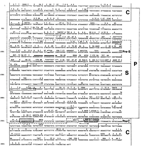
[image:3.612.343.540.76.331.2] [image:3.612.64.304.79.227.2]G T T GAAA C G GA C C GG T cc T G G A G C TT A G
CATACCCACT TACGTGTTTA TACTAAATTG AATGAACAAG CTTTGGACAA AGCTCGCAGA TTGCTTTGGT GGCATTACAA TTGCCTCCTC TGGGGAGAAG
T A Tc ATTA AG T G G A TGAA C
CCACGTTCAC ACAATACATT TCTCGCCTCC GTACTTGGCT GTCTACTCCC GAAAATATAC GAGGCATGGA TGCCCCAACC ATTGAAGCAA ACAGCATCC
C G c AAC T T A AC A
AATCCAGGTG GCTCAAGGAG GCAGAAATCA AACTAAGGGA ACTAGAAAAC CTCGAGGACT CGAACCTAGA AGAGATAAGG TCAAAACCAC AGTTGACTAT
A A G AA G cCA T C AT GcGA CCAC A T
GGGAGAAGACGTTCT AAGTC CCGAGGCAGG AGATCCTCTC CATCCCAACG TGCGGGCTTCACT CTCCCAC GCAATCGGGG AAACCAGACA CGATCCCCCT
G A AATTA TA C c A GT A Tc c CACA G A AG
ICACCTAGGGA F AGGGACCC TGCACGGCAT AAACTrGGGAA AATTAACTGG GTTGTATCAA ATGAAGGGCT GTGAGTTTAA CCCACACTGG AAGATACCAG
AA TC GA TCATGAG A C A TC c
ACATGGGCA CTACAAGCTAC ATCACGGACA GAAGGAGAG GAGCCCTGCC CGAAATTGGA AATAACTGAC TCCAGCCAAG ATGTGGCCCA AGAGCATTTC
cA T TG AA A T CAATGT A CGC GC GGGA A t A CC GC C SC
CTAGTGG CCA GACCAATCAG GGATTAAACC CAAGTACCATTCA AACTGC ATCACATCGA GTCACTGGTA AATGATTACG AACTAAAGCT CATTGAAGCA
T GC A S S C t TC CCAAT T C T AAC T ACATACCTG
GCAGGCGACTC ATAAGCGAGC TCCATATAC CTGGTCACAT TCAAGGGCCC GCACTTCACTC GGGAACAGA AGCACCTAGA CCCGCAACAA CA
O C A C t GACT A C CC t ACCA CT CC A.AAC
IAGGGGCATA CtCAAGCAAA AACGAACCA GACAGGAGAG CAGAAGGAGG AGAACCATtA CAGCAACATC TAGCAGGAAG AATGACTCCT CCCGAATTAT
'AC
A tC G tAC G C T A A AG T At A C ATG C AA A G GA AAAC AC GC CAAA C CAGGGGCCCAA AACAACGGCA GGAAAAATTC CTACCATCA ACACGTGATG GATCACATCG ACTCAGTGGA AGAACTACGG ACCTCACAAG CAGGGGGGCA IT TcA 6 GA A G C ATCAACT CAGT Ct AAGAGAT t AGCCTCA tCCC tC CGACCTGGCCGGAG GGGACAGCAC GCCGATAGGG CCTTTGATACAAATGACTC CTTCCCCTCC ACAACCATCACG TGGACATTC AGCACAGATCAAAAAAGACAAGPr
I GcT tC GA GT C G A AG GGAA CG ACCA t CtC GA tCt AA CI
IGAGTTCTTCA AGCAATATCA AGAGAACCGT CCGAAACCCG CCGAAACGGC ACCACCTCCC ATcAccGAGT TGCACGCTGC AGAACCTCCT CAGTGGAAGA1
ACA G GA CC CTG GG TCAATCACT ctT A AC CAT AT AT CC G_A 6 ACC G t 6 G CAGTr A AAAG ct CI
TTTCACCAGA AGACCCTTTA CTCAAAGCAA A _ wG6MGM ATCCCCGTCA AGGAA_CC AGAGGTACdG ATCCTCAAGG TCCCAAAGCT ICACCAACAAG AAGAA ArGG GAGCTACCTT CGGGGGAATA CTAGCTGGCC TAATCGGGTT ACtGG6tAGGA rTTTTCT6TG tGACAAAAAt TCTAGAAATA
CTGAGGAAGC TAGACTGGTG GTGGATTTCT CTCAGTTCTC CAAAGGAAAA AATGCTATGC GCTTTCCAAA ATACTGGTGC CCAAACCTCA CCACATTACG |
C r c AA C A
TAGGATCCTG CCCGtGGGGA TGCCCAGGAT TTCTCTGGAC TTATCTCAGG CTrTTTATCA TCTTCCTCTT GCTCCTGCTA GtAGCAGCAG GCTrTGCTGT
C TGG A G C t C C t
TCTrGACGGAA AACAAGTCTA CTATTTTCGA AAAGCTCCAA TGGGAGTCGG TCTCAGCCCT TcTCCTCCTC ATCTATTCAC TACTGCCATC GGAGCCGAAA
t C CA t C AC t
TCGCTAGTCG CTTTAACGTT TGGACTTTTT CTTATATGGA CGACTTCCTC CTCTGTCACC CAAGTGCTCG TCACCTTAAC ACAATTAGCC ACGCTGTCTG
T t A G A A C T CA A A t A C CGAA C T TC A TGATG C
ICACTTTTCTT
CAAGAArTG GArAGGAT CAACTTTGAC A^AAAGACTC CTTCCCCGGT GACAACGATC AGATTTCTCG GCTACGAGAT CAGCAAACAAT
TCGAAGAAAG 6 C C 6
CAATTTTGTT
CT AAAATCTTGT
CACATGGCGC
T A
CAAGTTAATG
A T S T A 6
CAGATGGAAT GAACTAAGAA CTGTAATCAA GAAAATCAAA GTTGGACAAT
C T 6 T T T A TC
TTACCTTTTA CGAAAGGAAA CATTGAAATG CTAAAACCTA TGTACGATGC
A C A T S A ASAT A TC
TGTATAAATT AACTATGGGA GTATGTAAGT TGACACTGGA TCCAAAGGTC
6 T
AATATCCCAT ATCACCGGCG GGAGCGCAGTK XXN GTTTACCTTTXNUXKNN TCAAACGGTANKX
TC CCCT AT A T C AACG
ATTAAACCCA GATGTATACT GACTGACTCT ACCTATGTTT GCCATAGGAA
p
T TO
GGTATGATTG GAAATGTATC CAAAGATTCA
TAT AC CA A T AG C
CTGTACTCAT AGAGTTAACT TTGCCTTTTC
0 TA
TCTTTACCTT TGCCACGTGT AGCCACGGAT
T T C AT A
GAGACATACA CATACAGGAA TTGCTGATOG
T ATCAG C T A TO
ATTCTCTAAG CTACCGTGGC ATTTTGCAAT
TG G GC C AACCTA ACA C T C G C C T 6 6
GOTATGCCAAA CAATTATTAA CCAGGCTGAC ATTGTACTAT GTACCCAGTA AATATAATCC TGCTGATGGC CCAACCAGGC ACAAACCTCC TGATTGGACG
250TG
CTGCCA
TT AACT Ar,:,::A:
:. .:T--
--2501 :::A*;:GCGTAC @ --T-::::G@-
@-
-;-@-
E.--**s@@--*@
--- CGAAGCAT ATATATATAC CACATAGGCT IATGTGGTCTC jTAAGAT A TTCGGAGCTG CGTGCCAAOO Pe-C--.- I*--@-CCAG Pre-C
r ---T- A T CGTG IN A T
TATCTTTACG TCGACCTCGC TGTTGTTCCT TGTGACTGTA CCTTTGGTAT GTACCATTGT TTATGATTCT TGCTTATAT TGGATGTCAA TGCTTCAAGA
C C G A AA A A A A G 7 A
GCTTTAGCAA ATGTATATGA TCTGCCAGAT GATTTCTTTC CTCAGATTGA TGATCTTGTT AGAGATGCTA AGGATGCTTT AGAACCTTAT TGGAAAGCCG
TT --- - ^A C T A T A CATS A C A AT 9
AAACAATAAA GAAACATGTT TTAATTGCTA CTCACTTTGT GGATTTGATT GAGGACTTCT GGCAGACCAC TCAGGGTATG AGCCAAATTG CAGACGCCCT
AA T A TACT T CCA A A CAG CGA C T T GSA T A AAC
CCGAGCAGTA ATTCCACCTA CTACCGTACC AGTACCGGAG GGTTTTCTCA TTACTCATAG TGAGGCAGAA GAGATCCCCT TGAACGATCT CTTTTCAAAT
01 G A A TOG T A
[image:4.612.61.555.66.581.2]3001 |CAAGAGGAGA GGATAGTCAA TTTCCAACCT GACTATCCCA TTACAGCTAG AATT
FIG. 4. Comparative DNAsequenceanalysisof the DHBV-3genome(upperline; 35) andthe HHBV-4genome(lowerline) linearizedat
the EcoRI site. Only nonconserved nucleotides areindicated forDHBV. Foroptimalalignment ofbothsequences, afewgaps(indicated by black bars)wereintroducedbyusingtheGAPprogramoftheUWGCG software(limit 1, 100; limit 2, 100). Agapof 3bp introducedinto both
sequencesatposition 1266 has been madebecause ofa3-nucleotide insertatthecorresponding position oftwoChinese DHBV isolateswhich have been recently sequenced(Sprengeletal., unpublished). Known andpredictedgenes areboxed. The directrepeatsequences important for viral replication are shown in shaded boxes. A putative enhancer sequence is marked (x). Transcription start sites ofDHBV (1) are
indicatedby arrows, and the consensus sequencefor processing andpolyadenylation is overlined.
ences between DHBV and HHBV, viral DNA was isolated
from a pool of five viremic heron sera without previous
repairof the gappedregion. Thevirus poolcontainedatleast two major sequence variants as revealed by restriction enzyme analysis of labeled virus DNA (data not shown). After digestion with restriction enzyme KpnI, three DNA fragments, 1.2, 1.8, and 3.0 kb in size, were observed (data
not shown), which suggests the existence of viral genomes
with one and two KpnI recognition sites. After insertion of
the KpnI fragments into phage vector M13mpl8, three
independent recombinants withafull-lengthgenome(3.0 kb)
and five subgenomic KpnI inserts (1.8 and 1.2 kb) were
obtained. As herons were not available for testing of the
infectivity of the cloned viralDNA, onephage
(mpl8HHBV-501
1001
1501
2001 TTC
CATATGAAGA
T O T
TAGGCCATC T OCT
TTCTAGGTAT
CCC
GCTACATTAA
CT 6
TATGTTTAGC
on November 10, 2019 by guest
http://jvi.asm.org/
1.0 2.0 3.0 kb
I
1111111 1 11111
I lliHiM
M
I
1111 1111 1111111111 1111.111111111
III11111111 111111 11D
N
A
I I ..
11 1
iIEIIEhli
11111l
ii 1IlIllIlIll 111111111111I IIEI IIIIIIIIIIIIIIDlII
I
111111111
I I
C-OR
F
EIlIo
Ii 111111111
11111
1
Il!III
fE
U1
II
111111101
I
11
III
111111
111111111111X111
IIII
lE
P-O RF
I I
a
S-O
R
F
FIG. 5. Schematicpresentation ofa DNA(first row) and protein sequencealignment(rows2,3, and4representreadingframesA, B, and
C) of the DHBV-3 and the HHBV-4genome. Base exchanges and amino acid substitutions are indicated byvertical bars. Known and predicted genes are boxed. Small filledboxesaboveand below thelinesindicate gaps introduced intothecorrespondinggenomesforoptimal
alignments.
4)carryingafull-lengthgenomicinsertwasselected arbitrar-ily for sequencing. Both strands of the HHBV DNA were
completely sequenced (Fig. 4; fordetails, seeMaterials and
Methods).
Comparativesequenceanalysis of HHBVand DHBV. The genome of HHBV is 3,027 bp in length, andcompared with the DHBV-3 isolate (35), it shows a nucleotide sequence
variabilityof21.6%.To getboth sequencesaligned,gapshad tobe introduced intothepre-Scoding region (Fig. 4, 5, and
6) of bothgenomes. Thecomputer-basedalignment ofboth sequences reveals a conserved genome organization with three major ORFs, referred to as pre-C/C, pre-S/S, and pol
(Fig.4).The beginning and theendpointsof thepre-C/C-and
pol-ORF were defined as described for DHBV (35). As initiation codon for the pre-S-ORF, the first ATG (position 801; Fig. 4) present on the analogous DHBV 2.1-kb pre-S
mRNAtranscript (1) was used. The S-ORF starts with an
ATG atposition 1317 (Fig. 4), whichwasshowntobe used to initiate protein synthesis of the 17-kDa envelope protein ofDHBV (29). Noadditional long ORFswere found in the HHBV genome(Fig. 5),and ORFsofthe DNA minus strand wereignored. They are not conserved between thedifferent DHBV isolates and HHBV, and for DHBV no transcripts
derived from the DNA plus strand were identified. The
comparison of DHBV and HHBV shows an asymmetric distribution of base exchanges which preferentially affect
ORFs without coding capacity (Fig. 5). There is, however,
oneremarkable exception: the region coding for pre-S and overlapping with the middle part of the pol-ORF. The
peptide sequence in the middle part ofthe pol-ORF is as
variableas a peptide stretch of a noncoding frame, and the
pre-S protein sequence is also strongly affected. Very long stretcheswithoutDNAsequencedivergence are not present
(Fig.
5).
Predicted proteins. As deduced from the nucleotide se-quence, the nucleocapsid antigen and the 17-kDa envelope protein are the most conserved viral gene products, with the
same sequence divergence of 16.5%. Similarily conserved
(16.8%)is the carboxy-terminal part of the pol-ORF-derived protein (nucleotide position 1316 to 2561; Fig. 4) which carries amino acid motifs characteristic for reverse tran-scriptase (41, 42) and RNase H(Schodelet al., in press). The amino-terminal part of the pol gene product (nucleotide
position 170 to 800; Fig. 4) is less conserved (divergence,
25.36%). The middle part of the pol sequences (nucleotide
position 800 to 1316; Fig. 4) is highly variable, with only 33.2% amino acid identity, and appears not to encode enzymatic functions as speculated previously for DHBV (35,
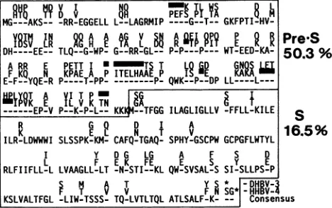
37).Asimilarhigh sequence divergence (50.3%) is apparent
in thepre-S partof the 36-kDaenvelope protein (Fig. 6). This may beassociated with the different host range of HHBV.
Transcription and replication signals. As described for
DHBV (1), there is aTATAbox sequence upstream of the
C-mRNA/pregenome transcription initiation site (nucleotide
position 2530 to 2540; Fig. 4 and 7) which probably
repre-sentspart ofthe coregene promoter. NeitheraTATA box upstream oftheputativeHHBVpre-S mRNAstart sitenor a putative S-promoter element with simian virus 40 late promoter sequencesimilarity, as suggested for human
hep-atitis B virus (2), was found upstream of the 5' end of the DHBV S-mRNA ofseveral DHBV isolates (Sprengel,
un-published data)oroftheanalogousregion ofthe HHBV. An
octamersequence(TGTTTGCT; nucleotideposition2250to
2258;Fig. 4) present in allhepadnaviruses sequenced sofar andrecently speculated to play a role in viral gene expres-sion (7, 32, 33) is only partially conserved in the fully sequenced HHBV genome (Fig. 4). It is unlikely that this sequence divergence renders the HHBV defective since several HHBV isolates whichweresequencedin thisregion exhibitthe same mutation (datanot shown). An AATAAA
signal sequenceforRNAprocessingandpolyadenylation is
strictlyconservedforDHBVandHHBV,suggestingthat the
transcripts ofboth virusesarecoterminalandareprocessed withintheCgene.
Two direct repeat sequences (DR1 and DR2) which are
important in initiatingDHBV DNA plus- and minus-strand synthesis (14, 15, 31, 44) are strictly conserved between
MG-_QAKS__ -RR-EGGELL L--LAGRMIP ----G--T--
GKFPTI-HV-I V LR AGiAE ATG L SRA 9T
PpT
PE
KK DH---EE-- TLQ--G-WP- G--RR-GL-- P-P---P---WT-EED-KA-ARR E PETT I * ETS T L'GD GN
g
FKQ N
KPAETA
PITELHAAETp
P QW &DED KA--LE-F--YQE-R P----T-PP---P- QWK--P--DP LL----L---iYP8TKAEIY. VT PK TN UAG G T ---EP-V P--K-P-L-- KK --TFGG ILAGLIGLLV -FFLL-KILE
PreS
50.3 %
S
R GQ D I A 1
K E L N T V
1
ILR-LDWWWI SLSSPK-KM- CAFQ-TGAQ- SPHY-GSCPW GCPGFLWTYL
I Y DOG LG A F S 0
RLFIIFLLLL
LVAAGLL-FT
EN-KSTIF--EKL
QW-ESVSAL-SS SI-ySLLPS-EP
KSLVALTFGL -LIW-TSSS- TQ-LVTLTQL ATLSALFK- Consensus
;.5%
FIG. 6. Protein sequence alignment of the pre-S/S proteins of DHBV-3 and HHBV-4. Gaps introduced for optimal alignment (UWGCG program GAP; gap weight, 2.0; gap length weight, 0.3)are indicatedbyblack bars.
m ill
illo o ligil ill
-111
11111
11111111
iiiijo
illimilli illoollillilli
on November 10, 2019 by guest
http://jvi.asm.org/
[image:5.612.105.524.71.189.2] [image:5.612.329.563.542.689.2]TATA
GCTTTTCCAACACCCCTCTCrCGAAGCAA
TATATATTCCACATAGGC1
:::::::::::::::::::: ::::::::::::::
GCCGTTACAACACCCCTCTgACGAGCATATATATATACCACATAGGC1
DR2
1
>'
M
W
N
L
R
I
T
P
L
S
F
G
A
ATGTGGMCTTMGMAA1ACACCCCTCTgCTTCGGAGCT
ATGTGGTCTCTMGA
ACACCCCTCTqCATTCGGAGCT
i
i
DR1
* * S * *
L
H *S
P * * *FIG. 7. Sequence comparison of the amino-terminal pre-C region of DHBV-3 (top) and HHBV-4 (bottom). The direct repeat sequences (DR1) of both viruses are boxed, and the inverted repeats are shown by arrows. Transcription initiation sites of the DHBV RNA pregenome upstream of DR1 and the site of DHBV DNA minus-strand initiation withinDR1 are indicated by leftward and rightward arrows, respectively. Thepredicted amino-terminal protein sequence of the DHBV precore protein is shown at the upper line of the open box. Amino acid sequence substitutions found in HHBV versus DHBV are indicated at the bottom line of the open box.
DHBV and HHBV (Fig. 4). This suggests that HHBV replicates like DHBV and the primers for DNA plus- and minus-strand synthesis are similar for both viruses. How-ever,thedistance betweenboth direct repeats is 1 nucleotide
shorter (45 instead of 46 nucleotides) for HHBV, which
affects predictably both the precore protein sequence and the structure ofreplicative intermediates. First, the
muta-tions induce five aminoacid exchanges at the amino
termi-nusofthe precoreprotein(Fig. 7). Second, if in analogy with DHBV the 5' end of the HHBV pregenome is at position 2570 within DR1 (Fig. 7), and ifinitiation of DNA minus-strandsynthesistakes place at an analogousposition in DR1, the DNAminus strand of HHBV would have a 1-nucleotide-shorterterminalredundancy.Thus, theterminal redundancy which is probably essential for template switching in DNA
plus-strand synthesis is predictably different in sequence between DHBV and HHBV. As in DHBV, there is an
inverted repeatsequencebetween DR1 and DR2 (24) which mayplay a role in the viral lifecycle. In HHBV, this repeat
is conserved but 1 nucleotide shorter (9 instead of 10
nucleotides) and differs in sequence at the first nucleotide becauseoftwocomplementarypoint mutations(Fig. 7). The
same sequence motifs have been observed in five further HHBV isolates which were sequenced in this region (data
notshown). Therefore, analtered precore proteinsequence
andsequence divergenceintheoriginof replication seem to
be characteristic for the HHBV genome. Interestingly, the
pre-C-ORF, which is dispensible forDHBV replication (4,
30), ispresentin all HHBVisolatessequenced, suggestinga
selective advantage for viruses retaining pre-C-ORFs.
DISCUSSION
Screening ofsera from severalavian species for
DHBV-relatedvirusesrevealedanewhepatitisBvirus in 20to50% ofgreyherons tested inGermany. Severallines ofevidence, including sequencing data, demonstrate a close relatedness
of HHBV with DHBV. Since the sera were not from
age-matched animals but were collected randomly in dif-ferent areas of northern Germany, the
high
frequency of infection suggests that these animals are chronically in-fected. Conceivably, herons become infectedcongenitally,
as do ducks (26, 43). In none of the livers of infected oruninfected herons were
signs
ofprimary
liver carcinoma observedby visualinspection,
norwasintegrated
viralDNAdetected by Southern blot
analysis
(datanotshown).Thus,
as for DHBV,integration
of HHBV DNA into the host chromosomes either is rare, if itoccurs atall,
oroccursonly
late in life. A gene, X, which is present in all mammalian
hepadnaviruses and which may play a role in hepatocarci-nogenesis isnotpresentin DHBV or HHBV. The lackofan Xgene may be a characteristic feature which distinguishes avian from mammalian hepadnaviruses, but further mem-bersofthe avianhepadnaviruses have tobediscovered and
characterized to confirm this. On the basis of genome sequences, HHBV and DHBV areless closely related than the two rodent hepadnaviruses woodchuck hepatitis virus andgroundsquirrel hepatitis virus(78.5versus83.6% nucle-otide identity). When the different hepadnavirus genomes were inspected, the most variable protein sequences were
foundinashortregion ofthepolframe and thepre-S region
(F. Schodel, R. Sprengel, T. Weiner, D. Fernholz, R.
Schneider, and H. Will, Adv. Viral Oncol., in press). The
comparative sequencedatapresentedhere are in agreement with this findingand support the previous speculation that thecorresponding pol regionmostlikelyisatether between different enzymatic activities encoded in the pol-ORF (35).
In contrast, the pre-S sequences are believed to play an
importantrole inbinding ofthe virus to thehepatocyte (25)
andthehighly restricted host rangeofhepadnavirusescould be determined by these sequences. Interestingly, despite a
high pre-S sequence divergence of both rodent
hepadnavi-ruses,GSHVis infectious in woodchucks (8), which renders therodent virusesdifficultto usefor definition ofsequences
determining host-specific hepatocyte binding. When DHBV and HHBV pre-S sequences are compared, there is less sequence homology than observed with woodchuck and
ground squirrel hepatitis viruses. Since HHBV appears not to be infectious for ducks, the pre-S sequence is the most
obvious candidate that could determine
host-specific
hepa-tocyte binding. Strikingly, the longest continuous pre-S
sequence identity ofHHBVand DHBV does not exceed a
7-aminoacid-long continuous peptide (Fig. 6).This suggests either an highly variable virus receptor protein or the in-volvementofonlyaveryshortpeptide(s)orscatteredamino acidsofthepre-Sproteinin receptorbinding.The availabil-ity oftwocloselyrelated avian
hepadnaviruses
withdifferent host ranges, and thepossibility
oftesting
recombinantvi-ruses in vitro and in vivo in the most convenient
hepadna-virusanimal system(7, 34, 36), suchasducks,will allowus toidentifysequenceswhichdetermine host range
specificity.
ACKNOWLEDGMENTS
We are grateful to Karin Weimer, Marita Schrenk, and Gaby
Sowa forexperttechnical assistance. This workwasmadepossible
through the kind support of W. Heidmann, A. Buthe, and M. Friederichs (Veterinarmedizinische Hochschule, Hannover,
on November 10, 2019 by guest
http://jvi.asm.org/
[image:6.612.97.521.78.169.2]eralRepublic of Germany), who generously provideduswith sera
and liversamples fromgreyherons.
Samples from herons were obtained in the course ofa heavy-metal pollution study of birds whichwas supported byfunds from
the Land Niedersachsen. Part of the workwassupported byagrant from the Deutsche Forschungsgemeinschaft (Fa 138/3-2).
LITERATURE CITED
1. Buscher, M., W.Reiser, H. Will, and H. Schaller. 1985. Tran-scripts and the putative RNA pregenome of duck hepatitis B virus: implications for reverse transcription. Cell 40:717-724.
2. Cattaneo, R., H. Will, N. Hernandez, and H. Schaller. 1983. Signals regulating hepatitis B surface antigen transcription. Nature (London) 305:336-338.
3. Cattaneo, R., H. Will, and H. Schaller. 1984. HepatitisB virus transcription in the infected liver. EMBO J. 3:2191-2196. 4. Chang, C., G. Enders, R. Sprengel, N. Peters,H. E. Varmus,
and D. Ganem. 1987. Expression of the precore region ofan
avian hepatitis B virus is not required for viral replication. J. Virol. 61:3322-3325.
5. Enders, G. H., D. Ganem, and H. Varmus. 1985.Mappingthe major transcripts of ground squirrel hepatitis virus: the pre-sumptive template forreversetranscriptaseisterminally redun-dant. Cell 42:297-308.
6. Feitelson, M. A., I. Millman, T. Halbherr, H. Simmons, and
B. S. Blumberg. 1986. A newly identified hepatitis B virus in
treesquirrels. Proc. Natl. Acad. Sci. USA 83:2233-2237. 7. Galle, P. R.,H.J. Schlicht,M. Fischer,and H. Schaller. 1988.
Production of infectious duck hepatitis B virus in a human hepatoma cell line. J. Virol. 62:1736-1740.
8. Ganem, D., and H. E. Varmus.1987. The molecular basis of the hepatitis B viruses. Annu. Rev.Biochem. 56:651-693. 9. Gerlich, W. H., and W. S. Robinson. 1980. Hepatitis B virus
contains protein attached to the 5'-terminus of its complete strand. Cell 21:801-809.
10. Gust, I.D., C. J. Burrell, A. G. Coulepis,W. S.Robinson,and
A. J. Zuckerman. 1986. Taxonomic classification of human hepatitis B virus. Intervirology 25:14-29.
11. Heermann, K. H., U. Goldmann, W. Schwartz,T.Seyffarth,H. Baumgarten, and W. H. Gerlich.1984.Largesurfaceproteinsof hepatitis B virus containing the pre-S sequence. J. Virol. 52: 396-402.
12. Howard, C. R. 1986. The biology of hepadnaviruses. J. Gen. Virol. 67:1215-1235.
13. Landers, T. A., H. B. Greenberg, and W. S. Robinson. 1977. Structure ofhepatitis B Dane particleDNA and natureofthe endogenous polymerase reaction. J. Virol. 23:368-376. 14. Lien, J. M., C.E.Aldrich, and W. S.Mason. 1986. Evidence
that acapped oligoribonucleotide is the primer for the duck
hepatitis Bvirusplus-strand synthesis. J. Virol. 57:229-236. 15. Lien, J. M., D. J. Petcu, C. E. Aldrich, and W. S. Mason. 1987.
Initiation and termination of duck hepatitis B virus DNA synthesis duringvirus maturation. J. Virol. 61:3832-3840. 16. Mandart, E., A. Kay, and F. Galibert. 1984. Nucleotide
se-quenceofacloned duckhepatitis B virusgenome: comparison
with woodchuck and human hepatitis B virus sequences. J. Virol. 49:782-792.
17. Marion, P. L., S. S. Knight,M. A.Feitelson,L.S. Oshiro,and W. S. Robinson. 1983. Major polypeptide of duck hepatitis B surfaceantigen particles. J. Virol. 48:534-541.
18. Marion, P. L., L. S. Oshiro,D.C.Regnery, G. H. Scullard, and W. S.Robinson. 1980. Avirus in Beechey ground squirrels that is relatedtohepatitis B virus of humans. Proc. Natl. Acad. Sci. USA77:2941-2944.
19. Mason, W.S.,C. Aldrich, J. Summers, and J. M. Taylor. 1982. Asymmetric replication of duck hepatitisB virusDNAinliver cells: freeminus-strand DNA. Proc. Natl. Acad. Sci. USA79: 3997-4001.
20. Mason, W. S., G. Seal,and J.Summers. 1980. Avirus of Pekin ducks with structural and biological relatedness to human hepatitis B virus. J. Virol. 36:829-836.
21. Messing,J.,andJ.Vieira.1982.Anewpairof M13vectorsfor
selecting either strand ofdouble-digest restriction fragments.
Gene 19:269-276.
22. Moroy, T., J. Etiemble, C. Trepo, P. Tiollais, and M. A. Buendia. 1985. Transcription of woodchuck hepatitis virus in thechronicallyinfected liver. EMBO J. 4:1507-1514.
23. Molnar-Kimber,K.L., J.W.Summers,and W. S. Mason. 1984.
Mappingof thecohesiveoverlapof duckhepatitisB virus DNA andof the site of initiationofreversetranscription.J. Virol. 51: 181-191.
24. Molnar-Kimber, K. L., J. Summers, J. M. Taylor, andW. S. Mason. 1983. Protein covalentlybound to minus-strand DNA intermediates of duckhepatitisB virus. J. Virol. 45:165-172. 25. Neurath,A.R.,S. B. H.Kent,N. Strick,andK. Parker. 1986.
Identification and chemical synthesis ofa host cell receptor
bindingsiteonhepatitisB virus. Cell46:429-436.
26. O'Connell, A. P., M. K. Urban, and W. T. London. 1983.
Naturally occurring infectionof Pekin duckembryos byduck
hepatitisB virus.Proc. Natl. Acad. Sci. USA 80:1703-1706. 27. Pugh, J. C., J. J. Sninsky, J. W. Summers, and E. Schaeffer.
1987.Characterization ofapre-S polypeptideonthe surfaces of infectious avian hepadnavirus particles. J. Virol. 61:1384-1390.
28. Sanger,F.,S.Nicklen,andA. R.Coulson.1977. DNA
sequenc-ing with chain-terminating inhibitors. Proc. Natl. Acad. Sci. USA 74:5463-5467.
29. Schlicht, H. J., C. Kuhn, B. Guhr, R.J. Mattaliano, and H. Schaller.1987.Biochemical andimmunologicalcharacterization of the duck hepatitis B virus envelope proteins. J. Virol. 61: 2280-2285.
30. Schlicht, H. J., J. Salfeld, and H. Schaller. 1987. The duck
hepatitisBviruspre-C regionencodesasignalsequence which isessential forsynthesis and secretion ofprocessedcore pro-teins butnotfor virus formation.J. Virol. 61:2208-2212. 31. Seeger, C., D. Ganem, and H. E. Varmus. 1986. Biochemical
andgeneticevidence for thehepatitis Bvirusreplication
strat-egy. Science 232:477-484.
32. Shaul,Y.,andR.Ben-Levy. 1987.Multiplenuclearproteinsare
bound tohepatitis B virus enhancer elementand its upstream sequences. EMBOJ.6:1913-1920.
33. Shaul, Y.,R. Ben-Levy, and T.De-Medina. 1986.Highaffinity
bindingsite for nuclearfactorInext tohepatitisBvirus Sgene promoter. EMBO J. 5:1967-1971.
34. Sprengel, R., C. Kuhn, C.Manso, and H. Will. 1984. Cloned duck hepatitis B virus DNA is infectious in Pekin ducks. J. Virol. 52:932-937.
35. Sprengel, R.,C.Kuhn,H.Will,and H.Schaller.1985.
Compar-ative sequence analysis of duck and human hepatitis B virus genomes.J. Med. Virol. 15:323-333.
36. Sprengel, R.,H. E.Varmus,andD.Ganem. 1987.Homologous
recombination betweenhepadnaviral genomesfollowingin vivo DNAtransfection: implicationsfor studies of viral infectivity.
Virology159:454-456.
37. Sprengel, R.,and H.Will.1987. DuckhepatitisBvirus,p. 363-386. InG. Darai(ed.), Virus disease inlaboratoryandcaptive
animals. MartinusNijhoff Publishing, Boston.
38. Stemler, M.,J.Hess, R. Braun,H. Will, andC. H.Schroder. 1988. Serological evidence for expression of the polymerase
geneof humanhepatitisB virus invivo.J.Gen. Virol. 69:689-693.
39. Summers,J.,and W. S. Mason.1982.Replicationofthegenome ofahepatitis B-like virusby reversetranscription ofanRNA intermediate. Cell 29:403-415.
40. Summers, J.,J.M.Smolec,and R.Snyder. 1978. Avirus similar
tohepatitisBvirus associated withhepatitisandhepatoma in woodchucks. Proc.Natl.Acad. Sci. USA 75:4533-4537. 41. Toh, H.,H.Hayashida,and T.Miyata.1983. Sequence
homol-ogybetweenretroviralreversetranscriptaseandputative poly-meraseofhepatitisBvirus and cauliflower mosaic virus.Nature (London)305:827-829.
42. Toh, H.,R.Kikuno,H.Hayashida,T.Miyata,W.Kugimiya,S.
Inouye, S. Yuki, and K. Saigo. 1985. Close structural resem-blance betweenputative polymeraseofaDrosophila
on November 10, 2019 by guest
http://jvi.asm.org/
able genetic element 17.6 and pol gene product of Moloney murine leukaemia virus. EMBOJ.4:1267-1272.
43. Urban, M. K., A. P. O'Connell, and W. T. London. 1985. Sequence ofeventsin naturalinfection of Pekin duck embryos withduck hepatitis B virus. J. Virol. 55:16-22.
44. Will, H., W. Reiser, T. Weimer, E. Pfaff, M. Buscher, R.
Sprengel, R.Cattaneo,and H.Schaller. 1987. Replication
strat-egyofhumanhepatitis B virus. J. Virol. 61:904-911.
45. Will, H., J.Salfeld, E.Pfaff, C. Manso, L. Theilmann, and H. Schaller. 1986. Putative reverse trarnscriptase intermediates of
human hepatitis B virus in primary liver carcinoma. Science 231:594-596.