0022-538X/86/100105-09$02.00/0
Copyright C) 1986, AmericanSocietyfor Microbiology
Isolation and
Characterization of NIH 3T3 Cells Expressing
Polyomavirus Small T Antigen
TETSUO NODA,t MASANOBU SATAKE,t TERRY ROBINS, AND YOSHIAKI ITOt*
MolecularMechanisms of Carcinogenesis Laboratory,
LBI-Basic
Research Program, Frederick Cancer Research Facility,National Cancer Institute,Frederick, Maryland 21701
Received 10 February 1986/Accepted 20 June 1986
The polyomavirus small T-antigen gene, together with the polyomavirus promoter, was inserted into a
retrovirus vector pGV16 which contains the Moloney sarcoma virus long terminal repeat and neomycin
resistancegene driven by the simianvirus40promoter. Thisexpression vector, pGVST, waspackaged into
retrovirus particles bytransfection ofx42 cellswhichharbor packaging-defective murine retrovirusgenome.
NIH 3T3 cells were infected by this replication-defective retrovirus containing pGVST. Of the 15
G418-resistant cellclones,8expresssmall Tantigenatvarious levelsasrevealed byimmunoprecipitation.A cellular
protein with anapparentmolecular weightofabout32,000 coprecipitateswith small T antigen.
Immunoflu-orescentstaining showsthatsmall T antigen is mainlypresentin the nuclei. Morphologically, cellsexpressing
small T antigenareindistinguishable from parental NIH 3T3cells and haveamicrofilamentpatternsimilarto
that inparental NIH3T3 cells. Cellsexpressing small T antigen formaflat monolayer but continueto grow
beyond the saturation density observed for parentalNIH3T3cellsand eventuallycomeoff the cultureplateas
aresultofoverconfluency. Thereissomecorrelationbetween the level of expression ofsmall Tantigen and the
growth rate of the cells. Small T-antigen-expressing cells form small colonies in soft agar. However, the
proportion of cells which form these small colonies is rather small. A clone of thesecells tested didnotform
tumorsin nudemice within 3 months after inoculation of106 cellsperanimal.Thus,presentstudiesestablish
that the smallTantigen of polyomavirusis asecondnucleus-localized transforminggeneproduct of the virus
(thefirstonebeing largeT antigen)andbyitself hasafunction which istostimulatethegrowthof NIH 3T3
cellsbeyond their saturation density in monolayer culture.
The early region of polyomavirus is essential for cell
transformation andistranscribed intoasingleRNAspecies. Three alternative splicing events take place in thisprimary
transcript, generating three mRNA species, each of which
codes for a distinct transforming protein. They are called
large, middle, and small T antigens (reviewed in reference
17). Ithas been
suggested
thateachofthese threetransform-ing proteins contributestocelltransformation independently
fromthe other two (2, 7). Itisthisfeature whichmakesthis
virus unique inthe studyof oncogenic celltransformation.It
is hoped, therefore, thatthe complex biological process of
cell transformation could be dissected by analyzing the
functions ofthese individual genesand theirproducts. Large T antigen is a DNA-binding protein (14) and is mainly localizedtothenuclei of cells.Inthelytic cycle,large Tantigen is required for viral DNAreplication and
regula-tion oftranscription. Itis also knowntotrans-activatesome
cellular genes. In cell transformation, only the
amino-terminal 40%of largeTantigen is required(28). It has been
suggestedthatlargeTantigen is abletoconvertprimaryrat
embryo fibroblasts
with
limiteddoubling potential toestab-lished cell lines with unlimiteddoubling potential (28) with-outaltering the morphology (22, 34). Itisnotclear whichof
thebiochemical functions knowntobeassociated withlarge
Tantigenisresponsible for this immortalization or
establish-mentfunction.
Middle T antigen is bound to the membrane and is
primarily responsible for inducing the phenotype of
trans-formedcells(16,18, 21). Middle Tantigenis associated with
*Correspondingauthor.
tPresent address: Institute forVirus Research, Kyoto
Univer-sity,Sakyo-ku, Kyoto 606, Japan.
an activity which phosphorylates middleT antigenon
tyro-sinein vitro(10, 32, 37). This activity isdue to anassociated
cellular tyrosine protein kinase, a product of the
proto-oncogene c-src(6). MiddleTantigen has been showntoalter
theproteintyrosine kinase activity ofc-src uponbinding(4).
There is an intimate correlation between the level of the
middleTantigen-associated protein tyrosine kinase activity
andthe extentofthetransformed phenotype (37). MiddleT
antigen is necessary and sufficient to transform established
rodentfibroblast celllines(40),althoughitisnotsufficientto
transformprimaryratembryo fibroblasts (27).
Small T antigen contains 196 amino acid residues. All
except the carboxy-terminal 4 amino acids are included in
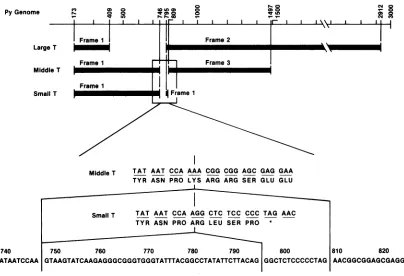
the amino-terminal half of middle T antigen (Fig. 1). This raises the question ofwhether small T antigen has its own
unique function which is notsharedby middleTantigen. It has been reported that allthree
T-antigen
genes,including
thesmallT-antigengene, arerequiredfor thetransformation
of primary rat embryo fibroblasts in vitro (7, 28). On the
contrary, onlytwoT-antigengenes, eitherlargeand middle
T-antigengenes ormiddle and smallT-antigengenes, appear tobe
required
forinvivo tumorformationwheninoculatedintonewborn rats (1,2). Although somewhatcontradictory,
these results suggest some important role that small T
antigen plays in cell transformation in vitro and in
oncogenesis
in vivo.Very little is known about the biochemical function of
small T antigen. One ofthe reasons forthis lack of
knowl-edge is thatcells expressing only small Tantigen have not
beenavailable until now, whereascells
expressing only
large
ormiddle Tantigens have been widelyavailable. It seems
that the calcium phosphate
coprecipitation
method(42)
results in the introduction of a
large
copy number ofthe105
on November 10, 2019 by guest
http://jvi.asm.org/
Py Genome Cl) 0) 0
0 0
w- qw to
Frame1
Large T
Frame 1 Middle T
SmallT
0
toLO0) 0
lr cn o a
r-hc _
I lr
F.- 0
, 0
lr I
N O
- 0
en 9
l
Frame2 I
kIm
I
aI
IFrame 3
MiddleT TAT AAT CCA AAA CGG CGG AGC GAG GAA TYR ASN PRO LYS ARG ARG SER GLU GLU
,
~~~~~~~~~~~~~~~~~~~~~~~~~~~~~~~~~~~~~I
Small T TAT AAT CCA AGO CTC TCC CCC TAG AAC
t ~~~~TYRASN PRO ARG LEU SER PRO*l
740 750 760 770 780 790 800 810 820
TATAATCCAA GTAAGTATCAAGAGGGCGGGTGGGTATTTACGGCCTATATTCTTACAG GGCTCTCCCCCTAG AACGGCGGAGCGAGG
FIG. 1. OverlappingcodingregionforthreepolyomavirusT antigens. small T-antigen gene into cells. When small T antigen
accumulates inlarge amounts in the cells, easydetachment of the cells from the plastic surface of the culture plates
appears tooccur (7, 45).
To isolate cells expressing only small T antigen, a
retro-virus vector as helper-free, replication-defective virus
con-tainingtheneomycin gene (13, 39) wasused totransfer the
small T-antigen gene into the recipient cells. This method
wasconsidered useful for introducing onlyalimitednumber
of small T-antigen genes per cell, thereby limiting the
pro-duction of small T protein. In addition, itwas necessary to
coselect small T-antigen-expressing cells by using a domi-nant selectable marker, since the phenotype of such cells
was not known. Each gene was expressed by using an
internal promoterwithin the retroviral genome(12). In this
way, we wereabletoisolate NIH 3T3 cells expressing only small Tantigen. In thispaper, wedescribe someproperties of suchcells. Themostimportant implicationof thisstudyis thatsmall Tantigenhas itsown biological function, namely
growth-stimulating activity.
MATERIALS ANDMETHODS
Cells and medium. NIH 3T3 cells were obtained from WallaceRowe,NationalInstitutes ofHealth, Bethesda,Md. Clones MT-A and MT-B of NIH 3T3 cells expressing only
middle T antigen were obtained by limiting dilution of the cells isolatedas twoindependent dense foci after transfec-tion of NIH3T3 cells with theplasmid pPyMT1 (40) which induces the synthesis of only middle T antigen. To obtain clone MT/ST-A of NIH 3T3 cells which expresses both
middle and small Tantigens, G418-resistant cellswere first
selected after infection of NIH 3T3 cells with pGVST
containingvirus withhelper Moloneymurineleukemia virus
(see below). Then the clone MT/ST-A was isolated by
limiting dilution of cells obtained as dense focus after
transfecting the population of G418-resistant cells with
pPyMT1.Thesecellsweremaintained inDulbecco modified
Eagle medium (DMEM; GIBCO Laboratories)
containing
10%calfserum (Colorado SerumCo.).Recombinant DNA technique. All the procedures used to construct pGVST were
performed essentially
as described previously (25). Restriction enzymes were obtained fromBethesda Research Laboratories, Inc., New
England
BioLabs, Inc., andBoehringer Mannheim Biochemicals. T4 DNA ligase and calf intestine alkaline phosphatase were
purchased from Boehringer Mannheim. All the enzymes
were used asrecommended by the manufacturers.
Helper-free defective retrovirus containing pGVST
con-struct. Onday 1, 3 x 10⁵ tj2 cells(obtained fromR.
Mulligan,
Massachusetts Institute ofTechnology, Cambridge, Mass.)
which harbor packaging-defective murine leukemia virus
genome (26) wereplatedper 100-mmplateandincubatedin
10 mlofDMEMcontaining10% calfserum.Onday2(about
16to20 hafterplating),1mlof0.25 M
CaCl2 containing
2 ,ugofpGVSTwas added slowly to 1 ml of sterile 2x FIBS (50
mM HEPES
[N-2-hydroxyethylpiperazine-N'-2-ethanesul-fonic acid], 280 mM NaCl, 1.4 mM phosphate) (42) with
constant agitation, poured into the culture plate without
removal of the medium, and incubated for 16 h. Onday 3,
cells were washed once with DMEM containing 5% calf
serum and fed with DMEM containing
10%
calfserum. Onday 4, G418 (13, 39)wasaddedtothemediumto400
pLg/ml.
After this, the medium was changed every 3 days with
DMEM containing 10% calfserum and 400
pig
ofG418per ml. Approximately 60 to 100 G418-resistant colonies perplate became recognizable. Thesecolonieswere isolatedon
day 10. These G418-resistant colonies were pooled and
seededat2x 106cells per T75flask. After 24 hof incubation
in the presence of 400
[Lg
of G418 perml, themedium waschangedtoDMEM
containing
10% calfserumwithout
G418. Cellswereincubated further for22 h. The culturesuperna-
r+-I Frame1 i
,
moo
FrameIon November 10, 2019 by guest
http://jvi.asm.org/
[image:2.612.105.509.72.348.2]0
Q -a C. I- BR322
IR
--Ea Amp)
zjc I
5o
\(N
o
coI
0-c4 PY\ E
/
0
/'
b pBR322
SV40 Ori
Neo
pPySTI
FIG. 2. Structureof recombinant plasmids pGV16 (30) (a) and pGVST (b). The origin of the sequences is asfollows: solid line together withlong terminal repeat (LTR); Moloney murine sarcoma virus, open box with or without hatched area;SV40orpolyomavirus as indicated, solidbox; pBR322, stippled box, neomycin resistance gene from transposonTn5 (13, 39). The polyomavirus sequence from theBcI site (nucleotide 5021)totheBgll sites(n'tcleotide87)containing the origin of replication was inserted at theBanmHl site in pGV16 after BainHI linkerswere attached at both ends of thisfragment. The EcoRI site was present in the polylinker of pGV16 (30).
tantwascollected and centrifuged for 4 min at 700 x g. This
supernatant was used as virus stock. Virus stocks prepared
this way usually contain about 104 infectious units per ml which can transfer G418 resistance to the recipient cells. To
transfer pGVST to NIH 3T3 cells, we seeded the cells 105
cells per100-mmplateinDMEMcontaining 10% calf serum.
On day 2, cells were inoculated with 1 ml of virus
prepara-tion which contains 5 p.g of polybrene per ml and were
incubated for 2 h with occasional rocking. Infected cells
wereincubated in fresh DMEM containing 10% calf serum.
Onday 3, G418 was added to the medium to 400 Fg/ml and
the culture was
further
incubated. The mediumwasreplacedwith a fresh medium containing 400
[.g
of G418 per ml everyday for2 days and every 3 d,ays thereafter. About 1,000 to
2,000 G418-resistant colonies per plate became recognizable
onday 8. These G418-resistant cells were pooled, and15cell
clones were isolated by limitingdilution.
Immunoprecipitation, PAGE, and fluorography.
Immuno-precipitation of
[35S]methipnine-labeled
small T antigen,subsequent analysis by sodium do,decyl
sulfate-polyacry-lamidegel electrophoresis (PAGE), and fluorography of the
gels were performed as described previously (16). Briefly, subconfluentcells ina60-mmplate(approximately 106cells)
were labeled with 300 lCj of
VSSmethionine
(800Ci/mmol:New England Nuclear Corp.) in 2 ml of DMEM lacking
methionine for 3 h at 37°C. Cell extracts made from these
labeled cells were subjected to immunoprecipitation withrat
anti-T-antigen serum obtained from tumor-bearing rats or
nonimmune control rat serum (20). Immunoprecipitates
were analyzed by sodium dodecyl sulfate-PAGE on 12%
polyacrylamide gels.
Double-immunofluorescent staining of cells for small T
antigenand microfilament. Cells grown on coverglass were
fixed with 3.7% formaldehyde in phosphate-buffered saline
for 20 min and permeabilized with 0.1%, Nonidet P-40 in
phosphate-buffered saline for 10 min. The fixed cells were
reactedfor40minat37°C withrat monoclonal antibodies Cl
or C4 (9), which recognize the amino-terminal common
region of three T antigens (36). Culture supernatant of
hybridoma cells was used undiluted. Ascites fluid was
di-luted50-fold with phosphate-buffered saline. After extensive
washing, cells were reacted for 40 min at 37°C with
appro-priately diluted fluorescein-tagged anti-rat immunoglobulin
G rabbit immunoglobulin G (Cooper Biomedical, Inc.) and
rhodamine-tagged phalloidin (43) (Molecular Probes, Inc.). Afterbeing washedwith phosphate-buffered saline, the cells on coverglasswere mounted on a slideglasswith
buffered-glycerol mounting medium and viewed through an
epifluo-rescence microscope. Pictures were taken on Kodak Tri-X
filmwith appropriate filters forfluorescein or rhodamine.
Chemicals. ThedrugG418(13) wasobtainedfromGIBCO Laboratories. Bacto-Agar (Difco Laboratories)was used for
soft-agar assay of anchorage-independent cell growth.
Polybrene (SigmaChemical Co.)was usedfor better
adsorp-tion of virus tocells.
RESULTS
Construction of a retrovirus vector which expresses
poly-omavirussmall Tantigen. Figure 2 shows a backbone
retro-virus vector, pGV16 (30), which contains an origin regionof
polyomavirus from BclI to BglI (for polyomavirus genetic
map, see reference 38). This 358-base-pair BclI-BglI
frag-ment has Ba,nHI linkers at both ends and iscloned at the
BamHI site present in thepolylinker located atthe junction
between the sequence derived from Moloney murine
sar-coma virusandtheplasmid pBR322 (about 8 o'clockin Fig.
2a). Small T antigen gene is derived from plasmid pPyST1
(45),whichcontains the entire polyomavirus genomelacking
theintron for smallT-antigen gene clonedattheBamHI site
of pAT153. The 2.2-kilobase BamHI-EcoRI fragment of
pPyS'r1
containingthecoding regionfor small Tantigenandtheearly promoterwasisolated. The358-base-pair BclI-BgIl
fragment which contains BamHI linker at both ends was
removed frompGV16 bycleavingwithBamHIand EcoRI at
positionsAand theEcoRIsite(Fig. 2)andwasreplacedwith
the 2.2-kilobase BamnHI-EcoRI fragment of pPyST1. The
polylinkerinpGV16contains anEcoRI site which accepted
one end of the 2.2-kilobase polyoma fragment. In this
construct, each gene was expressed by using an internal
on November 10, 2019 by guest
http://jvi.asm.org/
[image:3.612.104.519.79.265.2]92.5 K 68.0 K--43.0K
25.7
K---2 3 4 5 6 7 8 91S11 12 13 14
10 1 2 1 4 1
T N T NTNTNTNTNTNTN T N T N T N T N T N T N T N
925K
680K -430K
25.7K- X
m _ _ _
18.4
K--0
-18.4
K-FIG. 3. Autoradiogram ofimmunoprecipitated [35S]methionine-labeled small Tantigen. Fifteen G418-resistant cell clones isolated after infection ofNIH 3T3 cells with retrovirus containing pGVST were labeled with [35Slmethionine. Immunoprecipitation of small Tantigen, sodiumdodecyl sulfate-PAGE, and fluorography were performed as described previously (16). The numbers 1 to 15 indicate clones 1 to 15, respectively. The positions of molecular weight markers are indicated. Symbols: T. immunoprecipitated with anti-T antigen serum; N, immunoprecipitated with nonimmune control serum.
promoter within the retroviral genome as described
previ-ously for avian retrovirus vectors (12). This plasmid is
named pGVST and is shown in Fig. 2b.
Isolation of NIH 3T3 cells expressing small T antigen.
pGVSTwas introduced into NIH 3T3 cellsas a helper-free defective virus, as described in Materials and Methods. Of
15 G418-resistant cell clones isolated, 8 expressed
21-kilodalton (kDa) protein at various levels as revealed by immunoprecipitation (Fig. 3). This 21-kDa protein
comi-grates with authentic small T antigen present in
polyoma-virus-transformed TlAl rat cells (18).
[35S]methionine-1 2 3 4 5 6
T N T N T N T N TNT N
-Middle T
WI_T
-11.!...
F..4 -f. ftf*..
Small T
FIG. 4. Autoradiogram of V35Slmethionine-labeled cellular
pro-tein coprecipitated with small T antigen. [35Slmethionine-labeled cell extracts were subjected to immunoprecipitation for
polyoma-virus T antigen. sodium dodecyl sulfate-PAGE and fluorography
weredoneasdescribedpreviously (16). Lanes: 1. NIH3T3cells;2,
clone ST-A of NIH 3T3 cells expressing small Tantigen; 3, clone ST-B of NIH 3T3 cells expressing small T antigen: 4, clone MT/ST-A ofNIH 3T3 cells expressing both middle and small T antigens: 5. clone MT-A ofNIH 3T3cells expressingonlymiddle T antigen;6,clone MT-B ofNIH 3T3cells expressing only middle T antigen.Thearrow onthe left indicates the 32-kDa cellular protein.
labeled 21-kDa protein produced in clone 11 cells (Fig. 3)
was gel purified and analyzed by tryptic peptide finger
printing. It wasconfirmed that thefingerprint of the 21-kDa
protein is indistinguishable from that of genuine small T
antigen(data not shown). NIH 3T3 cells which express only
small tantigen were alsoisolated by usingmurine leukemia
virus as a helper without using
4,2
cells. COP-5 cells, whichconstitutively express polyomavirus large T antigen (41),
were infected with Moloney murine sarcoma virus. The
infected COP-5 cells were transfected with pGVST and
incubated for 48 h. The culture supernatant of these cells
was used as a virus stock. NIH 3T3 cells were infected with
this virus, andG418-resistantcellswereselected. The clones
ST-A and ST-B of NIH 3T3 cells that are shown in Fig. 4
were obtained this way (see below). The details of this
methodfor other genes have been described elsewhere (30).
In addition to small T antigen, there is at least one
component, the 32-kDa protein, which is specifically
im-munoprecipitated with the anti-T serum.This32-kDaprotein
is more clearly shown in Fig. 4, in which it is always
observed in clones which express small T antigen (ST-A,
ST-B; arrow in lanes 2 and 3) and is absent in those
expressing middle T antigen only (MT-A, MT-B; arrow in
lanes 5and 6). The 32-kDaprotein is also present in the clone
in which both middle and small T antigens are present
(MT/ST-A; lane 4). These results suggest that the 32-kDa
proteinis notrecognized directly by the anti-T antigen serum used but is coprecipitated with small T antigen, probably
because it is associated with small Tantigen. Figures 3 and
4also show protein bands at positions 43K and 35K which
appeartobe specific toanti-T serum. However, the43-kDa
protein is clearly not bound to any T antigen, since it is
present in the lanes in which there is no T antigen. The
35-kDa protein appears to be in the same category. The
antiserumused mustcontainantibody activity against these
cellular components.
Subcellularlocalization of small Tantigen.Clone11ofNIH
3T3 cells expressing small T antigen (Fig. 3) was found to
have the highest level ofsmall T antigen among the clones
that weisolated. Using this clone 11, subcellularlocalization
of small T antigen was examined by immunofluorescence.
on November 10, 2019 by guest
http://jvi.asm.org/
[image:4.612.105.523.75.259.2] [image:4.612.85.274.456.624.2]FIG. 5. Immunofluorescent labeling of smallTantigen and phalloidin labelingofmicrofilament of clone 11ofNIH 3T3 cells expressing small Tantigen.(A)Fluoresceinlabelingof the cells with anti-Tantigen rat monoclonalantibody C-1(9) andanti-ratimmunoglobulinGrabbit immunoglobulin G. (B) Cells in panel A labeled with rhodamine-phalloidin. (C) NIH 3T3 cells labeled with rhodamine-phalloidin.
Monoclonal antibodies which recognize the amino-terminal
commonregion of three species ofTantigens (9)detect small
Tantigen in the nuclei (Fig. SA). The staining pattern, i.e..
diffusefluorescence in the nucleoplasm and lack of
fluores-cence in the nucleoli, is very similar to that of large T
antigen. Thefollowing additional experiments confirmed this
nuclear fluorescence. A plasmid DNA, pPyST1, was
microinjected into the nuclei of NIH 3T3 cells. The
immu-nofluorescent staining patternofT antigen in the cellsat 48
haftermicroinjectionwasindistinguishable from that shown
in Fig. 5A (data not shown). Although there was some
fluorescence in the cytoplasm, similar fluorescencewasalso
present in the parental NIH 3T3 cells (data not shown). It is
therefore unclear whether some small T antigen is also
present in places other than the nuclei. There are some
variations in the intensity of nuclear fluorescence among members of the cell population. It is not clear at present
what causes this variation. We tested many other small
T-antigen-expressing cell clones by immunofluorescence. Essentially the same nuclear labelingpattern was observed in all cases tested.
Figure 5B shows bundle formation of actin-containing microfilament ofthe same set of cells shown inpanel A. The
cells expressing small T antigen have a well developed
microfilament bundle patternwhich isindistinguishablefrom
that inparental NIH 3T3 cells (Fig. SC). The results suggest
that small Tantigen haslittle,if any,effectonmicrofilament
bundle formation in NIH 3T3 cells.
Growth characteristics of cells expressing small T antigen.
NIH 3T3 cells expressing small T antigen were
morpholog-ically indistinguishable from parental NIH 3T3 cells. When
they approached confluency, cell-to-cell contact occurred
normally and they formed aflat cell sheet as parental cells
did. However,unlike parental cells, cells expressing smallT
antigen did not stop growing at confluency. They kept growing beyond the saturation density of normal NIH 3T3
cells and eventuallycameoff the plates, while parental NIH
3T3 cells stopped growing at their saturation density and
stayed as a monolayer for some weeks. Although the cell
density increased, small T-antigen-expressing cells did not
pile
up on top ofeach other. Figure 6 shows the growthcurve, in DMEM containing 10%/ calfserum, of each of the
15clones shown in Fig. 3. Clones 1,2,
5,
10, 12, 14, and 15,whichdid not express small Tantigen, stopped growing after
reaching their saturation densities and formed a flat
monolayer.
Despite
the fact that these clones underwentinfection and
neomycin selection,
they
all retained thegrowth
characteristics of theirparental
cells. Somediffer-encesinsaturation
density
observedamongtheclonesmight
100
50
if)
0
am
-i0
0 .0 E z
=
10
5
2
NIH 3T3 -0*- Cl.3
--*-- Ci. 1 --- Cl.4
--O-- Cl.2 A Cl. 6
--A-- Cl.5 --- Cl.7
---- CIl.o --- CI.8
--U-- Cl. 12 ---- CI. 9 --O-- Cl. 14 -v-- ci.11
Cl.15
TCl.13
Small T( -) Small T( +)
2 4 6
DaysAfter Plating
FIG. 6. GrowthcurveofNIH3T3 cellsexpressingpolyomavirus
small Tantigen. A total of 2 x 10⁵cells of eachclone, obtainedby
infection ofretrovirus containing pGVST to NIH 3T3 cells, were
plated in60-mm dishes and incubated in DMEM-10% calfserum.
Theculture mediumwaschanged every3days.Cells were trypsin-ized and counted ondays 2. 4. and 6by dyeexclusion.
on November 10, 2019 by guest
http://jvi.asm.org/
[image:5.612.78.547.68.223.2] [image:5.612.322.553.341.664.2]NIH 3T3
"
CI. 1
Cl. 10 Cl. 12
Cl. 3 Cl. 6
Cl. 2
Cl. 14
Ci. 7
Cl.5
Cl. 15
Cl. 9
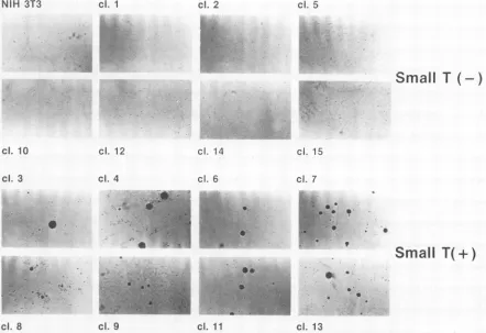
FIG. 7. Flat, dense foci formed by small T-antigen-expressing cellsoverthe backgroundofaflat NIH 3T3cell sheet. Eighty cells ofeach clone mixed with 10s NIH 3T3 cells were plated in 60-mm
dishes and incubated in DMEM-10% calfserum for 10 days. Cells were analyzedafterGiemsa staining. Uppereight panels: NIH 3T3
cell clones not expressing small T antigen. The upper left corner
shows parental NIH 3T3 cells. Lower eight panels: NIH 3T3 cell clones expressing small Tantigen.
be due to clonal variations present in the parental cell population.
Clones 3, 4, 6, 7, 8, 9, 11,and 13 producedsmallTantigen. In all cases, cells grew beyond the saturation density of
parental NIH 3T3 cells. The drop in cell number seen
between days 4 and6 wasdue to the detachment ofcells as aresult of overgrowth. Therewas some correlation between
theamount of small T antigen produced in the cellsand the growth rate of the cells. For example, clones 4, 7, and 11 produced low,intermediate, andhigh levels, respectively, of small Tantigen (Fig. 3), and these threeclones grew
increas-ingly more rapidly (Fig. 6). Although the growth curve of
clone 4 shown in Fig.6is similartothatof clones that donot
expresssmall T antigen, we were unabletomaintain clone4
as acell sheet for long after the cells reached confluency.
This increased growth potential of small T-antigen-producingcells beyondsaturationdensitycanalso beseenin
Fig. 7, which showsdishes in whichasmall number of small
T-antigen-expressing cellswere mixed with alargeexcessof
parental NIH 3T3 and incubated until the cells reached confluency. Theupperhalf of Fig. 7 shows the clonesof cells
notexpressing smallt antigen. These cells, including
paren-tal NIH 3T3 cells, formed a flat monolayer with very few
background foci. The lower eight panels represent cells expressing small Tantigen. In thesecases, clusters of small
T-antigen-expressing cells can be seen as flatbut dense foci over the background ofa flat sheet ofNIH 3T3 cells.
Since small Tantigen was found tostimulate thegrowth of
cells in monolayer, it is interesting to test whether small T
antigen induces anchorage-independent cell growth. The
upperhalf of Fig. 8 shows the clones of cells not expressing
small Tantigen and suspended in soft agar, while the lower
half represents those expressing small T antigen and
sus-pended in soft agar. The cells shown inthe upperhalf of Fig.
8 did notundergo more than two to three cell divisions in the
4-week period. On the contrary, cells expressing small T
antigen formed small colonies in soft agar. These colonies
usually contained about 20 to 30 cells after the 4-week
incubation. However, the properties of cells which formed
small colonies was rather small, ranging from about 0.1 to
10%, depending on the clones. In this case, we did not see a
clear correlation between the amount of small T antigen
producedand the frequency or size of small colonies formed
in soft agar. Eight of such small colonies were isolated from
three different clones of small T-antigen-expressing cells.
Only one of them formed small colonies at rather high
frequency, namely about 30%, while others showed a low
frequency of plating in soft agar, similar to that of the
parental clones, suggesting that a low frequency of colony
formation by cells expressing small T antigen is a stable
property of such cell clones (data not shown).
Small T-antigen-expressing cells were then tested to see
whether they formed tumors when inoculated into nude
mice. When the clone expressing the largest amounts of
small T antigen, namely clone 11, was used, no tumors were
observed during the 3 months after the inoculation of 106
cells per nude mouse.
DISCUSSION
By introducing the small T-antigen gene into cells with a
retrovirus vector as replication-defective retrovirus, we
were able to isolate NIH 3T3 cells expressing only
polyomavirus small T antigen. The reason we were able to
isolate such cells, while previous attemptsby others (7, 45)
using the calcium phosphate coprecipitation method were
unsuccessful, is most likely the introduction of the small
T-antigen gene into cells in a helper-free retrovirus form
which limits the number of gene copiesintroduced intocells
in the present studies. Thus, the amount of small T
antigen
produced in the cellswasbelow the toxic level. About halfof
the cell clones which expressed the neomycin gene also
expressed the small T-antigengene. It wasnotclearwhythe
other half of the clones thatexpressedtheneomycingene did
not express small Tantigen.It islikelythatapartorallofthe
small T-antigen gene is deleted in these cases. Precise
analysis of the structure of the integrated pGVST is
neces-sary to clarify this.
By examining the properties of these small
T-antigen-expressing cells, we were able to study the subcellular
localization of small T antigen and its effects on
microfil-ament bundleformation, cell morphology, andgrowth
char-acteristics.
Segawa and Ito (35) reported that small T antigen was
recovered mainly in the cytoplasmic soluble fraction in cell
fractionation studies. Immunofluorescent staining in the
present studies revealed, however, that it is present mainly
inthe nuclei (Fig. 4). Theseapparentlycontradictory results
suggest that small T antigen is present in the nuclei, not
boundtightly to DNAorothernuclearcomponents, but in a
soluble form. The immunofluorescent staining pattern of
small T antigen is indistinguishable from that of large T
antigen. It isinterestingtonotethat,although bothlarge and
on November 10, 2019 by guest
http://jvi.asm.org/
[image:6.612.56.294.71.368.2]NIH 3T3 cl. 1 cl. 2 cl. 5
Small
T (-)
cl. 10
cl. 3
cl. 12
cl. 4
cl. 14
ci. 6
cl. 15
cl. 7
;f
a W
4.
Cl. 8
.9
S
0*
cl. 9
,i^,.g..
.x
-.,..:
m.
6
Small
T(+)
cl. 13
FIG. 8. Colonyformation insoftagarof the clones of cells expressing and not expressing small T antigen. Cells(1(3)of 15 cellclones (Fig. 3)aswellasparental NIH 3T3 cells weresuspended in0.33'S agarin 60-mmplasticculture plates and incubated for 4 weeks at 37°C by the method of MacPherson and Montagnier(24). Cells cultured in suspension in soft agarwerephotographed through usingamicroscope with low-power magnification. Upper eight panels:NIH 3T3cellclones not expressing small T antigen. The upper leftcornershowsparental NIH 3T3 cells. Lowereight panels: NIH 3T3 cell clonesexpressingsmall T antigen.
small Tantigens show a similar immunofluorescent staining
pattern,largeTantigen, aDNA-binding protein, stays in the
nuclei, while small T antigen easily comes out of the nuclei
during the cell fractionation procedures. This may suggest
that small t antigen is not a DNA-binding protein. In any
event, itis important to note that polyomavirus transforming
genesproduce two distinct geneproducts, both of which are
localized mainly to the nuclei. It has been shown that large T
antigen has signal peptides necessary for its nuclear local-ization (29). The positions of such signals are outside the
large T-small Tcommon region. A similar peptide sequence
does not appearto be present in the small T-antigen unique
region. It remains to be seen whether small T antigen hasa
different type of signal for nuclear localization. Small T
antigenof simian virus40(SV40), which hasahighdegree of amino acid sequence homology with polyomavirus small T
antigen, has been showntobe present both in the nucleiand
in the cytoplasm (3, 11).
Rodent fibroblast cells, after transformation by
poly-omavirus, usually lose well-developed actin-containing
mi-crofilament bundles andtend to show diffuse actin staining.
particularly in membrane rufflesand at theedges ofthecells.
Established lines of rodent fibroblasts transformed by middle
Tantigen alone contain a greatly reduced amount of
micro-filament bundles indistinguishable from that seen in
polyomavirus-transformed cells (19,23).Incontrast,the hr-t
mutants, whichinduce normal largeTantigenbut notmiddle
or small T antigens, do not alter the microfilament bundle
pattern (19, 34). Using viral mutants which induce normal
large and smallTantigens but not middle T antigen, Liang et
al. (23) and Itoet al. (19) deduced that both large and small
Tantigens probably donot have any effect onmicrofilament
organization. We have shown unambiguously in this paper
that smallTantigen haslittle, if any. effect on microfilament
bundling. It is interesting to note that SV40 small t antigen
has been reported to alter microfilament pattern (3, 15).
Consideringthe high degree of sequence homology between the twosmallTantigens, it will be interesting to examine the
significance of this difference.
Polyomavirus small T antigen has been transiently
overexpressed inmonkey CV-1 cellsby Zhuet al.(45), who
used SV40-polyoma recombinant virus. They observed that
polyomavirus small t antigen is localized both in the nuclei
andinthecytoplasm,causesdrasticchangesinmorphology,
and makes cells detacheasily. It seemsthatthedifferencein
the results is attributable to differences in the two
experi-mental systems, monkey epithelial cells (CV-1) versus
mouse fibroblasts and overexpression versus low-level
ex-pression.
Rundell and co-workers have reported in their series of
papers that SV40, BK virus, human papovavirus, and
polyomavirus small T antigens are bound to two cellular
proteins of32 and 56 kDa (5, 31, 44). We are able to show
that small T antigen and the 32-kDa cellular protein are
coprecipitatedwith anti-Tantibodies,probablybecausethey
are bound. The 32-kDa protein that we observed might
correspond tothe 32-kDa protein describedby Rundell and
co-workers. Itis notclearinFig.4whethera56-kDa
protein
...."P
,.F
:-j".,
I 6.,
i.., -,
.., :..
"E
on November 10, 2019 by guest
http://jvi.asm.org/
[image:7.612.86.528.73.376.2]is coprecipitating with polyomavirus smallTantigenbecause of the high background. It remainstobe elucidated whether the association of small Tantigen witheither the 32-kDa or
the 32-and 56-kDa cellularproteins isimportant forsmall T antigen to exert its function.
Small T antigen has littleor noeffectonthemorphology of
NIH 3T3 cells. We made the most importantobservation in
the present studies in finding out the function of small T antigen when we compared the growth characteristics of
cellsexpressing small Tantigenwith thoseofparental cells. NIH 3T3 cells expressing small T antigen form a flat
monolayeratconfluency. However, these cellsaredriven to
grow without stoppingat the saturation density ofparental cells. Small T antigen is also able to induce anchorage-independent growthin NIH 3T3 cells, although this effectis
weak. Consistent withthis weakactivity ininducing anchor-age-independent growth,cells expressing small Tantigendo
notform tumors in nude mice. Theseproperties ofNIH 3T3 cells containing small T antigen are reminiscent ofthose of
NIH 3T3 cells stimulated to grow by mitogenic growth
factors, such as epidermal growth factor orplatelet-derived
growth factor (reviewed in reference 33). These growth factors are usually mitogenic to the cells in monolayer culture and increase the cell density. Except for some
transforming growth factors such as TGF-cx (8), however.
they do not usually stimulate cells to divide when the cells
are suspended in soft agar. Therefore, we suggest thatsmall Tantigen has a growthfactor-like activity and enables cells
to overcome a growth-inhibitory effect caused by high cell
density. It remains to be seen whether small T antigen will
induce cellular DNA synthesis inquiescentNIH 3T3cells. It has been suggested that SV40 small T antigen also has a
function associated with cellular growth control, as
dis-cussed by Bossert et al. (5).
Itisunlikely that small Tantigenstimulates cell growthby
actingoncells externally, since small T antigen is presentin
the nuclei. Although we have not tested whether small T
antigenis alsoreleased into theculturemedium, preliminary results suggest that the culture medium of small T-antigen-expressing cells doesnotcontain anactivity whichpromotes the growth of NIH 3T3 cells, suggesting that the growth-stimulating activity of small T antigen that we observed is
not due to action of small T antigen from outside the cells. Since small T antigen has a growth factor-like
growth-promoting activity and is present in nuclei, it is tempting to speculate that small T antigen may be involved ina nuclear
event that regulates cellular DNA synthesis. Since signals fromactivated growth-factor receptors induce cellular DNA synthesis and celldivision, small T antigen might share with growth-factor receptors some nuclear mechanism by which
cellular DNA synthesis is induced.
The fact that small T antigen has its own independent
function raises the question of whether middle Tantigenalso hasthatfunction, since the entire polypeptide chain ofsmall T antigen except for four carboxy-terminal amino acids is
completely included in middle T antigen (Fig. 1). We have
not yet done the experiments which directly address that question. However, we have observed that the addition of
small Tantigen tomiddle T-antigen-expressing cells results ina dramatic enhancement of the growth rate of the cells in
soft agar without enhancing the activity associated with
middle Tantigen. The results suggest that middle T antigen
does not exert small T-antigen function. This point, namely the question ofwhether small T antigen has its own unique
function not shared by large or middle T antigen. is the subject ofour paper in preparation.
We have established that small T antigen is the second
polyomavirus transforming gene product which is localized
to the nuclei. Sincethe behavior of the cellsexpressing only
large T antigenobservedpreviously(22,34) and that ofsmall
T antigen observed in the present studies arevery different
and the primary structures of theunique region oflargeand
small T antigens are verydifferent (Fig. 1), we assume that
the biochemical function oflargeand small Tantigenwill be
very different, too. We will continue oureffort to elucidate
whether large andsmallTantigens contributetothe process
of cell transformation by entirelydifferent mechanisms.
ACKNOWLEDGMENTS
We thank R. Kamen for plasmids pPyST1 and pPyMT1 and COP-5cells,R. Mulliganfor02cells, and B. E. Griffinfora set of rat monoclonal antibodiesagainstpolyomavirusTantigens.
The research was sponsored by the National Cancer Institute, under contract no. NO1-CO-23909 with Litton Bionetics, Inc.
LITERATURECITED
1. Asselin, C., C. Gelinas, and M. Bastin. 1983. Roleof the three polyoma early proteins in tumorigenesis. Mol. Cell. Biol. 3:1451-1459.
2. Asselin, C., C. Gelinas, P. E. Branton, and M. Bastin. 1984. Polyoma middle T antigen requires cooperation from another gene to express the malignant phenotype in vivo. Mol. Cell. Biol. 4:755-760.
3. Bikel,I.,T. M.Roberts,M. T.Bladon,R.Green,E.Amann,and D. M.Livingston. 1983.Purificationofbiologically activesimian virus 40 small tumor antigen. Proc. Natl. Acad. Sci. USA 80:906-910).
4. Bolen, J. B., C. J. Thiele, M. A. Israel, W. Yonemoto, L. A. Lipsich, and J. S. Brugge. 1984. Enhancement of cellular src gene product associated tyrosyl kinase activity following polyoma virus infection andtransformation. Cell38:767-777. 5. Bossert, A.,P.Mulgaonkar,and K.Rundell. 1985.Interaction of
simian virus 40 small-Tantigenproduced in bacteria with 56K and 32K proteins of animal cells. J. Virol. 56:325-327. 6. Courtneidge, S. A., and A. E. Smith. 1983. Polyoma virus
transforming protein associates with the product of the c-src cellular gene. Nature (London)303:435-439.
7. Cuzin,F., M.Rassoulzadegan,andL. Lemieux. 1984.Multigenic control oftumorigenesis: threedistinct oncogenes are required fortransformation ofratembryo fibroblasts by polyomavirus, p. 109-116.InG. F. Vande Woude, A.J. Levine, W.C.Topp, and J. D. Watson (ed.), Cancer cells, vol. 2. Oncogenes and viral genes. ColdSpringHarbor LaboratoryPress, ColdSpring Harbor, N.Y.
8. Delarco, J. E., and G. J. Todaro. 1978. Growth factors from murine sarcomavirus-transformedcells. Proc. Natl. Acad. Sci. USA 75:4001-4005.
9. Dilworth, S. M., andB. E.Griffin. 1982. Monoclonalantibodies against polyoma virus tumor antigens. Proc. Natl. Acad. Sci. USA 79:1059-1063.
10. Eckhart, W., M. A. Hutchinson, and T. Hunter. 1979. An activity phosphorylating tyrosine in polyoma Tantigen immu-noprecipitates. Cell 18:925-933.
11. Ellman, M., I. Bikel, J. Figge, T. Roberts, R. Schlossman, and D. M.Livingston. 1984. Localization of the simian virus 40 small Tantigen in the nucleus and cytoplasm ofmonkey and mouse cells. J. Virol. 50:623-628.
12. Emerman, M.,and H. M. Temin. 1984.Genes with promoters in retrovirus vectors can be independently suppressed by an epigenetic mechanism. Cell 39:459-467.
13. Garapin, F. C., F. Horodniceanu, P. Kourisky, and A. C. Garapis. 1981. A new dominant hybrid selective marker for highereukaryotic cells. J. Mol. Biol. 150:1-14.
14. Gaudray, P., C. Tyndall, R. Kamen, and F. Cuzin. 1981. The high affinity binding site on polyoma virus DNA for the viral large-T protein. Nucleic Acids Res. 9:5697-5710.
on November 10, 2019 by guest
http://jvi.asm.org/
15. Graessman,A.,M.Graessman,R.Tjian, and W. C. Topp. 1980. Simian virus 40 small-T protein is required for loss ofactincable networks inratcells. J. Virol. 33:1182-1191.
16. Ito,Y. 1979. Polyoma virus-specific 55K proteinisolated from plasma membrane of productively infected cells is viruscoded
and important for cell transformation. Virology 98:261-266. 17. Ito, Y. 1980. Organization and expression of the genome of
polyoma virus, p.447-480. lit G. Klein (ed.). Viral oncology. Raven Press, New York.
18. Ito, Y., J.R.Brocklehurst,and R. Dulbecco. 1977.Virus-specific proteins in the plasma membrane of cells lytically infected or
transformed by polyoma virus. Proc. Natl. Acad. Sci. USA 74:4666-4670.
19. Ito, Y., Y. Hamagishi, K. Segawa, T. Dalianis, E. Appella, and M. Willingham. 1983. Antibodies against a nonapeptide of
polyoma virus middleT antigen: cross-reaction withacellular
protein. J. Virol. 48:709-720.
20. Ito, Y., N. Spurr, and R. Dulbecco. 1977. Characterization of polyoma virus T antigen. Proc. Natl. Acad. Sci. USA 74:1259-1263.
21. Ito,Y., N. Spurr, and B. E.Griffin. 1980. MiddleTantigenas
primary inducer of full expression of the phenotypeof transfor-mation by polyoma virus. J. Virol. 35:219-232.
22. Lania, L., M. Griffiths, B. Cooke, Y. Ito, and M. Fried. 1979. Untransformedratcellscontaining freeandintegratedDNAof
apolyoma non-transforming (hr-t) mutant.Cell 19:793-802. 23. Liang, T.J., G. G. Carmichael, andT. L. Benjamin. 1984. A
polyomamutantthatencodes smallTantigenbutnotmiddleT antigen demonstrates uncoupling of cell surface and cytoskele-talchanges associated with cell transformation. Mol. Cell. Biol. 4:2774-2783.
24. MacPherson, I., and L. Montagnier. 1964. Agar suspension culturefor the selectiveassayof cells transformed by polyoma
virus. Virology 23:291-294.
25. Maniatis, T., E. F. Fritsch, andJ. Sambrook. 1982. Molecular cloning: alaboratory manual. Cold Spring Harbor Laboratory,
Cold SpringHarbor, N.Y.
26. Mann, R., R. C. Mulligan,and D.Baltimore.1983. Construction ofaretroviruspackagingmutantand itsusetoproduce helper-free defective retrovirus. Cell 33:153-159.
27. Rassoulzadegan, M., A. Cowie, A. Carr, N. Glaichenhaus, R. Kamen, and F. Cuzin. 1982. The role of individual polyoma virus early proteinsin oncogenic transformation. Nature
(Lon-don) 300:713-718.
28. Rassoulzadegan, M., Z. Naghashfar, A. Cowie, A. Carr, M. Grisoni,R. Kamen,and F.Cuzin. 1983. Expression ofthelarge T protein of polyoma virus promotes the establishment in culture of "normal" rodent fibroblast cell lines. Proc. Natl. Acad. Sci. USA 80:4354-4358.
29. Richardson, W. D., B. L. Roberts, and A. E. Smith. 1986. Nuclearlocalization signals in polyoma virus large Tantigen. Cell 44:77-85.
30. Robins, T., C. Jhappan, J. Chirikjan, and G.F.Vande Woude. 1986. Molecular cloning of the 'intronless" EJ ras oncogene
using a murine retrovirus shuttle vector. Gene Analysis Tech-niques 3:12-16.
31. Rundell, K.,E.0.Major, and M. Lampert. 1981. Association of cellular56,000-and 32.000-molecular-weight proteins with BK virus and polyomavirus t-antigens. J. Virol. 37:1090-1093. 32. Schaffhausen, B.S., and T. L. Benjamin. 1979. Phosphorylation
of polyoma T antigen. Cell 18:935-946.
33. Scher, C. D., R. C. Shepard, H. N. Antoniades,and C. D. Stiles. 1979. Platelet-derived growth factor and the regulation of the mammalian fibroblast cell cycle. Biochim. Biophys. Acta 560:217-241.
34. Schlegel, R., and T. Benjamin. 1978. Cellular alterations depen-dent upon the polyoma virus hr-t function: separation of mito-genic fromtransforming capacities. Cell 14:587-599.
35. Segawa, K., and Y. Ito. 1982. Differential subcellular localiza-tion of in viivo-phosphorylated andnonphosphorylated middle-sized tumor antigen ofpolyoma virus and its relationship to middle-sized tumor antigen phosphorylating activity in vitro. Proc.Natl. Acad. Sci. USA 79:6812-6816.
36. Smart, J. E., and Y. Ito. 1978.Three species ofpolyomavirus tumorantigens share common peptides probably near the amino termini ofthe proteins. Cell 15:1427-1437.
37. Smith, A. E., R. Smith, B. E. Griffin, and M. Fried. 1979. Protein kinaseactivityassociated withpolyoma virus middle T antigen inli/ro. Cell 18:915-924.
38. Soeda, E., J. R. Arrand, N. Smoller, J. E. Walsh, and B. E. Griffin. 1980. Coding potential and regulatory signals of the polyoma virus genome. Nature(London) 283:445-453. 39. Southern, P. J., and P. Berg. 1982.Transformation of
mamma-lian cells to antibiotic resistance with a bacterial gene under control of theSV40early region promoter. J. Mol.AppI.Genet. 1:327-341.
40. Treisman, R., U. Novak, J. Favaloro, and R. Kamen. 1981. Transformation of rat cells by an altered polyoma virus genome expressing only the middle T protein. Nature (London) 292:595-600.
41. Tyndall, C., G. L.Mantia, C. M. Thacker, J. Favaloro, andR. Kamen. 1981. A regionof the polyoma virus genome between the replication origin and late protein coding sequences is required in cis for both early gene expression and viral DNA replication. Nucleic Acids Res. 9:6231-6250.
42. Wigler, M., A. Pellicer, S. Silverstein, and R. Axel. 1978. Biochemicaltransfer ofsingle-copyeukaryotic genes using total cellular DNAas donor. Cell 14:725-731.
43. Wulf, E., A. Deboben, F. A. Bautz, H. Faulstich, and T. Wieland.1979. Fluorescentphallotoxin,atoolforvisualization of cellular actin. Proc. Natl. Acad. Sci. USA 76:4498-4502. 44. Yang,Y.-C.,P.Hearing, and K. Rundell. 1979.Cellular proteins
associated with simian virus 40 early gene products in newly infectedcells. J. Virol. 32:147-154.
45. Zhu, Z., G.M. Veldman, A. Cowie, A. Carr, B.Schaffhausen, and R. Kamen. 1984.Construction and functional characteriza-tionofpolyomavirusgenomes thatseparatelyencode the three early proteins. J.Virol. 51:170-180.

![FIG.3.sodiuminfectionrespectively.immunoprecipitated Autoradiogram of immunoprecipitated [35S]methionine-labeled small T antigen](https://thumb-us.123doks.com/thumbv2/123dok_us/1373610.90674/4.612.85.274.456.624/sodiuminfectionrespectively-immunoprecipitated-autoradiogram-immunoprecipitated-methionine-labeled-small-antigen.webp)