A novel genetic switch controls phase variable expression of
CwpV, a
Clostridium difficile cell wall protein
mmi_6812 541..556Jenny E. Emerson,†§Catherine B. Reynolds,§ Robert P. Fagan, Helen A. Shaw, David Goulding‡ and Neil F. Fairweather*
Division of Cell and Molecular Biology, Imperial College London, London SW7 2AZ, UK.
Summary
Clostridium difficile is a nosocomial pathogen that can cause severe gastrointestinal infections. C. difficile encodes a family of cell wall proteins, some of which are implicated in pathogenesis. Here we have characterized CwpV, the largest member of this family. CwpV is surface expressed and post-translationally processed in a manner analogous to the major S-layer protein SlpA. Expression ofcwpV is phase variable, with approximately 5% of cells in a population expressing the protein under standard laboratory growth conditions. Upstream of cwpV, inverted repeats flank a 195 bp sequence which undergoes DNA inversion. Use of a gusA transcrip-tional reporter demonstrated that phase variation is mediated by DNA inversion; in one orientationcwpV is expressed while in the opposite orientation the gene is silent. The inversion region contains neither the promoter nor any of the open reading frame, therefore this system differs from previously des-cribed phase variation mechanisms. The cwpV pro-moter is located upstream of the inversion region and we propose a model of phase variation based on intrinsic terminator formation in the OFF transcript. A C. difficilesite-specific recombinase able to catalyse the inversion has been identified.
Introduction
Clostridium difficile is a Gram-positive, spore-forming anaerobe that causes a range of gastrointestinal dis-eases, ranging from diarrhoea to pseudomembraneous colitis, collectively termedClostridium difficile-associated
disease (CDAD) (Poxton et al., 2001; Bartlett, 2006).
Infections in humans are most commonly associated with antibiotic therapy in nosocomial environments and have been increasing in both number and severity in recent
years (Barbut et al., 2007). Antibiotics are thought to
disrupt the normal intestinal flora, allowing C. difficile to colonize the gut. The principal virulence factors produced by C. difficile are two cytotoxins, TcdA and TcdB. The modes of action of TcdA and TcdB are well described: both toxins, which are highly related in structure and func-tion, are glucosyltransferases that target small GTPases resulting in alterations in the cytoskeleton, apoptosis, infiltration of neutrophils and damage to the gut mucosa
(Justet al., 1995; Voth and Ballard, 2005).
In order to colonize their hosts, pathogenic bacteria must both evade the immune response and interact with host cells, often adhering to specific surface-localized molecules (Pizarro-Cerda and Cossart, 2006). Bacterial surface proteins and structures play key roles in these
processes. In C. difficile the major surface proteins are
within the S-layer, a paracrystalline proteinaceous array that completely coats the bacterium. The S-layer is formed of two proteins, the high-molecular-weight S-layer protein (HMW SLP) and the low-molecular-weight (LMW) SLP,
which are products of the SlpA precursor (Calabi et al.,
2001). After cleavage of the SlpA precursor, the resulting HMW and LMW SLPs interact via defined domains to form
a stable heteromeric complex (Faganet al., 2009). In the
genome ofC. difficile630, 28 paralogues of the HMW SLP
have been identified (Sebaihia et al., 2006), and
trans-criptomic and proteomic studies have shown several of these proteins to be expressed under laboratory conditions
(Wright et al., 2005; Emerson et al., 2008). Antibodies
against some of these proteins are found in serum of
CDAD patients (Wrightet al., 2008) implying at least some
of this family of cell wall proteins (CWPs) are expressed in vivoduring infection. All of these CWPs contain two or three cell wall binding motifs (Pfam PF04122) in addition to a unique domain that is proposed to specify function. Accepted 15 July, 2009. *For correspondence. E-mail n.fairweather@
imperial.ac.uk; Tel. (+44) 20 7594 524; Fax (+44) 20 7594 3069. Present addresses: †Department of Biochemistry, University of
Oxford; South Parks Road, Oxford OX1 3QU; ‡The Wellcome
Trust Sanger Institute, Wellcome Trust Genome Campus, Hinxton, Cambridge, CB10 1SA, UK.§These authors contributed equally to
this work.
Re-use of this article is permitted in accordance with the Terms and Conditions set out at http://www3.interscience.wiley.com/ authorresources/onlineopen.html
Molecular Microbiology(2009)74(3), 541–556 䊏 doi:10.1111/j.1365-2958.2009.06812.x First published online 6 August 2009
In C. difficile several CWPs have been identified that may interact with the host to facilitate adherence. These
include the adhesin Cwp66 (Waligora et al., 2001), the
fibronectin binding protein Fbp (Hennequin et al., 2003)
and the protease Cwp84 (Janoiret al., 2007). The HMW
SLP also shows strong binding to the intestinal epithelium of both human and mouse and anti-HMW SLP antibodies
block adherence to HEp-2 cells (Calabiet al., 2002). SLPs
have been shown to induce inflammatory and regulatory cytokines in human monocytes and dendritic cells, sug-gesting that during infection they may interact with the
immune system (Ausielloet al., 2006).
In this study we investigate the expression, regulation and processing of another member of the CWP family, which we term CwpV. We show that CwpV is surface expressed and post-translationally processed in a manner
analogous to SlpA. We demonstrate that cwpV
expres-sion is phase variable and that the mechanism of tran-scriptional control differs from those previously described.
We propose a mechanism for this phase variation and
have identified one C. difficile site-specific recombinase
that mediates DNA inversion.
Results
Surface localization and processing of CwpV
In C. difficile 630, the cwpV gene (CD0514) encodes a predicted protein of 167 kDa, containing an N-terminal signal peptide, three PF04122 cell wall binding motifs presumed to mediate attachment to the underlying cell wall, a serine/glycine-rich region and finally nine repeats, each of 120 amino acids (Fig. 1). Analysis of other strains ofC. difficilerevealed that the number of repeats is vari-able, with CDKK371 having six repeats and R8366 and Y having four repeats. CwpV is annotated as a putative
adhesin (Sebaihiaet al., 2006) based on homology to a
known haemagglutinin fromSalmonella typhimurium. To
further characterizecwpV,the gene fromC. difficile 630
was cloned into pMTL960, anEscherichia coli–C. difficile
shuttle vector. The promoter Pcwp2 utilized was that of
another cell wall protein, Cwp2, that is moderately
expressed in C. difficile(Calabi and Fairweather, 2002).
We also constructed acwpVgene knockout inC. difficile
630Derm using the Clostron technique (Heap et al.,
2007). Expression of CwpV was investigated using two antibodies; one raised against the N-terminal cell wall binding domain (anti-CwpVNter) and a second raised against the first of the nine repeats (anti-CwpVrpt1). Cell
wall extracts of C. difficile were prepared using low pH
[image:2.595.57.287.64.140.2]glycine extraction, which enriches for the SLPs and other minor surface localized proteins including CwpV (Calabi et al., 2001; Wrightet al., 2005). Figure 2 shows that in strain 630, CwpV is detected as two fragments. The Fig. 1. Domain architecture of CwpV. The N-terminus of CwpV
contains a signal sequence (black), followed by three PF04122 cell wall anchoring motifs (grey) and a region of unknown function (white). A serine–glycine rich region (pink) precedes a number of repeats (blue). The repeats are almost identical in sequence with the exception of the N-terminal repeat which shows more sequence diversity. Different strains have different numbers of repeats. Sequences were deposited at EMBL with accession numbers FM17250, FM17252 and FM17254.
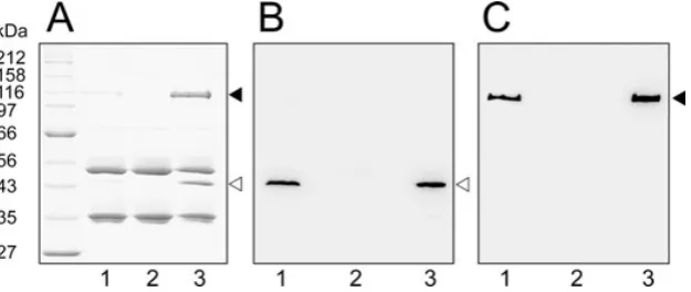
Fig. 2. Surface expression of CwpV.C. difficilestrains were grown overnight in BHI broth. S-layer extracts were prepared and analysed by SDS-PAGE and Western blotting.
A. Coomassie blue-stained gel.
B. Western blot using anti-CwpVNter (1:5000). C. Western blot using anti-CwpVrpt1 (1:5000).
Lane 1: 630; lane 2: 630DermDcwpV; lane 3: 630DermDcwpV(pCBR044). The 40 kDa (䉰) and 120 kDa (䉳) fragments of CwpV are indicated. For Western blots the total protein loaded in lane 3 was fivefold less than in lanes 1 and 2 to allow all bands to be visualized using one exposure time.
[image:2.595.144.457.488.620.2]smaller 40 kDa fragment represents the cell wall bind-ing domain and reacts with anti-CwpVNter. The larger 120 kDa fragment contains the repeat sequences and reacts with anti-CwpVrpt1. This indicates that CwpV is post-translationally cleaved in a manner analogous to
the major S-layer protein SlpA (Calabi et al., 2001).
Neither fragment is observed in thecwpVdeletion strain
630DermDcwpV. In the complemented mutant 630D erm-DcwpV(pCBR044) expression is clearly at a higher level than the wild-type strain, as together both fragments rep-resent a considerable percentage of the total protein in the glycine extract. Full-length CwpV was never observed either in cell wall extracts or in whole cell lysates (data not shown), even in the complemented mutant, indicating that processing is highly efficient and that it may be required for correct localization of the protein in the cell wall.
Expression of CwpV is phase variable
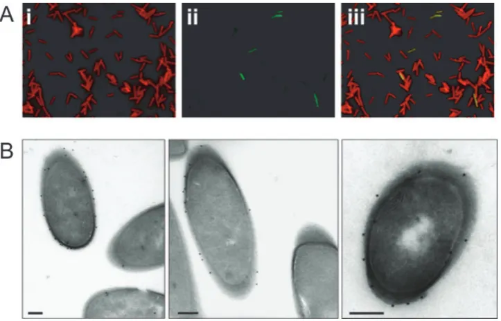
Localization of CwpV in 630 was investigated by immu-nofluorescence on intact bacteria using anti-CwpVrpt1 and co-staining with anti-LMW SLP antibody. All bacteria were labelled with anti-LMW SLP whereas only a small proportion of cells were stained with anti-CwpVrpt1 (Fig. 3A). In those bacteria that did express CwpV, the protein was localized to the cell surface as observed by immunogold electron microscopy (Fig. 3B). In cultures
grown in brain–heart infusion (BHI) broth the proportion of cells expressing CwpV was found to be consistently between 5% and 10%. A similar proportion of CwpV posi-tive cells was seen in colonies grown on BHI agar and we were unable to detect single colonies where all the cells were either expressing or not expressing CwpV (data not shown).
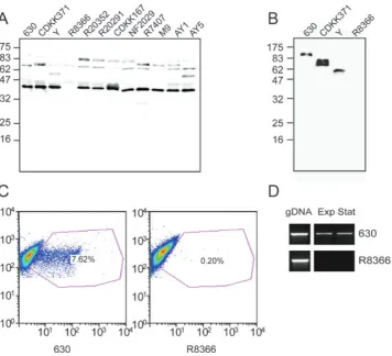
Expression of CwpV was analysed in a panel of strains using Western blotting with the anti-CwpVNter antibody. All strains analysed were found to express CwpV with the exception of R8366 (Fig. 4A). Representative strains encoding four, six and nine copies of the 120 bp repeat in their genome were further investigated using anti-CwpVrpt1 antibody. All these strains, with the exception of R8366, expressed this domain with a detected band size representative of the number of encoded repeats (Fig. 4B). Flow cytometry analysis of 630 and R8366 using anti-CwpVrpt1 antibodies revealed approximately 7% of 630 cells to express CwpV, whereas R8366 cells were not labelled (Fig. 4C). DNA sequence analysis
revealed that R8366 contained an intactcwpVgene (data
[image:3.595.117.479.405.637.2]not shown), suggesting that the expression defect might be at the level of transcription. Reverse transcription poly-merase chain reaction (PCR) analysis of 630 and R8366 showed that transcripts were detected in 630 grown to exponential and stationary phase, whereas no transcripts were detected in R8366 (Fig. 4D).
Fig. 3. Phase variable expression of CwpV.
A.C. difficile630 were grown in BHI broth and labelled with (i) rat anti-LMW SLP (red) showing that all bacteria are surface labelled and (ii) anti-CwpVrpt1 (green) showing a fraction of bacteria labelled. Labelling was visualized using Texas Red anti-rat conjugate and fluorescein anti-rabbit conjugate. (iii) merge of (i) and (ii).
B. Immunogold electron microscopy of 630 labelled with anti-CwpVrpt1 antibodies. Staining is seen only on the external surface of cells. Bar=200 nm.
Inverted repeats upstream of cwpVmediate DNA inversion
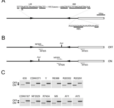
Examination of the DNA sequence upstream of thecwpV
open reading frame (ORF) revealed a pair of imperfect 21 bp inverted repeats (IRs) 57 bp upstream of the start codon and separated by 195 bp (Fig. 5A). A small number of previously described genetic switches in bacteria rely on DNA inversion catalysed by site-specific recombi-nases, which bind to IRs and catalyse strand exchange. The best-characterized example of such a system is the
phase variation of Type 1 fimbriae in E. coli, where the
fimA promoter is located between two IRs (Abraham
et al., 1985). Inversion of the sequence bounded by the IRs, which is mediated by specific recombinases FimB
and FimE, results in ON–OFF switching of expression
offimA(reviewed in Hendersonet al., 1999).
In order to determine whether the IRs upstream ofcwpV
[image:4.595.123.480.52.376.2]lead to inversion of the DNA sequence between the IRs, PCR was performed on genomic DNA using primer NF823 with either NF826, designed to detect the orien-tation as published in the genome sequence (Sebaihia et al., 2006) (hereafter referred to as OFF), or with NF825, designed to detect the inverted orientation (ON) (Fig. 5B). As shown in Fig. 5C, bands were detected with both primer pairs in all strains tested apart from R8366, indi-cating that the genomic DNA between the IRs of these strains does undergo inversion. DNA sequencing of these PCR products revealed that recombination occurs within the central region of identity of the IR as the outer T/G Fig. 4. Expression ofcwpVis seen in all strains except R8366.
A. Western blot analysis of a panel ofC. difficilestrains. Glycine extracts were prepared and probed with anti-CwpVNter antibody (1:1000). A band at 40 kDa was observed in all strains, with the exception of R8366.
B. Western blot analysis of stains using anti-CwpVrpt1 antibody. The sizes of bands (120 kDa in 630, 82 kDa in CDKK371 and 57 kDa in Y) are consistent with the predicted sizes of the repetitive regions of these strains. R8366 showed no reaction.
C. Flow cytometry analysis of 630 and R8366 stained using anti-CwpVrpt1 antibody and fluorescein anti-rabbit conjugate. Side scatter, which is representative of particle size, is shown on they-axis and fluorescence intensity on thex-axis. Frequency of events (bacteria) is indicated by colour. Unstained bacteria are visualized in a grouping at the left of the graph and the purple polygon represents the area in which events were scored as positive. 7.62% of 630 bacteria are positively stained, whereas negligible positive events are seen with R8366.
D. RNA was extracted from mid-exponential (Exp) and early stationary (Stat) phase cultures, reverse transcribed and the conserved 5′region ofcwpVwas amplified by PCR using primers NF654 and NF655. Transcription is seen in 630 but not in R8366. Genomic DNA (gDNA) was used a positive control.
mismatch does not differ between the ON and OFF sequences, whereas the inner C/T mismatch inverts along with the inter-IR DNA (data not shown).
In strain R8366, which does not express CwpV, DNA could only be detected in the OFF orientation. This sug-gests DNA inversion is greatly reduced or absent in this strain. Examination of the DNA sequence upstream of
cwpVin a number of strains revealed very high sequence
conservation (data not shown). However, in R8366, a two-nucleotide deletion was observed in the left IR (LIR) (Fig. 5A). We therefore hypothesize that this deletion pre-vents DNA inversion in strain R8366 and that inversion is
necessary for expression ofcwpV.
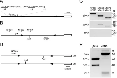
Initiation of transcription is upstream of the IRs
In order to locate the start site of transcription ofcwpVwe
performed 5′ RACE (Rapid Amplification of cDNA Ends)
analysis on mRNA isolated from strain 630. Several clones were obtained and were sequenced to determine the 5′ nucleotide of the transcript. Surprisingly, the 5′ end was not present within the sequence bounded by the IRs, but was localized 36 or 37 bases upstream of the
LIR (data not shown; Fig. 6A). Putative -10 and -35
sequences, closely matching those recognized by the
major bacterial sigma factor s70 in other Gram-positive
[image:5.595.112.480.60.408.2]promoters (Wosten, 1998), were identified upstream of Fig. 5. The region upstream region of cwpV contains inverted repeats and undergoes DNA inversion.
A. Diagrammatic representation of features upstream ofcwpVin 630. Two inverted repeats, LIR and RIR, are found upstream of thecwpV translational start site in the genomic DNA. DNA is shown in the published OFF orientation and the two base pair deletion in strain R8366 is indicated (D). The predicted RBS sequence and the ATG are shown.
B. Diagrammatic representation of the inversion region showing the two orientations. The annealing sites of primers used to probe the orientation are shown. NF823 anneals outside the inverted repeats. NF825 and NF826 anneal inside the inversion region in opposite orientations.
C. PCR amplification to detect the orientation of the inverted repeats in genomic DNA. Two PCR reactions were performed on genomic DNA from each strain ofC. difficile to determine the orientation of the inversion region. NF823 and NF825 give a product in the ON orientation; NF823 and NF826 give a product in the OFF orientation. All strains were found to contain the DNA switch in both orientations apart from strain R8366 in which no DNA in the ON orientation can be detected. DNA sequencing of genomic DNA of 630, R20352 and R20928 and several other strains revealed the majority of DNA is in the OFF orientation.
the site of initiation (see Fig. 6A). A second subset of clones revealed a start site four bases upstream of the
LIR, but no-35 or-10 sequences are seen upstream of
this site. It is therefore likely that these transcripts result from trimming of a longer transcript rather than the exist-ence of two transcriptional start sites. To further confirm the location of the transcriptional start site ofcwpV, cDNA was prepared from 630 and PCR-amplified using a reverse primer (NF826) located within the IRs (in the ON orientation) with a series of forward primers localized further upstream (Fig. 6B). Products were successfully amplified using forward primers NF863 or NF875 located within and downstream of the LIR respectively, but not with primer NF824 located further upstream (Fig. 6C). These results confirm that the promoter ofcwpV(PcwpV) is located proximal to and upstream of the LIR.
Interestingly, all RACE clones sequenced were
observed to be in the ON orientation; no clones were isolated that contained the sequence between the IRs in
the OFF orientation. To further probe the role of DNA
inversion in expression ofcwpV, RNA was extracted from
630, converted to cDNA and PCR-RFLP analysis was carried out. Using primers NF863 and NF823 a product was amplified extending from the observed transcriptional
start site to within thecwpVgene. This PCR product was
then digested with AseI, the cleavage site of which is
situated asymmetrically within the region bounded by the IRs (Fig. 6D). The results (Fig. 6E) clearly show that
the cwpV mRNA is almost entirely present in the ON
orientation. In contrast, the majority of the genomic DNA is present in the OFF orientation. It therefore appears that stable full-length transcripts can only be produced from genomic DNA in the ON orientation. This is consistent with the detection of CwpV expression in only around 5% of cells, corresponding to those few cells with genomic DNA in the ON orientation. The region of DNA bounded by the
IRs, located between the promoter and the cwpV ORF,
[image:6.595.60.541.62.384.2]was named thecwpVswitch.
Fig. 6. Transcription ofcwpVinC. difficile.
A. Diagrammatic representation of thecwpVpromoter and transcriptional start site as determined by 5′RACE.
B and C. PCR amplification to confirm the transcriptional start site. cDNA was prepared from 630 and amplified by PCR using primer NF826 together with primer NF824, NF863 or NF875. Amplification is seen with primers NF875 and NF863 but not NF824, confirming that the start site of transcription lies between the NF824 and NF863 binding sites. Positive (gDNA) and negative (RNA) controls behaved as expected. D and E. RFLP analysis ofcwpVcDNA. cDNA was prepared from 630, amplified by PCR using primers NF863 and NF823 and digested with AseI. Bands detected from DNA in the ON and OFF orientations are indicated. Densitometry analysis revealedcwpVmRNA is predominantly in the ON orientation (99%), and the genomic DNA (gDNA) is predominantly in the OFF orientation (96%).
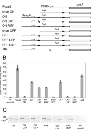
Characterization of the cwpVgenetic switch using a b-glucuronidase reporter
To study further the ability of thecwpVswitch to control
transcription, a series of transcriptional reporter
con-structs was created in E. coli using gusA, encoding
b-glucuronidase, which has been used previously to
measure activity of C. difficile promoters (Mani and
Dupuy, 2001). The promoterlessgusAgene together with
thetcdB ribosome binding site from pTUM177 was first
cloned into pUC19 downstream of Pcwp2. GusA expression
driven by Pcwp2 could then act as a positive control for
the reporter system. DNA fragments located upstream of
cwpV were then amplified from 630 genomic DNA and
cloned upstream ofgusAin order to assay their ability to
drive expression. Two different lengths of fragment were used: (i) long fragments containing DNA extending 432 bp
upstream of the start codon ofcwpVand including PcwpV;
(ii) short fragments which extended 273 bp upstream of
the start codon of cwpV and did not include the LIR.
Plasmids with the long upstream sequence were also constructed containing a 2 bp deletion in the LIR (LIR*) or right IR (RIR*), as found in the LIR of R8366 which does not undergo DNA inversion. All constructs were generated
such that thecwpVswitch was in each of the two
orien-tations, termed ON and OFF. This was possible because
thecwpVswitch does not invert inE. coli(see below). A
diagrammatic representation of the different constructs is shown in Fig. 7A.
Once constructed in pUC19 the promoter-gusA
frag-ments were then subcloned into the E. coli– C. difficile
shuttle vector pMTL960 (Purdy et al., 2002) and
trans-ferred toC. difficile 630 by conjugation. Exponential cul-tures were analysed for GusA activity. As expected, the plasmid containing the long upstream sequence in the ON orientation (pCBR037) was able to drive expression of
gusA(Fig. 7B). However, expression was also observed
from the long OFF plasmid (pCBR038). PCR analysis of the plasmids with long upstream sequences after growth in C. difficile confirmed that both originally ON and ori-ginally OFF plasmids contained DNA in both the ON and OFF orientations (Fig. 7C). This suggests that in C. difficilethecwpVswitch on this plasmid can undergo inversion, resulting in sufficient levels of the switch in
the ON orientation to allow expression ofgusA.
Strains containing LIR* or RIR* mutant derivatives of the ON plasmids (pCBR039 and pCBR041) also
ex-pressed gusA, whereas those in the OFF orientation
(pCBR040 and pCBR042) did not. PCR analysis showed that OFF plasmids with the LIR* and RIR* mutations con-tained DNA only in the OFF orientation, confirming that the 2 bp deletion did indeed lock the DNA in one
orienta-tion and prevent switching. This is consistent
with a complete lack of GusA activity detected in these
strains and a lack of expression of CwpV in strain R8366, which contains the same upstream sequence as OFF LIR*. DNA in the ON orientation and containing the 2 bp deletion in either IR drove expression of GusA, indicating that the deletion itself does not prevent expression. There-fore it is the inability to switch that causes the phenotype of strain R8366. PCR analysis of ON LIR* detected some
of thecwpVswitch in the OFF orientation showing that the
orientation of ON LIR* does not remain fully locked ON (Fig. 7C), suggesting that this 2 bp deletion inhibits the OFF to ON inversion more effectively than ON to OFF.
Plasmids containing the short fragment of DNA
upstream ofcwpV(pCBR035 and pCBR036) did not drive
the expression of gusA, regardless of the orientation of
the cwpV switch, consistent with the promoter being
located upstream of the LIR. Interestingly, deletion of the
entirecwpVswitch region (pCBR043) resulted in
expres-sion ofgusA, demonstrating that thecwpVpromoter
func-tions in the absence of the cwpV switch. This indicates
that thecwpVswitch is a negative regulator of expression
when present in the OFF orientation.
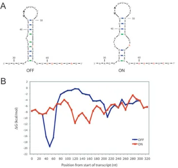
A model for the cwpVswitch mechanism
Considering the mechanisms whereby DNA inversion
could act as a negative regulator ofcwpVexpression, two
possibilities seem likely. First, one or more proteins could bind to the DNA or RNA in the OFF orientation and prevent transcription or translation. Second, intrinsic pro-perties of the DNA or RNA in the OFF orientation could prevent transcription or translation. These two mecha-nisms are not mutually exclusive as there may be a spe-cific structure in the OFF DNA/RNA to which protein binding could mediate the negative regulation.
To explore the second hypothesis and search for the presence of intrinsic terminators, the RNA structure of transcripts from the two orientations were predicted
using mfold, a program that predicts the structure and
free energies of RNA sequences (Zuker, 2003). A stable stem loop structure consisting of a 9 bp stem containing four G-C bp was predicted to form in the OFF orientation at the junction between the LIR and the region of inver-sion (Fig. 8A). This stem loop structure is followed by a poly-U tract. In the ON orientation the sequence of this region is altered by a number of base pairs (coloured
red in Fig. 8A) due to inversion of the cwpVswitch and
the predicted RNA structure is therefore different. A
model for predicting intrinsic terminators in E. coli has
been developed (Lesniket al., 2001) and has been used
to predict intrinsic terminators in a number of bacterial
species including Clostridium acetobutylicum (Paredes
et al., 2004). This model is built from sequence con-straints based on known terminator sequences from
E. coli. Briefly there must be a stem of 4–18 bp, contain-ing at least four G-C bp, a loop of 3–10 nt and followcontain-ing the stem a poly-U tract with sufficient U-content. Quan-titative models explaining how such structures destabi-lize the transcription complex have been reported (Yager and von Hippel, 1991). The structure predicted for the OFF orientation fulfils all the criteria for an intrinsic ter-minator, whereas the ON stem only contains two G-C bp and has a large internal loop and therefore does not fulfil the criteria. To further compare the stability of predicted RNA structures of the ON and OFF transcripts, quanti-tative predictions of free energy of RNA folding for the transcript in the two orientations were calculated for 60 nt long stretches of sequence, systematically
movi-ng alomovi-ng the transcript in 10 nt jumps. A peak in free energy, characteristic of an intrinsic terminator (Washio et al., 1998), is seen in the OFF orientation at the junction between the LIR and the inversion region; this peak is not present in the ON orientation (Fig. 8B). Based on these predictions our current hypothesis for
the mechanism underlying phase variation of cwpV is
that transcription initiates upstream of the LIR regardless
of the orientation of the cwpV switch, but in the OFF
[image:8.595.60.371.69.513.2]orientation transcription is terminated upon reaching the region of inversion due to an intrinsic terminator. In the ON orientation, transcription can continue through the inversion region to the ORF and produce a full-length transcript.
Fig. 7. Analysis of transcription from PcwpVin
C. difficileusing a GusA reporter.C. difficile 630 containing plasmids with different components of thecwpVswitch upstream of gusAwere grown in liquid medium, harvested and theb-glucoronidase activity measured. A. Diagrammatic representation of the promoter regions present in the plasmids used in this experiment. Plasmids used were: Pcwp2,pCBR034; short ON, pCBR035; ON,
pCBR037; ON LIR*, pCBR039; ON RIR*, pCBR041; short OFF, pCBR036; OFF, pCBR038; OFF LIR*, pCBR040; OFF RIR*, pCBR042;DIR, pCBR043.
B. Enzyme activities of cell lysates. Error bars represent the standard deviations (n=3). C. PCR analysis of DNA isolated from cultures using primers to detect the ON (left) (NF793 and 826) and OFF (NF793 and 825) orientations of thecwpVswitch.
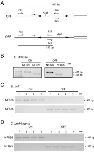
Inversion of thecwpVswitch does not occur in E. coliorClostridium perfringens
Our results suggest that one or more site-specific
recombinases in C. difficile mediate DNA inversion to
control expression of cwpV. The ability of different
species to mediate inversion of the cwpV switch was
assessed by PCR analysis of plasmids pCBR037 and
pCBR038. These plasmids contain the cwpV switch in
the ON and OFF orientations respectively. The
orienta-tion of the cwpV switch can be determined by PCR
using primers which anneal to opposite strands within the region of inversion, paired with a primer outside the region of inversion (see Fig. 9A). InC. difficile, inversion
of the cwpV switch occurred in both the ON and OFF
plasmids, as shown by the appearance of PCR products using either primer NF825 or NF826 (Fig. 9B). In
con-trast, in E. coli (Fig. 9C) or C. perfringens (Fig. 9D),
no inversion was seen in four independent isolates containing either plasmid. Therefore it is likely that C. difficile encodes one or more specific recombinases
which catalyse the inversion of the cwpV switch and
these enzymes are not present in C. perfringens or
E. coli.
Identification of aC. difficilerecombinase mediating the cwpVswitch
A search theC. difficile630 genome identified 22 putative recombinases. However, 16 of these are associated with the many mobile genetic elements (MGEs) in this strain, and are largely absent from other strains as determined by microarray analysis (Stableret al., 2006). AllC. difficile
strains analysed to date exhibit cwpV switch inversion
[image:9.595.116.483.52.398.2](except R8366), making it likely that the site-specific Fig. 8. RNA structure and free energy predictions of thecwpVDNA switch.
A. Predicted RNA structures found at the junction between the LIR and the region of inversion. Nucleotides that differ between the two orientations are shown in red. GC base pairing is shown in green and AU base pairing in blue. Numbering is from the predicted transcriptional start site. The two bases in bold are those deleted in strain R8366.
B. Free energy predictions for the two orientations, measuring theDG of 60 nt regions and sequentially moving along the sequence in 10 nt segments. Data were obtained using the on-line programmfold(Zuker, 2003). The free energy peak seen at 50 nt in the OFF orientation corresponds to the predicted RNA structure shown in A.
recombinase(s) responsible for inversion are well conserved. A total of seven putative site-specific recom-binase genes were chosen for analysis: three of the best-conserved MGE-associated recombinases, one putative housekeeping recombinase and all three non-MGE-associated recombinase genes. The genes were each cloned into pACYCDuet-1 to allow their expression as
His-tagged proteins, and then transformed into E. coli
BL21lDE3 containing pUC19-based plasmids with
the cwpV switch in either the ON or OFF orientation
(pCBR026 or pCBR027). Orientation-specific PCR
showed that only one recombinase, CD1167, mediated DNA inversion (Fig. 10). This gene, which we designate recV, is a tyrosine recombinase of the XerC/XerD family of DNA recombinases and can catalyse both ON to OFF and OFF to ON inversion. None of the other recombinases tested catalysed DNA inversion inE. coli. Western blotting using anti-His tag antibodies verified that all
recombi-nases were expressed in E. coli, producing bands of
[image:10.595.57.368.52.554.2]the expected size (data not shown).
Fig. 9. DNA inversion is specific to C. difficileand does not occur in
C. perfringensorE. coli. Orientation-specific PCRs were carried out to determine whether DNA inversion of thecwpVswitch occurs in E. coliorC. perfringens.
A. ThecwpVswitch region in the ON and OFF orientations, in pCRB037 and pCRB038, respectively, showing the position of the primers used.
B. PCR products of the ON and OFF plasmids after introduction intoC. difficile. DNA inversion occurs in each case as seen by the appearance of both PCR products. C and D. The plasmids were introduced into E. coli(C) andC. perfringens(D). Four independent transformants or transconjugants were picked and in each case no DNA inversion is seen.
Discussion
In this study we describe the phase variation ofcwpV, a
gene encoding a cell surface protein ofC. difficile. Phase variation mediated by DNA inversion has been described in a small number of bacterial systems (van der Woude and Baumler, 2004). The best-characterized systems involve inversion of a promoter or part of the ORF. In the former, a promoter in one orientation actively transcribes a gene whereas in the opposite orientation either no gene transcription occurs or an alternative gene is transcribed.
For example, in the E. coli fim system, the fimA gene
product is either expressed or not, depending on the
promoter orientation (Abraham et al., 1985). In the
Salmonella hin system, in one orientation the promoter directs expression of the H1 flagellin, whereas in the opposite orientation it directs expression of H2 flagellin
(Zieg et al., 1977). In both these systems the
recombi-nase(s) involved in DNA inversion are located either within the inverted DNA (S. typhimurium hin) or within the immediate vicinity of the switch (E. coli fim) (reviewed in van der Woude and Baumler, 2004). It is interesting that recVis not located in the vicinity ofcwpV. This fact, along
with the presence of the cwpV switch and recV in all
C. difficilestrains analysed within a diverse set of isolates,
suggests that this switch andrecVhave not been recently
acquired by horizontal gene transfer.
In the case of cwpV, we have established that the
promoter and transcriptional initiation site are situated
upstream of the cwpV switch and that DNA inversion
controls the expression ofcwpV.The production of stable
full-lengthcwpVtranscripts is only possible from template
DNA in the ON orientation. This mechanism for controlling gene expression involving DNA inversion encompassing neither the promoter nor the ORF is to our knowledge
completely novel. Furthermore, by using agusA
transcrip-tional reporter inC. difficile, we have shown that thecwpV
switch region is not required for expression from thecwpV
promoter, and that the cwpV switch must therefore act
in cis to negatively regulate expression. We propose a model to account for these observations based on pre-dicted secondary structure of the mRNA transcripts. This mechanism involving transcriptional termination modu-lated by DNA inversion is to our knowledge unique. Tran-scripts in the OFF orientation are predicted to form a stable stem loop structure followed by a poly-U tract, which induces transcriptional termination at some 60–70 nucleotides downstream of the transcript initiation site. In the ON orientation the predicted structure does not fulfil the criteria for an intrinsic terminator and transcription can proceed unhindered. We acknowledge the possibility that, in the OFF orientation, transcription could also be affected by binding a repressor molecule or that control could be mediated by a combination of these mechanisms.
Interestingly modulation of transcription termination
is also seen in the E. coli fim system, where a
Rho-dependent phase variable transcriptional terminator is
present downstream of the fimE recombinase gene in
phase OFF cells (Joyce and Dorman, 2002; Hindeet al.,
2005). In phase ON bacteria, termination of the fimE
transcript does not occur and the mRNA transcript is longer and more stable, leading to increased levels of FimE recombinase and biasing the switch to the phase
OFF. However, in thefimswitch, the primary level control
offimAexpression is inversion of the promoter, whereas
in thecwpVswitch described here, transcription
termina-tion is the key event determining expression ofcwpV.
[image:11.595.56.376.54.243.2]A common theory to account for phase variation in bacterial pathogens is that alteration of surface structures allows evasion of the immune responses. Other explana-tions include modulation of adhesin expression that could facilitate detachment of bacteria from host substrates, resulting in dissemination of the bacteria from the host. However, in many cases the biological significance
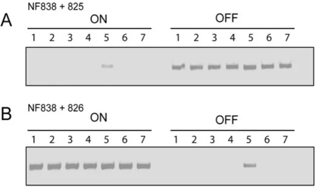
Fig. 10. Identification of a recombinase mediating DNA inversion of thecwpVswitch. Genes for seven recombinases (CD1932, CD1905, CD2066, CD1822, CD1167, CD3578 andCD1222) were cloned into pACYCDuet-1 and co-transformed intoE. coliBL21lDE3 with pUC19 plasmids carrying thecwpV switch in either the ON or OFF orientations (pCBR026 or pCBR027). PCR reactions with primer pairs NF838 and NF825 (A) and NF828 and NF826 (B) were then carried out. Recombinase 5 (CD1167) mediated inversion in both the ON to OFF and OFF to ON orientations.
of phase variation remains a mystery (van der Woude, 2006). To date we have been unable to define a functional role for CwpV. Although annotated as a haemagglutinin
(Sebaihia et al., 2007), we have not found
haemagglu-tination activity despite using blood from a number of species (data not shown). The presence of multiple repeats in a surface protein of a bacterial pathogen usually suggests adhesin activity, but to date we have been unable to define such a role for CwpV. It is possible, however, that CwpV does mediate either bacteria–host or bacteria–bacteria interactions, and this is currently under investigation.
For C. difficile, survival in the enteric system may require adaptation to changing environmental
condi-tions, and the regulated expression ofcwpVmay reflect
responses that allow adaption to particular niches during
infection. In the enteric symbiont Bacterioides fragilis,
DNA inversion is known to operate on a large scale, controlling multiple loci including biosynthetic operons for surface localized capsular polysaccharides (Patricket al.,
2003; Fletcher et al., 2007). Mutagenesis studies have
demonstrated that B. fragilis requires the capacity to
produce a repertoire of capsular polysaccharides and that mutants expressing a single polysaccharide are defective in intestinal colonization of mice (Liuet al., 2008). These
studies and others (Cerdeno-Tarragaet al., 2005)
illus-trate the importance of DNA inversion as a mechanism to modulate gene expression and ultimately to increase survival in such environments.
At this stage we can only speculate on the
advan-tages toC. difficileof expressing a phase-variable CWP.
Expression of cwpVmay be in response to a particular
growth condition or environmental stimulus, but to date we have been unable to detect any conditions under which the frequency of DNA inversion orcwpVtranscription is altered
(Emersonet al., 2008 and data not shown). CwpV is the
only CWP that is known to undergo phase variation in C. difficile.Analysis of the genome sequence of 630 did not
reveal copies of the IRs associated withcwpVat any other
locations in the genome. As recombinases are highly spe-cific for their target nucleotide sequences, this suggests
that cwpV is the only phase-variable gene in C. difficile
regulated by RecV. Further analysis may of course reveal other genes regulated by DNA inversion mediated by different recombinases. Our ongoing work is concentrating
on the detailed mechanisms underlying the control ofcwpV
expression as well as investigating the function of the CwpV protein.
It appears that the DNA inversion of thecwpVswitch is
controlled by one or more recombinases that are
specific to C. difficile. The recombinase identified here,
RecV, is present in all strains ofC. difficileso far
investi-gated (Stabler et al., 2006) implying that this control
mechanism of CwpV expression may be universal. The
transcription levels ofrecVare not seen to be altered by
any environmental stress so far investigated (Emerson et al., 2008). The recombinase RecV was able to mediate DNA inversion in both the ON to OFF and OFF to ON
orientations when expressed inE. coli, suggesting that if
other factors are necessary for DNA inversion they are highly conserved. InE. coli, thefimswitch is controlled by
two recombinases, fimBand fimE, which are located in
thefim locus (Klemm, 1986). Therefore, there could be
other recombinases inC. difficile that function alongside
RecV to control CwpV expression via control of thecwpV
switch orientation. Construction of arecVknockout strain
will shed light on this. However, genetic manipulation of C. difficileis still challenging and techniques for comple-mentation of knockout mutants by insertion of a single chromosomal copy of a wild-type or mutant allele have not yet been described. Such experiments would be highly informative and would allow us to study the behaviour of
a single copy of a mutatedcwpVgene or promoter.
MostC. difficileCWPs are not members of the LPxTG protein family which are attached to the peptidoglycan
via the sortase pathway (Marraffiniet al., 2006), but are
instead non-covalently anchored to the underlying cell wall by an as yet uncharacterized mechanism. The majority of
predictedC. difficileCWPs, including CwpV, are members
of a large family of CWPs which are localized to the cell wall via non-covalent interactions (Emerson and Fairweather, 2009). Other characterized members of this family include the S-layer proteins (SlpA), the adhesin Cwp66 and the protease Cwp84 (Waligoraet al., 2001; Janoiret al., 2007). Our results show that CwpV is processed by internal cleavage to produce two proteins that are localized to the cell wall. We have recently described a similar mechanism for the two SLPs, HMW SLP and LMW SLP, which after cleavage re-associate via defined domains to produce a
stable complex on the cell surface (Faganet al., 2009).
Whether CwpV is processed by the same machinery as that used for SlpA and whether the two CwpV cleavage products re-associate as a complex is unknown but is currently under investigation. We have shown that CwpV can constitute a large proportion of total cell surface protein and may therefore interact with SlpA in order to maintain the integrity of the S-layer. The level of CwpV expression in
single ON cells is unknown but construction of a recV
knockout strain will allow us to determine the level of
expression from cells with thecwpVswitch ON.
In this study we have shown that expression of the C. difficile cell wall protein CwpV is phase variable and controlled by a novel switch employing an intrinsic terminator. To our knowledge this is the first description of phase variation controlled by DNA inversion in Clostridia and it will be of interest to determine whether the mecha-nism described here is found in other members of the genus. The functional consequences of phase variation
of CwpV, particularly in the context of pathogenesis, remain to be elucidated and are currently under investi-gation in our laboratory.
Experimental procedures
Bacterial strains and growth conditions
Bacterial strains and plasmids used in this study are described in Tables 1 and 2 respectively. C. difficile and C. perfringenswere routinely cultured either on blood agar base II (Oxoid, Basingstoke, UK) supplemented with 7% defi-brinated horse blood (TCS Biosciences, Botolph Claydon, UK), BHI agar (Oxoid) or in BHI broth (Oxoid). Cultures were grown in an anaerobic cabinet (Don Whitley Scientific, Shipley, UK) at 37°C in an atmosphere of 10% CO2, 10% H2
and 80% N2.E. coliwas grown at 37°C on LB agar or in LB
broth supplemented with appropriate antibiotics.
DNA and RNA manipulations
DNA manipulations were carried out according to standard techniques (Sambrooket al., 1989). For use in DNA cloning, C. difficilegenomic DNA was isolated as described previously (Calabi et al., 2001). PCRs used Taq polymerase (Sigma), Expand Long-Template Polymerase (Roche Diagnostics) or KOD (Merck) in accordance with the manufacturers’ protocols. For PCR-RFLP the product of 40 rounds of PCR amplification were digested using AseI (New Englad Biolabs). Details of plasmid construction including all primer sequences (Table S1) are given in theSupporting information.
Conjugation intoC. difficileandC. perfringens
Plasmids were transformed into E. coli CA434 and then conjugated into C. difficile 630 or C. perfringens SM101 as described by Purdy et al. (2002) using thiamphenicol
(30mg ml-1
) to select for pMTL960-based plasmids and cycloserine (250mg ml-1) to counter-select forE. coli.
Construction of acwpVknockout mutant in C. difficile630
AcwpVmutant was generated inC. difficile630Derm(Hussain et al., 2005) by insertion of a bacterial group II intron containing a retrotransposition-activated marker (RAM) conferring eryth-romycin resistance (Zhonget al., 2003) using the ClosTron clostridial gene knockout system (Heapet al., 2007). An Ll.ltrB target site was identified within the 630cwpVgene and intron retargeting primers NF1003-NF1005 were designed using the online TargeTron algorithm (http://www.sigma-genosys.com/ targetron/). Plasmid retargeting was carried out as previously described (Heap et al., 2007) and the resulting plasmid, pIC007:Cdi-cwpV-1056s was transferred toC. difficile630 by conjugation from E. coli CA434. Four to five colonies of 630 (pIC007:Cdi-cwpV-1056s) were resuspended in 1 ml of anaerobic BHI with isopropyl-b-D-thiogalactopyranoside (IPTG) 1 mM and thiamphenicol (30mg ml-1) and incubated at
37°C for 3 h. The bacteria were then harvested, resuspended in 1 ml of fresh BHI broth and incubated for a further 2 h. Potential Ll.ltrB insertions were selected by plating bacteria on BHI agar supplemented with erythromycin (2.5mg ml-1
). Fol-lowing subculture, putativecwpVmutants were screened by colony PCR using primers specific for the inserted RAM (NF722 and NF723), thecwpVgene (NF1064 and NF1065) and each of the cwpV gene primers in combination with a primer internal to the Ll.ltrB intron (NF1063). A singlecwpV mutant was further characterized by western immunoblotting using antibodies specific for CwpV (see below) and desig-nated 630DermDcwpV.
b-Glucuronidase assays
[image:13.595.55.537.476.662.2]b-Glucuronidase enzyme activity was measured in lysates of C. difficile cultures as described previously (Dupuy and
Table 1. Strains used in this study.
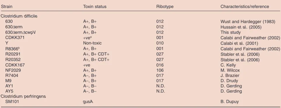
Strain Toxin status Ribotype Characteristics/reference
Clostridium difficile
630 A+, B+ 012 Wust and Hardegger (1983)
630Derm A+, B+ 012 Hussainet al. (2005)
630DermDcwpV A+, B+ 012 This study
CDKK371 +vea 001 Calabi and Fairweather (2002)
Y Non-toxic 010 Calabiet al. (2001)
R8366b A+, B+ 001 Calabi and Fairweather (2002)
R20291 A+, B+CDT+ 027 Stableret al. (2006)
R20352 A+, B+CDT+ 027 Stableret al. (2006)
CDKK167 +ve 016 C. Kelly
NF2029 A+, B+ 106 M. Wilcox
R7404 A-, B+ 017 J. Brazier
M9 A-, B+ 017 D. Drudy
AY1 A-, B- N.D. D. Gerding
AY5 A-, B- N.D. D. Gerding
Clostridium perfringens
SM101 gusA B. Dupuy
a. +ve, toxin A positive but toxin B unknown.
b. No expression ofcwpV. This strain was sourced from two independent labs. Neither isolate expressedcwpVand both contained the 2 bp deletion.
Sonenshein, 1998; Mani et al., 2002) using the substrate p-nitrophenyl-b-D glucuronide (Sigma).
Purification of recombinantCwpVprotein and generation of antibodies
The first repeat of cwpV from C. difficile 630 (CwpVrpt1) was amplified by PCR using primers NF345 and NF346 and cloned into pGEX-4T-1.E. colitransformants were grown in LB to OD6000.4 and expression was induced by addition of
1 mM IPTG. Bacteria were harvested after 2 h, lysed by sonication and the fusion protein purified by glutathione affinity chromatography. The GST tag was cleaved using thrombin and the proteins separated by size exclusion chromatography. The purified CwpV repeat was inoculated into rabbits to raise antisera recognizing CwpVrpt1. Antibod-ies were affinity purified using a column of CwpVrpt1-coated Affi-gel 10 (Bio-Rad).
The conserved N-terminal 48.9 kDa of CwpV (CwpVNter) was cloned from C. difficile 630 into pENTR-TEV-D-TOPO and subsequently subcloned to pDEST15 (Invitrogen).E. coli transformants were grown overnight at 25°C in Overnight Express media (Novagen/Merck Chemicals). Bacteria were harvested, lysed by sonication and the fusion protein purified
by glutathione affinity chromatography followed by size exclusion chromatography. The purified protein was inocu-lated into rabbits to raise antisera recognizing CwpVNter. LMW SLPs were produced as described previously (Fagan et al., 2009) and antisera generated in rats. All antisera were generated commercially (Eurogentec).
Immunodetection
Western immunoblotting. C. difficile were grown overnight in BHI broth and harvested by centrifugation. Glycine cell-surface extracts were performed as described previously (Calabiet al., 2001). Proteins were subjected to SDS-PAGE and Western blotting according to standard protocols. Anti-CwpVrpt1 and anti-CwpVNter were used at the dilutions indicated in the text, followed by anti-rabbit-HRP (Dako Cyto-mation) at 1/2000. Blots were developed using SuperSignal West Pico Chemiluminescent Substrate (Pierce).
Immunocytochemistry. C. difficile cells from liquid culture were washed with PBS then fixed in 8% formaldehyde in PBS, which was quenched using 20 mM NH4Cl for 15 min.
[image:14.595.60.542.70.397.2]The washed cell suspension was spotted onto a glass slide and allowed to dry. The bacteria were incubated with 1/10
Table 2. Plasmids used in this study.
Name Relevant characteristics Source/reference
pMTL007 E. coli-Clostridiumshuttle vector containing the Ll.ltrB group II intron with a retrotransposition-activatedermBcassette
Heapet al. (2007)
pIC007:
Cdi-cwpV-1056s
pMTL007 with the Ll.ltrB intron retargeted to insert after base 1056 inC. difficile630cwpVORF
This study
pTUM177 Contains promoterlessgusAgene withC. difficile tcdBRBS Mani and Dupuy (2001)
pMTL960 E. coli – C. difficileshuttle vector Purdyet al. (2002)
pCBR023 Contains modifiedgusAgene under control ofcwp2 promoter; pUC19 backbone
This study
pCBR026 pUC19 containinggusAand long upstream sequence ofcwpV with switch in ON orientation
This study
pCBR027 pUC19 containinggusAand long upstream sequence of cwpVwith switch in OFF orientation
This study
pCBR034 Insert from pCBR023 cloned into pMTL960 This study
pCBR037 Insert from pCBR026 cloned into pMTL960 This study
pCBR038 Insert from pCBR027 cloned into pMTL960 This study
pCBR039 Derivative of pCBR037 containing 2 bp deletion in LIR (LIR*) ofcwpVswitch This study pCBR041 Derivative of pCBR037 containing 2 bp deletion in RIR (RIR*) ofcwpVswitch This study pCBR040 Derivative of pCBR038 containing 2 bp deletion in LIR (LIR*) ofcwpVswitch This study pCBR042 Derivative of pCBR038 containing 2 bp deletion in RIR (RIR*) ofcwpVswitch This study pCBR035a pMTL960 containinggusAand short upstream sequence.
cwpVswitch in ON orientation
This study
pCBR036 pMTL960 containinggusAand short upstream sequence. cwpVswitch in OFF orientation
This study
pCBR043 Derivative of pCBR037 with entirecwpVswitch deleted This study pCBR044 pMTL960 containing the full-lengthcwpVgene from 630 under
the control ofcwp2promoter
This study
pCBR045 pACYC-Duet1 containingCD1932 This study
pCBR046 pACYC-Duet1 containingCD1905 This study
pCBR047 pACYC-Duet1 containingCD2066 This study
pCBR048 pACYC-Duet1 containingCD1882 This study
pCBR049 pACYC-Duet1 containingCD1167(recV) This study
pCBR050 pACYC-Duet1 containingCD3578 This study
pCBR051 pACYC-Duet1 containingCD1222 This study
a. pCBR035–043 have the pMTL960 backbone and were constructed by insertion of Acc65I-BamHI fragments from pCBR024 to pCBR032 which have a pUC19 backbone.
rabbit anti-CwpVrpt1 and 1/20 rat anti-LMW-SLP, then washed and incubated with 1/10 anti-rabbit-FITC (Dako Cytomation) and 1/40 anti-rat-rhodamine red (Jackson ImmunoResearch Laboratories, West Grove, PA).
Flow cytometry. Ten microlitres of an overnight liquid culture ofC. difficilewas washed, resuspended in 1/10 rabbit anti-CwpVrpt1, washed and finally resuspended 1/10 anti-rabbit-FITC (all washes and dilutions in PBS). Cells were washed and resuspended in 200ml PBS, and analysed using a Becton Dickinson FACS Calibur.
Immunogold electron microscopy. Colonies were scraped
from blood agar plates and resuspended in cold 4% paraformaldehyde (PFA) and 0.2% glutaraldehyde in PBS, and were fixed on ice for 10 min. The cells were harvested, resuspended in 4% PFA and 0.2% glutaraldehyde and fixed for a further 1 h. The bacteria were washed three times in PBS, infiltrated with 2.3 M sucrose in PBS overnight at 4°C, attached to a specimen stub and plunge-frozen in liquid nitrogen. The 65 nm ultrathin cryosections were cut on a Leica UCT ultramicrotome with EM FCS cryoattachment. Sections were mounted on grids with formvar support and blocked with 0.02 M glycine in PBS followed by 10% FCS in PBS; this was the diluent for all subsequent steps. Sections were incubated in anti-CwpVrpt1 for 1 h followed by three 5 min PBS rinses. Sections were incubated in 15 nm Protein A gold for 30 min followed by three 5 min PBS rinses. Sec-tions were rinsed in double distilled H2O six times over 5 min.
The sections were then contrasted with uranyl acetate in methyl cellulose on ice for 10 min. The sections were air dried and viewed in a CM100 transmission cryo-electron micro-scope (Philips, Guildford, UK) at 16 000¥ to 32 000¥
magnification.
Acknowledgements
J.E. and C.R. were funded by the Wellcome Trust and R.F. by the MRC. We thank Aaron Rae for help and advice on flow cytometry; Nigel Minton and John Heap for plasmids pMTL960 and pMTL007; Bruno Dupuy for plasmid pTUM177; and Deirdre Ni Eidhin, Dale Gerding, Brendan Wren, Denise Drudy and Mark Wilcox for strains.
References
Abraham, J.M., Freitag, C.S., Clements, J.R., and Eisenstein, B.I. (1985) An invertible element of DNA controls phase variation of type 1 fimbriae ofEscherichia coli.Proc Natl
Acad Sci USA82:5724–5727.
Ausiello, C.M., Cerquetti, M., Fedele, G., Spensieri, F., Palazzo, R., Nasso, M.,et al.(2006) Surface layer proteins fromClostridium difficileinduce inflammatory and regula-tory cytokines in human monocytes and dendritic cells. Microbes Infect8:2640–2646.
Barbut, F., Mastrantonio, P., Delmee, M., Brazier, J., Kuijper, E., and Poxton, I. (2007) Prospective study ofClostridium difficileinfections in Europe with phenotypic and genotypic characterisation of the isolates.Clin Microbiol Infect 13:
1048–1057.
Bartlett, J.G. (2006) Narrative review: the new epidemic of Clostridium difficile-associated enteric disease.Ann Intern Med145:758–764.
Calabi, E., and Fairweather, N. (2002) Patterns of sequence conservation in the S-layer proteins and related sequences inClostridium difficile.J Bacteriol184:3886–3897. Calabi, E., Ward, S., Wren, B., Paxton, T., Panico, M., Morris,
H.,et al.(2001) Molecular characterization of the surface layer proteins fromClostridium difficile.Mol Microbiol40:
1187–1199.
Calabi, E., Calabi, F., Phillips, A.D., and Fairweather, N. (2002) Binding ofClostridium difficilesurface layer proteins to gastrointestinal tissues.Infect Immun70:5770–5778. Cerdeno-Tarraga, A.M., Patrick, S., Crossman, L.C., Blakely,
G., Abratt, V., Lennard, N., et al.(2005) Extensive DNA inversions in theB. fragilisgenome control variable gene expression.Science307:1463–1465.
Dupuy, B., and Sonenshein, A.L. (1998) Regulated transcrip-tion ofClostridium difficiletoxin genes.Mol Microbiol27:
107–120.
Emerson, J., and Fairweather, N.F. (2009) Surface structures ofC. difficileand other clostridia. InClostridia – Molecular
Biology in the Post-Genomic Era. Bruggemann, H., and
Gottschalk, G. (eds). Norfolk: Caister Academic Press, pp. 157–167.
Emerson, J.E., Stabler, R.A., Wren, B.W., and Fairweather, N.F. (2008) Microarray analysis of the transcriptional responses of Clostridium difficile to environmental and antibiotic stress.J Med Microbiol57:757–764.
Fagan, R.P., Albesa-Jove, D., Qazi, O., Svergun, D.I., Brown, K.A., and Fairweather, N.F. (2009) Structural insights into the molecular organization of the S-layer fromClostridium difficile.Mol Microbiol71:1308–1322.
Fletcher, C.M., Coyne, M.J., Bentley, D.L., Villa, O.F., and Comstock, L.E. (2007) Phase-variable expression of a family of glycoproteins imparts a dynamic surface to a symbiont in its human intestinal ecosystem. Proc Natl
Acad Sci USA104:2413–2418.
Heap, J.T., Pennington, O.J., Cartman, S.T., Carter, G.P., and Minton, N.P. (2007) The ClosTron: a universal gene knock-out system for the genus Clostridium.J Microbiol
Methods70:452–464.
Henderson, I.R., Owen, P., and Nataro, J.P. (1999) Molecular switches – the ON and OFF of bacterial phase variation. Mol Microbiol33:919–932.
Hennequin, C., Janoir, C., Barc, M.-C., Collignon, A., and Karjalainen, T. (2003) Identification and characterization of a fibronectin-binding protein from Clostridium difficile. Microbiology149:2779–2787.
Hinde, P., Deighan, P., and Dorman, C.J. (2005) Charac-terization of the detachable Rho-dependent transcription terminator of the fimE gene in Escherichia coli K-12. J Bacteriol187:8256–8266.
Hussain, H.A., Roberts, A.P., and Mullany, P. (2005) Generation of an erythromycin-sensitive derivative of Clostridium difficile strain 630 (630Derm) and demons-tration that the conjugative transposon Tn916E enters the genome of this strain at multiple sites. J Med Microbiol
54:137–141.
Janoir, C., Pechine, S., Grosdidier, C., and Collignon, A. (2007) Cwp84, a surface-associated protein ofClostridium
difficile, is a cysteine protease with degrading activity on extracellular matrix proteins.J Bacteriol189:7174–7180. Joyce, S.A., and Dorman, C.J. (2002) A Rho-dependent
phase-variable transcription terminator controls expression of the FimE recombinase inEscherichia coli.Mol Microbiol
45:1107–1117.
Just, I., Selzer, J., Wilm, M., von Eichel-Streiber, C., Mann, M., and Aktories, K. (1995) Glucosylation of Rho proteins byClostridium difficiletoxin B.Nature375:500–503. Klemm, P. (1986) Two regulatoryfimgenes,fimBandfimE,
control the phase variation of type 1 fimbriae inEscherichia coli.EMBO J5:1389–1393.
Lesnik, E.A., Sampath, R., Levene, H.B., Henderson, T.J., McNeil, J.A., and Ecker, D.J. (2001) Prediction of rho-independent transcriptional terminators inEscherichia coli. Nucleic Acids Res29:3583–3594.
Liu, C.H., Lee, S.M., VanLare, J.M., Kasper, D.L., and Maz-manian, S.K. (2008) Regulation of surface architecture by symbiotic bacteria mediates host colonization. Proc Natl
Acad Sci USA105:3951–3956.
Mani, N., and Dupuy, B. (2001) Regulation of toxin synthesis inClostridium difficileby an alternative RNA polymerase sigma factor.Proc Natl Acad Sci USA98:5844–5849. Mani, N., Lyras, D., Barroso, L., Howarth, P., Wilkins, T.,
Rood, J.I.,et al.(2002) Environmental response and auto-regulation ofClostridium difficileTxeR, a sigma factor for toxin gene expression.J Bacteriol184:5971–5978. Marraffini, L.A., DeDent, A.C., and Schneewind, O. (2006)
Sortases and the art of anchoring proteins to the envelopes of Gram-positive bacteria.Microbiol Mol Biol Rev70:192– 221.
Paredes, C.J., Rigoutsos, I., and Papoutsakis, E.T. (2004) Transcriptional organization of theClostridium acetobutyli-cumgenome.Nucleic Acids Res32:1973–1981. Patrick, S., Parkhill, J., McCoy, L.J., Lennard, N., Larkin,
M.J., Collins, M., et al. (2003) Multiple inverted DNA repeats ofBacteroides fragilisthat control polysaccharide antigenic variation are similar to the hin region inverted repeats of Salmonella typhimurium. Microbiology 149:
915–924.
Pizarro-Cerda, J., and Cossart, P. (2006) Bacterial adhesion and entry into host cells.Cell124:715–727.
Poxton, I.R., McCoubrey, J., and Blair, G. (2001) The patho-genicity ofClostridium difficile.Clin Microbiol Infect7:421– 427.
Purdy, D., O’Keeffe, T.A., Elmore, M., Herbert, M., McLeod, A., Bokori-Brown, M., et al. (2002) Conjugative transfer of clostridial shuttle vectors from Escherichia coli to Clostridium difficilethrough circumvention of the restriction barrier.Mol Microbiol46:439–452.
Sambrook, J., Fritsch, E.F., and Maniatis, T. (1989)Molecular Cloning.Cold Spring Harbor, NY: Cold Spring Harbor Press. Sebaihia, M., Wren, B.W., Mullany, P., Fairweather, N.F., Minton, N., Stabler, R.,et al.(2006) The multidrug-resistant human pathogenClostridium difficilehas a highly mobile, mosaic genome.Nat Genet38:779–786.
Sebaihia, M., Peck, M.W., Minton, N.P., Thomson, N.R., Holden, M.T., Mitchell, W.J., et al. (2007) Genome sequence of a proteolytic (Group I)Clostridium botulinum strain Hall A and comparative analysis of the clostridial genomes.Genome Res17:1082–1092.
Stabler, R.A., Gerding, D.N., Songer, J.G., Drudy, D., Brazier, J.S., Trinh, H.T.,et al.(2006) Comparative phylo-genomics ofClostridium difficile reveals clade specificity and microevolution of hypervirulent strains. J Bacteriol
188:7297–7305.
Voth, D.E., and Ballard, J.D. (2005) Clostridium difficile toxins: mechanism of action and role in disease. Clin Microbiol Rev18:247–263.
Waligora, A.J., Hennequin, C., Mullany, P., Bourlioux, P., Collignon, A., and Karjalainen, T. (2001) Characterization of a cell surface protein ofClostridium difficilewith adhe-sive properties.Infect Immun69:2144–2153.
Washio, T., Sasayama, J., and Tomita, M. (1998) Analysis of complete genomes suggests that many prokaryotes do not rely on hairpin formation in transcription termination. Nucleic Acids Res26:5456–5463.
Wosten, M.M. (1998) Eubacterial sigma-factors. FEMS Microbiol Rev22:127–150.
van der Woude, M.W. (2006) Re-examining the role and random nature of phase variation. FEMS Microbiol Lett
254:190–197.
van der Woude, M.W., and Baumler, A.J. (2004) Phase and antigenic variation in bacteria.Clin Microbiol Rev17:581– 611.
Wright, A., Wait, R., Begum, S., Crossett, B., Nagy, J., Brown, K., and Fairweather, N. (2005) Proteomic analysis of cell surface proteins from Clostridium difficile. Proteomics5:
2443–2452.
Wright, A., Drudy, D., Kyne, L.K.B., and Fairweather, N.F. (2008) Immunoreactive cell wall proteins of Clostridium difficileidentified by human sera.J Med Microbiol57:750– 756.
Wust, J., and Hardegger, U. (1983) Transferable resis-tance to clindamycin, erythromycin, and tetracycline in Clostridium difficile. Antimicrob Agents Chemother 23:
784–786.
Yager, T.D., and von Hippel, P.H. (1991) A thermodynamic analysis of RNA transcript elongation and termination in Escherichia coli.Biochemistry30:1097–1118.
Zhong, J., Karberg, M., and Lambowitz, A.M. (2003) Targeted and random bacterial gene disruption using a group II intron (targetron) vector containing a retrotransposition-activated selectable marker.Nucleic Acids Res31:1656– 1664.
Zieg, J., Silverman, M., Hilmen, M., and Simon, M. (1977) Recombinational switch for gene expression.Science196:
170–172.
Zuker, M. (2003) Mfold web server for nucleic acid folding and hybridization prediction.Nucleic Acids Res31:3406– 3415.
Supporting information
Additional supporting information may be found in the online version of this article.
Please note: Wiley-Blackwell are not responsible for the content or functionality of any supporting materials supplied by the authors. Any queries (other than missing material) should be directed to the corresponding author for the article.