International Journal of Innovative Technology and Exploring Engineering (IJITEE) ISSN: 2278-3075,Volume-8, Issue-8S, June 2019
Abstract: In this article we present Microaneurysms is an eye-created blood dot, which is the result of eye disease indication. The resultant of microaneurysms is due to sugar in the eyes which is “DIABETES”, which is a characterized by uncontrolled hyperglycaemia. Diabetes is caused by a failure in our body to suppress endogenous glucose production due to insulin shortage. Diabetes may also cause due to gene from the ancestors. Our project is used to light the microaneurysms (blood dots), and to classify the severity of the Diabetic Retinopathy. Due to this we can able to control and save the patients by the best treatment and food control. If we leave this disease, then will lead to increase of sugar in body and also leads to death so this project is used to save the youngsters and old people. If a Diabetic Patient injured, even it is a small wound, it is difficult to cure. So it is must for all people to check their eyes using automatic detection of microaneurysms, and cure the disease earlier, if the blood dots found in the fundus image. PCA is used to classify the severity of Diabetic Retinopathy.
Keywords: Blood vessel; micro-aneurysms; fundus; hemorrhage
I. INTRODUCTION
Diabetes is a serious complex disease. This disease not only affects old people and youngster but also children. It is a group of diseases. It is mainly due to excess of sugar in the blood. This disease may also causes due to genetic disorder. It is a metabolic disease. The glucose levels in the blood will be high over prolonged period of time. It causes many defects to people. It will leads to body loss eventhough they eat. Then leads to tiredness and feel thirsty. Blood sugar levels are controlled by a hormone called insulin. The key to early microaneurysm detection, which is a small circular dark spots on the retina surface and its count indicates the severity of the disease.
Diabetes is categorized in many forms they are, 1.Diabetes Mellitus
2.Diabetes Insipidus 3.Gestational Diabetes
Revised Manuscript Received on Mayr 22, 2019.
A.Mohanbabu, Assistant professor, Branch of Electronics and Communication Engineering, Karpagam College of Engineering, Coimbatore
S.Deepika, Student, Branch of Electronics and Communication Engineering, Karpagam College of Engineering, Coimbatore
R.Janani vidhya, Student, Branch of Electronics and Communication Engineering, Karpagam College of Engineering, Coimbatore
M.Santhana, Student, Branch of Electronics and Communication Engineering, Karpagam College of Engineering, Coimbatore
R.Sruthilakshmi, Student, Branch of Electronics and Communication Engineering, Karpagam College of Engineering, Coimbatore
Diabetes Mellitus
Diabetes Mellitus is a disease that causes the human body not to make enough insulin. It is commonly referred to as Diabetes, it is a group of metabolic disorders that have high levels of blood sugar over a long period of time. Too much of sugarvin the blood is called as “HYPERGLYCEMIA”. It is further divided into two types
1. Type 1 Diabetes.
2. Type 2 Diabetes. Both are severe diseases.
Diabetes Insipidus
Antidiuretic hormone is major in this type. This antidiuretic hormone (ADH) is also known as vasopressin. It helps keep the kidneys in the body with the correct quantity of water. This condition is also called “water diabetes”.
Gestational Diabetes
This happens when a high level of blood glucose during pregnancy is present. Changes in the hormone may also cause gestational diabetes during pregnancy.
II. DIABETIC RETINOPATHY
Diabetic Retinopathy is a retina disease due to the retinal blood vessel effects of diabetes. It is the leading cause of blindness in the 20-60 age groupIt is a systemic disease complication in people with diabetes mellitus of either type 1 or type 2. The primary factor leading to diabetic retinopathy is chronic hyperglycemia. Hyperglycemia is increase of sugar content in blood vessels. It also causes Haemorrhages. Landmark studies like the Diabetes Control and Complications Trial identified risk factors for diabetic retinopathy development and progression, including glycemic control, duration of diabetes mellitus, hypertension, and male sex. It affects the retina by damaging the retinal microvasculature. It is usually of two types, 1. Non-proliferative Diabetic Retinopathy (NPDR). 2. Proliferative Diabetic Retinopathy (PDR).
Non-proliferative Diabetic Retinopathy
i) The earliest stage of diabetic retinopathy is also called non-proliferative diabetic retinopathy as background retinopathy. Some microscopic changes occurs in the blood vessels of the eye. These changes are cannot be seen by our nacked eye and they do not produce any symptoms. They are raised from a mild state and then moderated to a severe state.
Retinal Microaneurysms Detection using Local
Convergence Index Features
ii) It is first characterized by microaneurysms which gets burst and then get leaked into the retina. The retina is then accumulated with drops or tiny spots of blood but at the very early stage, it does not produce any symptoms. The blood dot is pointed as hard exudates. It not only a sight-threatening disease but also trigger macular odema orischaemia, that are various forms of retinopathy, which causes sudden loss of vision.
Proliferative Diabetic Retinopathy
i) It is characterized by neovascularization that causes loss of vision. It also affects people with pre-diabetic metabolic abnormalities from time to time. The condition is characterized by the growth in the eye of small abnormal blood vessels (a process called neovascularization) and fibrous growth in the retina and surrounding vitreous fluid (a layer of jelly-like substance that protects and separates the retina from the lens).
ii) Neovascularization in diabetic retinopathy (an abnormal growth of the blood vessel) occurs in response to retinal ischaemia (lack of blood flow to the retina). Because the blood vessels are abnormal, new vessels may grow on the optic disk, they may bleed into the retina or vitreous fluid causing blood spots in the eye that block vision. It is also a condition that is sight-threatening. If the fluid from the vessels leaks and disturbs vision in the macula, macular oedema occurs, it is the leading cause of blindness in working-age people.
Diabetic Macular Oedema
This is also a risk of Macular Oedema at all stages of non-proliferative diabetic retinopathy, a sight-threatening condition that occurs due to vascular changes, i.e. abnormal new blood vessels burst into the macula and bleed into it.
Ischaemic Maculopathy
Ischaemic maculopathy is an untreatable form of diabetic retinopathy characterized by capillary loss in the macula and impaired blood flow to the macula.
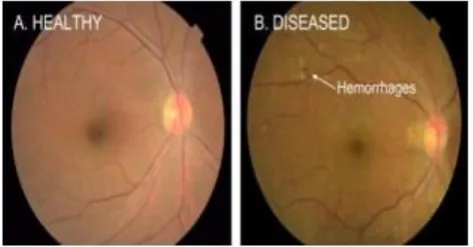
III. HAEMORRHAGES
[image:2.595.52.289.587.710.2]Haemorrhages is represented as long arrow and microaneurysms is represented as short arrow.It causes distorting vision.
Fig. 1 Sample digital fundus image with a Micro Aneurysm (MA)
IV. FLOW CHART
Flowchart Description
International Journal of Innovative Technology and Exploring Engineering (IJITEE) ISSN: 2278-3075,Volume-8, Issue-8S, June 2019
V. GENERAL DESCRIPTION
In our project we use SVM (Support Vector Machine). It is a classifier which is used to classify the severity of the disease. It is supervised learning models associated with algorithms that is used to analyze data which is used for classification. It is divided into 4 types they are,
Classification SVM Type 1 (also known as C-SVM classification)
Classification SVM Type 2 (also known as nu-SVM classification)
Regression SVM Type 1 (also known as epsilon-SVM regression)
Regression SVM Type 2 (also known as nu-SVM regression).
VI. SOFTWARE IMPLEMENTATION
Matlab
It is an environment for multi-paradigm numerical computing and proprietary programming language developed by works of math.
Syntax
Its application is built around the Matlab script language. It is an application using the command window as an interactive.
VII. ALGORITHMS
To reduce the imperfections such as poor contrast and noise and to form an image more suitable for extracting pixels features required for classification stage preprocessing is done.
Walter Klein Clahe
Vessel Removal
Illumination Equalization
Table. 1
Algorithm Purpose
Walter–Klein Enhancing Contrast
CLAHE Enhancing Major Objects
Vessel Removal Enhancing MA Near
Vessels
Illumination Enhancing MA at The
Equalization Border
Walter–Klein Contrast Enhancement
Contrast of retinal images is improved by this method. It can be done by applying gray level transformation. It is defined the local contrast enhancement operator.
Clahe
Clahe works in the image on small regions instead of the whole image called "tiles". It is used to analyze the small portion. For an every individual tile, the contrast transform function is calculated by adapthisteq. The contrast of each
tile is enhanced to approximately match the histogram of the output region specified by the value of ' Distribution'. The neighboring tiles are then combined to eliminate artificially induced boundaries using bilinear interpolation. In homogeneous, to avoid amplifying any noise in the image, the contrast can be limited.
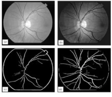
Vessel Removal
[image:3.595.313.539.381.577.2]A retinal image has varying thickness and foreground lighting and blood vessel contrast. For thick vessels, the contrast is higher and lower for thin vessels. Extraction of tree structure becomes more difficult due to the presence of anomalies in the retinal images. The literature contains a variety of methods for vessel tree extraction. They are kernel-based methods such as filters for edge detection and matched filters. When selecting a larger kernel, thick vessels are accurately obtained in matched filter methods, while thin vessels with increased thickness are obtained. Using smaller kernels can help precisely select thin vessels with higher correlation, but with reduced thicknesses the thick vessels are obtained. The conventional matched filtering technique requires a large number of different size kernels with different orientations. By combining different images corresponding to different kernels, the final tree structure is obtained. Local and region-based properties have been reported using a sampling technique to segment blood vessels.
Fig. 2 Vessel Removal
Illumination Equalization
space, which are the equalization of histograms applied by trahanias and venetsanopoulos. If the probability of bright pixels is higher than that of dark ones, "whitening" will result.
Fig. 3 Illumination Equalization
VIII. SEGMENTATION AND FEATURE EXTRACTION
Image segmentation is dividing the detected image into a region that is mutually exclusive and exhausted. The various features of segmented image is evaluated by feature extraction in order to form a feature vector required to input the SVM classifier. Various features of segmented images are extracted by the feature extraction.
Walter: It is done by using gray scale diameter closing. This is used to find out the small dark patterns on the green channels.
Flem: It uses contrast normalization method which increases the capability to differentiate MAs and other dark spots which appear on the retina.
Zhang: All MAs from color retinal images is detected by using Hierarchical approach based on multiscale correlation filtering.
IX. IMAGE ACQUISITION
In this process normal image is converted into digital image. This type is the photodiode built using silicon materials and the output is the proportional voltage waveform to the light. There must be relative displacements in both x and y directions to generate the 2D image.
X. PREPROCESSING
For all images preprocessing should be done to get a better result. It consist of resizing, noise removal, and color transformation for the further process of detection of Diabetic Retinopathy. Noise is appeared with low signal in the form of shadow regions. To make better the quality and accuracy of the result preprocessing shoud be done on the input during the comparison of normal eye image and diabetic eye image. Filtering can also be done to avoid the noises. Here we used Weiner Filter for deblurring the image.
XI. IMAGE ENHANCEMENT
In this system, Max flow-min cut graph cut segmentation is preferred.
Further there are two methods, 1) Boundary based segmentation. 2) Region based.
but in the above two methods there are some drawbacks. Such as non continuous closed edge, small holes in the
[image:4.595.100.242.102.190.2]segmented image etc.,. so Max flow-min method is preferred.
Fig. 4 Graph Cut Segmentation
XII. SEGMENTATION USING ADAPTIVE K MEANS
Image segmentation in computer vision is the process of digital image partitioning into different segments.
The goal of segmentation is to simplify or change the representation of the image into something that is easier to understand and that is more meaningful.
Here we use Adaptive K-means algorithm for the segmentation of affected portion of the retina.
XIII. RESULT AND DISCUSSIONS
In this Fig. 5 (a) and Fig. 5 (b) shows input fundus image. In this retinal image is detected and it is converted to 2D image for the comparison of defected image to the normal eye image can be analyzed and detected.
Fig. 5 (a) Fig. 5 (b)
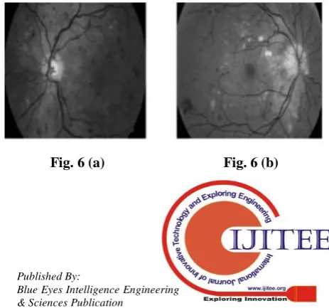
In this model Fig. 6 (a) and Fig. 6 (b) shows green channel extraction. This model contribute a low contrast image does not contain much information. The color retina image's green component gives the best result in blood vessel contrast (darker blood vessels on a bright background). Thus, the blood vessel contrast is verified.
[image:4.595.315.540.419.530.2] [image:4.595.311.543.614.830.2]International Journal of Innovative Technology and Exploring Engineering (IJITEE) ISSN: 2278-3075,Volume-8, Issue-8S, June 2019
In this Fig. 7 (a) and Fig. 7 (b) shows adaptive histogram equalized Image. It is a technique of computer image processing used to improve image contrast. It is used to enhance local contrast and edges.
Fig. 7 (a) Fig. 7 (b)
In this Fig. 8 (a) and Fig. 8 (b) shows output from graph cut segmentation. It is the output from the graph cut segmentation. Microaneurysms clearly through the nacked eye can be viewed.
Fig. 8 (a) Fig. 8 (b)
In this Fig. 9 (a) and Fig. 9 (b) shows output from classifier. This result is such as a notification. It is in the form of text notification can be analyzed.
Fig. 9 (a) Fig. 9 (b)
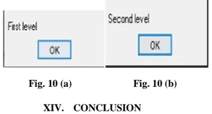
In this Fig. 10 (a) and Fig. 10 (b) shows severity of the disease. It is used to find the severity of the disease. If it is smaller it displays First level. If it is larger it displays second level. It is also a text notification. Due to this we can verify our severity of the disease and we can view correctly.
Fig. 10 (a) Fig. 10 (b)
XIV. CONCLUSION
This proposed system will help diabetic retinopathy patients for periodic eye screening.In this project,we are using green channel detection which is used to view the
microaneurysms clearly,using this channel we can analyze the disease quickly.Nowadays,this disease is very common in all ages.So this software helps to analyze the disease.This process of this project is ease and fast speed of eye screening and providing accurate results.
REFERENCES
1. Behdad Dashtbozorg, Jiong Zhang.Retinal Micro aneurysms Detection using Local Convergence Index Features. IEEE Transactions on Image processing Volume: PP, Issue: 99, July 2018.
2. Renu, Sachin Kumar. Automated Detection of Diabetic Retinopathy through Blood Vessel and Micro-aneurysms. International Journal of Computer Sciences and Engineering. Volume.6(2), Feb 2017, E-ISSN: 2347-2693.
3. Deepali D. Rathod, Ramesh R. Manza. Extraction of Non Proliferative Diabetic retinopathy using new designed wavelet and classification using KNN classifier. Int. Journal of Engineering Research and Application. ISSN : 2248-9622, Volume. 7, Issue 12, ( Part -4) December 2017, pp.06-13.
4. Dilip Singh Sisodia, Shruti Nair.Diabetic Retinal Fundus Images: Preprocessing and Feature Extraction For Early Detection of Diabetic Retinopathy. Biomedical & Pharmacology Journal. Volume. 10(2), 615-626 (2017).
5. Lin Li,Juan Shan. Automated Microaneurysm Detection in Fundus Images through Region Growing.2017.IEEE Transactions on Image processing. 2471-7819/17/31.00.
[image:5.595.51.290.111.222.2] [image:5.595.51.290.286.405.2] [image:5.595.66.270.455.552.2] [image:5.595.66.278.629.742.2]