to probe novel α4β2 nicotinic
acetylcholine receptor (nAChR)
binding sites
A thesis submitted for the degree of
Doctor of Philosophy at
the Australian National University
By Ryan Gallagher
Research School of Chemistry
Declaration
I certify that the work in this thesis has not previously been submitted for a degree nor has it been submitted as part of requirements for a degree except as fully acknowledged within the text.
I also certify that the thesis has been written by me. Any help that I have received in my research work and the preparation of the thesis itself has been acknowledged. In addition, I certify that all information sources and literature used are indicated in the thesis.
Sections of original work described in this thesis have been accepted for publication in the Australian Journal of Chemistry;
Gallagher, R. Chebib, M. Balle, T. and McLeod, M.D., Thiol-Reactive Analogues of Galanthamine, Codeine and Morphine as Potential Probes to Interrogate Allosteric Binding within Nicotinic Acetylcholine Receptors. Australian Journal of Chemistry, Accepted for publication 7th September 2015.
A copy of the manuscript is included along with the supporting information as electronic supplementary material at the end of this thesis.
Ryan Gallagher
__________________________________
Acknowledgements
First and foremost I wish to thank my supervisor, Associate Professor Malcolm McLeod. Your continued guidance and encouragement over the past three and a half years is much appreciated, as is the considerable time spent reading through the many drafts of this thesis. I would not have been able to achieve what I have without your help. I also wish to thank the remaining members of my supervisory panel, Associate Professor Mary Collins, Dr Thomas Balle and Professor Chris Easton.
I wish to thank Dr Nick Kanizaj, for his general help in the lab and for demonstrating many of the organic chemistry techniques that were new to me including sealed tube reactions and the use of solvent purifiers. I wish to thank Dr Bradley Stevenson for his help with running the LCMS and troubleshooting when things went wrong. I also wish to thank the various technical staff at the Research School of Chemistry, especially Anitha and Elizabeth for running my mass spectrometry samples as well as Chris and Peta for their help with the NMR.
I wish to thank the past and present members of the McLeod group; Paul, Chris, Luke, Dwain, Lucy, Natasha, Jacob, Keshav and Andy. It was great to be able to bounce ideas off each other and you made life in the lab a lot more entertaining. Special thanks goes to Chris and Paul for proof reading sections of this thesis.
Table of Contents
Declaration ... i
Acknowledgements ... ii
Table of Contents ... iii
List of Abbreviations ... v
Abstract ... vii
Chapter 1: Introduction ... 1
1.1. Nicotinic Acetylcholine Receptors – Structure, Location and Function: ... 1
1.1.1. General Structure: ... 1
1.1.2. Muscular vs. Neuronal nAChRs: ... 4
1.1.3. Ligand Binding: ... 6
1.2. Agonists, Antagonists and Allosteric Modulators: ... 8
1.2.1. Acetylcholine: ... 8
1.2.2. Agonists: ... 10
1.2.3. Antagonists: ... 13
1.2.4. Allosteric Modulators: ... 16
Chapter 2: Investigating the binding site of Codeine and Galanthamine ... 18
2.1. Introduction: ... 18
2.1.1. Galanthamine: ... 18
2.1.2. Substituted Cysteine Accessibility Method: ... 23
2.1.3. Project Aims:... 27
2.2. Synthesis of α,β-Unsaturated Ketones: ... 29
2.2.1. Codeinone: ... 29
2.2.2. Morphinone: ... 32
2.2.3. Narwedine: ... 35
2.3. Synthesis of Chlorinated Derivatives of Codeine: ... 37
2.3.1. Codeine Mustard: ... 37
2.3.2. Benzyl Chloride Derivative: ... 42
2.4. Reaction Kinetics: ... 49
2.4.1 Solution Kinetics: ... 49
2.4.2. Synthesis of Adducts:... 52
2.4.3. Codeinone and Morphinone: ... 60
2.4.4. Narwedine: ... 64
2.5. Potential application of the analogues as thiol reactive probes: ... 67
Chapter 3: Analogues of Methyllycaconitine ... 74
3.1. Introduction: ... 74
3.1.1. Delphinium Alkaloids: ... 74
3.1.2. Methyllycaconitine and nAChRs: ... 75
3.1.3. Simplified Analogues of MLA: ... 80
3.1.4. Project Aims: ... 84
3.2. Synthetic Strategies: ... 86
3.3. Synthesis of Bicyclic Alcohol: ... 88
3.4. Ester Coupling Route: ... 93
3.4.1. Synthesis of Biaryl Acids: ... 93
3.4.2. Esterification of Bicyclic Alcohol with Biaryl Acids: ... 100
3.5. Azabicyclo Arylboronate Route: ... 104
3.5.1. Synthesis of the Azabicyclo Arylboronate: ... 104
3.5.2. Suzuki Coupling with the Azabicyclo Arylboronate: ... 107
3.6. Preliminary Screening Results: ... 110
3.7. Conclusions and Future Work: ... 114
Chapter 4: Experimental ... 117
4.1. General Experimental: ... 117
4.2. Experimental for Chapter 2: ... 119
4.2.1: Synthesis of Ketones: ... 119
4.2.2: Synthesis of Codeine Mustard: ... 126
4.2.3: Synthesis of 3-chloromethyl-3-deoxymorphine:... 128
4.2.4: Synthesis of N-acetylcysteine methyl ester adducts: ... 133
4.2.5: Reaction Kinetics: ... 137
4.3. Experimental for Chapter 3: ... 138
4.3.1: Synthesis of Bicyclic Alcohol: ... 138
4.3.2: Synthesis of Biaryl Acids: ... 141
4.3.3: Synthesis of Azabicyclo Aryl Iodide: ... 157
4.3.4: Synthesis of Azabicyclo Esters: ... 159
Appendix A: Kinetics Data... 172
Appendix B: Selected NMR Spectra ... 187
List of Abbreviations
ACE-Cl α-chloroethyl chloroformate
ACh acetylcholine
AChBP acetylcholine binding protein AChE acetylcholine esterase
AP-ESI atmospheric pressure electrospray ionisation
b.p. boiling point
CDI carbonyl diimidazole
COSY correlation spectroscopy
DAN 14-deacetylnudicauline
DCC 1,3-dicyclohexylcarbodiimide
DCM dichloromethane
DCU 1,3-dicyclohexylurea
DHBE dihydro-β-erythroidine DMAP 4-(dimethylamino)pyridine
DMF dimethylformamide
DMP Dess-Martin periodinane
DMSO dimethylsulfoxide
Dppf bis-diphenylphosphinoferrocene
EC50 agonist concentration required to elicit half the maximum response
EI electron ionisation
ESI electrospray ionisation
GCMS gas chromatography – mass spectrometry
HEPES 4-(2-hydroxyethyl)-1-piperazineethanesulfonic acid HMBC heteronuclear multiple bond correlation
HRMS high resolution mass spectrometry HSQC heteronuclear single quantum correlation
IC50 antagonist concentration required for 50% inhibition
IR infrared
Kd equilibrium dissociation constant
Ki inhibition constant
LD50 dose that is lethal to 50% of a population of test animals LRMS low resolution mass spectrometry
mAChR muscarinic acetylcholine receptor m-CPBA meta-chloroperbenzoic acid
MeOH methanol
MePPh3Br methyl triphenylphosphonium bromide
MLA methyllycaconitine
MTSEA 2-aminoethylmethanethiosulfonate nAChR nicotinic acetylcholine receptor
NAM negative allosteric modulator
NMR nuclear magnetic resonance
nOe nuclear Overhauser effect
NOESY nuclear Overhauser effect spectroscopy
NUD naudicauline
PAM positive allosteric modulator
PCC pyridinium chlorochromate
p-TsCl para-toluenesulfonyl chloride
Rf retention factor
rhAChE recombinant human acetylcholinesterase
RT room temperature (25 °C)
SCAM substituted cysteine accessibility method
SOCl2 thionyl chloride
t½ half-life
TBAF tetrabutylammonium fluoride TBDMS tert-butyldimethylsilyl t-BuOK potassium tert-butoxide
TcAChE Torpedo californica acetylcholinesterase
THF tetrahydrofuran
TLC thin layer chromatography
UHPLC ultra high pressure liquid chromatography
Abstract
Nicotinic acetylcholine receptors (nAChRs) are a complex class of ligand gated ion channels consisting of multiple subtypes with different stoichiometries. Of these, the two most commonly found in the brain are the α7 and α4β2 nAChRs. The binding site of agonists and competitive antagonists at nAChRs is well established. However, the binding site of many allosteric modulators remains unknown. Additionally, despite their importance in brain function, the role of specific subtypes and stoichiometries is largely unknown. This thesis deals with the synthesis of alkaloid derivatives that can be used to establish the binding sites of allosteric modulators and study the role of specific subtypes and stoichiometries in the brain. Such information can be used to develop better drugs and drug targets for the treatment of neurological diseases.
Galanthamine and codeine are reported to be positive allosteric modulators at nAChRs. In order to establish their binding site, thiol reactive analogues of galanthamine, codeine and the structurally similar alkaloid, morphine, were synthesised for use as probes in covalent trapping experiments. The α,β-unsaturated ketone derivatives of each alkaloid; narwedine, codeinone and morphinone were synthesised along with the codeine mustard and a protected derivative of the benzyl chloride analogue of codeine. While the chlorinated derivatives were too unstable for use as probes, the α,β-unsaturated ketone derivatives were stable in aqueous solution and their reactivity towards thiols was assessed by monitoring their reaction with a cysteine derivative. All of the α,β-unsaturated ketone derivatives displayed sufficient reactivity for use as thiol reactive probes.
Methyllycaconitine (MLA) is an antagonist at nAChRs that is known to bind at the α7–α7 interface of α7 nAChRs and at the α4–α4 and α4–β2 interfaces of α4β2 nAChRs. Small bicyclic ester analogues of MLA were synthesised with functional groups targeting key residues that are unique to the binding sites at the α4–α4 and α4–β2 interfaces of α4β2 nAChRs. Esters with the pyridine moiety were synthesised to target an aspartic acid residue in the α4–β2 binding site via salt bridge interactions. Esters with the acetamide moiety were synthesised to target a tryptophan residue in the α4–α4 binding site via
Chapter 1:
Introduction
1.1. Nicotinic Acetylcholine Receptors – Structure, Location and Function:
1.1.1. General Structure:
Nicotinic acetylcholine receptors (nAChRs) are a class of pentameric ligand-gated ion channels composed of five homologous subunits organised around a central pore1-3. They are involved in neuron-muscle and neuron-neuron communication. The first nAChR subtype studied extensively was isolated from the electric organ of Torpedo marmorata
consisting of four subunits assigned α, β, γ and δ in order of increasing apparent molecular weight4. The receptor structure could be broken down into three domains (Figure 1.1); a large extracellular domain (E) consisting of β-strands organised into a β-barrel configuration, a membrane spanning domain and a smaller intracellular domain (I) consisting of an α-helix from each subunit2,5. Each subunit has four α-helix
[image:11.595.109.536.422.718.2]trans-membrane domains (M1–M4) arranged such that the M2 domain forms the wall of the central pore. An excess of negatively charged groups lines the extracellular and intracellular surfaces resulting in a cation stabilising environment that repels anions5.
Figure 1.1: Structure of the muscle nAChR viewed from above (left) and in the plane of the cell membrane (right); α, β, γ and δ are the subunits of the muscle
nAChR, E is the extracellular domain and I is the intracellular domain5
Subunits are classed as either α-type or non α-type based on the presence or absence of a pair of adjacent cysteine residues, crucial to the formation of the agonist binding site1,2. Studies of the Torpedo nAChR by electron microscopy in the closed and open states revealed that the α-type subunits exist in a ‘distorted’ state, when compared to the non α-type subunits, when the channel was closed2,5. However, binding of agonists such as
acetylcholine (ACh) causes a local disturbance along with a larger scale conformational change to a ‘relaxed’ conformation. The inner M2 domain changes shape, widening the pore and allowing the flow of ions.
[image:12.595.136.415.478.694.2]The agonist binding site, located at the interface between an α-subunit and an adjacent subunit, is made up of a total of 6 loops (A–F) containing the residues that interact with agonists6. Three of these loops (A–C) are found on the α-type subunit termed the ‘principal’ subunit, while the remaining three loops (D–F) are found on the adjacent subunit, termed the ‘complementary’ subunit. In addition to the crucial pair of adjacent cysteine residues, found at the apex of loop C, five aromatic amino acids are also found in the agonist binding site forming what is known as the ‘aromatic cage’. These include three tyrosines (Tyr) and one tryptophan (Trp) from the ‘principal’ subunit and one tryptophan from the ‘complementary’ subunit (Trp). Acetylcholine and related agonists such as carbamoylcholine are able to interact with these residues through cation-π interactions between the quaternary ammonium ion and the electron-rich aromatic rings (Figure 1.2).
Figure 1.2: Five aromatic amino acids forming the ‘aromatic cage’; three tyrosines (Tyr)
and twotryptophans (Trp), carbamylcholine is also shown in white, image generated from X-Ray crystallography data7, Lymnaea stagnalis AChBP, PDB Code 1UV6, using PyMol
Further insight into the structure of the agonist binding site was obtained with the discovery of the ACh binding protein (AChBP) isolated from snails (Lymnaea stagnalis) and sea slugs (Aplysia californica). The AChBP is a soluble homopentamer released by these species in response to increased ACh levels in the synaptic cleft. It resembles the extracellular domain of nAChRs providing a good model for the agonist binding site of nAChRs2,6. It is most closely related to the human α7 nAChR, sharing 24% sequence identity in the ligand binding domain.
[image:13.595.106.542.435.647.2]The binding site of the AChBP is found in cavities at the interface between subunits formed from three loops on the ‘principal’ face of one subunit and three loops on the ‘complementary’ face of the adjacent subunit. There are five of these cavities per pentamer, located close to the outer edge of the ring (Figure 1.3)6. The key residues, including those that constitute the ‘aromatic cage’ and the pair of adjacent cysteine residues, are conserved between the agonist binding site of nAChRs and the binding site of the AChBP. As such, the AChBP exhibits similar ligand specificity to that of nAChRs binding known agonists such as ACh and nicotine as well as antagonists such as methyllycaconitine (MLA)6.
Figure 1.3: Structure of the agonist binding site in the AChBP, adapted from Brejc, K. et. al.6
1.1.2. Muscular vs. Neuronal nAChRs:
Since the analysis of nAChRs from Torpedo marmorata, a total of 17 distinct subunits have been identified. Different combinations of subunits give rise to differing functionality and affinity for certain agonists and antagonists. The different nAChRs that result can be classified based on their location in the body, as muscular or neuronal.
Figure 1.4: Muscle nAChRs at the neuromuscular junction, adapted from Physioweb8
Muscular nAChRs are found in skeletal muscles at the neuromuscular junction (Figure 1.4) where they mediate neuromuscular transmission by allowing the passage of ions to generate an action potential9. This action potential is propagated along the muscle fibres causing the muscle to contract. There are two subtypes of muscular nAChRs (Figure 1.5), foetal and adult. Foetal muscular nAChRs, like the Torpedo nAChRs, consist of two α1
subunits and one each of β1, γ and δ subunits2-4,9. In adult muscular nAChRs the γ subunit
is replaced with a ε subunit.
Figure 1.5: The two subtypes of muscular nAChRs, foetal (left) and adult (right)
Neuronal nAChRs are found in the central and peripheral nervous systems as well as in some non-neuronal tissues. Neuronal nAChR subtypes are composed of different subunits and are denoted by their composition, for example α7. Unlike muscular nAChRs, there are a significant number of different neuronal nAChR subtypes owing to the many subunits expressed. There are twelve known neuronal nAChR subunits, nine α-type (α2–α10) and three non α-type (β2–β4). Two different classes of receptor (Figure 1.6) can be formed from these subunits, namely homomeric and heteromeric pentamers2-4,9,10. Homomeric pentamers are made up of five of the same α-type subunit (α7, α9 or α10). Heteromeric pentamers are made up of α-type (α2 – α6) and non α-type subunits usually of the form (αx)2(βy)33.By far the most common subtypes found in the brain are the homomeric α7 and
the heteromeric α4β2 nAChRs. These receptors are commonly differentiated by their affinity for different ligands with α4β2 nAChRs characterised by high affinity for nicotine and α7characterised by high affinity for α-bungarotoxin (α-BTX).
Figure 1.6: The two types of neuronal nAChRs, heteromeric (left) and homomeric (right) The role of neuronal nAChRs in the body is not as well understood as muscular nAChRs, because of the significant number of different receptor subtypes and the lack of subtype specific agonists and antagonists to study them10. However, they are believed to modulate the release of neurotransmitters and influence physiological functions including sleep, anxiety and several cognitive functions10. As a consequence of the diversity of neuronal nAChR subtypes and their roles they are implicated in a variety of neurological conditions9 including addiction, schizophrenia, Alzheimer’s disease and epilepsy.
1.1.3. Ligand Binding:
[image:16.595.105.446.375.662.2]Nicotinic acetylcholine receptors can exist in three distinct states; resting, active and desensitized (Figure 1.7). Receptors alternate between these three states via conformational changes in response to ligand binding. In the absence of an agonist such as acetylcholine, receptors are predominantly in the resting state. In this state the ion channel is closed but the receptor is responsive to the application of an agonist. When an agonist is bound to the receptor the proteins are stabilised in a conformation in which the ion channel is open. If the agonist is allowed to remain for too long, the receptor undergoes a conformational change to the desensitized state in which the ion channel is again closed but the agonist remains bound. When the agonist is removed the receptor eventually reverts to the resting state. The equilibria between the resting, active and desensitised states can be shifted by binding of allosteric modulators. For example positive allosteric modulators, discussed later in this chapter, can cause a shift in the equilibrium from the closed states to the active state in the presence of an agonist.
Figure 1.7: The three states of nAChRs
The equilibrium established when a ligand (L) binds to a receptor (R) to generate a complex (L•R) is defined as follows:
L + R L R
The equilibrium constant (Kd) for the reverse reaction is commonly used to describe the
affinity of that ligand for the receptor (i.e. how well the ligand binds to the receptor). A low value for Kd corresponds to a high affinity. It has units of concentration and is
calculated as follows:
Kd =[L][𝑅𝑅][L ∙ R]
Where [L], [R] and [L•R] are concentrations of the ligand, receptor and ligand-receptor complex respectively. The value of Kd also corresponds to the ligand concentration at
which half the receptor population will be occupied (i.e. [R] = [L•R]). The affinity of a ligand for a receptor is commonly measured by carrying out direct binding assays (measuring Kd) or competition binding assays (measuring the inhibition constant, Ki) with
radiolabelled ligands. Because desensitisation is fast relative to the time-scale of binding assays, the affinity measured in this manner reflects binding in the desensitised state rather than the active state. The values estimated by binding assays will generally also be lower than those corresponding to binding in the active state because the desensitised state has a higher affinity for agonists.
The response a receptor has to binding of a ligand can be described in terms of efficacy and potency. Efficacy is the maximum possible response that a ligand can elicit from the receptor. Potency is a measure of the concentration of ligand required to elicit a response of a given magnitude. Potency is generally quantified as the agonist concentration required to elicit half of the maximum response (EC50), or the antagonist concentration required for
50% inhibition (IC50). The EC50 value for an agonist is generally much lower than the Kd
or Ki value estimated from binding assays, sometimes by orders of magnitude, reflecting
the fact that EC50 values correspond to binding in the active state. This binding perturbs the
equilibrium between resting and active states leading to a functional response that can be detected.
1.2. Agonists, Antagonists and Allosteric Modulators:
1.2.1. Acetylcholine:
O N
O
Figure 1.8: Chemical structure of ACh
Acetylcholine (ACh) is an endogenous neurotransmitter acting on nicotinic (nAChR) and muscarinic (mAChR) acetylcholine receptors in the central and peripheral nervous system. As the name suggests it is the ester formed between choline and acetic acid (Figure 1.8). It is synthesised in nerve cells via reaction between choline and acetyl coenzyme A, catalysed by the enzyme choline acetyl transferase. Acetylcholine is then released from nerve cells into the extracellular space where it can interact with nAChRs on nearby cells. It is later removed from the extracellular space by acetylcholinesterase (AChE) catalysed hydrolysis back to choline, which is then transported back into the nerve.
[image:18.595.181.373.575.749.2]When ACh acts on a homogenous receptor population consisting of a single type of binding site, a monophasic response is observed and a plot of this response to ACh against the dose follows a sigmoidal curve (Figure 1.9). Alternatively, when ACh acts on a receptor population consisting of two different types of binding site, a biphasic response may be observed and a plot of this response to ACh against the dose gives rise to a curve with two sigmoidal regions corresponding to the two different binding sites. This can occur when the receptor population is heterogeneous, consisting of two different receptors or when the population is homogenous where the receptors contain two different binding sites. The effect is only clearly seen if there is a significant difference in the potency of the agonist at the two binding sites leading to a difference in their sensitivity to agonist binding.
Figure 1.9: Monophasic and biphasic response curves
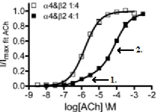
Figure 1.10: The stoichiometries of α4β2 receptors, (α4)2(β2)3(left) and (α4)3(β2)2 (right)
Of the two predominant nAChR subtypes present in the nervous system, the α4β2receptors display the most interesting response to ACh. It is known that α4β2receptors exist in two stoichiometries, and both appear to be naturally expressed in the human brain11. These are the high sensitivity (α4)2(β2)3 form and the low sensitivity (α4)3(β2)2 form (Figure 1.10).
As a result, when studying the response of α4β2 receptor populations to ACh, a biphasic dose-response curve is observed reflecting, in part, these mixed populations.
Figure 1.11: Dose-response curves for α4β2 receptor populations of the (α4)2(β2)3form
(white squares) and (α4)3(β2)2form (black squares), adapted from Harpsoe, K. et. al.11
The response of individual nAChR stoichiometries can also be studied by controlling receptor expression in vitro. While receptor populations consisting solely of the (α4)2(β2)3
form give rise to a monophasic response, receptor populations of the (α4)3(β2)2 form give
rise to a biphasic response (Figure 1.11) suggesting that this form contains high and low sensitivity binding sites11. In addition to the two high sensitivity ACh binding sites found at the α4-β2 interfaces, the (α4)3(β2)2 form has a low sensitivity ACh binding site at the
α4-α4 interface. Activation of the α4-α4 in addition to the α4-β2 binding sites gives rise to a higher response but this is not evident in the dose-response curves since the responses are normalised (Figure 1.11). It has been hypothesised that the lower sensitivity of the (α4)3(β2)2 form results from the need for agonist binding at the α4-α4 interface to fully
activate the receptor11. By targeting the α4-α4 interface, ligands could be developed that
selectively inhibit or modulate agonist activity at α4β2 receptors of the (α4)3(β2)2
stoichiometry.
1.2.2. Agonists:
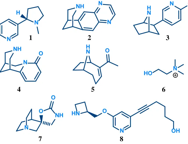
[image:20.595.130.424.293.496.2]An agonist is any compound that binds to a receptor to elicit a response. An agonist is a full agonist if it elicits the maximal possible response from the receptor, defined relative to an agonist such as ACh. Alternatively, it is a partial agonist if it elicits a fraction of the maximal response. In general, agonists of nAChRs contain common key features which have enabled the development of a pharmacophore model. The initial model developed by Beers and Reich 12 was based on the structure of ACh and compared the structures of other agonists and antagonists. Two key features were identified, a cationic nitrogen involved in cation–π interactions with the previously discussed ‘aromatic cage’, and a hydrogen bond acceptor separated by a distance of approximately 5.9 Å (Figure 1.12).
Figure 1.12: Pharmacophore model developed by Beers and Reich12 applied to nicotine (left) and ACh (right)
More recently, Nicolotti et al.13 analysed a selection of 11 agonists with sufficient structural variation to develop an updated pharmacophore model. The two key features identified by Beers and Reich are included in the model along with a hydrophobic centre generally occupied by aliphatic rings. The hydrogen bond acceptor identified by Beers and Reich is defined in this model as the lone pair of either a pyridyl nitrogen or a carbonyl oxygen. It has recently been determined that this hydrogen bond is formed with a backbone -NH, often through a water molecule14. A selection of nAChR agonists, both natural and synthetic, is shown in Figure 1.13 on the following page.
N
N
H
N O
N NH
O
H N
N Cl
NH
N N
HO N
6
1 3
5 4
2
N
NH O
O
7
N OH
O HN
8
[image:21.595.153.477.81.329.2]H
Figure 1.13: Selection of natural and synthetic nAChR agonists
Nicotine (1) is the main alkaloid in tobacco responsible for its addictive effects, and is the prototypic agonist of nAChRs15,16. It acts on all subtypes of nAChRs, although of the two
most prominent subtypes it has a significantly greater binding affinity at α4β2 receptors (Ki = 1–11 nM) where it is a potent full agonist (EC50 = 1 μM)16. Long-term exposure of
α4β2 receptors to nicotine (1) results in an increase in the number of receptors in a process known as upregulation2,17. Additionally, the exposure causes a shift in assembly from the (α4)3(β2)2 form to the (α4)2(β2)3 form18. Varenicline (2), a smoking cessation aid sold
under the tradename Champix, is a partial agonist (45% efficacy) with significantly higher binding affinity at α4β2 receptors (Ki = 110–170 nM) than other nAChR subtypes19.
Epibatidine (3), a toxin secreted by various species of poison dart frogs, is the most potent nAChR agonist known. It is approximately 100 times more potent than ACh at α4β2 receptors (EC50 = 4–20 nM)16. Like ACh, the dose-response curve obtained for brain α4β2
receptor populations is biphasic suggesting mixed populations and differences in its action on the two stoichiometries expressed20. The upregulation effect seen after exposure to nicotine (1) is not found after exposure to epibatidine (3). While it is also an agonist at α7 receptors, it has greater binding affinity at α4β2 receptors (Kd = 19 pM)16.
Other natural agonists such as cytisine (4) and anatoxin-A (5) generally show this same trend in binding affinity16. One natural agonist which doesn’t conform to the same trend in binding affinity is choline (6), a selective agonist for the α7 receptor21. It has 10-fold lower potency than ACh at the α7 receptor (EC50 = 1.6 mM)16 but is a full agonist. This suggests
that the carbonyl oxygen of ACh may not be necessary for agonist binding at the α7 receptor.
The synthetic compound AR-R17779 (7), a conformationally restricted analogue of ACh, is another agonist that is selective for the α7 receptor. It has more than 100 times greater binding affinity at the α7 receptor (Ki = 92 nM) than at the α4β2 receptor22. Studies on
analogues of AR-R17779 (7) reveal that replacement of the proton on the carbamate nitrogen with an alkyl group dramatically increased the binding affinity at α4β2 receptors whilst simultaneously decreasing the binding affinity at the α7 receptor. This suggests a lipophilic pocket may be present in the binding site of the α4β2 receptor that is not present in the α7 receptor binding site.
Sazettidine-A (8), is a synthetic nAChR agonist, selective for the α4β2 receptor. It was originally thought of as a ‘silent desensitiser’ causing desensitisation of receptors without activating them23. However, more recent studies have revealed it is an agonist at α4β2 receptors with different activity at the two stoichiometries. It is a full agonist on the (α4)2(β2)3 form, but only a partial agonist on the (α4)3(β2)2 form (6% efficacy)24. The same
upregulation effect noted after exposure to nicotine (1) is also found after exposure to sazettidine-A (8)23.
In addition to the agonists discussed here there are numerous other agonists of nAChRs and these have been reviewed extensively in the literature13-16,25.
1.2.3. Antagonists:
Figure 1.14: Dose-Response curves reflecting the effect of competitive antagonists (left)
and non-competitive antagonists (right) on the response to agonist binding.
An antagonist is any compound that binds to a receptor without eliciting a response, but in doing so it blocks the response elicited by an agonist. Antagonists can be either competitive or non-competitive depending on their effect on agonist binding (Figure 1.14). Competitive antagonists (surmountable) are those which can be overcome by increasing the agonist dose. Non-competitive antagonists (insurmountable) are those which cannot be overcome by increasing the agonist dose. In the case of competitive antagonists the effect on the dose-response curve is a shift to the right such that the agonist maintains efficacy at higher doses. In the case of non-competitive antagonists the effect is a shift downward such that the agonist shows lower efficacy. In general, competitive antagonists bind at the same site as agonists and therefore conform to the agonist pharmacophore model. As non-competitive agonists generally bind at a different site to agonists they do not conform to the agonist pharmacophore model.
Some of the most potent and selective nAChR antagonists are peptide based neurotoxins found in venoms including α-bungarotoxin from the Taiwanese Banded Krait and the α-conotoxins from Conus snails15,16,26. α-Bungarotoxin, consisting of 75 amino acids, is a selective antagonist at the α7 nAChR that does not appear to interact with heteromeric nAChRs such as the α4β2 subtype. Similarly, the α-conotoxins, consisting of 14-17 amino acids, are selective towards specific subtypes of receptor. For example, α-conotoxin ImI is a selective antagonist of the α7 nAChR. Some non-peptidic antagonists are shown in Figures 1.15 and 1.16.
O
N MeO
O
N MeO
MeO
HO
N
O OH
N
OH H
H OMe
OMe
OH
MeO H MeO
O O N
O O
O OH
OMe
N O
OH
OMe N
9 10
14
13 12
N
O N
O H
H
H
[image:24.595.79.472.77.401.2]11
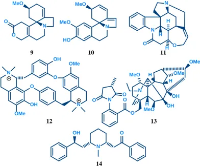
Figure 1.15: Selection of competitive nAChR antagonists
Dihydro-β-erythroidine (DHBE, 9) and erysodine (10) are alkaloids found in the seeds of
Erythrina species26,27. Erysodine (10) is a more selective antagonist of neuronal nAChRs over muscle nAChRs when compared with DHBE (9)27. Erysodine (10) has greater binding affinity than DHBE (9) at α4β2 receptors (Ki = 5 nM vs 35nM) and, to a lesser
extent, at α7 receptors (Ki= 4 μM vs 9 μM)26. Strychnine (11), a potent convulsant known
for its action as a glycine receptor antagonist, is a potent competitive antagonist at the α7 nAChR (IC50 = 1.9 μM, Ki = 1.1 μM)), and a non-competitive antagonist at the α4β2
nAChR (IC50 = 118 μM)28. d-Tubocurarine (12), an alkaloid from the bark of the
Chondodendron tomentosum plant used as an arrow poison, is a non-selective antagonist active at muscle and neuronal nAChRs26. Methyllycaconitine (MLA, 13) from the
Delphinium brownii plant is one of the most potent competitive antagonists at the α7 nAChR (IC50 = 140 pM, Ki = 1 nM)15,29 that also acts as an antagonist at the α4β2
receptor26. A more detailed analysis of MLA and its analogues is covered in chapter 3.
Lobelline (14), an alkaloid found in Indian tobacco, was originally considered an nAChR agonist as it stimulates the release of dopamine26,30. However, more recent evidence shows
that this stimulation is not nAChR mediated and it acts as an nAChR antagonist.
H N
N N N
N Cl
Cl
Cl
Cl
Cl
O H N
15 16
18
17
N H Cl
O N
20 19
O
O
O
OH
[image:25.595.114.522.76.391.2]21 22
Figure 1.16: Selection of non-competitive nAChR antagonists
Mecamylamine (15), originally developed as a ganglionic blocker to treat hypertension, is the archetypal non-competitive antagonist for neuronal nAChRs15,16,31. It inhibits activity at most subtypes of neuronal nAChRs functioning as an open channel blocker as evidenced by its voltage-dependent antagonism31. The upregulation effect noted following chronic exposure to nicotine (1) also occurs following chronic exposure to mecamylamine (15)32. Other compounds, originally designed as ganglionic blockers for the treatment of hypertension, also function as non-selective non-competitive antagonists at nAChRs and include hexamethonium (16) and chlorisondamine (17)15,16. Racemic bupropion (18), originally developed as an antidepressant and now used as a smoking cessation aid, is a non-competitive antagonist at α4β2 and α7 nAChRs that does not appear to act within the channel pore15,16,33. Additionally, dissociative anaesthetics such as phencyclidine (19) and (S)-ketamine (20) and steroids such as progesterone (21) and testosterone (22) have also been reported as non-competitive antagonists at neuronal nAChRs15,16.
1.2.4. Allosteric Modulators:
Allosteric modulators are compounds that bind to sites other than the agonist binding site to alter the response to agonist binding. Positive allosteric modulators (PAMs) increase the response at a given agonist concentration, while negative allosteric modulators (NAMs) decrease the response. They can alter the efficacy of the agonist resulting in an upward (PAM) or downward (NAM) shift in the dose-response curve. Alternatively, they can alter the affinity of the agonist for the receptor resulting in a shift in the dose-response curve to the left (PAM) or right (NAM). Non-competitive antagonists, discussed in the previous section, are negative allosteric modulators that affect the efficacy of the agonist without affecting the affinity. Negative allosteric modulators that affect the affinity of the agonist have a similar effect to competitive antagonists but can be distinguished from these based on the fact that they do not compete with the agonist for binding. A selection of positive allosteric modulators of nAChRs, both natural and synthetic, is shown in Figure 1.17 below.
O
O
N
HO
N N O
H N
O
O
O
HO
N
HO
OH N
H HO
N O N N
CN
Br NH
HN
23 24 25
29 28
27 26
H
[image:26.595.66.487.380.702.2]H
Figure 1.17: Selection of nAChR positive allosteric modulators
Galanthamine (23) and physostigmine (24) are AChE inhibitors that are reported to act as positive allosteric modulators of α4β2 and α7 nAChRs15,16,34. They increase the affinity of
ACh for the receptors without affecting the efficacy35. Despite interest in their activity at nAChRs the binding site remains unknown. A more detailed analysis of the effect of galanthamine on nAChRs is covered in Chapter 2. Codeine (25), an opioid with a chemical structure similar to that of galanthamine has the same effect on nAChRs without the inhibition of acetylcholinesterase15,16,35. Desformylflustrabromine (26) is a positive
allosteric modulator, selective for the α4β2 receptor, which acts to increase the maximum response that can be elicited by acetylcholine36. It has the opposite effect at α7 nAChRs
where it acts as an inhibitor. 5-Hydroxyindole (27), a metabolite of serotonin, is a positive allosteric modulator that is selective for the α7 receptor, increasing the efficacy of acetylcholine37. The synthetic compound, NS9283 (28), selectively modulates heteromeric nAChRs, including the (α4)3(β2)2 receptor, but not the (α4)2(β2)3 receptor38,39. Like
galanthamine and physostigmine, it increases the affinity of acetylcholine without affecting the efficacy. While the majority of naturally synthesised steroids act as antagonists of neuronal nAChRs, estradiol (29) is a positive allosteric modulator at the α4β2 receptor34,40. Its effects on α7 nAChRs have not been established.
Nicotinic acetylcholine receptors are a complex class of receptors consisting of multiple subtypes with different stoichiometries. While the binding site of agonists and competitive antagonists at nAChRs has been studied extensively, the binding sites of allosteric modulators such as those described in this section generally remains unknown. Chapter 2 of this thesis deals with work developing analogues of galanthamine (23) and codeine (25) that can be used to establish their binding site at nAChRs. An understanding of the role of specific subtypes and stoichiometries in the brain requires subtype and stoichiometry specific ligands. Chapter 3 of this thesis deals with work developing ligands that bind selectively to either the (α4)2(β2)3 or the (α4)3(β2)2 stoichiometry of the α4β2 nAChR.
Chapter 2:
Investigating the binding site of Codeine and Galanthamine
2.1. Introduction:
2.1.1. Galanthamine:
Galanthamine (23) is an alkaloid present in many plant species from the Amaryllidaceae family including Galanthus, Narcissus and Leucojum. It was first isolated by Proskurnina and Yakovlena in 1952 from the Caucasian snow drop (Galanthus woronowii)41. The
chemical structure was determined, with the incorrect stereochemistry, by Uyeo and Kobayashi in 195642. The structure was confirmed, and the stereochemistry was corrected,
when galanthamine (23) was synthesised for the first time by Barton and Kirby43 in an 8-step synthesis culminating in the reduction of (–)-narwedine (30) to a separable mixture of (–)-galanthamine (23) and (–)-epigalanthamine (31) with lithium aluminium hydride (Scheme 2.1). Since then there have been numerous attempts to improve upon this synthesis such that it would be viable to prepare (–)-galanthamine (23) on an industrial scale44. The reduction of narwedine (30) can be completed stereoselectively to afford galanthamine (23) as the sole product using L-selectride45.
O
O
N
O
O
O
N
HO
30 23
O
O
N
HO 31
+
LiAlH4
Scheme 2.1: Reduction of (–)-narwedine to (–)-galanthamine and (–)-epigalanthamine
During initial studies into the synthesis of galanthamine (23) by Barton and Kirby43 an interesting method to obtain enantiomerically pure narwedine (30) was discovered. When crystallising (–)-narwedine (30) prepared from (–)-galanthamine (23) the sign and magnitude of the optical rotation of the material obtained was dependent on the solvent used. When crystallising from acetone a product with a negative optical rotation was obtained corresponding to (–)-narwedine (30). However, when crystallising from ethanol a positive optical rotation was obtained, corresponding to (+)-narwedine (30a).
O
O
N
O
O
N
O O
30 30a
O
N O
O
Figure 2.1: Racemisation of narwedine (30) via an intermediate dienone
In protic solvents such as ethanol, narwedine (30) can racemise via an intermediate dienone as shown in Figure 2.1 above. It was therefore hypothesised that during crystallisation from ethanol narwedine (30) racemised completely. This hypothesis was confirmed by crystallising racemic narwedine (30 + 30a) from ethanol with (–)-galanthamine (23) in a 2:1 ratio to obtain pure (+)-narwedine (30a). As crystallisation proceeded, traces of (–)-galanthamine (23) remaining after oxidation induced separation of (+)-narwedine (30a) from the racemate giving crystals of (+)-narwedine (30a)43. This is believed to result from (–)-galanthamine (23) interacting with (–)-narwedine (30) preventing it from crystallising. It has since been demonstrated that only traces of either enantiomer of galanthamine (23) are needed to obtain high yields of enantiomerically pure narwedine (30 or 30a)45 by a dynamic kinetic resolution process. Furthermore, racemic narwedine (30 + 30a) can be seeded with a small amount (1%) of the desired enantiomer of narwedine (30 or 30a) instead of galanthamine (23) as in the industrial synthesis 46. In this process, crystallisation of the added enantiomer is induced in preference to the other enantiomer. Galanthamine (23) is currently obtained either industrially46, or isolated from
Narcissus or Leucojum species in addition to the original Galanthus species.
Galanthamine (23) was initially used in Eastern European countries as a curare reversal agent in anaesthetic practice and to assist in recovery from paralysis associated with poliomyelitis and other neuromuscular disorders47-49. Once it was established that
galanthamine (23) could penetrate the blood-brain barrier its use in the treatment of neurological disorders associated with decreased cholinergic transmission, particularly Alzheimer’s disease, was investigated. Galanthamine (23) is currently approved to treat the symptoms of Alzheimer’s disease in many countries world-wide, including Australia50.
Galanthamine (23) is reported to have a dual mode of action on the cholinergic system with the overall effect of increasing nAChR activity44,47. It increases ACh levels by
competitively inhibiting AChE, the enzyme responsible for breaking down ACh (IC50 ≈ 3 μM)51. At low concentrations (0.02-2 μM), it is also reported to act as a positive
allosteric modulator increasing the response of neuronal nAChRs to ACh. Galanthamine (23) binds to pre-synaptic and post-synaptic nAChRs increasing their sensitivity to ACh. Since pre-synaptic nAChRs are involved in the release of neurotransmitters, including ACh, galanthamine (23) has the additional effect of enhancing the release of neurotransmitters such as ACh. At higher concentrations (>10 μM) galanthamine (23) acts as an inhibitor at nAChRs52.
The binding site of galanthamine (23) on the AChE enzyme is well established. As a competitive inhibitor it binds in the same location as ACh. Crystal structures have been obtained for galanthamine (23) bound to Torpedo californica acetylcholinesterase (TcAChE)53 and recombinant human acetylcholinesterase (rhAChE)54 demonstrating that it interacts with the choline binding site and the acyl-binding pocket to prevent ACh from binding to the enzyme. However, since galanthamine (23) acts as a positive allosteric modulator at nAChRs, its binding site at these receptors is more difficult to locate. While there have been numerous studies into potential binding sites of galanthamine (23) at nAChRs a definitive binding site has not yet been determined.
An X-ray crystal structure of galanthamine (23) bound to the AChBP has been obtained, demonstrating that galanthamine (23) binds in the same region as ACh55. However, it was
noted that galanthamine (23) did not interact with the pair of adjacent cysteine residues in the C-loop of the binding site that are crucial for agonist binding. Mutants of the AChBP, with these cysteine residues replaced by alanine or serine, were prepared and binding studies were carried out with agonists and allosteric ligands, including galanthamine (23). While agonists like ACh showed a significant reduction in binding affinity for these mutants when compared with the wild type, there was no change observed with allosteric ligands. Based on these findings it was suggested that galanthamine (23) and other allosteric ligands may bind at interfaces not containing the principle face of the α-subunit of heteromeric nAChRs55. The residues that galanthamine (23) was found to interact with are still present at these interfaces but the pair of cysteine residues are not.
Blind docking experiments have also been used to gain insight into potential binding sites for galanthamine (23) and other allosteric modulators at nAChRs. Studies on the AChBP identified three potential binding sites for galanthamine (23), physostigmine (24) and codeine (25), within the channel pore56. Two were located close to the agonist binding site (I and II), the third was located further down in a region corresponding to just above the transmembrane domain in functional receptors (III) as shown in Figure 2.2 below.
Figure 2.2: Location of potential binding sites of galanthamine identified by blind docking studies on the inner (left) and outer (right) surfaces of two adjacent residues
of the AChBP. ACh is shown in the agonist binding site in black. Image generated from X-Ray crystallography data57 PDB Code 3WIP, using PyMol
Following further modelling studies on the AChBP and the extracellular domain of human α7 and α4β2 nAChRs another two potential binding sites on the outer surface were identified in addition to the agonist binding site58. Both sites were at the interface between two subunits, one was above the agonist binding site (IV), the other was below (V) as shown in Figure 2.2 above. Based on studies with an antibody known to inhibit the binding of galanthamine (23) and other nAChR allosteric modulators59, the lower binding site on
the outer surface of the protein (V) was identified as the most likely binding site of those identified by blind docking experiments.
More recently, photolabelling studies with [3H]galanthamine and [3H]physostigmine were
employed to directly identify amino acids contributing to their binding sites on the muscle-type Torpedo nAChR60. Based on these studies three binding sites were identified for galanthamine (23) and physostigmine (24) in the presence of agonist (Figure 2.3). These were at the δ-β interface at a site equivalent to the benzodiazepine binding site in GABAA receptors (I) and at the α-γ interface in the entry to the agonist
[image:32.595.197.353.272.441.2]binding site (II) and within the ion channel near the level of the agonist binding site (III). Additionally, in the absence of agonist, these ligands were found to bind in the α-δ agonist binding site (IV).
Figure 2.3: Location of binding sites of galanthamine (23) and physostigmine (24) on the muscle-type Torpedo nAChR, physostigmine (24) is shown in pink at each
binding site, adapted from Hamouda, A.K. et.al.60
While the results of these studies provide some insight into the binding site locations of galanthamine (23) and other nAChR positive allosteric modulators they do not definitively establish the binding site at neuronal nAChRs, especially the α7 and α4β2 receptors. Identification of the binding site at these receptors would aid in the development of a proposed mechanism of allosteric modulation and allow for the design of more effective allosteric modulators. Photoaffinity labelling, as applied in the study of the muscle type
Torpedo nAChR60, is limited by the fact that photoreactive ligands only photolabel certain residues. For example, [3H]galanthamine only photolabeled tyrosine and cysteine residues. A more promising method is covalent trapping which is an extension of the substituted cysteine accessibility method. The technique has already been successfully applied to identify the binding site of small analogues of MLA (13) at nAChRs61,62, and to demonstrate that MLA (13) binds at the α7-α7 interface of α7 nAChRs62 and at the α4-α4 interface of α4β2 nAChRs29.
2.1.2. Substituted Cysteine Accessibility Method:
The substituted cysteine accessibility method (SCAM) is a technique for studying protein structure that was originally developed to locate the residues lining the ion channel of nAChRs within the membrane spanning segment of the receptor63. Consecutive residues in
the protein are mutated, in turn, to cysteine and the resulting mutants are exposed to small, hydrophilic, thiol reactive compounds such as 2-aminoethylmethanethiosulfonate (MTSEA, 32). When these thiol reactive compounds encounter a cysteine residue, they form a covalent bond with the thiol as shown in Figure 2.4.
S
S O
O
NH3
S S S
NH3 S
O O
32
Figure 2.4: Reaction of MTSEA (32) with ionised cysteine residues
In a membrane-embedded channel protein, the cysteine residues introduced will be in one of three regions: the water accessible surface, the lipid accessible surface or the protein interior. The lining of the channel is part of the water accessible surface and in the membrane spanning domain it is the only water accessible surface. At the lipid accessible surface and within the protein interior, deprotonation of the thiol is suppressed as a result of the low dielectric constant of these environments64. Since the thiol reactive compounds are generally electrophilic, the reaction is significantly faster with the ionised form of the thiol (-S–) than with the non-ionised form (-SH). This, in combination with the partitioning of the generally charged reagents within water rather than the lipid region, results in a significantly greater rate of reaction at the water accessible surface.
The formation of a covalent bond is generally detected as an irreversible change in the function of the receptor64,65. In the case of residues lining the channel pore a decrease in
response to agonist binding is often observed, as detected by electrophysiology experiments, resulting from the introduced bulk attached to the cysteine residue restricting the flow of ions through the channel (Figure 2.5). However, it is theoretically possible that the covalent attachment does not induce a measurable effect on receptor function. Therefore the fact that receptor function remains unchanged following exposure to the thiol reactive compound is not necessarily an indication that the mutated residue is not in the water accessible region.
Figure 2.5: Blocking effect of the increased bulk introduced within the channel as a result of the reaction between cysteine residues in the channel and MTSEA (32) Endogenous cysteine residues located in the water accessible regions of a protein may also impact on the results of experiments with cysteine mutagenesis. If covalent attachment of thiol reactive compounds to endogenous cysteine residues results in a measurable change in receptor function it may be necessary to engineer a pseudo wild-type protein with the endogenous cysteine residues replaced with a suitable, less reactive residue such as alanine or serine64,65.
Since each substitution can have significant effects on the structure and function of a receptor it is necessary to assay the function of mutant receptors prior to experiments with thiol reactive compounds. If a mutant has near wild-type functionality it is likely that the 3D structure of the mutant receptor is close to that of the wild-type receptor. It is then assumed that the introduced cysteine is in nearly the same location in 3-dimensional space as the original residue.
The technique can also be extended to determine residues within ligand binding sites65.
When a covalent bond is formed at a cysteine residue within a ligand binding site, the introduced bulk associated with the covalent attachment will generally prevent ligand binding through steric blockade. In this manner an irreversible change to the binding of a ligand to a receptor indicates that the mutated residue is likely to reside in the binding site of that ligand. However, binding of the thiol reactive compound in a different region to the ligand can lead to a conformational change that reduces ligand binding. If the ligand is already in the binding site when the thiol reactive compound is introduced, the ligand will protect any cysteine residues in the binding site preventing covalent attachment. While a protecting effect resulting from ligand binding may indicate that a particular residue resides in the binding site, the possibility that allosteric effects associated with the ligand binding at some distance from a particular cysteine residue result in that residue being obscured cannot be ruled out65. Ligands may protect residues located deep within a binding pocket by binding above the residues blocking entry of thiol reactive compounds. Alternatively, binding of the ligand may result in a conformational change in the protein that obscures the residue of interest.
Residues contributing to the binding site of a ligand can be more reliably determined by using thiol reactive analogues of the ligand. If the analogue is close enough in structure to the ligand it will bind in the same location as the ligand and may even retain the same biological effect. Once bound, the analogue can react with appropriately placed cysteine residues forming a covalent bond that traps the analogue within the binding site. To effectively distinguish cysteine residues within the binding site from those in other water accessible regions of the protein the analogue must not react too quickly. If the reaction is too fast the analogue will react with water accessible cysteine residues before it has the chance to bind in the ligand binding site. As a result the analogue may be trapped by a cysteine residue that is nowhere near the binding site of the original ligand. However, this is more of an issue in the traditional application of SCAM where higher concentrations of the methanethiosulfonate reagents (in the mM range) are required. For thiol reactive ligand analogues, their affinity for the receptor binding site allows for much lower concentrations to be used thereby limiting the non-selective reaction.
N
OH H
H OMe
OMe
OH
MeO H MeO
O O N
O O
S
33
Figure 2.6: Covalent trapping of MLA maleimide 33 with cysteine.
This covalent trapping technique has been applied to demonstrate that MLA (13) binds at the agonist binding site of α7 nAChRs62, and at the α4–α4 binding site of α4β2 nAChRs29,
a binding site that had not previously been proposed for MLA (13). Reactivity towards thiols was induced by replacing the succinimide ring with a maleimide ring giving analogue maleimide 33. Mutants of the α7 and α4β2 nAChRs with a free cysteine residue in the respective binding sites were exposed to a solution of analogue maleimide 33. The free cysteine residue reacted with analogue maleimide 33 via conjugate addition (Figure 2.6) trapping it within the binding site. This was observed as an irreversible reduction in the current elicited by ACh applied to the receptors after removal of any unreacted analogue maleimide 33. Using the same principles, it was demonstrated that the small, bicyclic MLA analogue 34 shown in Figure 2.7 also binds at the agonist binding site of α7 nAChRs62.
N
OH H
H OMe
OMe
OH
MeO H MeO
O O N
O O
N
O O N
O
O
34
Figure 2.7: Structure of bicyclic MLA analogue34, the structure of MLA is shown on the left for comparison.
2.1.3. Project Aims:
O
O
N
HO O
O
HO
N
23 25
HO
O
HO
N
35
H H
Figure 2.8: Structures of the alkaloids that are the focus of this study; codeine (25), morphine (35) and galanthamine (23)
It is clear from the available literature that the mechanism of allosteric modulation of nAChRs is not fully understood and the binding site of allosteric ligands such as those in Figure 2.8 has not been located with certainty. Covalent trapping provides a promising solution to the problem but requires thiol reactive analogues of the positive allosteric modulators. To ensure that the analogues bind in the same location as the original ligand, the modifications to the ligand structure must not be so drastic as to alter the binding properties of the ligand. To this end, the conjugated ketone analogues of codeine (25), morphine (35) and galanthamine (23), namely codeinone (36), morphinone (37) and narwedine (30) were targeted as thiol reactive probes. Additionally the chlorinated derivatives of codeine (25); mustard 38 and benzyl chloride 39 were also targeted. The structures of all target analogues are shown in Figure 2.9 below.
O
O
N
O O
O
O
N
30 36
HO
O
O
N
37
H H
O
O
HO
N
38
H
Cl
O
HO
N
39
[image:37.595.136.501.495.725.2]H Cl
Figure 2.9: Structures of the targeted analogues of galanthamine (23), codeine (25), and morphine (35)
These target analogues have been designed such that a single minor adjustment is made to the structure of the ligand. In the case of the α,β-unsaturated ketones, this adjustment is an oxidation of the allylic alcohol. As shown in the space filling models of codeine (25) and codeinone (36) in Figure 2.10 the two compounds are virtually indistinguishable in terms of the spacial arrangement of atoms. As a result, the two compounds are likely to have similar binding properties and bind in the same location on mutant nAChRs. However, it should be noted that conversion of the hydrogen bond donor (OH) to a hydrogen bond acceptor (C=O) may alter the binding properties. The modifications to the structure of codeine (25) to generate chlorinated analogues 38 and 39 are slightly more significant. However, the spacial arrangement of atoms is largely conserved and although the introduction of chlorine may have an effect on binding affinities the location of the binding site should still be the same. Although the binding site is likely to remain unchanged for the target analogues, their mode of action may change. For example, while codeine (23) is a positive allosteric modulator, its analogues may be inhibitors.
Figure 2.10: Space-filling models of codeine (25, left) and codeinone (36, right) viewing from the bottom face of the aromatic ring
Once the analogues had been prepared their reactivity towards thiols was evaluated in the first instance by studying the kinetics of the conjugate addition reaction with a protected cysteine derivative in solution. Additionally, the mode of reaction of the analogues with free cysteine was established by isolating and fully characterising the products of their reaction with the protected cysteine derivative.
2.2. Synthesis of α,β-Unsaturated Ketones:
2.2.1. Codeinone:
A number of different conditions were trialled to oxidise codeine (25) to codeinone (36) and these are summarised in Table 2.1 below. Of the four oxidising agents tested, Dess-Martin periodinane (DMP) was found to be the most suitable forming the cleanest product in the highest yield. The chromium based reagents, pyridinium chlorochromate (PCC) and CrO3, required multiple applications of flash chromatography to successfully
remove all of the chromium residues and the overall yields of the desired product suffered as a result. Additionally, CrO3 failed to completely oxidise codeine further limiting the
overall yield of the product. When MnO2 was used as the oxidising agent codeinone (36)
[image:39.595.119.518.347.564.2]was oxidised further generating 14-hydroxycodeinone (40) as the major product, identified by comparison of 1H-NMR data with the literature66.
Table 2.1: Conditions trialled for the oxidation of codeine (25) to codeinone (36) O
O
HO
N
Conditions
O
O
O
N
H H
25 36
O
O
O
N OH
40
+
5
8 6
5
8 6
Conditions Conversion Yield Product
PCC / DCM Reflux Complete 51% 36 CrO3 / H2SO4 / Acetone 0 °C – RT Incomplete 31% 36
DMP / DCM RT Complete 81% 36
MnO2 / DCM RT Incomplete 38% 40
An added advantage of the oxidation with DMP was that when pure DMP was used and the reaction allowed to proceed to completion, pure codeinone could be obtained without the need for flash chromatography. Instead, the oxidising agent and its by-products were converted to water soluble salts by stirring vigorously with 2 M aq NaOH. These were removed during the subsequent extraction. If freshly prepared DMP was not used a small portion of the codeinone (36) was oxidised further, generating 14-hydroxycodeinone (40).
Oxidation of the allylic alcohol to afford codeinone (36) was confirmed by analysis of the
1H-NMR and 13C-NMR spectra. In addition to the absence of the H
6 peak around 4.2 ppm,
the H5 peak changes from a doublet to a singlet indicating the removal of H6. The H8 peak
shifts further downfield to a position characteristic of the β-proton in an α,β-unsaturated ketone. A peak also appears in the 13C-NMR spectrum at 194.6 ppm which can be attributed to the ketone. In general, the 1H-NMR and 13C-NMR data were in agreement with literature data67.
In addition to simplifying the purification of codeinone (36), the ability to isolate the product without flash chromatography allowed an unwanted side reaction to be avoided. Under acidic conditions, codeinone (36) can isomerise via double bond migration to neopinone (41) as shown in Figure 2.11. This product is inseparable from codeinone (36). Following protonation of the ketone, the γ-proton is lost to form the intermediate dienol. This can then tautomerise to neopinone (41). It has been demonstrated that the isomerization described above can occur in aqueous solutions under acidic, basic or neutral conditions68,69. Under these conditions, codeinone (36) and neopinone (41) exist in an equilibrium favouring codeinone (36) which is the more stable compound due to conjugation of the alkene with the ketone. The effect of this equilibrium on the kinetics studies will be discussed later in this chapter.
O
O
O
N
O
O
O
N H
36
O
O
O
N
O
O
HO
N
41
H
-H+
Tautomerism
H
H
[image:40.595.94.463.463.744.2]Figure 2.11: Mechanism of double bond migration of codeinone (36)
The presence of neopinone (41) in samples of codeinone (36) was readily apparent in the
1H-NMR spectrum with duplicate methyl peaks at 3.90 ppm and 2.47 ppm corresponding
[image:41.595.129.509.256.655.2]to the O-methyl and N-methyl protons respectively, along with a number of small doublets. An assignment of the peaks in the 1H-NMR spectrum of a sample of codeinone (36) contaminated with neopinone (41) is given in Table 2.2. Regions where peaks corresponding to the two isomers overlap are highlighted in bold. Literature data for a mixture of the two isomers70 was used to aid in the assignment.
Table 2.2: 400 MHz 1H-NMR data for codeinone (36) and neopinone (41) O O O N 1 2 3 3a 4 5 6 7 8 9 10 11 12 13 14 15 16 17 17a H O O O N 1 2 3 3a 4 5 6 7 8 9 10 11 12 13 14 15 16 17 17a 36 41
Codeinone (36) Neopinone (41)
1 6.58-6.71 (m) 6.58-6.71 (m)
2 6.58-6.71 (m) 6.58-6.71 (m)
3a 3.83 (s) 3.90 (s)
5 4.69 (s) 4.99 (s)
7 6.07 (dd, J = 10.4, 2.8) 3.30 (d, J = 17.2)
2.74 (dd, J = 17.2, 6.4)
8 6.58-6.71 (m) 5.49 (d, J = 6.0)
9 3.44 (m) 3.63 (d, J = 6.4)
10 3.10 (d, J = 18.4) 2.25-2.35 (m)
3.19-3.26 (m) 2.53-2.66 (m)
14 3.19-3.26 (m) -
15 2.08 (td, J = 12.4, 4.8) 1.81-1.92 (m)
2.25-2.35 (m) 1.81-1.92 (m)
16 2.53-2.66 (m)
2.25-2.35 (m)
2.53-2.66 (m) 2.25-2.35 (m)
17a 2.46 (s) 2.47 (s)