Open Access
Research
The strength of the HIV-1 3' splice sites affects Rev function
Susanne Kammler
1,3, Marianne Otte
1,4, Ilona Hauber
2, Jørgen Kjems
3,
Joachim Hauber
2and Heiner Schaal*
1Address: 1Institut für Virologie, Heinrich-Heine-Universität Düsseldorf, Universitätsstr. 1, Geb. 22.21, D-40225 Düsseldorf, Germany, 2
Heinrich-Pette-Institute for Experimental Virology and Immunology, Martinistrasse 52, D-20251 Hamburg, Germany, 3Department of Molecular Biology,
University of Aarhus, C.F. Møllers Allé, Bldg. 1130, DK-8000 Aarhus C, Denmark and 4Institut für Genetik, Heinrich-Heine-Universität Düsseldorf,
Universitätsstr. 1, Geb. 26.03, D-40225 Düsseldorf, Germany
Email: Susanne Kammler - [email protected]; Marianne Otte - [email protected]; Ilona Hauber - [email protected]; Jørgen Kjems - [email protected]; Joachim Hauber - [email protected]; Heiner Schaal* - [email protected] * Corresponding author
Abstract
Background: The HIV-1 Rev protein is a key component in the early to late switch in HIV-1 splicing from early intronless (e.g. tat, rev) to late intron-containing Rev-dependent (e.g. gag, vif, env) transcripts. Previous results suggested that cis-acting sequences and inefficient 5' and 3' splice sites are a prerequisite for Rev function. However, we and other groups have shown that two of the HIV-1 5' splice sites, D1 and D4, are efficiently used in vitro and in vivo. Here, we focus on the efficiency of the HIV-1 3' splice sites taking into consideration to what extent their intrinsic efficiencies are modulated by their downstream cis-acting exonic sequences. Furthermore, we delineate their role in RNA stabilization and Rev function.
Results: In the presence of an efficient upstream 5' splice site the integrity of the 3' splice site is not essential for Rev function whereas an efficient 3' splice site impairs Rev function. The detrimental effect of a strong 3' splice site on the amount of Rev-dependent intron-containing HIV-1 glycoprotein coding (env) mRNA is not compensatable by weakening the strength of the upstream 5' splice site. Swapping the HIV-1 3' splice sites in an RRE-containing minigene, we found a 3' splice site usage which was variably dependent on the presence of the usual downstream exonic sequence. The most evident activation of 3' splice site usage by its usual downstream exonic sequence was observed for 3' splice site A1 which was turned from an intrinsic very weak 3' splice site into the most active 3' splice site, even abolishing Rev activity. Performing pull-down experiments with nuclear extracts of HeLa cells we identified a novel ASF/SF2-dependent exonic splicing enhancer (ESE) within HIV-1 exon 2 consisting of a heptameric sequence motif occurring twice (M1 and M2) within this short non-coding leader exon. Single point mutation of M1 within an infectious molecular clone is detrimental for HIV-1 exon 2 recognition without affecting Rev-dependent
vif expression.
Conclusion: Under the conditions of our assay, the rate limiting step of retroviral splicing, competing with Rev function, seems to be exclusively determined by the functional strength of the 3' splice site. The bipartite ASF/SF2-dependent ESE within HIV-1 exon 2 supports cross-talk between splice site pairs across exon 2 (exon definition) which is incompatible with processing of the intron-containing vif mRNA. We propose that Rev mediates a switch from exon to intron definition necessary for the expression of all intron-containing mRNAs.
Published: 04 December 2006
Retrovirology 2006, 3:89 doi:10.1186/1742-4690-3-89
Received: 18 September 2006 Accepted: 04 December 2006
This article is available from: http://www.retrovirology.com/content/3/1/89
© 2006 Kammler et al; licensee BioMed Central Ltd.
Background
During replication of the human immunodeficiency virus type 1 (HIV-1) the viral (+)RNA genome is reverse tran-scribed and integrated into the host cell genome. Tran-scription of this provirus by the cellular RNA polymerase II generates a polycistronic pre-mRNA that contains at least four 5' splice sites (5'ss) D1-4 and eight 3' splice sites (3'ss) A1, 2, 3, 4c, 4a, 4b, 5 and 7 that enable alternative splicing of more than 40 different mRNAs. Additionally, isolate specific (D5 and A6) and subgenomic construct-specific usage of cryptic splice sites has also been reported [1-4] (for a recent review see [5] and Fig. 1). Beside these well-known 5'ss, additional sites might be present prefer-entially serving as U1 snRNA binding sites to stabilize the viral RNA rather than serving for transcript diversity (e.g., 5'ss of exon 1a, [6]).
Replication of HIV-1 requires an early to late switch in splicing from early intronless to late intron-containing Rev-dependent mRNAs. The intronless transcripts of the 1.8-kb or "multiply spliced" class code for the regulatory and accessory proteins Tat, Rev and Nef. Processing of these transcripts is fully compatible with the model of exon recognition. In the late phase, all transcripts of the 4.0-kb class coding for the Env, Vpu, Tat, Vpr and Vif pro-teins contain at least one intronic sequence. However, due to the variable inclusion of the small non-coding leader exons 2 and/or 3 in some cases these so-called "partially or incompletely spliced" mRNAs are even more often spliced than the early "multiply spliced" Rev-independent mRNAs (cf., the "partially or incompletely spliced" 1.2.3.5I env RNA vs. the "multiply spliced" Rev-independ-ent 1.7 nef mRNA). Thus, the number of intron removals is not decisive for Rev-dependence but rather the imple-mentation of intron definition.
Whereas the early 1.8-kb mRNA species readily exit the nucleus and undergo translation, the 4.0-kb and non-spliced 9.0-kb mRNAs require Rev which overcomes the restriction of nuclear export of intron-containing tran-scripts by accessing the CRM1 nuclear export pathway [7,8]. In particular, the viral transcripts encoding the Env, Vpu, Vpr, Vif and structural viral Gag, Pro, and Gag-Pro-Pol proteins include the tat/rev intron flanked by D4 and A7, which contains a complex secondary structure, i.e., the Rev response element (RRE) which functions as high-affinity binding site for Rev.
Even though the interactions between splicing and Rev-dependent mRNA export are still not totally understood it is commonly accepted that cis-acting sequences in gag/pol and env [9-11], as well as inefficient splice sites [12,13], are prerequisites for the Rev-regulated HIV-1 gene expres-sion. In fact, based on their sequence-mediated intrinsic strength, the HIV-1 splice acceptors are predicted to be
inefficient. They all contain suboptimal polypyrimidine tracts (PPTs) interrupted by purines and, in some cases, by other AG dinucleotides and branch point sequences (BPSs) with 1–4 mismatches to U2 snRNA. For A2, A4a, A5 and A7 even branching on uracil or guanine instead of the typically used adenine has been reported [14,15].
Determination of the strength of a splice site however, is exacerbated by the fact that its intrinsic strength can be greatly modified, both positively as well as negatively, by cis-acting splicing regulatory sequences called splicing enhancers and silencers. Several cis-acting elements i.e. splicing silencer elements, have been identified in the HIV-1 genome. These serve as protein binding sites for members of the heterogenous nuclear ribonucleoprotein (hnRNP) family by down regulating splicing at the 3' splice sites A2 [16], A3 [17,18], the HXB2-specific A6 [19,20] and A7 [17,21,22]. Interestingly, and also a priori unexpectedly for inefficient splice sites, previous studies have also mapped splicing enhancer sequences as binding sites for SR proteins in exon 5 [23], the HXB2 specific exon 6 [20] and downstream of A7 [17,21,22,24-26]. Binding of SR proteins downstream of a splice acceptor can increase the efficiency of U2AF binding to the polypyrimi-dine tract either by displacement of hnRNP A1 protein that blocks access of spliceosomal components to the 3'ss or by direct interaction between the RS domains of the SR protein and U2AF35.
Previous experimental studies examining the strength of HIV-1 3' splice sites support the predicted inefficiency of these sites but did not take into account the influence of all the cis-acting sequences which had not been identified at that time [27]. Therefore, we were interested in examin-ing the impact of the intronic sequence versus the cis-act-ing, mostly exon-located, enhancer and silencer elements on the strength of the retroviral 3'ss.
In a splice site swapping strategy we compared the splicing efficiency of the HIV-1 3'ss A1, 2, 3, 4cab, 5 and 7 in the presence and absence of their natural downstream exonic sequences. Since HIV-1 exon 2 drastically increased usage of the intrinsic weak splice site A1, which is required for the vif mRNA, we characterized this newly identified bipartite ESE and show that inactivation of the heptameric sequence M1 within an infectious molecular clone specif-ically impedes exon definition.
Results
A functional 3' ss is not necessary for Rev function
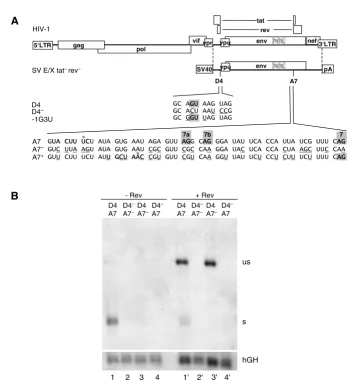
Rev. To analyze whether 3'ss A7 also contributes to the steady-state level of the glycoprotein mRNA we inacti-vated 3'ss A7 and the two upstream minor 3'ss, A7a and A7b [2-4], by silent point mutations (A7 -, Fig. 2A) ena-bling analysis of these mutations in the glycoprotein-mediated syncytia assay.
To verify that the introduced mutations did not lead to activation of a cryptic 3'ss we additionally compared the subgenomic HIV-1 transcripts by Northern blot analysis of the respective poly(A)+ RNA fractions following
tran-sient transfection of HeLa-T4+ cells with either the subge-nomic env expression vector SV E/X tat- rev- or SV E/X tat -rev- A7- (Fig. 2B). Due to mutations of the tat and rev ATG translational initiation codons, these vectors express nei-ther Tat nor Rev. Thus, in the absence of Rev transfection with SV E/X tat- rev- led almost exclusively to detection of spliced mRNA (Fig. 2B, lane 1). In contrast, after cotrans-fection with a Rev-expressing plasmid the majority of the detected mRNA was the unspliced poly(A)+ glycoprotein mRNA (Fig. 2B, lane 1'). As expected, mutations of all three 3'ss, A7, A7a and A7b, led to complete loss of any Alternative splicing of HIV-1
Figure 1
Alternative splicing of HIV-1. (A) Organization of the HIV-1 genome. Filled boxes indicate open reading frames present in all isolates, light grey boxes indicate the Tev orf which is isolate specific. The long terminal repeats (LTR) are present at both ends of the proviral DNA. (B) Localization of splice sites, splicing regulatory elements and the Rev responsive element (RRE). 5' splice sites: D1a-5; 3' splice sites: A1-7. Splice sites A6/D5 are isolate specific and not functional in the isolate NL4/3 used in this study. Splice sites A1a/D1a defining exon 1a have been recently described [6]. The nomenclature of the 3'ss is according to Stoltzfus [17,18] and Purcell and Martin [2] (in brackets). Splicing regulatory elements: M1, M2 (this report); ESSV [16,64]; ESS2p [18]; ESE2/ESS2 [17,32,43,44,49]; GAR [23,28]; ESS/ESE [19,20]; ISS [22]; ESE3 [17,21,24,25,33,65]; ESS3a, b
[17,21,24,33,66]. (C) Splicing pattern and proteins encoded by the different mRNA classes. The 1.8 and 4 kb mRNAs contain obligatory sequences (dark grey) as well as alternative sequences (light grey) due to alternative usage of the splice sites. The nuclear export of the 4 kb mRNAs and the genomic full-length 9 kb mRNA is dependent on Rev binding.
A
B
C
4 kb
Vif Vpr Vpu Env Tat-1
Rev
9 kb
Gag Pol (Genome)
Rev
1.8 kb
Tat Rev Nef
Tev
tev
rev
env nef vif
pol gag
tat
LTR
vpr vpu
R U3 U5
LTR
R U3 U5
D1 D2 D3 D4 D5
A1 A2 A3 A4cabA5 A6 A7
RRE
ESSV ESS2p ESE2 ESS2
GAR ESS
ESE
ISS ESE3
ESS3a, b M1/M2
(A2) (A3)(A4)
detectable spliced transcript indicating that no cryptic splice acceptor was significantly activated (lane 3). How-ever, in the presence of Rev the amount of unspliced poly (A)+ env mRNA was unaffected by the presence or absence of a functional splice acceptor (cf. lane 1' with 3') demon-strating that the 3'ss mutations did not decrease the pool of unspliced poly (A)+ transcripts. This contrasted the pre-viously shown 5'ss dependency of spliced and unspliced
[image:4.612.127.481.82.470.2]transcripts [28] (cf. lane 1 with 4 and 1' with 4'), i.e. the lack of U1 snRNA-binding to the 5'ss leads to env RNA degradation (see hGH detectability in lanes 2, 4, 2', and 4'). The results of the Northern analysis were confirmed by glycoprotein expression analyzed by Western blot and syncytium formation (data not shown). Together these results demonstrate that a 3'ss is dispensable for Rev-mediated env expression. Moreover, these results show 3'ss A7 is nonessential for RNA stability and Rev responsiveness
Figure 2
3'ss A7 is nonessential for RNA stability and Rev responsiveness. (A) Schematic drawing of the HIV-1 genome and of the subgenomic env expression plasmid SV E/X tat- rev-. LTR: long terminal repeat, SV40: SV40early promoter, pA: SV40
poly-adenylation sequence. Nucleotide sequences of the 5'ss D4 and its mutations D4- and -1G3U as well as the 3'ss A7 and its
mutations A7- and A7+ are shown beneath. The splice sites (grey squares, including the minor 3'ss 7a and 7b), the reported or
supposed branch point sequence (bold, asterix indicates the branch point nucleotide) and the mutated nucleotides (underlined) are marked. In the 3'ss mutants the reading frame was kept unchanged except for position 703 (Val→Ala) in A7+. (B)
HeLa-T4+ cells were transiently transfected with the subgenomic env expression plasmids (SV E/X tat - rev -) containing either the
wild type 5'ss D4 or the non functional D4- mutation combined with either the wild type 3'ss A7 or the A7- mutation in
pres-ence or abspres-ence of a Rev expression plasmid (SVcrev) as indicated above the lanes. The poly(A)+ RNA was analyzed by
North-ern blotting. s: spliced, us: unspliced transcript. Transfection efficiency was monitored by co-transfection of a human growth hormone (hGH) expressing plasmid (pXGH5).
us
s
hGH D4
A7 D4– A7–
D4 A7–
D4– A7 D4
A7 D4– A7–
D4 A7–
D4– A7
- Rev + Rev
1 2 3 4 1' 2' 3' 4'
B
GC GGU UAG UAG
A
vpu vpr
vif env nef
3‘LTR gag
pol 5‘LTR
tat
rev
RRE
D4 A7
SV40 vpu env RRE pA
SV E/X tat–rev–
A7 A7– A7+
GUUA CUU UCU AUA GUG AAU AGA GUU AAGG CAAG GGA UAU UCA CCA UUA UCG UUU CAAG GUC UUA AGU AUA GUG AAU CGC GUU CGC CAA GGA UAC UCA CCA CUA AGC UUC CAA GUU CUU UCU AUUU GCU AAC CGU GUU CGU CAA GGU UAU UCU CCU CUU UCU UUU CAAG
7a 7b 7
D4 D4– -1G3U
GC AGGU AAG UAG GC ACU AAU CCG
HIV-1
*
that the protective function of U1 snRNA binding is inde-pendent of the recognition of the 3'ss during progression of spliceosome formation.
To exclude the possibility that the requirement for U1 snRNA complementarity for protection of the transcript was caused by an RNA surveillance mechanism detecting a functional 3'ss in the absence of a 5'ss we mutated both the 5'ss (D4-, Fig. 2A) and 3'ss (A7-) and analyzed the steady-state levels of total poly(A)+ RNA. In the absence of both the 5' and 3'ss RNA could still not be detected irre-spective of the presence or absence of Rev (Fig. 2B, lanes 2 and 2'). This indicates that the U1 snRNA dependency for the expression of this subgenomic env mRNA was not due to an unpaired/cryptic splice site but was intrinsic to the transcript sequence.
3'ss efficiency competes with Rev function
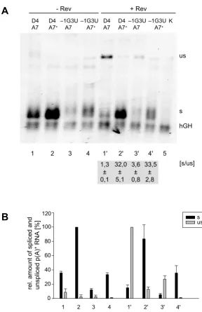
Increasing the complementarity between the 5'ss D4 and U1 snRNA did not lead to a decrease in env expression, indicating that even in the presence of a strong 5'ss Rev-regulated env mRNA transport was not impaired [23,29]. To specifically investigate the influence of the strength of the 3'ss on Rev-mediated glycoprotein expression we improved the strength of 3'ss A7 in its context of a subge-nomic glycoprotein expression vector. To achieve this, the suboptimal BPS was attenuated and a new BPS with higher complementarity to U2 snRNA was created further downstream. Additionally, the canonical AG dinucle-otides of the cryptic sites A7a and A7b were mutated to prevent an interference with potentially binding splicing factors and the pyrimidine content of the PPT (in the region between the new BPS and the intron/exon border) was increased from 48 % to 77 %. All these nucleotide changes were introduced as silent mutations except for one (Val to Ala at position 703), which was not expected to influence the fusogenic activity of the glycoprotein (Fig. 2A, A7+).
As expected, analysis of HeLa-T4+ cells transfected with this vector revealed that the introduced mutations improved the efficiency of A7 as evident by a dramatic increase in the amount of spliced transcript (Fig. 3A and 3B, cf. lane 1 with 2). This was also confirmed by in vitro splicing experiments with the respective splicing con-structs (data not shown). In the presence of Rev however, almost no unspliced poly(A)+ message was observed (Fig. 3A, B, cf. lane 1' with 2'), suggesting that splicing, enhanced by the strength of the 3'ss, competes with Rev activity.
To address the question of whether a suboptimal 5'ss could compensate for an efficient 3'ss in Rev function we combined a 5'ss of intermediate complementarity to U1 snRNA (-1G3U, Fig. 2A) [28] with the efficient 3'ss A7+. In
agreement with our previous results, in the presence of A7 this intermediately strong 5'ss led to a 2–3 fold decrease in the amount of RNA (Fig. 3A, cf. lanes 1 and 3, 1' and 3'). However, while the ratio of spliced to unspliced tran-scripts (Fig. 3A, s/us) was altered only 3-fold, in the pres-ence of A7+ this ratio increased up to 25-fold irrespective of the strength of the 5'ss (Fig. 3). This finding demon-strates that Rev activity is specifically and inversely dependent on the efficiency of the 3'ss A7.
To determine the sequence requirements of a 3'ss compat-ible with Rev function in more detail, we constructed a single-intron splice reporter based on a truncated HIV-1 tat/rev intron harbouring the RRE (Fig. 4A) and analyzed 3'ss A5 because of its complexity. A5 exhibits a discontin-uous pyrimidine stretch and overlaps with the competing alternative 3'ss 4c, 4a and 4b. Moreover, ten BPSs have been experimentally mapped in this region, five of which are associated with splicing at 3'ss A5 [14,30] (see Fig. 5, constructs A4cab and A5). Since the AG-dinucleotides and BPSs can compete for binding of splicing factors we mutated them consecutively (Fig. 4A): First the AG dinu-cleotides of 3'ss A4c, a and b were changed to CG (AG-) to exclude splicing at these positions. Next, the complemen-tarity between the 5' BPS (named BPS1 in Fig. 4A) and U2 snRNA was reduced while the complementarity of the 3' BPS (BPS2) was enhanced (b1- b2+). Thirdly, the pyrimi-dine content was increased from 52% in the wild type 3'ss A5 to 60% (Py+) and 72 % (Py++), respectively.
Following transient transfection of HeLa-T4+ cells with these constructs the poly(A)+ RNA was analyzed by North-ern blot. Neither the mutations of the upstream AGs (Fig. 4B, lane 2) nor of the branch sites (lane 3) led to splicing at the 3'ss A5 but efficiently allowed Rev-dependent detectability of the unspliced transcript (lanes 2' and 3').
Spliced RNA was not detected until the pyrimidine con-tent was further increased (lane 4 and 5). Remarkably, a pyrimidine content of 60% (Py+) was still compatible with a low-level of Rev function (lane 4') but in contrast, a highly efficient 3'ss due to a further increase in the pyri-midine content of only 12% (Py++) was not (lane 5').
Removing the improvement of BPS2 (SA5 b1- AG- Py++) reduced splicing efficiency 3-fold (cf. lane 5 with 6) and concomitantly restored Rev-compatibility (cf. lanes 5' and 6') in spite of the high pyrimidine content. This suggests a comprehensive effect of overall 3'ss strength on Rev activ-ity.
the construct with only one predicted suboptimal branch site (cf. lane 7 with 6). Reconstruction of the AGs of 3'ss A4c, a, b further decreased the level of spliced transcripts (cf. lane 7 with 8) but increased the level of the Rev-dependent unspliced RNA (cf. lane 7' with 8'). In general,
the amount of unspliced transcript in the presence of Rev (Fig. 4B and 4C, lanes 1'–8') was inversely proportional to that of the spliced transcript. This confirms our findings shown in Fig. 3, that splicing efficiency driven by the 3'ss competes with Rev function.
[image:6.612.168.456.90.535.2]Weakening of the 5'ss D4 does not compensate for the strength of 3'ss A7 Figure 3
Weakening of the 5'ss D4 does not compensate for the strength of 3'ss A7. HeLa-T4+ cells were transiently
trans-fected with the subgenomic HIV-1 constructs (SV E/X tat - rev -) combining an efficient (A7+) or inefficient (A7) 3'ss with a 5'ss
with high (D4) or lower (-1G3U, cf. Fig. 2A) complementarity to U1 snRNA. The p(A)+ RNA was analyzed by Northern
blot-ting (cf. Fig. 2). (A) Northern blot with indication of the ratio of spliced (s) and unspliced (us) RNA in presence of Rev ([s/us], mean ± standard error) from three independent experiments. (B) Mean values of the relative amounts of spliced (s, black) and unspliced (us, grey) transcripts from three independent experiments, normalized to transcription efficiency (hGH). The spliced (s) and unspliceds (us) RNA populations were quantified from different exposure times of the blots to adjust for the different levels of signal intensities. The maximum values of both RNA populations were defined as 100%.
A
B
sus
rel. a
m
o
unt
of s
p
lice
d
a
n
d
uns
pli
c
ed
p(A)
+RNA [%]
0 20 40 60 80 100 120
1 2 3 4 1' 2' 3' 4'
us
s
hGH
1 2 3 4 1' 2' 3' 4' 5
D4 A7
D4 A7+
–1G3U A7
–1G3U A7+
- Rev + Rev
D4 A7
D4 A7+
–1G3U A7
–1G3U A7+
K
[s/us] 33,5
± 2,8 32,0
± 5,1 1,3
± 0,1
The strength of the 3'ss competes with Rev responsiveness Figure 4
The strength of the 3'ss competes with Rev responsiveness. (A) Nucleotide sequence of one-intron constructs with mutations in the 3'ss A5. The reported branch point sequences for A5 (grey boxes, asterix indicates the branch point nucle-otide), the 3'ss A4c, a, b, A5 (black boxes) and the PPT (hatched) are marked. Mutated nucleotides compared to the wild type are underlined. (B) Northern blot analyses of the p(A)+ RNA after transient transfection of HeLa-T4+ cells with one-intron
constructs carrying SA5 mutations (cf. Fig.2). (C) Diagram of the hGH standardized relative amounts of spliced (left) and unspliced (right) transcripts from (B). The maximal amount was defined as 100%. The numbers below correspond to the lanes of the Northern blot.
B
1 2 3 4 5 6 7 8 1' 2' 3' 4' 5' 6' 7' 8'
- Rev + Rev
SA 5P y ++ SA 5A
G-P y ++
SA 5b
1 -AG
-Py ++
SA 5b
1 -b2
+A G-P
y ++
SA 5b
1 -b2
+A G-P
y +
SA 5b
1 -b2
+A G -SA 5A G -SA 5 SA 5P y ++ SA 5A
G-P y ++
SA 5b
1 -AG
-Py ++
SA 5b
1 -b2
+AG -Py
++
SA 5b
1 -b2
+A G-P
y +
SA 5b
1 -b2
+AG -SA 5A G -SA 5 s us hGH
C
re l. am ount of unspl iced p( A)+RNA [
% ] 0 20 40 60 80 100 0 20 40 60 80 100 re l. am ount of splic ed p( A) +RN A [ % ] 6 3
1 2 4 5 7 8 1' 2' 3' 4' 5' 6' 7' 8' BPS 2
A4c A4a A4b SA5
BPS 1 PPyPPy
AAAAAGTGTTGCTTTCATTGCCAAGTTTGTTTCATGACAAAAGCCTTAGGCATCTCCTATGGCAAG AAAAAGTGTTGCTTTCATTGCCACGTTTGTTTCATGACAAACGCCTTCGGCATCTCCTATGGCAAG AAAAAGTGTGGCGTCCGTTGCCACGTTTGTTTACTAACAAACGCCTTCGGCATCTCCTATGGCAAG AAAAAGTGTGGCGTCCGTTGCCACGTTTGTTTACTAACAAACGCCTTCGGCATCTCCTATTTCAAG AAAAAGTGTGGCGTCCGTTGCCACGTTTGTTTACTAACAAACGCCCTCGCCTTCTCCTCTTTCAAG AAAAAGTGTGGCGTCCGTTGCCACGTTTGTTTCATGACAAACGCCCTCGCCTTCTCCTCTTTCAAG AAAAAGTGTTGCTTTCATTGCCACGTTTGTTTCATGACAAACGCCCTCGCCTTCTCCTCTTTCAAG AAAAAGTGTTGCTTTCATTGCCAAGTTTGTTTCATGACAAAAGCCCTAGCCTTCTCCTCTTTCAAG SA5
SA5 b1–b2+AG–
SA5 AG
-SA5 b1–b2+AG–Py+
SA5 b1–b2+AG–Py++
SA5 b1–AG–Py++
SA5 AG–Py++
SA5 Py++
A
* * * * *
D4 A
The intrinsic strengths of the HIV-1 splice acceptor sites differ largely
The observation that the efficiency of the 3'ss competes with Rev function implicates that all HIV-1 3'ss should be inefficient to allow the export of unspliced transcripts nec-essary for virus replication. Indeed, this has been already reported by O'Reilly and coworkers [27] however, at the time of publication the knowledge of HIV-1 splice site reg-ulation by cis-acting sequences was rather incomplete.
To differentiate between the contribution to the overall splice site strength of the splice site regulating elements in the 3' exonic sequences and the intrinsic strength of the HIV-1 3'ss we used a splice site swapping strategy and
ana-lyzed the HIV-1 3'ss with or without their natural down-stream exonic sequences (Aex and A, respectively, Fig. 5). Each 3'ss included the experimentally defined or assumed, by complementarity to U2 snRNA, branch point sequence, the polypyrimidine tract and the AG dinucle-otide. Because of their functional and spatial overlap the 3'ss A4c, a and b were experimentally considered as an entity. The 3'ss A6, which is located in the tat/rev intron, was not included in this analysis because its activity has been described in isolate HIV HXB2 but not in HIV NL4/ 3 which was used in this study [2,31]. As reference sequences the non functional (A7-) and the efficient 3'ss (A7+) mutants shown in Fig. 2 and 3 were also included. Schematic drawing of the one-intron splicing reporter
Figure 5
Schematic drawing of the one-intron splicing reporter. Diagram of the one-intron construct used for comparison of the HIV-1 3'ss by a splice site swapping strategy. SV40: SV40early promoter, pA: SV40 polyadenylation sequence. RRE: Rev response element. Fragments including the different 3'ss (grey boxes) and branch sites (dashed line: assumed from consensus; underlined: reported BP, numbers are referring to the associated 3'ss, BP A2 [15], BP 4cab and A5 [14,30], BP A7 [14]) were inserted into the cassette. The ISS has been described by [22]. The 3'extended versions of the splice acceptor constructs addi-tionally include the downstream exon sequences with cis-acting splicing regulating sequences (M1, M2 [this report]; ESSV [16,64]; ESS2p [18]; ESE2/ESS2 [17,32,43,44,49]; GAR [23,28]; ESE3 [17,21,24,25,33,65]; ESS3a, b [17,21,24,33,66]; splicing silencer (light grey boxes); splicing enhancer (dark grey boxes)).
A7 [7] 7b 7
[5] [5]
A5 AAAAAGTGTTGCTTTTCAATTGCCAAAGTTTGTTTCAATGAACAAAAAGCCTTAAGGCATCTCCTATGGCAAG
4b
4c 4a 5
[5]
A7+ GTTCTTTCTATTGCTAAACCGTGTTCGTCAAGGTTATTCTCCTCTTTCTTTTCAAG
ACCCACCTCCCAATCCCGAGGAT
A7¯ GTCTTAAGTATAGTGAATCGCGTTCGCCAAGGATACTCACCACTAAGCTTCCAA
[4c] [4c] [4ab] 4c 4a 4b
A4cab TGTACCAATTGCTAATTGTAAAAAAGTGTTGCTTTTCAATTGCCAAAGTTTGTTTCAATGAACAAAAAGCCTTAAG
[4ab] [4ab]
A3 ACATATCTATGAAACTTATGGGGATACTTGGGCAGGAGTGGAAGCCATAAATAAAGAATTCTGCAACAACTGCTGTTTATCCATTTCAAG3
2
A2 ACCCTGAATTAGCAGGACCAACTAATTCATCTGTATTACTTTGACTGTTTTTCAAG
1
A1 TAGCAACAGACATACAAAACTAAAGAATTACAAAAACAAATTACAAAAATTCAAAATTTTCGGGTTTATTACAAG
A1ex
A2ex
D4 A
SV-SD4/RRE/SA-pA
A3ex
A5ex
A7ex
ISS
A4cab5
ex
A4cab
AGGGACAGCAGAGATCCAGTTTGGAAAGGACCAGCAAAGCTCCTCTGGAAAG1
AGACTCTGCTATAAGAAAGGCCTTATTAGGACACATAGTTAGCCCTAGGTGTGAATATCAAGCAGGACATAACAAG2 ESSV
AGAATTGGGTGTCGACATAGCAGAATAGGCGTTACTCGACAGAGGAGAGCAAGAAATGGAGCCAGTAGATCCTAGACTAGAGCCCT3 ESS2p ESE2 ESS2
AGGAAGAAGCGGAGACAGCGACGAAGAGCTCATCAGAACAGTCAGACTCATCAAGCTTCTCTATCAAAGCA5 GAR
CGATTAGTGAAC
AGACCCACCTCCCAATCCCGAGGGGACCCGACAGGCCCGAAGGAATAGAAGAAGAAGGTGGAGAGAGAGACAGAGACAGATCCATT7 ESE3 ESS3a
ESS3b
GAR
AGGCATCTCCTATGGCAAGGAAGAAGCGGAGACAGCGACGAAGAGCTCATCAGAACAGTCAGACTCATCAAGCTTCTCTATCAAAGCA5
4b
AGGCATCTCCTATGGCA4b AG5
ex
[5] [5] [5] [5]
[5] [5] [5]
[4ab] [4ab]
M1 M2
GTACTTTTCTATAGTGAATAGAGTTAGGCAGGGATATTCACCATTATCGTTTCA7a AG
D4 A
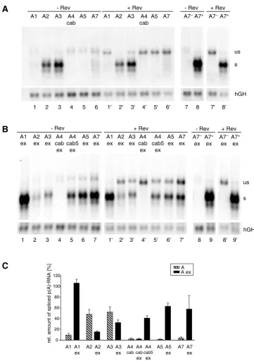
Northern blot analyses of these constructs revealed that only 3'ss A2 and A3 led to detection of significant amounts of spliced mRNA in the absence of their natural 3' exonic sequences (Fig. 6A, lanes 2 and 3). Consistent with the results shown in Fig. 3 and 4 these two constructs showed the lowest number of unspliced transcripts in the presence of Rev (Fig. 6A, cf. lanes 2' and 3' with 1', 4'–6'). Thus, these results indicate that 3'ss A2 and A3 are the most efficient core 3'ss, here referred to as the intrinsic efficiency of the 3'ss. For all other 3'ss the intrinsic effi-ciency was low and significant amounts of unspliced mes-sage could be detected in the presence of Rev.
Interestingly, the opposite picture was obtained for the series of constructs where the downstream exonic sequences were included (Fig. 6B and 6C). Compared to their respective intrinsic efficiencies, splicing at A2 and A3 was decreased 3-fold and 1.5-fold in the presence of their downstream exons (Fig. 6C, cf. A2 with A2ex and A3 with A3ex). This is in accordance with the described ESS ele-ments, consisting of three hnRNP A1 binding sites within exon 3 [16], and hnRNP H and hnRNP A/B binding sites within exon 4 [18,32]. Therefore, significant amounts of Rev-dependent, unspliced messages are only detectable if the intrinsic strength of these 3'ss is silenced by their downstream exonic sequence (cf. Fig. 6A, lane 2' and 3' with Fig. 6B, lane 2' and 3').
Since the alternative 3'ss A4c, A4a, A4b and A5 are all in close proximity to each other we tested whether these sites are regulated by the same bidirectional enhancer in exon 5 (A4cab5ex), which also leads to efficient splicing of the flanking 3'ss A5 and 5'ss D4 [23]. Alternatively, additional sequences upstream of this ESE may be sufficient to influ-ence the strength of at least one of the 3'ss A4c, A4a and A4b (A4cabex). The result showed that in the absence of the bidirectional ESE in exon 5 none of these 3'ss could be adequately activated as evident by the absence of any spliced transcript (Fig. 6B, lane 4). Hence, the alternative 3'ss A4c, A4a, A4b and A5 seemed to be moderately acti-vated by the same bidirectional enhancer in exon 5, still allowing Rev-mediated nucleocytoplasmic transport of unspliced transcripts (cf. Fig. 6B, lane 4' with lanes 5' and 6'). Comparison of the amount of spliced transcript from the constructs carrying either the BPS of all 3'ss A4c, A4a, A4b and A5 (Fig. 6C, A4cab5ex) or only the BPS for the 3'ss A4a, A4b and A5 (A5ex) showed a slight increase (30%) in the amount of spliced transcript of the latter. This suggests that competition of the four alternative 3'ss might also contribute to the inefficiency of splicing and that this is also supportive for the Rev-mediated export of the unspliced message.
To date A7 is the only splice site with a known splicing silencer in the intronic region and therefore we cannot
distinguish between the impact of the suboptimal PPT and this ISS on the intrinsic inefficiency of this 3'ss (Fig. 6A, lane 6). However, splicing at A7 depends on activa-tion by its flanking downstream sequences carrying the bipartite ESE3/ESS3 regulatory sequence (cf. Fig. 6A lane 6 with Fig. 6B, lane 7) [17,21,25,33]. Thus, in this experi-mental context, the ESE clearly dominates over the ESS function.
Most strikingly, 3'ss A1 extended by its natural exonic sequence turned out to be the most efficient 3'ss of all (Fig. 6B, cf. lane 1 with 2–7). Even in the presence of Rev, only a very small amount of unspliced message was detected comparable to 3'ss A7+ (Fig. 6B, cf. lane 1' with 9'). Therefore, from these experimental results we con-clude that exon 2 contains a strong splicing regulatory ele-ment, which has not been identified so far.
These results combined show that, although all HIV-1 3'ss are predicted to be weak on the basis of their intronic sequences, there are distinct differences in their intrinsic splicing efficiency. To co-ordinate both splicing and Rev function the strength of the individual 3'ss is finally regu-lated by cis-regulating ESEs and ESSs in their 3' exons.
An SF2/ASF-dependent splicing enhancer in exon 2
The strength of the 3'ss competes with Rev responsiveness Figure 6
The strength of the 3'ss competes with Rev responsiveness. (A) Northern blot analysis (cf. Fig. 2) from HeLa-T4+ cells
transfected with constructs containing the HIV-1 3'ss in absence of their authentic 3' exon sequences. The particular 3'ss and the co-transfection of a rev expressing plasmid (SVcrev) are given above the lanes. The 3'ss A7- and A7+ were used as reference
constructs for a nonfunctional and an efficient 3'ss. All lanes were derived from the same Northern blot. (B) Northern blot analysis from cells transfected with 3'ss in presence of their authentic 3' exon sequences (ex). All lanes were derived from the same blot. (C) Mean values of the relative amounts of spliced transcripts in absence and presence of the 3' exon sequences from three independent experiments, normalized to transcription efficiency (hGH, cf. Fig. 2). The amount of spliced transcripts derived from the construct containing the improved A7+ was defined as 100% (not shown).
A4 cab5
ex A4 cab ex
C
re
l. a
m
ount
o
f
sp
lic
ed
p
(A)
+RNA [%
]
0 20 40 60 80 100 120
A1 A2 A3 A4 A5 A7
cab
A A ex
A5 ex
A7 ex A3
ex A2 ex A1 ex
B
A4 cab5
ex
1 2 3 4 5 7 1' 2' 3' 4' 5' 6' 7' A1
ex
A1 ex A2
ex
A2 ex A3
ex
A3 ex A4
cab ex
A4 cab ex
A5 ex A7
ex
A7 ex A4
cab5 ex
- Rev + Rev
A5 ex
6
us
s
hGH - Rev + Rev A7–
ex
A7+
ex A7+
ex A7–
ex
8' 9' 8 9
A
1 2 3 4 5 6 1' 2' 3' 4' 5' 6' A1 A2 A3 A4
cab A5 A7 - Rev
A1 A2 A3 A4 A5 A7 cab + Rev
7 8 7' 8' - Rev + Rev A7–A7+ A7–A7+
us
s
carrying mutations Δ M1 and Δ M2 revealed that the intro-duced mutations led to reintro-duced reactivity of a polyclonal SF2/ASF antibody with a band of the corresponding molecular mass of SF2/ASF-binding sites. To confirm the SF2/ASF-dependent exon 2 recognition two additional mutations were analyzed which were predicted by ESE-finder to specifically abolish SF2/ASF-binding. As expected these two constructs (Δ M1 SF2-, Δ M2 SF2-) led to a complete lack of exon 2 recognition (Fig. 8B) support-ing the observation that M1 and M2 represent the SF2/ ASF-dependent exon 2 splicing enhancer. Since we were interested in analyzing this newly identified ESE within an infectious molecular clone we set out to test for a silent point mutation predicted to specifically inactivate this enhancer. Based on computer analysis only one mutation was found that fulfilled the desired criterion (Δ M1 – 43). The mutation Δ M1 – 43 resulted in a slightly reduced loss of exon 2 recognition compared to the other mutations (Fig. 8B). The most obvious difference was the appearance of a comparable amount of unspliced transcript. Never-theless, this result confirmed that the Δ M1 – 43 mutation affected exon 2 recognition.
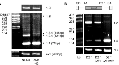
Therefore, we inserted the Δ M1 – 43 mutation into the molecular clone NL4/3 and investigated its effect on viral replication in PM1 cells. Unexpectedly, no difference in the replication kinetic was observed throughout the infec-tion period of up to 10 days (data not shown) suggesting that Δ M1 – 43 could not dramatically impair viral gene expression. However, analyzing the viral mRNA of the infected cells at day 5 post-infection by RT-PCR using primers specific for the upstream region of the HIV-1 genome revealed a different splicing pattern for the non-coding leading exons (Fig. 8A). As observed within the splicing reporter, the presence of the Δ M1 – 43 mutation led to a significant loss of exon 2 recognition (cf. band 1.2.4 in lanes NL4/3 and Δ M1-43) confirming that the newly identified ESE within exon 2 supports exon defini-tion within the context of the viral genome. Furthermore, the lack of exon 2 recognition was accompanied by an increase in exon 3 recognition (cf. band 1.3.4 in lanes NL4/3 and Δ M1-43). These results support recent find-ings that the optional leader exons might not play a direct role in viral gene expression [36]. The most abundant detectable spliced isoform corresponded to a 1.4 tran-script which was not expected to be affected by the muta-tion. However, most surprisingly, the amount of vif mRNA was also not altered by the Δ M1 – 43 mutation (cf. band 1.2l in lanes NL4/3 and Δ M1-43), demonstrating that this mutation exclusively impaired exon 2 definition but not intron (D1 – A2) definition. To confirm these results functionally we compared the replication kinetics of NL4/3 and NL4/3 Δ M1-43 in the vif non-permissive and permissive cell lines CEM and CEM-SS respectively. Consistent with the PM1 infection experiments no
differ-ence in the viral replication kinetics could be observed between CEM and CEM-SS (data not shown) indicating that the level of vif expression is not impaired by the Δ M1 – 43 mutation during the infectious experiment up to 16 days.
To further investigate the apparent discrepancy between the transient transfection experiment using the splicing reporter (cf. Fig. 8B, lane Δ M1-43) and the infection experiments we specifically analyzed the effect of the Δ M1 mutation on splice site usage of A1 in the splicing reporter by excluding exon definition through deletion of 5'ss D2. Similar to the infection experiments 3'ss A1 usage involving in intron definition still occurs in the presence of Δ M1 mutation although to a somewhat lesser extent (Fig. 9B, D2- Δ M1). In contrast, mutating both hepta-meric sequences (Fig. 9B, cf. D2- Δ M1 with D2- Δ M1/2) resulted in a total failure of 3'ss A1 recognition. Thus, the
intron-containing Rev-dependent vif mRNA is less
dependent on the strength of the bipartite ESE (if defined through the number of SR binding sites).
Discussion
Efficient 3'ss compete with Rev function but retroviral rep-lication requires an equilibrium between splicing and nuclear export of intron-containing transcripts and there-fore the low efficiency of the 3'ss is a key factor for viral replication. Under such suboptimal conditions a slow down of the first transesterification reaction is likely [14,41,42]. Nevertheless, a prediction of 3'ss efficiency based only on the evaluation of the sequence by available algorithms is still not reliable in all cases due to the com-plex interplay of the U snRNAs and proteins involved in 3'ss recognition. An experimental assessment of the splic-ing efficiency of the HIV-1 3'ss was performed by O'Reilly and coworkers [27]. In a heterologous β-globin/HIV-1 construct they evaluated the relative efficiencies of the HIV-1 3'ss compared to the β-globin 3'ss A1 which was used as a reference for an efficient 3'ss. The outcome of this study was a relative homologous clustering of the HIV-1 3'ss between 40% and 60% splicing efficiency with exception of 3'ss A1 (in the original publication referred to as 3'ss A2, see also Fig. 1) which showed a significantly lower efficiency (26%). Unfortunately, at that time the relevance and multitude of cis-acting sequences regulating alternative splicing in the HIV-1 transcripts are just start-ing to be unfolded. Therefore, only the downstream exonic cis-acting sequence involved in regulation of 3'ss A7 [21,33] was completely included in the investigated fragment (101 nt 3' of the intron/exon border). However, in the past, it had become evident that trans-acting factors are involved in constitutive as well as alternative splicing and that almost all HIV-1 exons and also some intron sequences include splicing enhancers and/or silencers [16-18,21-23,28,33,43,44] (see Fig. 1, for recent reviews see [5,45]. Hence, from this apparent ubiquitous presence of cis-acting sequences the question arose as to how the
classical elements defining a canonical splice site (BPS, PPT, AG-dinucleotide) and the cis-acting elements con-tribute to the overall strength of the different HIV-1 3'ss and their response to Rev. To evaluate the impact on the intrinsic strength of the 3'ss, i.e., the intronic sequence versus the cis-acting, exon-located enhancer and silencer elements, we compared the efficiency of the HIV-1 3'ss A1, 2, 3, 4cab and 5 in the presence and absence of their nat-ural downstream exonic sequences in a splice site swap-ping strategy.
[image:12.612.56.550.87.260.2]Our comparison of the HIV-1 3'ss with an optimized 3'ss as an internal reference with almost no response to Rev, revealed significant variation in the strength of the viral 3'ss with relative splicing efficiencies from 1% to 52% in the absence and 15% to 106% in the presence of their nat-ural 3' sequences. Based on these data we grouped the HIV-1 3'ss into two different categories. The first category includes 3'ss A1, A4cab, A5 and A7 which were all but inactive in the absence of their 3' exonic sequences. Their 3' sequences had an overall stimulatory effect on splicing efficiency at these sites. This is especially interesting for the increase in splicing efficiency at A7 (from 4 to 57%) since this 3' exon includes both a splicing enhancer and a silencer [21,25,43] that has been shown to compete with ASF/SF2-binding at the ESE and hnRNPA1 binding at the ESS [24,26,46]. Thus, different ratios of ASF/SF2 and hnRNP A1 in different cells may lead to differences in the activation of A7. Under our experimental conditions, however, a dramatic influence of the ESS on the strength of A7 was not detectable (data not shown). Hence, the ESE was clearly dominant over the ESS function. Addi-tionally, we identified a new cis-acting sequence within exon 2, which profoundly increased splicing efficiency at Alignment of exon 2 of HIV-1 M group consensus sequences
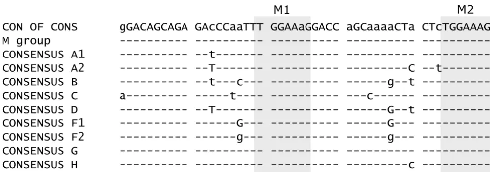
Figure 7
Alignment of exon 2 of HIV-1 M group consensus sequences. Exon 2 sequences were obtained from the HIV Sequence Database [67] flanked by A1 and D2. The heptameric sequences (M1 and M2) found to enhance splicing are highlighted by grey boxes.
CON OF CONS
gGACAGCAGA GAcCCaaTTT GGAAaGGACC aGCaaaaCTa CTcTGGAAAG
M group
---CONSENSUS A1
--t---
---CONSENSUS A2
--- --T--- --- ---C
--t---CONSENSUS B
--t---c--- ---g--t
---CONSENSUS C
a--- ---t---- ---c---
---CONSENSUS D
--T--- ---G--t
---CONSENSUS F1
---G--- ---G---
---CONSENSUS F2
---g--- ---g---
---CONSENSUS G
---CONSENSUS H
---c
An ASF/SF2-dependent ESE within exon 2 Figure 8
An ASF/SF2-dependent ESE within exon 2. (A) Sequence of exon 2 (top line) and analyzed mutations. The heptameric sequences (M1 and M2) of the bipartite ESE are boxed and the mutations are given below exon 2 sequence. A schematic draw-ing of the two-intron minigene with exon 2 as internal exon flanked by A1 and D2 is given below the sequences. (B) HeLa-T4+
cells were transfected with LTR ds ex2 (ex2) or its mutations (cf. (A)), cotransfected with SVctat and pXGH5 (hGH) and ana-lyzed by RT-PCR as described in Materials and Methods. (C) Pull-down analysis of in vitro transcripts of either exon 2 or exon 2 carrying both mutations, M1 and M2 in HeLa cell nuclear extract. The immunoblot was detected with a polyclonal SF2/ASF antibody.
Exon 2 GGACAGCAGAGATCCACTTTGGAAAGGACCAGCAAAGCTCCTCTGGAAAG
ΔM1
---CT-TC---ΔM2 ---CT-TC
ΔM1 SF2- ---T--A---ΔM2 SF2- ---T---G---ΔM1-43
---G---A
kb 396 346 298
220 201
154 134
187
ΔM1
ex2 ΔM2 ΔM1
SF2– Δ ΔM1-43 M2 SF2–
splicing pattern size [bp]
311 398
281
194
144
hGH
B
A1
SD
SA
Exon2
D2
pA LTR
M1
M2
33 kDa 40 kDa
NE ex2 ΔM1/
3'ss A1. This regulatory element includes two TGGAAAG heptameric sequences which constitute key elements of a bipartite ESE and support our observation that a func-tional enhancer seems to be defined by at least two indi-vidual binding sites [34]. The identification of ASF/SF2 as the respective splicing regulatory protein is in agreement with the finding, that overexpression of ASF/SF2 stimu-lated splicing at site A1 in HIV-1 pNL4/3 transfected 293T cells [47].
The second group of 3'ss, A2 and A3, showed approxi-mately 50% splicing efficiency in the absence of exon sequences and a 2–3 fold decrease in efficiency in the presence of exon sequences, consistent with published ESS sequences in exon 3 (ESSV) and 4 (ESS2p, ESS2) (see Fig. 1) [16-18,43,48]. Despite the presence of these ESSs usage of both 3'ss could also be stimulated in vivo follow-ing overexpression of ASF/SF2 [47] and in vitro upon addi-tion of recombinant SC35 [49] disclosing the ambivalence of HIV-1 3'ss regulation. A reason for the restrictive control of 3'ss A2 and A3 might be the
cytotox-icity of Tat [50] and Vpr [51] which are translated as first reading frames of the appropriate mRNAs.
[image:14.612.72.544.87.345.2]Comparing the two splice site groups we noticed that the pyrimidine content of the PPT was highest in 3'ss A2 and A3 (65 and 69%) and lower in the other 3'ss (40% up to max. 62%). This encouraged us to analyze the contribu-tion of the intronic sequence elements, i.e., BPS, PPT and the AG-dinucleotide, to the efficiency of the HIV-1 3'ss. To this end, taking the A5 sequence as an example, we increased the pyrimidine content stepwise from 52/55% in the wild-type sequence to 60% (Py+) and 72% (Py++) and combined it with an improved complementarity of the branch site to U2 snRNA and removal of competing AG dinucleotides A4c, a, b (Fig. 4). Simultaneous improvement of these elements led to enhanced splicing at A5 with no response to Rev. Moreover, if only one of the elements was altered exclusively the increase of the pyrimidine content to 72% but not to 60% led to enhanced splicing at A5 (data not shown). The altered branch point and removed AGs were only able to increase Mutation of M1 by a single, silent point mutation does not alter vif mRNA expression within an infectious molecular clone Figure 9
Mutation of M1 by a single, silent point mutation does not alter vif mRNA expression within an infectious molecular clone. HIV-1 splicing pattern of PM1 cells infected with NL4/3 or NL4/3 carrying a silent point mutation within M1 (ΔM1-43, cf. Fig. 8A). The upper part shows an RT-PCR amplification for the vif 1.2I mRNA (the letter I denotes incompletely spliced or intron-containing mRNA) using primer pair #1544/#2183. The middle part was obtained using primer pair #1544/ #1542. The stars indicate that these bands were isolated from the gel and confirmed by sequencing. To compare the total amount of HIV-1 transcripts within both mRNA preparations a separate RT-PCR was performed using primer pair #2335/ #480 amplifying exon 1 sequence. (B) RT-PCR analysis of RNA from HeLa-T4+ cells transfected with the minigene lacking D2
(cf. figure legend 8B).
A
396
346
298
220
201
154
506/517
*
**
*
B
1.2I
1.3.4 (145bp)
1.2.4 (121bp)
1.4 (71bp)
ex1 (263bp)
1.2I
hGH
346
298
220
201
154
134
kb
D2
–D2
–Δ
M1
396
D2
–Δ
M1/M2
A1
D2
–SA
Exon2
SD
1.2
1.4
Δ
M1
splicing in the presence of 60% or more pyrimidines. These findings show that the differences in the pyrimidine content of the HIV-1 3'ss only as a first approximation could explain their different splicing efficiencies.
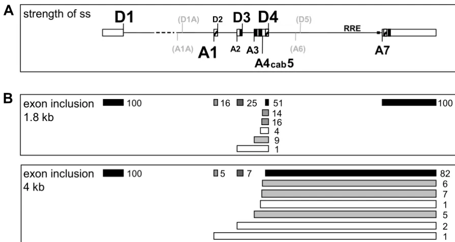
As previously determined experimentally for all HIV-1 5'ss [28,29] and here for all HIV-1 3'ss, there is no strict alter-nation between strong 5'ss and weak 3'ss as discussed recently [52]. On the contrary, D1 and A1 are the most efficient splice sites defining the first intron. Nevertheless, this splice site pair is not the most frequently recognized pair (based on the frequencies of only 16% exon 2 inclu-sion and 1% vif mRNA, Fig. 10B) most likely due to the intrinsically weakest 5'ss D2 which opposes cross-talk of the splice site pair across exon 2. This suggests that, although harboring a bipartite ESE HIV-1 exon 2 will be efficiently included only if the intrinsic strength of D2 is increased. Indeed, this has been shown most recently resulting in decreased virus production [36]. However, our finding that a switch in splice site pair recognition can crucially depend on exonic cis-acting regulatory sequences supports the possibility that Rev by interacting with trans-acting splicing regulatory proteins could switch cross-talk of splices site pairs. Alternatively, by interacting with trans-acting splicing regulatory proteins, Rev could functionally substitute for an ESE. Consistent with this, Rev has been found to bridge p32 which co-purifies with ASF/SF2 to the RRE thereby possibly stabilizing the interaction of U1 snRNP with the 5'ss and arresting further spliceosome for-mation [53]. This model provides an explanation for our finding that partially inactivating the ESE (Δ M1 – 43 mutation) did not affect processing of the Rev-dependent 1.2I vif mRNA but specifically leads to loss of exon 2 rec-ognition within the Rev-independent class of mRNAs.
Although it seems that HIV-1 splice site regulation is highly complex as outlined here (without even addressing the possible impact of secondary structures or superordi-nated hairpin structures on splice sites usage [54,55]) it is this complexity that provides a possibility to selectively inhibit HIV-1 splice site usage as a potential treatment strategy without cellular side effects [56,57].
Methods
OligonucleotidesOligonucleotides were synthesized and purified as previ-ously described [58]. Sequences are given in Tab. 1.
Recombinant plasmids
SVcrev was constructed by cloning the EcoRI-XhoI frag-ment from pUHcrev [59] into pSVT7. The SV40 early env expression vector SV E/X tat- rev- contains the EcoRI-XhoI fragment of pNLA1 [60], a cDNA derivative of pNL4/3. The 5' ss mutations were constructed as previously described [28]. To introduce the mutations A7- and A7+ in
3'ss A7, overlapping oligos were PCR amplified (A7-: #626, #628; A7+: #797, #798), the PCR products were restricted with AvaI/AflII (A7-) and SpeI/XmaI (A7+), respectively, and cloned into the appropriate vector back-bones.
The one-intron constructs (SV-SD/RRE/SA-pA) and muta-tions in the 5'ss were constructed as previously described [28]. Mutations in the 3'ss were introduced by swapping the BstEII-XmaI fragment except for A7ex which was cloned as a BstEII-BamHI fragment. For A7+ overlapping oligos (#796/#797) were amplified. For A7 - a fragment was amplified using primer pair #985/#986 and plasmid SV E/X tat -rev - SA7 - as template. HIV 1 3'ss A1 sequence was amplified using pNL-gpt (kindly provided by Valerie Bosch, ATV-DKFZ Heidelberg) as template both with 5' primer #948 and 3' primer either #947 (A1) or #1089 (A1ex). All other HIV 3' ss were amplified using pNLA1 as template and primers #946 and either #945 (A2) or #1091 (A2ex), #936 and #939 (A3) or #1492 (A3ex), #940 and #941 (A4cab) or #943 (A4cab5) or #1088 (A4cab5ex), #942 and #943 (A5) or #1088 (A5ex), #800 and #1065 (A7) or #139 (A7ex). The A5 mutations were constructed by introducing PCR products from overlap-ping oligos (A5 AG -: #1591, #1483; A5 b1-b2+AG -: #1586, #1484; A5 b1-b2+AG -Py+: #1586, #1592; A5 b1 -b2+AG -Py++: #1586, #1587; A5 b1-AG -Py++: #1482, #1389; A5 AG -Py++: #1388, #1590). For A5 Py++ (#942, #1481) and A5 Py+ (#942, #1486) pNLA1 was used as a PCR template.
To clone the three-exon-two-intron minigene reporter construct LTR ds ex2 two PCR fragments containing a 5'ss (#377, #918; BssHII-SalI) and a 3'ss (#931, #932; PstI-SalI) were inserted into LTR 1.4tatCAT, a vector coding for transcripts with the native HIV-1 tat 1.4 mRNA leader sequence [61], to generate LTR SD SA tatCAT.
Exon 2 including flanking intronic sequences of pNL-gpt was PCR-amplified with primers #1183 and #1913 and cloned into LTR SD SA tatCAT via EcoRI and PstI. Simi-larly, to insert the mutations of heptamer 1 and 2 the 3' PCR primer was substituted for #1913a (LTR ds ex2 hept.1) and #1913b (LTR ds ex2 hept.2) respectively.
All plasmid sequences can be obtained on request.
Cell culture, transfection and Northern blot analysis
HeLa-T4+ cells [62] were transfected with FuGENE™ 6 (Roche Molecular Biochemicals) and total RNA was pre-pared 30 h after transfection by a modified guanidinium isothiocyanate protocol [63] using RNA-clean (Hybaid-AGS, Heidelberg). The poly(A)+ RNA from 80–100 μg
total RNA was isolated with Dynabeads® oligo(dT)
aga-rose-1% formaldehyde-gel, and blotted onto a positively charged nylon membrane (Roche Molecular Biochemi-cals). After UV crosslinking (0.5 J/cm2), the membrane was hybridized with digoxigenin-(DIG) labeled antisense RNA probes in a buffer containing 50% (v/v) formamide, 5 × SSC, 50 mM sodium phosphate (pH 7), 0.1% (w/v) N-lauroylsarcosine, 7% (w/v) SDS, 2% (w/v) blocking rea-gent (Roche Molecular Biochemicals), 50 mg yeast RNA/L at 68°C. To monitor transfection efficiency and RNA loading, the membrane was hybridized with a DIG-labeled antisense RNA probe specific for exon 5 of human growth hormone (hGH, expressed from cotransfected plasmid pXGH5 as a transfection control). HIV-specific RNA was detected by a DIG-labeled antisense RNA probe specific for the 3' end of env (LTRcenvpA-, nt 8648–8887) and detection with anti-Digoxigenin-AP-Fab fragments (50 mU/ml; Roche Molecular Biochemicals) and
chemi-luminescence substrate (250 μM CDP-Star™; Roche
Molecular Biochemicals) as previously described [28]. Quantification was done with the Lumi-Imager F1 (Roche
Molecular Biochemicals) and the LumiAnalyst™ 3.1 soft-ware.
RT-PCR assay
[image:16.612.76.524.93.331.2]Isolation of total RNA was performed using a modified guanidinium isothiocyanate protocol [63]. Cells were washed twice with 2 ml of PBS and cell lysis was per-formed with 500 μl of buffer D [4 M guanidinium-isothi-ocyanat, 25 mM Na-Citrat pH 7, 0,5% N-Laurylsarkosin]; 7.6 μl of 2-mercaptoethanol, 50 μl of 3 M sodium acetate (pH 4), 500 μl of phenol and 100 μl of a chloroform-iso-amyl alcohol mixture (24:1) were added and mixed for 15 s. After incubation on ice for 15 min, phases were seper-ated by centrifugation (10,600 × g, 4°C, 20 min). RNA was precipitated in 1 volume of isopropanol overnight. After centrifugation (10,600 × g, 4°C, 20 min) the RNA pellet was washed twice with 70% ethanol and dissolved in 10 μl of DMDC-ddH2O. Prior to reverse transcription, 4 μl of RNA samples were subjected to DNase I digestion using 10U DNase I (Roche Molecular Biochemicals) with Schematic overview of HIV-1 splice sites and their strength
Figure 10
Schematic overview of HIV-1 splice sites and their strength. (A) Schematic drawing of the HIV-1 pre-mRNA and the distribution of the splice sites. The relative strength of the splice sites, based on splice site swapping strategies in this and pre-vious publications [27,29], is represented by the size of the letters. D5 and A6 (grey) are marked for better orientation but they are not used in HIV-1 NL4/3 and therefore their relative strength was not tested. D1A and A1A are recently published splice sites preferentially involved in RNA stabilization [6]. ESEs (hatched) and ESS/ISS (black) are marked. (B) Schematic draw-ing of the exon structure in the 1.8 kb and 4 kb HIV-1 RNA classes. The numbers represent the relative incidence of exon inclusion [%] calculated from previously published RT-PCR analyses [2]. Differences to 100% in the total amount of the possi-bilities of overlapping exon recognition are due to rounding errors. The darker the colour of the exons the more efficiently they are included in the alternatively spliced HIV-1 transcripts.
B
A
exon inclusion
4 kb
100
1 2 82
6 7 1 5 100 51
16 25
14 16 4 9 1
exon inclusion
1.8 kb
100
A1
D1
D2D3
D4
A2 A3
A4
cab5
(D5)
(A6)
A7
RRE
strength of ss
5 7
(D1A)
Table 1: Oligonucleotides used throughout these experiments
Oligo no. sequence
#139 5'-CCCAAGCTTTCTAGACTCGAGCTACAAAATCCTTTC #377 5'-CTGAAGCGCGCACGGCAAGAGGCGAGGGGAGGCGACTG #480 5'-GCGCGCTTCAGCAAGC
#481 5'-GCGCGCACGGCAAGA #559 5'-CTTTACGATGCCATTGGGA
#626 5'-TGTCTTAAGTATAGTGAATCGCGTTCGCCAAGGATACTCACCA #628 5'-CCCCTCGGGATTGGGAGGTGGGTTTGGAAGCTTAGTGGTGAGTATCC #796 5'-TGAGGTTACCGTTCTTTCTATT
#797
5'-GATCCCGGGATCCTCGGGATTGGGAGGTGGGTCTGAAAAGAAAGAGGAGAATAACCTTGACGAACACGGTTAGCAAT AGAAAGAAC
#798 5'-TGAACTAGTAGGTTTAAGAATAGTTTTTGCTGTTCTTTCTATT #800 5'-TGAGGTTACCGTACTTTCTATAGTGAAT
#918 5'-GCTATGTCGACAAGGAGCTGCAGATCGATGAATTCGATACTTACCAGTCGCCTCCCCTC #931 5'-GCTATGTCGACACCCAATTCTGAAACGATAATGGTGAATATCCCTGCCTAACTCTATTCACTATA #932 5'-ATCCTGCAGAATAGTTTTTGCTGTACTTTCTATAGTGA
#936 5'-ACTGGTTACCACATATCTATGAAACT
#939 5'-ACAGGATCCATCCCCGGGCTGAAATGGATAAACA #940 5'-ACTGGTTACCTGTACCAATTGCTATT
#941 5'-ACAGGATCCATCCCCGGGCTAAGGCTTTTGTCAT #942 5'-ACTGGTTACCAAAAAGTGTTGCTTTC
#943 5'-ACAGGATCCATCCCCGGGCTGCCATAGGAGATGC #945 5'-ACAGGATCCATCCCCGGGCTGAAAAACAGTCAAA #946 5'-ACTGGTTACCACCCTGAATTAGCAGA
#947 5'-ACAGGATCCATCCCCGGGCTGTAATAAACCCGAA #948 5'-ACTGGTTACCTAGCAACAGACATACA
#985 5'-GATGGATCCCGGGCTCGGGATTGGGAG #986 5'-TGAGGTTACCGTCTTAAGTATA
#1065 5'-ATCCCCGGGCTGAAACGATAATGGTGA
#1088 5'-ACAGGATCCATCCCCGGGTGCTTTGATAGAGAAG #1089 5'-ACAGGATCCATCCCCGGGCTTTCCAGAGGAGCTT #1091 5'-ACAGGATCCATCCCCGGGCTTGTTATGTCCTGCT #1183 5'-CTAGAATTCAGCAACAGACATACA
#1224 5'-TCTTCCAGCCTCCCATCAGCGTTTGG #1225 5'-CAACAGAAATCCAACCTAGAGCTGCT
#1388 5'-TGAGGTTACCAAAAAGTGTTGCTTTCATTGCCACGTTTGTTTCATGAC #1389 5'-ATCCCCGGGCTGAAAGAGGAGAAGGCGAGGGCGTTTGTCATGAAAC #1481 5'-ATCCCCGGGCTGAAAGAGGAGAAGGCTAGGGCTTTTGTCATGAAAC #1482 5'-TGAGGTTACCAAAAAGTGTGGCGTCCGTTGCCACGTTTGTTTCATGAC #1483 5'-ATCCCCGGGCTGCCATAGGAGATGCCGAAGGCGTTTGTCATGAAAC #1484 5'-ATCCCCGGGCTGCCATAGGAGATGCCGAAGGCGTTTGTTAGTAAAC #1486 5'-ATCCCCGGGCTGAAATAGGAGATGCCTAAGGCTTTTGTCATGAAAC #1492 5'-ACAGGATCCATCCCCGGGAGGGCTCTAGTCTAGG
#1542 5'-CACCTTCTTCTTCTATTCCTT #1544 5'-CTTGAAAGCGAAAGTAAAGC
#1586 5'-TGAGGTTACCAAAAAGTGTGGCGTCCGTTGCCACGTTTGTTTACTAAC #1587 5'-ATCCCCGGGCTGAAAGAGGAGAAGGCGAGGGCGTTTGTTAGTAAAC #1590 5'-ATCCCCGGGCTGAAAGAGGAGAAGGCGAGGGCGTTTGTCATGAAAC #1591 5'-TGAGGTTACCAAAAAGTGTTGCTTTCATTGCCACGTTTGTTTCATGAC #1592 5'-ATCCCCGGGCTGAAATAGGAGATGCCGAAGGCGTTTGTTAGTAAAC
#1913 5'-TACTGCAGTACTTTTATGTCACTATTATCTTGTATTACTACTGCCCCTTCACCTTTCCAGAGGAGCTTTGCTG #1913a
5'-CTACTGCAGTACTTTTATGTCACTATTATCTTGTATTACTACTGCCCCTTCACCTTTCCAGAGGAGCTTTGCTGGTCGA TAGCAAACTGGATCTCTG
#1913b 5'-CTACTGCAGTACTTTTATGTCACTATTATCTTGTATTACTACTGCCCCTTCACGATAGCAGAGGAGCTTTGCTG #2183 5'-GGTCAGGGTCTACTTGTGTGC
50 mM Tris (pH 7.5) and 10 mM MgCl2 in a total volume of 10 μl at room temperature for 1 h. After DNase I inac-tivation at 80°C for 10 min, 4.5 μl of the DNase digested RNA samples were reversed transcribed with 200U Super-Script III RNase H-Reverse Transcriptase (Invitrogen) according to the manufacturer's protocol using 0.375 mM oligo(dT)15 (Roche Molecular Biocemicals) or 0.02 μM sequence-specific oligo #1542 as primer. As a negative control for the remaining plasmid DNA contamination of each sample, a second assay was performed as described above but replacing reverse transcriptase with ddH2O.
PCR was carried out with 1.25U AmpliTaq (Applied Bio-systems) in a total volume of 50 μl according to the man-ufacturer' protocol in a Robocycler Gradient 96 Temperature Cycler (Stratagene). All primers were used at a final concentration of 0.2 μM. Spliced and skipped RNA was detected with the primer pair #1544/#1542 and hGH mRNA with primer pair #1225/#1224. Prior to PCR the cDNA reaction mixture was denatured at 94°C for 3 min. To determine the linear PCR-amplification range allowing a semi-quantitative estimation of the relative abundance of pSV-1-env and hGH mRNA, a preliminary PCR test series was carried out using the same cDNA sample but varying the PCR cycle numbers between 15 and 30 [94°C, 0.5 min; 52°C (pSV-1-env) and 56°C (hGH), respec-tively, 1 min; 72°C, 1 min]. The reactions were completed with a final elongation step of premature amplified prod-ucts at 72°C for 10 min. Accordingly to the obtained results, PCR analysis was performed with 26 cycles for pSV-1-env as well as hGH PCR amplification.
PCR products were separated on 6% non-denaturating polyacrylamide gels, stained with ethidium bromide (10 min) and visualized with the Lumi-Imager F1 (Roche Molecular Biochemicals).
Pull-down Assay of ASF/SF2
Periodat-labeled in vitro transcripts (1 nmol) of Eco47lII-linearized T7 Ex2 and T7 Ex2 Δ M1/Δ M2 plasmids carry-ing mutations of the heptameric motive were prepared using T7-MEGAshortscript™ (Ambion) and 0.1 M sodium m-periodat. The binding reaction to the adipic acid dihy-drazide agarose (Sigma) was performed over night in 0.1 M NaOAc pH 5.0. Complex formation was performed in a 650 μl reaction volume containing 500 μl of HeLa nuclear extract (Cell Culture Center, Belgium) and 150 μl buffer D (20 mM HEPES-KOH pH 7.9, 100 mM KCl, 20% glycerol, 0.2 mM EDTA, 0.5 mM DTT) by incubation at 30°C for 30 min. After washing five times in 1 ml of buffer D the complexed RNAs were eluted by incubating the beads in 60 μl of 2 × sample buffer, heating at 90°C for 10 min, followed by fractionation on an 7% denaturing poly-acrylamide gel. The proteins were blotted onto a PVDF membrane (Immobilon™ P, pore size 0.45 μm; Millipore)
by electroblotting with 70V in transfer buffer (200 mM glycine, 25 mM Tris, 20% methanol) for 1 h. Blots were blocked o/n in PBS with 10% bovine serum albumin (BSA), 10% Tween®-20. Protein detection was performed in PBS, 1% BSA, 1% Tween®-20, with a goat polyclonal antibody raised against a peptide mapping near the C-ter-minus of SF2/ASF of human origin (Santa Cruz Biotech-nology, Inc. C-19:sc-10255) for 1 h, washed three times, incubated with a horseradish peroxidase conjugated AffiniPure donkey anti-goat IgG (Jackson ImmunoRe-search Laboratories, Inc. #705-035-147), washed four times, rinsed with water and visualized by a chemilumi-nescence detection system (ECL™-system and ECL™ hyper-film, Amersham; Super Signal® ultra, Pierce).
HIV-1 infection experiments
Cell cultures were maintained in RPMI 1640 medium containing 10% fetal calf serum (Pansystems GmbH) and antibiotics (penicillin and streptomycin). Viral stocks were prepared by transfecting 293T cells with provirus expression vectors, followed by ELISA (Innotest HIV p24 Antigen mAb; Innogenetics N. V.) of culture supernatants for p24 content. For HIV-1 infection, 5 × 10⁶ PM1, CEM or CEM-SS cells were resuspended in 500 μl culture medium and incubated at 37°C for 3 hours with 100 ng of each HIV-1 viral stock (HIV-1 NL4/3 wild type and ΔM1-43). After infection, cells were washed twice with PBS without Ca2+ and Mg2+ and further cultured in 5 ml medium for another 5 days. Subsequently, total RNA was isolated using Trizol reagent according to the protocol of the manufacturer (Invitrogen).
Competing interests
The author(s) declare that they have no competing inter-ests.
Authors' contributions
SK and MO performed the cloning work. Northern blot analyses were carried out by SK, RT-PCR and pull-down by MO. All infectious experiment were designed and con-ducted in a P3 facility by IH and JH. JK conceived optimi-zation of A7. HS devised and coordinated the study. SK and HS drafted the manuscript. All authors read and approved the final manuscript.