R E S E A R C H
Open Access
A novel method for downstream characterization
of breast cancer circulating tumor cells following
CellSearch isolation
Henrik Frithiof
1, Charlotte Welinder
1, Anna-Maria Larsson
1,2, Lisa Rydén
3,4*†and Kristina Aaltonen
1†Abstract
Background:Enumeration of circulating tumor cells (CTCs) obtained from minimally invasive blood samples has been well established as a valuable monitoring tool in metastatic and early breast cancer, as well as in several other cancer types. The gold standard technology for detecting CTCs in blood against a backdrop of millions of leukocytes is the FDA-approved CellSearch system (Janssen Diagnostics), which relies on EpCAM-based immunomagnetic separation. Secondary characterization of these cells could enable treatment selection based on specific targets in these cells, as well as providing a real time window into the metastatic process and offering unique insights into tumor heterogeneity. The objective of this study was to develop a method for downstream characterization of CTCs following isolation with the CellSearch system.
Methods:Anin vitroCTC model system focusing on clinically useful treatment predictive biomarkers in breast cancer, specifically the estrogen receptorα(ERα) and the human epidermal growth factor receptor 2 (HER2), was established using healthy donor blood spiked with breast cancer cell lines MCF7 (ERα+/HER2−) and SKBr3 (ERα−/HER2+). Following CTC isolation by CellSearch, the captured CTCs were further enriched and fixed on a microscope slide using the in-house-developed CTC-DropMount technique.
Results:The recovery rate of CTCs after CellSearch Profile analysis and CTC-DropMount was 87%. A selective and consistent triple-immunostaining protocol was optimized. Cells positive for DAPI, cytokeratin (CK) 8, 18 and 19, but negative for the leukocyte-specific marker CD45, were classified as CTCs and subsequently analyzed for ERαand HER2 expression. The method was verified in breast cancer patient samples, thus demonstrating its clinical relevance. Conclusions:Our results show that it is possible to ascertain the status of important predictive biomarkers expressed in breast cancer CTCs using the newly developed CTC-DropMount technique. Downstream characterization of multiple biomarkers using a standard fluorescence microscope demonstrates that important clinical and biological information may be obtained from a single patient blood sample following either CellSearch epithelial or profile analyses.
Trial registration:Clinical Trials NCT01322893
Keywords:Circulating tumor cells (CTC), Metastatic breast cancer, CellSearch, Biomarkers, Estrogen receptor (ER), Human epidermal growth factor 2 (HER2), Immunofluorescence
* Correspondence:[email protected]
†Equal contributors
3Division of Surgery, Department of Clinical Sciences, Lund University, Lund,
Sweden
4Department of Surgery, Skåne University Hospital, Lund, Sweden
Full list of author information is available at the end of the article
Background
During the last decade, enumeration of circulating tumor cells (CTCs) in peripheral blood was established as a prognostic tool for predicting time-to-recurrence and survival in metastatic and early breast cancer, as well as in several other cancer types [1-4]. The gold standard technology, and the only platform implemented on a lar-ger scale, is the FDA-approved semi-automated CellSearch technology (Jansen Diagnostics, Raritan, NJ, USA). In CellSearch, enriched EpCAM-positive CTCs are defined as nucleated cells positive for cytokeratin (CK) 8, 18 and 19, but negative for the leukocyte-specific surface protein, CD45. However, the field of CTC research is now moving beyond solely quantifying cells in peripheral blood. Pheno-typic and molecular characterization of CTCs has the po-tential to provide clinically important information from an easily accessible blood sample, a ‘liquid biopsy’. Serial blood sampling followed by molecular characterization can provide insights into tumor progression and enable early detection of treatment resistance.
In breast cancer, assessment of estrogen receptor α (ERα) status in the primary tumor is crucial in classifica-tion and treatment predicclassifica-tion [5]. Determining receptor status identifies patients eligible for endocrine therapy, which remains the mainstay adjuvant treatment for ERα+ breast cancers, either as monotherapy or in con-junction with chemotherapy. Although an ERα+ primary tumor is a common trait and found in approximately 80% of patients with primary breast cancer, it is no guar-antee for a favorable outcome following endocrine treat-ment as recurrence rates of 19–41% are observed at 10 years following 5 years of tamoxifen [6-8]. Moreover, in metastatic breast cancer, approximately 40–50% of patients fail to respond to endocrine treatment, despite an initially positive assay [9]. The causes of this consid-erable inconsistency are multifactorial and have not been entirely elucidated, but discordance in ERα status be-tween the primary tumor and involved lymph nodes or distant metastases has been established in 6–30% of studied cases and may contribute to treatment resistance [10-14]. In fact, this phenotypical shift is associated with significantly shorter median survival for patients with metastatic disease when compared with consistent ERα -positivity in disease progression [10]. Additionally, it has been reported that the majority of CTCs in patients with ERα+ primary tumors are in fact ERα− prior to therapy, with a concordance of less than 30% [15-17].
Human epidermal growth factor receptor 2 (HER2) is a tyrosine kinase receptor encoded by a proto-oncogene lo-cated on chromosome 17 (17q12), and is the second most important predictive biomarker in breast cancer [18,19]. Amplification of this gene occurs in approximately 10–30% of primary breast cancers, correlating with poor prognosis and an aggressive phenotype [18,19]. This subgroup of
patients benefits from immunotherapy with an HER2-targeted monoclonal antibody, trastuzumab, in combin-ation with chemotherapy in adjuvant, neoadjuvant and metastatic settings [20,21]. Similar to the dynamic pro-gression observed in ERα+tumors, the HER2 status of me-tastases can differ from that of the primary tumor [22]. Discordance has been observed in 7–14% of studied cases [11,23-25]. It has been shown that patients with HER2− tumors might acquire HER2 amplification during disease progression, as demonstrated by isolation of HER2+CTCs in patients with an HER2− primary tumor [26-29]. An-other explanation for the discrepancy in biomarker ex-pression between primary tumors and CTCs may be tumor heterogeneity. Tumor clones shed into the blood stream are more likely to represent those with most malig-nancy, exemplified by HER2-amplified clones, despite the primary tumor being diagnosed as HER2 normal. These patients are less likely to receive HER2-targeted treatment, although a complete or partial response has been observed in selected cases [30]. Two prospective trials including pa-tients with negative primary tumors and HER2-positive CTCs are currently open for recruitment and aim to elucidate whether trastuzumab will have a beneficial effect on these cases [31].
Thus, treatment decisions based on the phenotype of the primary tumor alone might omit critical facts rele-vant to the prognosis and choice of treatment. Biopsies from metastatic sites are not always available for prac-tical reasons and are inevitably accompanied by an inva-sive procedure. CTCs are easily accessible from a normal blood sample, and since CTCs are shed from multiple metastatic sites as well as from the primary tumor, characterization of these cells could provide im-portant information for treatment prediction.
The aim of this study was to establish a method for downstream characterization of multiple treatment pre-dictive markers expressed by CTCs after CellSearch-based selection, without the necessity of additional patient sam-ples. Validation of the method in samples from patients with metastatic breast cancer highlights the potential of the clinical utility of this technique.
Methods In vitromodel
Breast cancer cell lines MCF7 and SKBr3 were obtained from the American Type Culture Collection (ATCC/LGC Standards GmbH, Wesel, Germany) and were used to es-tablish anin vitromodel system for CTC characterization following CellSearch isolation. MCF7 expresses ERαbut is negative for HER2 amplification. Contrary, SKBr3 cells are HER2-positive and negative for ERα. MCF7 cells were grown in a 5.0% CO2 incubator under UV-light at
supplemented with 1% sodium pyruvate, 1% non-essential amino acids, 10% fetal bovine serum (FBS) and 1% penicil-lin streptomycin mixture (Pen-Strep) for MCF7, and RPMI 1640 (HyClone Laboratories, Inc.), while SKBr3 cells were cultured under the same conditions in 5 mL MEM/EBSS plus 10% FBS and 1% Pen-Strep. Harvesting of cells was performed at approximately 80–90% con-fluency after 5–10 min trypsinization.
Healthy donor blood samples were processed within 24 h from withdrawal, and spiking of cells occurred in conjunction with subsequent CellSearch analyses. Two different spiking methods were used. First, dilution of cells resulted in approximately 2000 cells per 7.5 ml blood, and using the CTC-DropMount technique (described below), approximately 200 cells were applied to 10 individual slides, which were later used in the optimization of stain-ing procedures. Second, to ascertain the recovery rate of the method, a specific number of cells were harvested in-dividually with a 10μL pipette under a bright-field micro-scope equipped with a standard achromatic × 10/0.25 objective. In detail, a fraction of the cell culture was trans-ferred to a Petri dish containing cell culture medium. While observing the cell culture suspension through the eyepieces of the microscope, suitable individual cells were selected and carefully extracted using a 10μL pipette be-fore transfer to a healthy donor blood sample. Since the process is continuously monitored by microscopy, one can confirm that the cell has been properly extracted. Refer-ence values of 5, 15, and 50 cells were selected. Independ-ently collected duplicates of each of the three respective cell quantities were added to 7.5 mL of healthy donor blood samples and processed according to the specified method. The agreement between the measured results and the reference values was calculated to define the re-covery rate.
Fixation of samples using CTC-DropMount
CellSearch Profile (Jansen Diagnostics) analysis was per-formed according to the manufacturer’s protocol, which involves enrichment of CTCs with magnetic ferrofluid-associated anti-EpCAM antibodies but no consecutive staining. The enriched samples were mounted on slides using a specific procedure developed in-house, CTC-DropMount. The solution containing isolated CTCs (ap-proximately 900 μL) was transferred to an 1.5 mL Eppendorf tube and placed in a magnetic tray. After 10 min incubation, the non-adherent solvent was ex-tracted. The cells were resuspended in 10 μL 1 × PBS, mounted on superfrost slides (ThermoScientific, Germany) and incubated at 37°C for 30 min. Fixation was accomplished by immersing slides in pure methanol for 5 min. The samples were stored at−20°C.
The CTC-DropMount method was also used for enriched cells after standard CellSearch epithelial cell
analysis (i.e. all cells were semi-automatically stained with CK-phycoerythrin (PE), CD45-allophycocyanin (APC) and DAPI in a procedure described previously (3)). In this case, the solution containing enriched CTCs was extracted from the CellSearch cartridge after complete analyses, and the cartridge was carefully rinsed with 1 × PBS buffer to ensure maximum extraction be-fore transfer to a 1.5 mL Eppendorf tube in a magnetic tray. An overview of the CTC-DropMount method is provided in Figure 1.
Immunostaining protocol for ERαand HER2
Following cell permeabilization using Dako Target Retrieval solution containing Tris/EDTA buffer solution pH 9.0 and detergent (S2368, Dako Denmark A/S, Glostrup, Denmark), slides were stained according to the optimized protocols detailed in Tables 1 and 2, respectively. Briefly, to define CTCs against the backdrop of remaining leukocytes, a CD45-specific AlexaFluor647-labeled mouse monoclonal antibody (F10894, AbD Serotec, Oxford, UK) was assessed at dilutions of 1:1, 1:5 and 1:10 [32]. The primary antibody used against ERα was a rabbit monoclonal antibody (RM-9101-S1, Thermo Scientific, Fremont, CA, USA), diluted 1:50 [33]. Secondary detection was accom-plished with AlexaFluor488-labeled goat anti-rabbit antibody (A-11034, Life Technologies, Carlsbad, CA, USA) at dilutions of 1:200, 1:300, 1:400, and 1:500. For detection of cytokeratin, slides were incubated with CellSearch Staining Reagent containing a PE-labeled mouse monoclonal antibody specific to CK 8, 18 and 19, at a concentration of 0.0006% (Janssen Diagnostics). Antibody dilutions were made with DAKO Antibody Diluent containing 1% FBS in PBS and 0.1% detergent (S2022, Dako). Slides were finally mounted with cover-slips and counterstained with an antifade reagent contain-ing the nucleic acid dye, DAPI, (S36942, Life Technologies), thus enhancing resistance to photobleaching.
The level of HER2-expression was investigated using a primary monoclonal rabbit antibody specific for the hu-man HER2 oncoprotein (1:250; EP1045Y, Abcam plc, Cambridge, UK), along with a polyclonal porcine anti-rabbit secondary antibody conjugated with alkaline phosphatase (ALP) (1:50; D0306, Dako). Detection was accomplished based on the ALP-Fast Red reaction, using the substrate and chromogen in Liquid Permanent Red (LPR) (K0640, Dako [34]). Incubation times assessed for the ALP-Fast Red reaction were 5, 10 and, 15 min. LPR was evaluated by both fluorescence and bright-field microscopy.
Alexa647/Cy5 were used for staining evaluation. In a few cases, an Olympus BX51 microscope (Olympus Optical CO) and a Zeiss Axio Observer Z1 microscope (Carl Zeiss Microscopy, LLC., NY, USA) outfitted with single pass filters for each individual fluorochrome were used for examination.
In vivovalidation
Patient blood samples were investigated for clinical validation of the technique. All patients had metastatic breast cancer and had been included in the ongoing CTC-MBC trial (Clinical Trial Id. NCT01322893) at Lund University, Sweden. In total,
[image:4.595.57.540.87.241.2]nine clinical samples of 7.5 mL whole blood from nine individual patients were assessed by CellSearch profile analysis. CTC-DropMount and subsequent staining according to the staining protocols detailed in Tables 1 and 2 were performed prior to evaluation by fluorescence and bright-field microscopy. All pa-tient sample analyses were processed in conjunction with positive and negative controls, decreasing the risk of methodological errors, as well as confirming successful staining reactions. Ethical permission for the CTC-MBC study was obtained from Lund University Ethical Board (EPN 2010/135) and all patients gave writ-ten informed consent.
Figure 1Overview of the method. Enriched CTCs were collected after CellSearch analysis using the Profile or Epithelial cell kit. The solution containing CTCs and leukocytes was placed in a magnetic tray. Following incubation, the non-adherent solvent was removed and the ferrofluid-attached cells were re-suspended in a smaller volume of PBS, thus permitting further enrichment. This solution was dropped and fixed on a glass slide before subsequent staining according to protocols 1 or 2. Visualization is possible with a fluorescence or bright-field microscope, depending on the staining method applied. (Drawing of magnetic stand reprinted with permission from IFI CLAIMS Patent Services.)
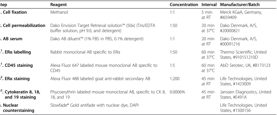
Table 1 Optimized staining protocol for ERα, CK, and CD45 expression in CTCs and leukocytes
Step Reagent Concentration Interval Manufacturer/Batch
1. Cell fixation Methanol 1:1 5 min
at RT
Merck KGaA, Germany, #I659409
2. Cell permeabilization Dako Envision Target Retrieval solution™(50x) (Tris/EDTA buffer solution, pH 9.0, and detergent)
1:50 20 min
at 37°C
Dako Denmark, A/S, #20000821
3. AB serum Dako AB diluent™(1% FBS in PBS, 0.1% detergent) 1:1 20 min at RT
Dako Denmark, A/S, #00091216
41. ER
αlabelling Rabbit monoclonal AB specific to ERα 1:50 60 min at 37°C
Thermo Scientific, United States, #9101S1210D
51. CD45 staining Alexa Fluor 647 labeled mouse monoclonal AB specific to CD45
1:5 60 min
at 37°C
AbD Serotec, UK, #B173123
62. ERαstaining Alexa Fluor 488 labeled goat anti-rabbit secondary AB 1:200 45 min at RT
Life Technologies, United States, #1423009
72. Cytokeratin 8, 18, and 19 staining
Phycoerythrin labeled mouse monoclonal AB, specific to CK 8, 18, and 19
0.0006% 45 min at RT
Janssen Diagnostics, United States, #E491A
8. Nuclear counterstaining
Slowfade® Gold antifade with nuclear dye, DAPI Life Technologies, United States, #1500156
Washing with iced PBS 10.0%, v/v, 3 × 3 min between each step.
Abbreviations:AB - antibody, DAPI - 4′,6-diamidino-2-phenylindole dihydrochloride, EDTA - Ethylenediaminetetraacetic acid, ERα- estrogen receptorα, PBS - phosphate buffered saline, RT - room temperature, Tris - 2-Amino-2-hydroxymethyl-propane-1,3-diol.
1
Step 4 and 5 can be performed simultaneously.
2
[image:4.595.60.540.492.691.2]Results CTC-DropMount
An overview of the CTC-DropMount technique is shown in Figure 1. Using CellSearch Profile analysis from whole blood, the recovery rate was found to be 87% on average (80% for 5 cells, 97% for 15 cells and 84% for 50 cells).
Immunofluorescence
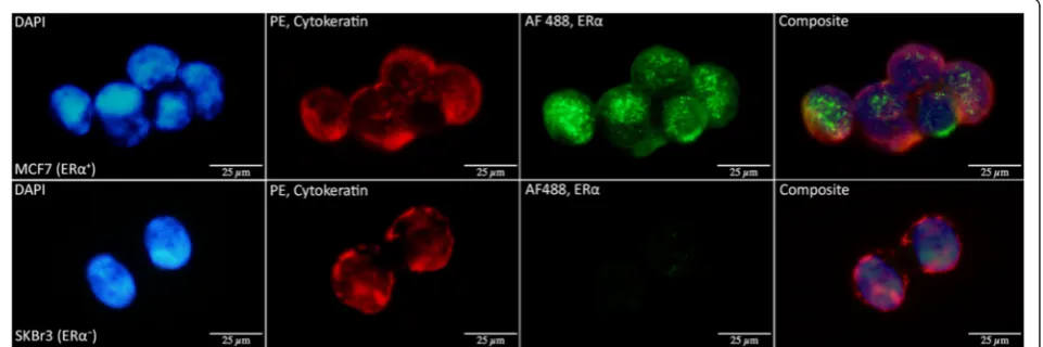
Using the AlexaFluor647-labeled monoclonal CD45-antibody, it was possible to separate leukocytes from CTCs under standard fluorescence microscopy. This re-sult provides an important prerequisite for further stain-ing and demonstrated sufficient selectivity of the method (see Figure 2). Optimal distinction between CTCs and leukocytes was achieved when combining the two filters for CK-PE and CD45-AlexaFluor647.
Criteria for ERα-positivity defined staining of the nu-clear region. The process was considered satisfactory when cells in the ERα+ cell line (MCF7) consistently stained positive for ERα with low background and marked nuclear intensity, while slides with ERα− cells
(SKBr3) simultaneously stained negative. Representative images are displayed in Figure 3.
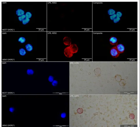
HER2-staining with LPR proved highly selective (see Figure 4). The ALP-based reaction suits CTCs particularly well since endogenous enzymatic activity is negligible in these samples. Hence, the risk of false positives is insignifi-cant. LPR permits assessment by both fluorescence and bright-field microscopy. Bright-field microscopy has the advantage of being easily accessible in most laboratories and the staining is impervious to fading. Despite the high background caused by ferrofluid remnants from Cell-Search analysis, LPR staining was clearly visible. Also, if combined with CK-PE staining, bright-field microscopy was preferable due to the risk of bleed-through between PE and LPR staining in immunofluorescence analyses.
[image:5.595.55.536.99.275.2]Staining of the fixed cells was optimized mainly using cells from CellSearch Profile analyses where no previous staining and permeabilization had affected the cells. However, the staining procedure was also tested follow-ing CellSearch epithelial cell analysis, and although these cells had been previously stained, the results were con-sistent with previously unstained cells.
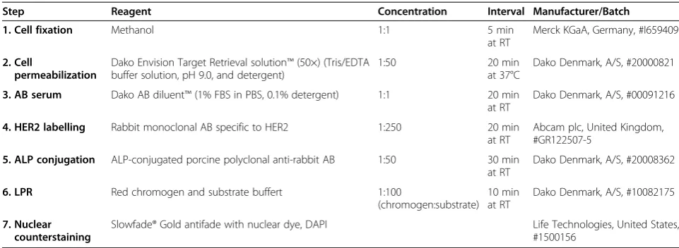
Table 2 Optimized staining protocol for HER2 expression
Step Reagent Concentration Interval Manufacturer/Batch
1. Cell fixation Methanol 1:1 5 min
at RT
Merck KGaA, Germany, #I659409
2. Cell
permeabilization
Dako Envision Target Retrieval solution™(50×) (Tris/EDTA buffer solution, pH 9.0, and detergent)
1:50 20 min
at 37°C
Dako Denmark, A/S, #20000821
3. AB serum Dako AB diluent™(1% FBS in PBS, 0.1% detergent) 1:1 20 min at RT
Dako Denmark, A/S, #00091216
4. HER2 labelling Rabbit monoclonal AB specific to HER2 1:250 20 min at RT
Abcam plc, United Kingdom, #GR122507-5
5. ALP conjugation ALP-conjugated porcine polyclonal anti-rabbit AB 1:50 30 min at RT
Dako Denmark, A/S, #20008362
6. LPR Red chromogen and substrate buffert 1:100
(chromogen:substrate) 10 min at RT
Dako Denmark, A/S, #10082175
7. Nuclear counterstaining
Slowfade® Gold antifade with nuclear dye, DAPI Life Technologies, United States, #1500156
Washing with iced PBS 10.0% (v/v), 3 × 3 min between each step except after LPR when the slides are only quickly rinsed in with PBS before nuclear counterstaining.
[image:5.595.57.541.610.686.2]Abbreviations:AB, antibody; ALP, Alkaline phosphatase; DAPI, 4′,6-diamidino-2-phenylindole dihydrochloride; EDTA, Ethylenediaminetetraacetic acid; LPR, liquid permanent red; PBS, phosphate buffered saline; RT, room temperature; Tris, 2-Amino-2-hydroxymethyl-propane-1,3-diol.
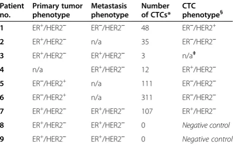
The CTC-DropMount method could be confirmed in nine patient samples after CellSearch Profile analysis, and examples of positive ERα and HER2 staining can be found in Figure 5. Table 3 outlines the patients’ charac-teristics with respect to their primary tumors, metasta-ses, and CTC phenotypes, as well as the total number of CTCs detected by CellSearch. The majority of detected CTCs were negative for both ERαand HER2 expression. We observed considerable intrapatient heterogeneity in levels of biomarker expression and cell morphology, ob-servations that are in concordance with previous re-search [33]. A visual comparison to corresponding CTCs in the picture galleries from CellSearch epithelial cell analyses suggested that the most intensely stained CTCs were also the most distinctly stained with the CTC-DropMount method for all investigated markers.
Discussion
In this study, we present a method for secondary characterization of breast cancer CTCs after CellSearch analysis. Protocols for the clinically important predictive markers ERαand HER2 were optimized in breast cancer cell lines and subsequently verified in samples from pa-tients with metastatic breast cancer. Fixation of CTCs was performed with the CTC-DropMount method de-scribed here, and ERα and HER2 staining protocols proved selective and consistent in our in vitro model system.
Secondary phenotypic characterization of fixed CTCs on standard microscope slides provides the possibility of concurrent morphological evaluation, assessment of the total number of cells and an estimation of the fraction of CTCs with expression of the analyzed biomarker. This gives unique information on the heterogeneity of marker expression, which is not available using PCR-based
molecular methods, for example [28,35,36]. Assessment of ERαstatus in CTCs could identify patients eligible for endocrine treatment that otherwise may be overlooked (i.e. ERα− primary tumor/ERα+ CTCs). Two of the nine patients included in the in vivo validation experiments presented ERα+ CTCs (see Table 3, and representative images in Figure 5). Both of these samples were drawn at or just prior to initiation of treatment against meta-static disease. The phenotype of the primary tumor from one patient was classified as ERα+, while the second pa-tient had a confirmed ERα+ metastatic biopsy (Table 3). Conversely, detection of ERα−CTCs in a patient with an ERα+ primary tumor might, in part, explain the lack of treatment response observed in this cohort. A similar assumption regarding HER2 gene amplification seems reasonable, since a subset of patients acquire oncogene amplification during disease progression [30]. The true number of patients suited for HER2-targeted treatment may in fact be higher than the number treated at present. This is currently being investigated in the ongoing European DETECT III and CIRCE T-DM1 studies, where the CTC HER2-positive phenotype is used as a treatment predictive marker [31]. In this study, HER2+ CTCs were identified after 6 months of chemo-therapy in a metastatic breast cancer patient with a HER2− primary tumor and a HER2− metastasis biopsy (see Table 3 and Figure 5).
[image:6.595.59.540.91.251.2]A few previous studies have used immunological stain-ing methods for secondary phenotypic characterization. Swennenhuiset al.fixed CTCs within the cartridge after complete CellSearch analysis using immunofluorescence and FISH analysis for successful characterization of HER2-status [37]. However, the CellTracks II analyzer had to be modified to improve the resolution and light collection. Other studies have used the FITC-channel in
the CellSearch system, where the intensity of HER2 staining is scored as negative (0), very weak (1+), moder-ate (2+) or very bright (3+) [27,38,39]. The clinical value of specific cut-off thresholds remains to be determined. Paoletti et al. recently reported a method utilizing the CellSearch-integrated FITC-channel for analysis of ERα, HER2, Ki67, and BCL-2 in individual blood samples with the intention of predicting resistance to endocrine ther-apy [40]. By implementing this approach, 7.5 mL of blood is required for analysis of each respective bio-marker. Few studies have described methods for second-ary characterization of ERα and HER2 in CTCs after
Ficoll density gradient separation and cytospin prepara-tions [17,33]. An advantage in circumventing immuno-logical enrichment before fixation of the cells onto microscope slides is the exemption from EpCAM-dependent selection. On the other hand, the number of cells that have to be screened manually by standard microscopy is very high, thus hampering the clinical feasibility and cost effectiveness if introduced into rou-tine clinical practice.
[image:7.595.59.540.91.527.2]Fixation of CTCs on microscope slides with the de-scribed CTC-DropMount method provides the possibil-ity to use a standard fluorescence microscope for CTC
characterization after immunological enrichment with the FDA-cleared CellSearch system. An advantage of the described method for secondary characterization is the scope to expand the CTC analysis to other putative pre-dictive markers as well as to more experimental markers, for example stem cell markers, epithelial mesenchymal transition (EMT) markers or markers associated with metastasis, proliferation or apoptosis, thus increasing our knowledge of metastasis biology. Selection of single CTCs or a subset of CTCs is also possible after CTC-DropMount using laser capture microdissection, for ex-ample. Subsequent single cell genomic analyses could
open the door to an even more detailed molecular characterization of CTCs [41].
The informative advantage of heterogeneity in marker expression using secondary staining methods is also as-sociated with the need for a prognostic cut-off value for the fraction or intensity of expression within the CTC population [17]. Using different cut-offs for marker posi-tivity has given conflicting results regarding discordance of marker expression between primary tumors and CTCs [17,33,42]. Thus, the prognostic significance of marker heterogeneity in breast cancer CTCs has to be determined, and reliability on the staining methodology is of immense importance. Using the CTC-DropMount technique, we found distinct nuclear staining of ERα in the MCF7 cell line and used nuclear staining as a criter-ion for ERα-positivity. However, to effectively determine the clinical implications, defined criteria, such as the number of CTCs to be evaluated and the fraction of ERα- or HER2-positive CTCs, has to be decided in fu-ture clinical studies.
The cell recovery rate after CTC-DropMount fixation was 87%, which is at the high end of recovery compared with studies using different methods for enrichment, fix-ation and detection [33,43-48]. The 80% recovery rate of five spiked cell line cells further indicates that this method could be useful in the clinical setting, where number of CTCs at the established CellSearch cut-off value (≥5 CTCs in metastatic breast cancer) is common.
Conclusions
[image:8.595.54.540.90.238.2]In conclusion, our results indicate that by retrieval of a single blood sample from patients with metastatic breast cancer it is possible to ascertain the status of important
Figure 5Immunostaining of metastatic breast cancer patient blood samples. Representative images of positive ERαand HER2 staining in clinical samples. RowA, from left to right: DAPI counterstain (fluorescent blue), cytokeratins 8, 18, and 19 (CK) stained with Phycoerythrin (red), estrogen receptor (ERα) stained with AlexaFluor 488 (green), and a composite of all channels. This patient sample (no. 4, see Table 3) was collected prior to initiation of therapy, illustrating two clustered ERα+CTCs, adjacent to a solitary leukocyte located in the lower left corner. This patient was diagnosed with an ERα+metastasis. RowB, from left to right: DAPI counterstain (fluorescent blue), HER2 stained with Liquid Permanent Red (red). This patient sample (no. 1, see Table 3) was obtained following 6 months of chemotherapy, illustrating HER2+CTCs identified by combination of fluorescence and bright-field microscopy. This patient was diagnosed with a HER2−primary tumor and HER2−metastasis.
Table 3 Patient data forin vivovalidation procedures
Patient no.
Primary tumor phenotype
Metastasis phenotype
Number of CTCs*
CTC phenotype§
1 ER+/HER2− ER−/HER2− 48 ER−/HER2+
2 ER+/HER2− n/a 35 ER−/HER2−
3 ER+/HER2− ER+/HER2− 3 n/a‡
4 n/a ER+/HER2− 12 ER+/HER2−
5 ER−/HER2+ n/a 111 ER−/HER2−
6 ER−/HER2+ n/a 311 ER−/HER2−
7 ER+/HER2− ER+/HER2− 107 ER+/HER2−
8 ER+/HER2− ER+/HER2− 0 Negative control
9 ER+/HER2− ER+/HER2− 0 Negative control
*As defined by CellSearch, single samples assessed 0–6 months from initiation of therapy against metastatic disease.
§
Phenotype according to CTC-DropMount. Criteria for biomarker positivity were≥1 ER+
CTC, and≥1 HER2+
CTC.
‡In this patient no CTCs were identified following secondary staining.
[image:8.595.57.291.526.673.2]predictive biomarkers expressed in breast cancer CTCs. The discordance of expression between primary tumors and metastases urgently informs us that new diagnostic tools are required for optimal treatment selection in both primary and metastatic breast cancer.
Abbreviations
ALP:Alkaline phosphatase; APC: Allophycocyanin; CK: Cytokeratins; CTCs: Circulating tumor cells; DAPI: 4′,6-diamidino-2-phenylindole; EDTA: Ethylenediaminetetraacetic acid; EpCAM: Epithelial cell adhesion molecule; ERα: Estrogen receptorα; FBS: Fetal bovine serum; HER2: Human epidermal growth factor receptor 2; LPR: Liquid permanent red;
PBS: Phosphate buffered saline; PE: Phycoerythrin; Tris: 2-Amino-2-hydroxymethylpropane-1,3-diol.
Competing interests
The authors declare that they have no competing interests.
Authors’contributions
HF performed thein vitroexperiments, immunofluorescent studies, figure designs and drafted the manuscript. KA conceived the study, performed the
in vivoexperiments and helped to draft the manuscript. LR initiated and conceived the CTC-MBC study and helped to draft the manuscript. A-MA initiated and planned the CTC-MBC study. CW initiated the conceptual design of the method. All authors read and approved the final manuscript.
Acknowledgements
We are thankful for skilled technical assistance in the CTC-lab by Kristina Lövgren and Sara Baker. Research nurse Anette Ahlin-Gullers collected blood samples from the participating patients. We are also indebted to Sofia Gruvberger-Saal for providing us with the cell lines.
This work was supported by funding from the Swedish Cancer Foundation, BioCare, the Swedish Medical Association, the Gunnar Nilsson Cancer Foundation, the Mrs Berta Kamprad Foundation, the Gyllenstierna Krapperup Foundation, and the Crafoord Foundation.
Grant numbers and sources of support
Swedish Cancer Foundation (CAN 2010/501, CAN 2010/1234, CAN 2013/533), the Gunnar Nilsson Cancer Foundation (2013/1224), the Mrs Berta Kamprad Foundation (36/2014), the Gyllenstierna Krapperup Foundation (2014/1702), the Crafoord Foundation (20100563).
Author details
1
Division of Oncology and Pathology, Department of Clinical Sciences Lund, Lund University, Lund, Sweden.2Skåne Department of Oncology, Skåne
University Hospital, Lund, Sweden.3Division of Surgery, Department of Clinical Sciences, Lund University, Lund, Sweden.4Department of Surgery,
Skåne University Hospital, Lund, Sweden.
Received: 4 February 2015 Accepted: 15 April 2015
References
1. de Bono JS, Scher HI, Montgomery RB, Parker C, Miller MC, Tissing H, et al. Circulating tumor cells predict survival benefit from treatment in metastatic castration-resistant prostate cancer. Clin Cancer Res. 2008;14(19):6302–9. 2. Cohen SJ, Punt CJA, Iannotti N, Saidman BH, Sabbath KD, Gabrail NY, et al.
Prognostic significance of circulating tumor cells in patients with metastatic colorectal cancer. Ann Oncol. 2009;20(7):1223–9.
3. Cristofanilli M, Budd GT, Ellis MJ, Stopeck A, Matera J, Miller MC, et al. Circulating tumor cells, disease progression, and survival in metastatic breast cancer. N Engl J Med. 2004;351(8):781–91.
4. Bidard F-C, Peeters DJ, Fehm T, Nolé F, Gisbert-Criado R, Mavroudis D, et al. Clinical validity of circulating tumour cells in patients with metastatic breast cancer: a pooled analysis of individual patient data. Lancet Oncol. 2014;15(4):406–14.
5. Goldhirsch A, Winer EP, Coates AS, Gelber RD, Piccart-Gebhart M, Thurlimann B, et al. Personalizing the treatment of women with early breast cancer: highlights of the St Gallen International Expert Consensus on the Primary Therapy of Early Breast Cancer 2013. Ann Oncol. 2013;24(9):2206–23.
6. Nadji M, Gomez-Fernandez C, Ganjei-Azar P, Morales AR. Immunohistochemistry of estrogen and progesterone receptors reconsidered: experience with 5,993 breast cancers. Am J Clin Pathol. 2005;123(1):21–7.
7. Davies C, Godwin J, Gray R, Clarke M, Cutter D, Darby S, et al. Relevance of breast cancer hormone receptors and other factors to the efficacy of adjuvant tamoxifen: patient-level meta-analysis of randomised trials. Lancet. 2011;378(9793):771–84.
8. Moy I, Lin Z, Rademaker AW, Reierstad S, Khan SA, Bulun SE. Expression of estrogen-related gene markers in breast cancer tissue predicts aromatase inhibitor responsiveness. PLoS One. 2013;8(11), e77543.
9. Osborne CK, Schiff R. Mechanisms of endocrine resistance in breast cancer. Annu Rev Med. 2011;62:233–47.
10. Lower EE, Glass EL, Bradley DA, Blau R, Heffelfinger S. Impact of metastatic estrogen receptor and progesterone receptor status on survival. Breast Cancer Res Treat. 2005;90(1):65–70.
11. Aitken SJ, Thomas JS, Langdon SP, Harrison DJ, Faratian D. Quantitative analysis of changes in ER, PR and HER2 expression in primary breast cancer and paired nodal metastases. Ann Oncol. 2010;21(6):1254–61.
12. Gomez-Fernandez C, Daneshbod Y, Nassiri M, Milikowski C, Alvarez C, Nadji M. Immunohistochemically determined estrogen receptor phenotype remains stable in recurrent and metastatic breast cancer. Am J Clin Pathol. 2008;130(6):879–82.
13. Iguchi C, Nio Y, Itakura M. Heterogeneic expression of estrogen receptor between the primary tumor and the corresponding involved lymph nodes in patients with node-positive breast cancer and its implications in patient outcome. J Surg Oncol. 2003;83(2):85–93.
14. Simmons C, Miller N, Geddie W, Gianfelice D, Oldfield M, Dranitsaris G, et al. Does confirmatory tumor biopsy alter the management of breast cancer patients with distant metastases? Ann Oncol. 2009;20(9):1499–504. 15. Fehm T, Hoffmann O, Aktas B, Becker S, Solomayer EF, Wallwiener D, et al.
Detection and characterization of circulating tumor cells in blood of primary breast cancer patients by RT-PCR and comparison to status of bone marrow disseminated cells. Breast Cancer Res. 2009;11(4):R59.
16. Aktas B, Muller V, Tewes M, Zeitz J, Kasimir-Bauer S, Loehberg CR, et al. Comparison of estrogen and progesterone receptor status of circulating tumor cells and the primary tumor in metastatic breast cancer patients. Gynecol Oncol. 2011;122(2):356–60.
17. Bock C, Rack B, Kuhn C, Hofmann S, Finkenzeller C, Jager B, et al. Heterogeneity of ERalpha and ErbB2 status in cell lines and circulating tumor cells of metastatic breast cancer patients. Transl Oncol. 2012;5(6):475–85. 18. Slamon DJ, Godolphin W, Jones LA, Holt JA, Wong SG, Keith DE, et al.
Studies of the HER-2/neu proto-oncogene in human breast and ovarian cancer. Science. 1989;244(4905):707–12.
19. Sjogren S, Inganas M, Lindgren A, Holmberg L, Bergh J. Prognostic and predictive value of c-erbB-2 overexpression in primary breast cancer, alone and in combination with other prognostic markers. J Clin Oncol. 1998;16(2):462–9.
20. Baselga J. Herceptin alone or in combination with chemotherapy in the treatment of HER2-positive metastatic breast cancer: pivotal trials. Oncology. 2001;61 Suppl 2:14–21.
21. Davoli A, Hocevar BA, Brown TL. Progression and treatment of HER2-positive breast cancer. Cancer Chemother Pharmacol. 2010;65(4):611–23. 22. Lindstrom LS, Karlsson E, Wilking UM, Johansson U, Hartman J, Lidbrink EK,
et al. Clinically used breast cancer markers such as estrogen receptor, progesterone receptor, and human epidermal growth factor receptor 2 are unstable throughout tumor progression. J Clin Oncol. 2012;30(21):2601–8. 23. Zidan J, Dashkovsky I, Stayerman C, Basher W, Cozacov C, Hadary A.
Comparison of HER-2 overexpression in primary breast cancer and metastatic sites and its effect on biological targeting therapy of metastatic disease. Br J Cancer. 2005;93(5):552–6.
24. Gancberg D, Di Leo A, Cardoso F, Rouas G, Pedrocchi M, Paesmans M, et al. Comparison of HER-2 status between primary breast cancer and corresponding distant metastatic sites. Ann Oncol. 2002;13(7):1036–43. 25. Regitnig P, Schippinger W, Lindbauer M, Samonigg H, Lax SF. Change of
HER-2/neu status in a subset of distant metastases from breast carcinomas. J Pathol. 2004;203(4):918–26.
26. Meng S, Tripathy D, Shete S, Ashfaq R, Saboorian H, Haley B, et al. uPAR and HER-2 gene status in individual breast cancer cells from blood and tissues. Proc Natl Acad Sci U S A. 2006;103(46):17361–5.
circulating tumor cells in advanced breast cancer patients. Breast Cancer Res Treat. 2009;118(3):523–30.
28. Fehm T, Muller V, Aktas B, Janni W, Schneeweiss A, Stickeler E, et al. HER2 status of circulating tumor cells in patients with metastatic breast cancer: a prospective, multicenter trial. Breast Cancer Res Treat. 2010;124(2):403–12. 29. Liu Y, Liu Q, Wang T, Bian L, Zhang S, Hu H, et al. Circulating tumor cells in HER2-positive metastatic breast cancer patients: a valuable prognostic and predictive biomarker. BMC Cancer. 2013;13:202.
30. Meng S, Tripathy D, Shete S, Ashfaq R, Haley B, Perkins S, et al. HER-2 gene amplification can be acquired as breast cancer progresses. Proc Natl Acad Sci U S A. 2004;101(25):9393–8.
31. Bidard FC, Fehm T, Ignatiadis M, Smerage JB, Alix-Panabieres C, Janni W, et al. Clinical application of circulating tumor cells in breast cancer: overview of the current interventional trials. Cancer Metastasis Rev.
2013;32(1–2):179–88.
32. Marrinucci D, Bethel K, Kolatkar A, Luttgen MS, Malchiodi M, Baehring F, et al. Fluid biopsy in patients with metastatic prostate, pancreatic and breast cancers. Phys Biol. 2012;9(1):016003.
33. Babayan A, Hannemann J, Spotter J, Muller V, Pantel K, Joosse SA. Heterogeneity of estrogen receptor expression in circulating tumor cells from metastatic breast cancer patients. PLoS One. 2013;8(9), e75038. 34. Speel EJ, Schutte B, Wiegant J, Ramaekers FC, Hopman AH. A novel
fluorescence detection method for in situ hybridization, based on the alkaline phosphatase-fast red reaction. J Histochem Cytochem. 1992;40(9):1299–308.
35. Van der Auwera I, Peeters D, Benoy IH, Elst HJ, Van Laere SJ, Prove A, et al. Circulating tumour cell detection: a direct comparison between the cell search system, the AdnaTest and CK-19/mammaglobin RT-PCR in patients with metastatic breast cancer. Br J Cancer. 2010;102(2):276–84.
36. Lowes LE, Allan AL. Recent advances in the molecular characterization of circulating tumor cells. Cancers (Basel). 2014;6(1):595–624.
37. Swennenhuis JF, Tibbe AGJ, Levink R, Sipkema RCJ, Terstappen LWMM. Characterization of circulating tumor cells by fluorescence in situ hybridization. Cytometry Part A. 2009;75A(6):520–7.
38. Ignatiadis M, Rothe F, Chaboteaux C, Durbecq V, Rouas G, Criscitiello C, et al. HER2-positive circulating tumor cells in breast cancer. PLoS One. 2011;6(1), e15624.
39. Riethdorf S, Muller V, Zhang L, Rau T, Loibl S, Komor M, et al. Detection and HER2 expression of circulating tumor cells: prospective monitoring in breast cancer patients treated in the neoadjuvant GeparQuattro trial. Clin Cancer Res. 2010;16(9):2634–45.
40. Paoletti C, Muniz MC, Thomas DG, Griffith KA, Kidwell KM, Tokudome N et al. Development of circulating tumor cell-endocrine therapy index in patients with hormone receptor positive breast cancer. Clin Cancer Res. 2014. Nov 7, e pub ahead of print doi: 10.1158/1078-0432.CCR-14-2781. 41. Heitzer E, Auer M, Gasch C, Pichler M, Ulz P, Hoffmann EM, et al. Complex
tumor genomes inferred from single circulating tumor cells by array-CGH and next-generation sequencing. Cancer Res. 2013;7:7.
42. Punnoose EA, Atwal SK, Spoerke JM, Savage H, Pandita A, Yeh RF, et al. Molecular biomarker analyses using circulating tumor cells. PLoS One. 2010;5(9), e12517.
43. Yusa A, Toneri M, Masuda T, Ito S, Yamamoto S, Okochi M, et al. Development of a new rapid isolation device for circulating tumor cells (CTCs) using 3D palladium filter and its application for genetic analysis. PLoS One. 2014;9(2), e88821.
44. Riethdorf S, Fritsche H, Müller V, Rau T, Schindlbeck C, Rack B, et al. Detection of circulating tumor cells in peripheral blood of patients with metastatic breast cancer: a validation study of the cell search system. Clin Cancer Res. 2007;13(3):920–8.
45. Murlidhar V, Zeinali M, Grabauskiene S, Ghannad-Rezaie M, Wicha MS, Simeone DM et al. A radial flow microfluidic device for ultra-high-throughput affinity-based isolation of circulating tumor cells. Small. 2014. 46. Lee A, Park J, Lim M, Sunkara V, Kim SY, Kim GH et al. All-in-one centrifugal
microfluidic device for size-selective circulating tumor cell isolation with high purity. Anal Chem. 2014.
47. Hillig T, Nygaard AB, Nekiunaite L, Klingelhofer J, Soletormos G. In vitro validation of an ultra-sensitive scanning fluorescence microscope for analysis of circulating tumor cells. Apmis. 2014;122(6):545–51. 48. Zhao M, Schiro PG, Kuo JS, Koehler KM, Sabath DE, Popov V, et al.
An automated high-throughput counting method for screening circulating tumor cells in peripheral blood. Anal Chem. 2013;85(4):2465–71.
Submit your next manuscript to BioMed Central and take full advantage of:
• Convenient online submission
• Thorough peer review
• No space constraints or color figure charges
• Immediate publication on acceptance
• Inclusion in PubMed, CAS, Scopus and Google Scholar
• Research which is freely available for redistribution