S H O R T R E P O R T
Open Access
Microbial population analysis of the midgut
of
Melophagus ovinus via
high-throughput
sequencing
De-Yong Duan
1,2, Guo-Hua Liu
1,2, Tian-Yin Cheng
1,2*and Ya-Qin Wang
1,2Abstract
Background:Melophagus ovinus, one of the most common haematophagous ectoparasites of sheep, can cause anaemia and reductions in weight gain, wool growth and hide value. However, no information is available about the microfloral structure of the midgut of this ectoparasite. In the present study, we investigated the microbial community structure of the midgut contents of fully engorged female and maleM. ovinususing Illumina HiSeq. Results:The phylum showing the highest abundance was Proteobacteria (99.9%). The dominant bacterial genera in females and males wereBartonella, ArsenophonusandWolbachia. Some less abundant bacterial genera were also detected, includingEnterobacter, Acinetobacter,Halomonas,Shewanella,BacillusandStaphylococcus.
Conclusions:Bartonella,ArsenophonusandWolbachiawere the dominant bacterial genera in the midgut of female and maleM. ovinus. Although detected,Enterobacter, Acinetobacter,Halomonas,Shewanella,Bacillusand
Staphylococcusshowed low abundances. Importantly, this is the first report of the presence ofArsenophonus,
Wolbachia,Enterobacter,Halomonas,Shewanella,BacillusandStaphylococcusin the midgut ofM. ovinus.
Keywords:Microbial community structure, Midgut,Melophagus ovinus, Illumina HiSeq, 16S rDNA
Background
Melophagus ovinus, also termed the sheep ked, is a wingless fly that belongs to the order Diptera, family
Hippoboscidae. Melophagus ovinus is one of the most
common haematophagous ectoparasites of sheep [1] and is mainly found on the animal’s neck, shoulder and peri-neal regions and between the hind legs. The life-cycle of M. ovinus includes four developmental stages: larva, pupa, nymph and adult, all of which occur in the wool of the host [2]. Although sheep are generally considered to be a definitive host of this ectoparasite,M. ovinuscan also parasitize the body surfaces of goats [3], European bison [4], rabbits and humans [5] and red foxes [6]. The
number of M. ovinus individuals within a flock varies
significantly over the annual cycle, increasing on ewes throughout winter until the lambing season in March and M. ovinus and decreasing on adults while rapidly
increasing on lambs until a peak in May; the flock’s total population reaches a minimum from April-May [7].
The blood-feeding process of M. ovinus causes harm
to the host in two ways. First, M. ovinus can cause
pruritus and inflammation; as the host attempts to rub, scratch and bite the parasitized location, some wool will be lost, and the skin will also be damaged, leading to secondary microbial infections and establishing condi-tions for cutaneous myiasis [2, 7–10]. A large number of M. ovinus parasitizing a single sheep can result in anaemia and reductions in weight gain and wool growth [11] as well as hide value [2, 7, 12].
Secondly, as a vector,M. ovinuscan transmit
Trypano-soma melophagium [12], Anaplasma ovis [13], Acineto-bacter[2, 13] and Borrelia burgdorferi [14]. In addition,
Luedke et al. [15] reported thatM. ovinuscan
mechanic-ally transmit blue-tongue virus, which causes a serious infectious disease in sheep. Recently, Rickettsiawas also
detected at a high prevalence (12.63%, 12/95) in M.
ovinusfrom Taklimakan Desert in China, and phylogen-etic analysis confirmed the presence ofR.raoultii andR. slovaca[8].
* Correspondence:[email protected]
1College of Veterinary Medicine, Hunan Agricultural University, Changsha,
Hunan Province 410128, China
2Hunan Co-Innovation Center of Animal Production Safety, Changsha, Hunan
Province 410128, China
Some studies have reported negative results for
vector-borne pathogens via molecular screening of M.
ovinus. For example, Hubálek et al. [16] found that tick-borne encephalitis virus and other arboviruses were not
present in M. ovinus. Similarly, Nelder et al. [17]
screened a 150 M. ovinus individuals for Coxiella
burnetii, with negative results. Rudolf et al. [18] also failed to find evidence of flaviviruses, phleboviruses, bunyaviruses, Borrelia burgdorferi, Rickettsia spp., Ana-plasma phagocytophilumorBabesiaspp. inM. ovinus.
However, previous studies on M. ovinus, including
those with positive or negative results, have focused on one specific pathogen, whereas a comprehensive study
on bacteria associated with M. ovinus(including
patho-genic and symbiotic bacteria) has yet to be conducted. Illumina HiSeq is an expedient and efficient method for analysing microbial community structure, with the following advantages: (i) bacterial identification does not depend on bacterial culture, and (ii) these techniques can identify bacterial species present at low relative abundance, providing more precise microbial popula-tion informapopula-tion [19, 20]. Investigapopula-tion of the 16S rDNA region can be used for species identification and as an index for microbial systematics, classifica-tion and identificaclassifica-tion. The V3-V4 hypervariable
re-gions are the most accurate and can identify
organisms at the genus level [21].
The aim of this study was to apply Illumina HiSeq based on the V3-V4 hypervariable regions of the 16S rDNA region to examine the microbial community
structure of the midgut ofM. ovinusto determine which
pathogenic and symbiotic bacteria were carried by M.
ovinus. The findings may lead to a strategy for
preventing infestations of M. ovinus and vector-borne
pathogens.
Methods
Sample collection and DNA extraction
All of the M. ovinus individuals used in this study were
obtained from sheep bred in the city of Jiuquan in
Gansu Province (1500 m above sea level; 39°71′N, 98°
50′E), China, in October 2016. The samples ofM. ovinus
were removed using forceps from wool at the neck, shoulder and perineal region and between the hind legs (the sheep were all bred by the same farmer and had
same environment). TheM. ovinussamples were
imme-diately transported to the Laboratory of Molecular Physiology of the College of Veterinary Medicine, Hunan
Agricultural University. Five adult females (fully
engorged) and 5 adult males (fully engorged)M. ovinus
were examined in this study. All M. ovinus specimens
were processed as individual samples. DNA extraction procedures were performed in a biosafety cabinet to en-sure protection of the samples from environmental
contamination. First, theM. ovinussamples were washed
three times in 70% ethanol for 2 min, followed by a wash with sterile deionized water to remove environmental
debris and to disinfect the surface. The M. ovinus
individuals were stabilized with fine-tipped forceps by holding their rear portions; the forceps were inserted
into the rear of each M. ovinus, and the dorsum was
sliced to expose the organs. The midgut was removed, and the contents were extruded and suspended in 0.01 M phosphate-buffered saline (PBS, pH 7.3,
includ-ing NaCl, KCl, KH2PO4 and Na2HPO4.12H2O). The
contents were then centrifuged at 300 r/min for 5 min, and the supernatant was retained. A 1 ml aliquot of the supernatant was centrifuged at 10,000 r/min for 1 min, the precipitates were subjected to DNA extraction using a TIANamp Bacteria DNA kit (TianGen Biotech
Corpor-ation, Beijing, China) according to the manufacturer’s
protocol. The DNA concentration was determined by 1% agarose gels electrophoresis. The DNA samples were diluted to a concentration of 1 ng/μl using sterile water and stored at -20 °C until analysis.
Sequencing
Amplicon generation
The primers 341F (5′-CCT AYG GGR BGC ASC AG-3′)
and 806R (5′-GGA CTA CNN GGG TAT CTA AT-3′)
with sample-identifying barcodes were used to amplify the V3-V4 hypervariable regions of the bacterial 16S rDNA.
A polymerase chain reaction (PCR) mixture (20μl per
reaction) was prepared with 15μl Phusion® High-Fidelity
PCR Master Mix (New England BioLabs, Ipswich, USA),
0.2μM forward and reverse primers, and 10 ng template
DNA. The reaction was as follows: initial denaturation at 98 °C for 1 min, followed by 30 cycles of 98 °C for 10 s, 50 °C for 30 s and 72 °C for 60 s, and a final elongation at 72 °C for 5 min. A negative control (sterile water) was included with each reaction. The PCR products were stored at 4 °C overnight or were frozen until use.
PCR product quantification and qualification
The PCR products (with 5 μl of 1× gel loading buffer)
were subjected to 2% agarose gel electrophoresis at 70 V for 50 min; each sample separated by an empty well. The bands were excised (the expected size was between 200 and 250 bp) using a clean scalpel, weighed, and extracted and purified using a GeneJET Gel Extraction Kit (Thermo Fisher Scientific, Waltham, USA). Each PCR product was quantified, and dilutions were performed to obtain a stock solution at 1μg/ml.
Library preparation and sequencing
Diago, USA) following the manufacturer’s protocol; index codes were added. The library quality was assessed using a Qubit @ 2.0 Fluorometer (Thermo Fisher Scientific) and quantitative PCR (Q-PCR). The libraries were sequenced on the Illumina HiSeq platform, and 250 bp paired-end reads were generated.
Data analysis
Paired-end reads with unique barcodes were trimmed to remove the barcodes and primers. To obtain raw tags, the trimmed reads were assembled using the FLASH software package (Version 1.2.7) [22]. This program was used to merge the paired-end reads when at least some overlapped with the reads produced by the same DNA
fragment’s opposite end. Raw tags were analysed using
QIIME software (Version 1.7.0) [23] under specific filter-ing conditions to obtain high-quality clean tags (effective tags). Additionally, chimeric sequences were removed using the UCHIME algorithm (http://www.drive5.com/ usearch/manual/uchime_algo.html), comparing the tags to Unite Reference Database (https://unite.ut.ee/) to detect chimeric sequences; effective tags were ultimately obtained. Uparse software (Version 7.0.1001) [24] was used to cluster the sequences with 97% similarity into operational taxonomic units (OTUs).
A representative sequence for each OTU was screened for further annotation. The taxonomy of the OTUs was obtained using QIIME software (Version 1.7.0) against Unite Database (https://unite.ut.ee/). MUSCLE software (Version 3.8.31) [25] was used to derive the phylogenetic relationships among OTUs via multiple sequence align-ment. Alpha and beta diversities were calculated based on normalized OTU abundance information, which was obtained using the sample with the fewest sequences as a standard. Indices, including observed species, Shan-non’s diversity index and Simpson’s diversity index and
Good’s coverage, were calculated with QIIME (Version
1.7.0), and the results are displayed using R software (Version 2.15.3).
Results
Morphological characteristics ofMelophagus ovinus Melophagus ovinus is an entirely wingless brown fly (Dipteran: Hippoboscidae). Both sexes have three pairs of legs, and the tibial ends have large claws that enable M. ovinus to maintain its position in the sheep’s fleece (Figs. 1a, 2a). The insect has piercing-sucking mouth-parts, reduced compound eyes and antennae. Body length ranges from approximately 4.0 to 6.2 mm; the body wall is leathery, and the entire surface exhibits dense setae. The head is short and embedded in the chest; the abdomen is wide and ameristic, with an oval
or round shape. The M. ovinus abdomen differs
depending on the sex: the female’s abdomen is large,
round, and invaginated on the back (Fig. 1b), whereas
the male’s abdomen is small, round, and embossed on
the back (Fig. 2b).
General statistics
Sequencing of the amplicons of the V3-V4 hypervariable regions of the bacterial 16S rDNA produced a number
of reads for the female and male M. ovinus samples
evaluated. A total of 145,679 sequences were obtained after barcodes and primer sequences were trimmed, and following quality control, 124,707 high-quality, effective tags were generated for analysis using OTU selection and taxonomic assignments.
The alpha diversity indices of the bacterial
communi-ties of female and maleM. ovinusare shown in Table 1.
Shannon’s diversity index and Simpson’s diversity index were similar for the female and male samples, suggesting that fully engorged females and males have similar
[image:3.595.58.540.532.711.2]bacterial community distributions. Good’s coverage rates of all samples were 100%, indicating that the sequencing depths were sufficient to saturate the bacterial diversity, with the majority of bacteria in the two samples being previously described.
OTU cluster analysis is shown as a Venn diagram in Fig. 3. Nine and 8 OTUs were obtained for female and maleM. ovinus, respectively. Seven of the OTUs showed high similarity between the two groups, indicating that fully engorged females and males have similar microbial populations.
In all samples, the majority of tags were classified, and more than 99% of tags were assigned to a genus. Only a small fraction of tags could be classified at the species level.
Microbial population characteristics
Proteobacteria and Firmicutes were the main bacterial
phyla in fully engorged female and maleM. ovinus.
Pro-teobacteria comprised 99.9% of the OTUs in the two groups, showing a marked predominance. The microbial population characteristics at the genus level are shown
in Table 2. Bartonella, Arsenophonus, Wolbachia,
En-terobacter, Acinetobacter,Halomonas,Shewanella, Bacil-lus and Staphylococcus were detected in all samples, with Bartonella, Arsenophonus and Wolbachia
predom-inating. Although Enterobacter, Acinetobacter,
Halomo-nas, Shewanella, Bacillus and Staphylococcus were
detected, abundance for these taxa was low.
Arsenopho-nusandWolbachiawere abundant in females (55.69 and
3.34%, respectively) but were less abundant in males
(38.14 and 1.67%, respectively). In contrast, Bartonella
was more abundant in males (59.91%) than in females (40.56%), though the number of samples was limited. Some of the bacterial genera appeared to show a sex-based selectivity, as they were only present in one sex.
For example,Novosphingobiumwas exclusively found in
females, whereas Salinicoccus was only detected in
males, both with low abundance. At the species level,
only Pseudomonas aeruginosa, Shewanella algae and
Staphylococcus xylosuswere identified, all of which were detected in both fully engorged females and males.
Discussion
Microbial community structures in the midgut of fully
engorged female and male M. ovinus were investigated
using Illumina HiSeq high-throughput sequencing. Our results showed certain bacterial genera that had already
been reported in previous studies of M. ovinus, such as
Bartonella and Acinetobacter, and we also detected
bac-terial genera that have not yet been reported in M.
ovi-nus, including Arsenophonus, Wolbachia, Enterobacter,
Halomonas,Shewanella,BacillusandStaphylococcus. Bartonellais a Gram-negative, haemotropic, fastidious, aerobic bacterium capable of intracellular parasitism
[26]. PathogenicBartonellacan cause many diseases,
in-cluding Salonica fever, cat-scratch fever, and Carrin’s
dis-ease. Bartonellahas a wide range of parasitic hosts and
can infect humans and a variety of other vertebrates [27]. The main vectors are blood-sucking parasitic
[image:4.595.59.541.88.255.2]Fig. 2Photomicrographs of maleMelophagus ovinus.aVentral view of the male.bPosterior end of the male
Table 1Indices of bacterial abundance and diversity in samples
Sample No. of species observed Shannon’s index Simpson’s index Good’s coverage
Female 9 1.199 0.524 1
Male 8 1.098 0.495 1
[image:4.595.57.546.674.714.2]arthropods, such as lice, chiggers [28] and ticks [29], and
mammals are the main reservoir hosts. WhenBartonella
infect a host, the bacteria first proliferate in endothelial cells and are later released into the blood, thereby infect-ing erythrocytes. When a haematophagous arthropod
feeds on an infected host, Bartonella bacteria enter the
vector, which then bites a healthy host, transferring the bacteria to the new host via the saliva and causing disease [28]. In the present study, we found a higher
abundance of Bartonella in the midgut of M. ovinus
than other relatively common bacterial genera, with males (59.91%) showing higher levels than females
(40.56%). This finding suggests that M. ovinus can
harbour a mass of Bartonella, which can adapt well to
the midgut of this insect.
Arsenophonus, an intracellular symbiotic bacterium with a wide range of hosts and a high degree of
biological diversity [30], was also present in the M.
ovinus midgut, with females (55.69%) having higher
levels than males (38.14%). Duron et al. [31] examined Arsenophonus 16S rDNA in 136 species of wild
arthro-pods and found six species to be infected.Arsenophonus,
which is extensively and non-specifically distributed in different tissues and organs of host insects, plays an
important role in killing male hosts, allowing female hosts to obtain more resources and reduce adverse im-pacts due to inbreeding within the population. Gherna
et al. [32] discovered that when Nasonia vitripennis
become infected withArsenophonus, approximately 80%
of male progeny die; this phenomenon can result in a
preference for female hosts. In previous studies,
Arseno-phonuswas demonstrated to provide vitamins and other nutrients to host insects [33]. However, further research is required to determine whether the high abundance of Arsenophonus in the midgut of M. ovinus is related to nutrient provision.
Wolbachia is a cytoplasmically inherited intracellular
symbiotic bacterium that can cause changes in a host’s
reproductive behaviour [34, 35].Wolbachia naturally
in-fects a wide range of hosts, including nematodes [36], crustaceans and mites [37, 38] and dipteral insects [39]. According to a previous report, 60% of terrestrial insect
species are infected with Wolbachia [40], which exists
mainly in the ovaries and spermaries of insect hosts. In
the ovary, Wolbachia mainly affects trophoblast cells,
which participate in oogenesis as nutrient carriers.
Wolbachia can induce reproductive manipulation of
cytoplasmic incompatibility and parthenogenesis in the process of transport, thus regulating reproductive behav-iour [41]. These bacteria are also found in other tissues, for instance, in the head, chest, midgut, Malpighian
tu-bules and blood lymph tissues of adult Drosophila[42].
In the present investigation, we identified Wolbachia in
the midgut ofM. ovinus, with females (3.34%) exhibiting
higher levels than males (1.67%).
Acinetobacteris a Gram-negative, obligate aerobic coc-cobacillus present in the normal flora of humans and an-imals and is widely distributed in natural environments (i.e. water, soil, mud, living organisms and vegetables)
[43–46]. An opportunistic pathogenic bacterium,
Acine-tobacter frequently causes various types of infections, especially in immunocompromised individuals and in patients in intensive health care units.Acinetobacterhas been detected in ticks and many types of arthropods. Kumsa et al. [47] performed molecular detection of
Acinetobacter species in lice and M. ovinus from
domestic animals in Oromia Regional State and found
Acinetobacter lwoffii and a new Acinetobacter spp.
[image:5.595.56.291.86.285.2](Acinetobactersp. G13) inM. ovinus from sheep. In the
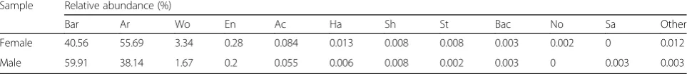
Table 2The relative abundances of bacteria at the genus level inMelophagus ovinus.“Other”indicates the sum of relative genus abundance for the genera excluding the top 11
Sample Relative abundance (%)
Bar Ar Wo En Ac Ha Sh St Bac No Sa Other
Female 40.56 55.69 3.34 0.28 0.084 0.013 0.008 0.008 0.003 0.002 0 0.012
Male 59.91 38.14 1.67 0.2 0.055 0.006 0.008 0.002 0.003 0 0.003 0.003
Abbreviations: Bar Bartonella,Ar Arsenophonus,Wo Wolbachia,En Enterobacter,Ac Acinetobacter,Ha Halomonas,Sh Shewanella,St Staphylococcus,Bac Bacillus,No Novosphingobium,Sa Salinicoccus
[image:5.595.57.539.663.715.2]present study, Acinetobacter was found in high
abundance in the midgut ofM. ovinus, though we could
not identify the species.
Rickettsiais an obligate, intracellular, parasitic bacter-ium that is transmitted mainly through arthropods. In
previous studies, Rickettsia was detected in M. ovinus
collected in Hungary, with a prevalence of 1.67% (1/60) [13]. Recently, high prevalence rates (12.63%, 12/95) for R. raoultii and R. slovaca were reported for M. ovinus from Taklimakan Desert in China [8]. However, two stud-ies reported a failure to detectRickettsiaspp.inM. ovinus collected in Ethiopia and the Czech Republic [18, 47]. Using next-generation sequencing, we similarly were
un-able to detect Rickettsia in two groups ofM. ovinus
col-lected from Jiuquan in Gansu Province, China. This
finding suggests that the capacity of M. ovinus to carry
Rickettsiamay depend on the geographical region.
Conclusions
Bartonella, Arsenophonus and Wolbachia were domin-ant bacterial genera in the midgut of fully engorged
female and male M. ovinus. Although detected,
Entero-bacter, Acinetobacter, Halomonas, Shewanella, Bacillus
and Staphylococcus showed low abundances.
Import-antly, this is the first report of Arsenophonus,
Wolba-chia,Enterobacter,Halomonas,Shewanella, Bacillusand Staphylococcusin the midgut ofM. ovinus.
Abbreviations
OTUs:Operational taxonomic units
Acknowledgements
Not applicable.
Funding
This research was financially supported by a grant from the National Natural Science Foundation of China (No. 31372431), the Youth Science Foundation of Orient Science and Technology College of Hunan Agricultural University (No. 16QNZ13).
Availability of data and materials
The raw tags have been deposited in Sequence Read Archive (SRA) of NCBI under BioProject accession number PRJNA381251. The individual run files received the accession numbers SRR5417208 and SRR5417209.
Authors’contributions
DDY and CTY conceived and designed the study. DDY performed the laboratory analyses and wrote the manuscript. DDY, LGH and WYQ critically revised the manuscript. All authors read and approved the final manuscript.
Ethics approval
This study was approved by the Animal Ethics Committee of Hunan Agricultural University (No. 43321503).
Consent for publication
Not applicable.
Competing interests
The authors declare that they have no competing interests.
Publisher’s Note
Springer Nature remains neutral with regard to jurisdictional claims in published maps and institutional affiliations.
Received: 18 May 2017 Accepted: 3 August 2017
References
1. Gibson W, Pilkington JG, Pemberton JM.Trypanosoma melophagiumfrom the sheep kedMelophagus ovinuson the island of St Kilda. Parasitology. 2010;137:1799–804.
2. Kumsa B, Parola P, Raoult D, Socolovschi C.Bartonella melophagiin
Melophagus ovinus(sheep ked) collected from sheep in northern Oromia, Ethiopia. Comp Immunol Microbiol Infect Dis. 2014;37:69–76.
3. Bequaert J. A monograph of theMelophaginae, or ked-flies, of sheep, goats, deer and antelopes (Diptera, Hippoboscidae). Entomol Am. 1942;22:1–220. 4. Izdebska JN. European bison arthropod parasites from closed polish
breeding facilities. Acta Parasitol. 2001;46:135–7.
5. Tetley JH. The sheep ked,Melophagus ovinusL. I Dissemination potential. Parasitology. 1958;48:353–63.
6. Lassnig H, Prosl H, Hinterdorfer F. Parasites of the red fox (Vulpes vulpes) in Styria. Wien Tierarztl Monat. 1998;85:116–22.
7. Small RW. A review ofMelophagus ovinus(L.), the sheep ked. Vet Parasitol. 2005;130:141–55.
8. Liu D, Wang YZ, Zhang H, Liu ZQ, Wureli HZ, Wang SW, et al. First report of
Rickettsia raoultiiandR. slovacainMelophagus ovinus, the sheep ked. Parasit Vectors. 2016;9:600.
9. Soulsby EJL. Helminths, arthropods and protozoa of domesticated animals. Seventh ed. Philadelphia: Lea and Febiger; 1982.
10. Soulsby EJL. Helminths, arthropods and protozoa of domesticated animals (sixth edition of Mönnig’s Veterinary Helminthology and Entomology). Can Vet J. 1969;10:223.
11. Nelson WA, Slen SB. Weight gains and wool growth in sheep infested with the sheep kedMelophagus ovinus. Exp Parasitol. 1968;22:223–6.
12. Martinkovic F, Matanovic K, Rodrigues AC, Garcia HA, Teixeira MM.
Trypanosoma(Megatrypanum)melophagiumin the sheep kedMelophagus ovinusfrom organic farms in Croatia: phylogenetic inferences support restriction to sheep and sheep keds and close relationship with
Trypanosomesfrom other ruminant species. J Eukaryot Microbiol. 2012;59: 134–44.
13. Hornok S, de la Fuente J, Biro N. Fernandez de Mera IG, Meli ML, Elek V, et al. first molecular evidence ofAnaplasma ovisandRickettsiaspp. in keds (Diptera: Hippoboscidae) of sheep and wild ruminants. Vector Borne Zoonotic Dis. 2011;11:1319–21.
14. Chu CY, Jiang BG, Qiu EC, Zhang F, Zuo SQ, Yang H, et al.Borrelia burgdorferi sensu latoin sheep keds (Melophagus ovinus), Tibet. China Vet Microbiol. 2011;149:526–9.
15. Luedke AJ, Jochim MM, Bowne JG. Preliminary bluetongue transmission with the sheep kedMelophagus ovinus(L.). Can J Comp Med Vet Sci. 1965; 29:229–31.
16. Hubalek Z, Cerny V, Mittermayer T, Kilik J, Halouzka J, Juricova Z, et al. Arbovirological survey in Silica plateau area, Roznava District, Czechoslovakia. J Hyg Epidemiol Microbiol Immunol. 1986;30:87–98. 17. Nelder MP, Lloyd JE, Loftis AD, Reeves WK.Coxiella burnetiiin wild-caught
filth flies. Emerg Infect Dis. 2008;14:1002–4.
18. Rudolf I, Betasova L, Bischof V, Venclikova K, Blazejova H, Mendel J, et al. Molecular survey of arthropod-borne pathogens in sheep keds (Melophagus ovinus), Central Europe. Parasitol Res. 2016;115:3679–82.
19. Degnan PH, Ochman H. Illumina-based analysis of microbial community diversity. ISME J. 2012;6:183–94.
20. Caporaso JG, Lauber CL, Walters WA, Berg-Lyons D, Huntley J, Fierer N, et al. Ultra-high-throughput microbial community analysis on the Illumina HiSeq and MiSeq platforms. ISME J. 2012;6:1621–4.
21. Ahn J, Yang L, Paster BJ, Ganly I, Morris L, Pei Z, et al. Oral microbiome profiles: 16S rRNA pyrosequencing and microarray assay comparison. PLoS One. 2011;6:e22788.
22. MagočT, Salzberg SL. FLASH: fast length adjustment of short reads to improve genome assemblies. Bioinformatics. 2011;27:2957–63.
24. Edgar RC. UPARSE: highly accurate OTU sequences from microbial amplicon reads. Nat Methods. 2013;10:996–8.
25. Edgar RC. MUSCLE: multiple sequence alignment with high accuracy and high throughput. Nucleic Acids Res. 2004;32:1792–7.
26. Kaiser PO, Riess T, O'Rourke F, Linke D, Kempf VA.Bartonellaspp.: throwing light on uncommon human infections. Int J Med Microbiol. 2011;301:7–15. 27. Breitschwerdt EB, Maggi RG, Chomel BB, Lappin MR. Bartonellosis: an
emerging infectious disease of zoonotic importance to animals and human beings. J Vet Emerg Crit Care (San Antonio). 2010;20:8–30.
28. Li XL, Yin CH. Recognition ofBartonellainfection. J Pathog Biol. 2012;7:872–5. 29. Angelakis E, Pulcini C, Waton J, Imbert P, Socolovschi C, Edouard S, et al.
Scalp eschar and neck lymphadenopathy caused byBartonellahenselae after tick bite. Clin Infect Dis. 2010;50:549–51.
30. Novakova E, Hypsa V, Moran NA.Arsenophonus, an emerging clade of intracellular symbionts with a broad host distribution. BMC Microbiol. 2009; 9:143.
31. Duron O, Bouchon D, Boutin S, Bellamy L, Zhou LQ, Engelstädter J, et al. The diversity of reproductive parasites among arthropods:Wolbachiado not walk alone. BMC Biol. 2008;6:27.
32. Gherna RL, Werren JH, Weisburg W, Cote R, Woese CR, Mandelco L, et al. Notes:Arsenophonus nasoniaegen. nov., sp. nov., the causative agent of the son-killer trait in the parasitic waspNasonia vitripennis. Int J Syst Bacteriol. 1991;41:563–5.
33. Kirkness EF, Haas BJ, Sun W, Braig HR, Perotti MA, Clark JM, et al. Genome sequences of the human body louse and its primary endosymbiont provide insights into the permanent parasitic lifestyle. Proc Natl Acad Sci USA. 2010; 107:12168–73.
34. O'Neill SL, Giordano R, Colbert AM, Karr TL, Robertson HM. 16S rRNA phylogenetic analysis of the bacterial endosymbionts associated with cytoplasmic incompatibility in insects. Proc Natl Acad Sci USA. 1992;89: 2699–702.
35. Juchault P, Frelon M, Bouchon D, Rigaud T. New evidences of feminizing bacteria in terrestrial isopods: evolutionary implications. C R Acad des Sci III-Vie. 1994;317:225–30.
36. Bandi C, Anderson TJ, Genchi C, Blaxter ML. Phylogeny ofWolbachiain filarial nematodes. Proc Biol Sci. 1998;265:2407–13.
37. Breeuwer JA, Stouthamer R, Barns SM, Pelletier DA, Weisburg WG, Werren JH. Phylogeny of cytoplasmic incompatibility microorganisms in the parasitoid wasp genusNasonia(hymenoptera: Pteromalidae) based on 16S ribosomal DNA sequences. Insect Mol Biol. 1992;1:25–36.
38. Cordaux R, Michel-Salzat A, Frelon-Raimond M, Rigaud T, Bouchon D. Evidence for a new feminizingWolbachiastrain in the isopod
Armadillidium vulgare: evolutionary implications. Heredity (Edinb). 2004;93: 78–84.
39. Dong P, Wang JJ. Reproductive manipulation ofWolbachiato its hosts. Chin Bull Entomol. 2006;43:288–94.
40. Werren JH, Windsor D, Guo L. Distribution ofWolbachiaamong Neotropical arthropods. Proc R Soc Lond B Biol Sci. 1995;262:197–204.
41. Kose H, Karr TL. Organization ofWolbachia pipientisin theDrosophila
fertilized egg and embryo revealed by an anti-Wolbachiamonoclonal antibody. Mech Dev. 1995;51:275–88.
42. Dobson SL, Bourtzis K, Braig HR, Jones BF, Zhou W, Rousset F, et al.
Wolbachiainfections are distributed throughout insect somatic and germ line tissues. Insect Biochem Mol Biol. 1999;29:153–60.
43. Zordan S, Prenger-Berninghoff E, Weiss R, van der Reijden T, van den Broek P, Baljer G, et al. Multidrug-resistantAcinetobacter baumanniiin veterinary clinics, Germany. Emerg Infect Dis. 2011;17:1751–4.
44. Peleg AY, Seifert H, Paterson DL.Acinetobacter baumannii: emergence of a successful pathogen. Clin Microbiol Rev. 2008;21:538–82.
45. Turton JF, Shah J, Ozongwu C, Pike R. Incidence ofAcinetobacterspecies other thanA. baumanniiamong clinical isolates ofAcinetobacter: evidence for emerging species. J Clin Microbiol. 2010;48:1445–9.
46. Kempf M, Rolain JM, Diatta G, Azza S, Samb B, Mediannikov O, et al. Carbapenem resistance andAcinetobacter baumanniiin Senegal: the paradigm of a common phenomenon in natural reservoirs. PLoS One. 2012; 7:e39495.
47. Kumsa B, Socolovschi C, Parola P, Rolain JM, Raoult D. Molecular detection ofAcinetobacterspecies in lice and keds of domestic animals in Oromia regional state, Ethiopia. PLoS One. 2012;7:e52377.
• We accept pre-submission inquiries
• Our selector tool helps you to find the most relevant journal
• We provide round the clock customer support
• Convenient online submission
• Thorough peer review
• Inclusion in PubMed and all major indexing services
• Maximum visibility for your research
Submit your manuscript at www.biomedcentral.com/submit