R E S E A R C H
Open Access
Toxoplasma gondii
prevalence in food animals
and rodents in different regions of China:
isolation, genotyping and mouse pathogenicity
Lin Wang
1,2†, Hua-Wei Cheng
3,4†, Kai-Quan Huang
1, Yuan-Hong Xu
5, Yong-Nian Li
6, Jian Du
2, Li Yu
2, Qing-Li Luo
2,
Wei Wei
4, Ling Jiang
3,4*and Ji-Long Shen
2*Abstract
Background:Recent studies ofToxoplasma gondiiisolates from animals in different regions of China have shown a limited genetic diversity and type China 1 was the dominant genotype ofT. gondiiprevalent in Chinese animals. However, little has been known concerning the isolation and genotyping ofT. gondiicirculating in chickens, pigs and rodents in China. The aim of the study was to characterize samples ofT. gondiiisolates obtained from naturally infected cats, pigs and free-range chickens slaughtered for human consumption in China.
Methods:In the present study, brain tissues of 77 animals collected from different areas of China, including 24 free-range chickens (Gallus domesticus) , 13 voles (Rattus flavipectus), 23 pigs and 17 cats, were bioassayed in mice and viableT. gondiiwere isolated from the brains of eleven. These elevenT. gondiiisolates were maintained in Kunming (KM) outbred mice and DNA isolated from tissues of infected mice was characterized using 11 PCR-restriction fragment length polymorphism (PCR-RFLP) markers: SAG1, SAG2, SAG3, BTUB, GRA6, c22-8, c29-2, L358, PK1, Apico, and CS3. Moreover, to determine mouse virulence of China 1 lineage of parasites, a TgCtgy5 genotype isolate was selected randomly and assessed in KM mice with different inoculation doses.
Results:Results of genotyping revealed that ten isolates were type China 1 (ToxoDB PCR-RFLP genotype #9), and TgCksz1 was a new genotype that was reported for the first time designated here as ToxoDB PCR-RFLP #225. No clonal types I, II and III lineages were found. DNA sequencing of four introns (EF1, HP2, UPRT1 and UPRT7) and two genes (GRA6 and GRA7) from representative isolates confirmed the results of PCR-RFLP genotyping. The TgCtgy5 isolate was highly virulent in KM mice; all infected mice died of acute toxoplasmosis, irrespective of the inoculation dose. The results indicate that mouse virulent isolates ofT. gondiiare predominantly circulating in cats in China.
Conclusions:T. gondiiisolated from chickens, pigs, cats and rodents in different locations in China were genotyped and the results reconfirmed the limited diversity ofT. gondiiin China and showed that type China 1 lineage was dominant in this country.
Keywords:Toxoplasma gondii, Genotyping, Virulence, PCR-RFLP, Type China 1
* Correspondence:[email protected];[email protected] †Equal contributors
3Department of Pharmacy, the Affiliated Provincial Hospital of Anhui Medical University, Hefei, Anhui Province 230001, People’s Republic of China 2Department of Parasitology, Provincial Laboratory of Microbiology & Parasitology and the Key Laboratory of Zoonoses Anhui, Anhui Medical University, Hefei, Anhui Province 230032, People’s Republic of China Full list of author information is available at the end of the article
Background
Toxoplasma gondii is an important water- and food-borne obligate intracellular parasite, which infects al-most all homoeothermic animals including humans, birds and domestic animals [1]. It has been estimated that up to 30% of the human population worldwide and 8% of population in China is chronically infected [2-4]. Felids are considered as the key in the transmission ofT. gondii to humans and other animals because they are the only definitive hosts that excrete the environmentally resistant oocysts through defecation. Humans become infected postnatally by ingesting tissue cysts from raw or undercooked infected meat, or by consuming food or drink contaminated with T. gondii oocysts. However, only a small percentage of exposed adult humans or ani-mals develop clinical disease. It is not clear why some hosts become ill with toxoplasmosis whereas most re-main asymptomatic. Recently, attention has been fo-cused on the genetic diversity among T. gondii isolates from apparently healthy and sick hosts. Severe cases of toxoplasmosis that have been reported in immunocom-petent individuals are considered to be due to infection with atypical T. gondii isolates [5-7]. Type II strains are the most prevalent in humans and animals in Europe [8].
Globally, genetic diversity of T. gondii consists of six major clades originating from a small number of distinct ancestral lineages [8]. The clonal population structure with three major types I, II and III is more frequently observed in North America and Europe [9]. These three genotypes differ in their virulence and/or pathogenicity to mice. Type I strains are highly virulent, whereas type II and III strains are intermediately or non virulent. Moreover, infections with the atypical types A and X were also observed in wildlife in North America and have recently been described as members of a fourth clonal lineage, designated Haplogroup 12 [10]. However, subsequent studies using multilocus markers have re-vealed a greater genetic diversity of T. gondii, particu-larly isolates from humans and animals in South America [11,12]. These isolates have been historically considered as atypical in order to differentiate them from the dominantly described archetypes. Recent stud-ies of T. gondii in humans and animals in Africa sug-gested the dominance of type II, III, Africa 1 and Africa 3 [13,14]. Some genotypes of T. gondii isolates from humans and cats in China have been previously reported, and the type China 1 (also known as ToxoDB#9) is widespread and likely the major lineage in mainland China [15-18].
However, the data of T. gondii isolates from food ani-mals, such as chickens and pigs in China are limited. Food animals are the main meat source for human con-sumption in China, and risk factor analysis indicated
that 30 to 63% of human infections can be attributed to the consumption of undercooked meat [19]. Serological and parasitological surveys in China indicate a high prevalence of infection of these animals [20,21], and a vi-able T. gondii isolate was isolated from tissues of pigs [22]. T. gondiiinfection in food animals continues to be a significant food safety problem, and has become to a major concern for Chinese consumers over the last dec-ade. Additionally, toxoplasmosis also causes considerable economical loss to the animal husbandry industry world-wide due to increased mortality, abortion, and medical costs [23].
In the present study, we report the isolation and geno-typing of T. gondiiisolates from naturally infected cats, pigs, voles and free-range chickens in central and south-western China using 11 polymerase chain reaction-restriction fragment length polymorphism (PCR-RFLP) markers. Additionally, the virulence of the isolates was determined in a mouse model. These results provide a new insight on the distribution of T. gondii genotypes and onT. gondiidiversity in this country.
Methods Ethics statement
All animals were treated in strict accordance to the guidelines for the Laboratory Animal Use and Care from Chinese CDC and the Rules for Medical laboratory Ani-mals (1998) from Ministry of Health, China. The proto-cols were approved by the Institutional Review Board (IRB) of the Institute of Biomedicine at Anhui Medical University. All efforts were made to minimize animal suffering during the course of these studies.
Animal samples and bioassay in mice
Experimental samples (brain tissues) were obtained from 24 free-range chickens (from Anhui province), 13 voles (from Hubei province), 23 pigs and 17 cats (from Guizhou province). To avoid any chance of cross-contamination, each sample was first separated from the body and placed into a labeled sterile plastic bag, and then the bag was placed in a cooler with ice packs to maintain the temperature at 4°C and taken to the labora-tory. Samples were transported by train within 24 h of collection to the laboratory for T. gondii examination and isolation. All animals were anesthetized before being sacrificed.
were killed on day 45 postinoculation and brains of all mice were microscopically examined for tissue cysts as a squash preparation as described [25]. The inoculated mice were considered infected with T. gondii when stages of the parasite (tachyzoites and/or tissue cysts) were demonstrated in their tissues. T. gondii isolates were cryopreserved in liquid nitrogen for future studies using homogenates of brains from infected mice, or vi-able tachyzoites from intraperitoneal fluids.
DNA extraction and genotyping
DNA was extracted from ascitic fluids or tissues of all infected mice using QIAamp DNA Mini kit (Qiagen, Hilden, Germany) according to the manufacturer’s in-structions. The purified DNA was dissolved in 20 μl of double-distilled water and stored at−20°C for multilocus genotyping studies. A negative control for DNA extrac-tion was included for each group of five samples.
Genotyping of T. gondii isolates was performed using multilocus PCR-RFLP with 11 genetic markers as previ-ously described: SAG1, SAG2, SAG3, BTUB, GRA6, c22-8, c29-2, L358, PK1, Apico [26,27] and marker CS3 [28]. Reference strains of T. gondii were also used in genotyping, including type I (GT1), type II (PTG), type III (CTG) and other strains (TgCgCa1, MAS, TgCatBr5, TgCatBr64, TgRsCr1). Briefly, the target DNA sequences were first amplified using a set of mixed external primers in a single reaction. Then multiplex products were 1:1 diluted in water and served as template DNA for nested PCR with internal primers for each marker separately. A known purified genomic DNA ofT. gondii isolate (RH) was used as a positive control and DNA from non-infected mice were included as negative con-trols. To confirm successful amplification, 5 μl of the nested PCR products was run on a 1% agarose gel containing ethidium bromide prior to purification and digestion. The remaining products were purified using AxyPrepTMPCR Cleanup Kit (Axygen, Union, USA) and digested with appropriate restriction endonucleases [29]. The restriction fragments were run by electrophoresis through a 2.5% ethidium bromide stained agarose gel for all markers except Apico, which required a 3% gel, and then visualized under UV light. A low DNA (25–700 bp) ladder (Ferments, Vilnius, Lithuania) was employed as size standard. The data were analyzed using ToxoDB (www.toxodb.org) database and compared with the ref-erence strain profiles [30].
SAG3 marker sequencing and phylogenetic analysis ofT. gondiiisolates
The nested PCR products for the marker SAG3 from T. gondii isolate TgCksz1 were sequenced in both directions using the internalT. gondiiprimers on an au-tomated sequencer (Applied Biosystems, USA), as
described previously [31]. Nucleotide sequences were analyzed and aligned using BioEdit Sequence Align-ment Editor [16]. For additional comparison, sequences fromT. gondiiRH strain (GenBank: JX218225),T. gondii PTG strain (GenBank: JX218226), and T. gondii CTG strain (GenBank: JX218227) available in the GenBank database (http://blast.ncbi.nlm.nih.gov/Blast.cgi) were also inputted.
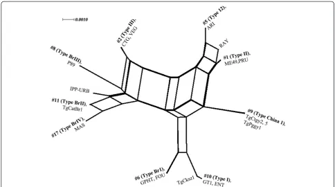
To further characterize these isolates, three isolates (TgCtgy2, TgCtgy5 and TgPggy1) were selected ran-domly and sequences were generated at four introns (EF1, HP2, UPRT1 and UPRT7) and two dense granule proteins (GRA6 and GRA7) as described previously method [10]. The evolutionary history was inferred using a reticulated network by SplitsTree4 [32], combining the RFLP genotype results from this paper and predominant genotypes described to date in South America, Europe and North America.
T. gondiiisolate virulence in mice
To determine the pathogenicity ofT. gondiiisolates with type China 1 widespread in China, TgCtgy5 was selected randomly to study its virulence phenotype in mice. For this, tachyzoites were collected from peritoneal cavities of infected mice and purified as described [16]. Subse-quently, parasites were diluted in phosphate buffered sa-line (PBS) and groups of ten five-week-old KM female mice were inoculated intraperitoneally with 101, 102, 103, and 104tachyzoites, in a final volume of 200μl PBS (per injection), as defined previously [33]. Five animals inoculated intraperitoneally with PBS were maintained as negative controls. The amount of tachyzoites was de-termined in a haemocytometer counting chamber. Mice mortality and morbidity was recorded daily for 45 d after infection, and survivors were killed under anesthesia and brains were examined for tissue cysts described as above. Meanwhile, the rest of the brains were tested for 529 bp repetitive fragment of T. gondii DNA by PCR using the primer pairs TOX4/TOX5 [34].
Results
Multilocus PCR-RFLP genotyping ofT. gondiiisolates
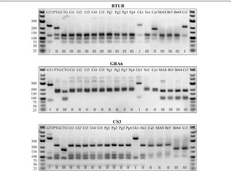
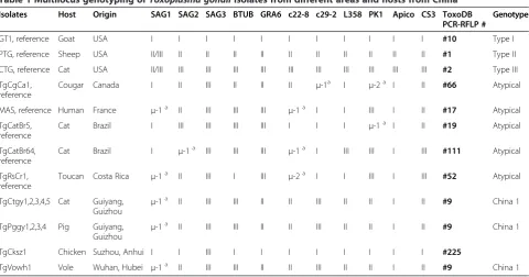
The present results showed that 10 isolates distributed in two provinces were grouped into one genotype (type China 1) based on the 11 markers analyzed. The geno-type presented geno-type II patterns at SAG2, GRA6, L358, PK1, c22-8 and CS3, but c29-2, SAG3, BTUB loci displayed a type III pattern and type I at the Apico locus. This genotype has already been found in three other hosts (cats, pigs and humans) but in voles for the first time from China. Type China 1 has been described not only in isolates from eastern China (Shandong, Jiangsu, Anhui and Zhejiang) but also in isolates from southern China (Guangdong), Central China (Henan and Hubei), northwest (Gansu) and southwest (Yunnan) of China. Therefore, the genotype is most likely a common geno-type circulating in mainland China.
The remaining isolate (TgCksz1) was a variant of the archetypal type I, as all alleles observed were of type I except for the SAG3 locus where an allele of type III was observed. The SAG3 fragment from GT1 strain
(type I) contained two restriction sites for the enzyme NciI, whereas the homologous PCR products from the CTG strain (type III) consisted of one restriction site. The nested PCR product for the marker SAG3 from PTG (type II) was not cleaved by NciI. The RFLP ana-lysis of the SAG3 fragment from TgCksz1 revealed an identical pattern with the type III lineages (Table 1). The data were designated here as genotype #225. Further-more, we determined the genotype by sequencing of a SAG3 gene of T. gondii TgCksz1, reconfirmed that the isolate contained a type III sequence without evidence of any additional mutations (Figure 2). The genotype was new and reported for the first time. This is the first re-port of genetic typing ofT. gondiiisolates from chickens and voles in China.
[image:4.595.64.538.88.439.2]The phylogenetic network of the representative isolates of T. gondii obtained from chickens, pigs and cats, to-gether with previously published data were compared using SplitsTree4 software [32], with taxa positioned in
the typical star-like network (Figure 3). A cluster analysis of the data showed that isolates TgCtgy2, 5 and TgPggy1 formed a single group, and a few major clonal lineages of T. gondiidominant in different geographical regions.
Virulence ofT. gondiiisolates with type China 1
Thein vivovirulence of the T. gondiiisolate TgCtgy5 in KM mice was determined using different tachyzoite
[image:5.595.58.539.94.346.2]doses. Mortality data after inoculation with tachyzoites are shown in Figure 4. All mice inoculated with tachyzoites died of toxoplasmosis between 5 d and 15 d post-infection, irrespective of the inoculation dose, indi-cating that the isolate was virulent in mice. The tachyzoites were found in peritoneal fluid of all dead mice, and the day of death was correlated with the in-oculum dose. This result indicates that highly virulentT.
Table 1 Multilocus genotyping ofToxoplasma gondiiisolates from different areas and hosts from China
Isolates Host Origin SAG1 SAG2 SAG3 BTUB GRA6 c22-8 c29-2 L358 PK1 Apico CS3 ToxoDB
PCR-RFLP #
Genotype
GT1, reference Goat USA I I I I I I I I I I I #10 Type I
PTG, reference Sheep USA II/III II II II II II II II II II II #1 Type II
CTG, reference Cat USA II/III III III III III III III III III III III #2 Type III
TgCgCa1, reference
Cougar Canada Ι II III II II II μ-1a Ι μ-2a Ι II #66 Atypical
MAS, reference Human France μ-1a II III III III μ-1a Ι Ι III Ι II #17 Atypical
TgCatBr5, reference
Cat Brazil Ι III III III III Ι Ι Ι μ-1a Ι II #19 Atypical
TgCatBr64, reference
Cat Brazil Ι μ-1a III III III μ-1a Ι III III Ι III #111 Atypical
TgRsCr1, reference
Toucan Costa Rica μ-1a II III Ι III μ-2a Ι Ι III Ι III #52 Atypical
TgCtgy1,2,3,4,5 Cat Guiyang, Guizhou
μ-1a II III III II II III II II Ι II #9 China 1
TgPggy1,2,3,4 Pig Guiyang,
Guizhou μ
-1a II III III II II III II II Ι II #9 China 1
TgCksz1 Chicken Suzhou, Anhui I I III I I I I I I Ι I #225
TgVowh1 Vole Wuhan, Hubei μ-1a II III III II II III II II Ι II #9 China 1
a
u-1 and u-2 are new alleles that are different from the clonal type I, II and III alleles.
[image:5.595.57.540.533.705.2]gondiiisolates are circulating in cats and they may cause more severe toxoplasmosis when spreading into human population.
Discussion
We investigated the genetic diversity ofT. gondiiisolates from chickens, pigs, cats and voles from central and southwestern China. Among food animals, chickens and pigs are considered the most important reservoirs forT. gondii transmission and the isolation of T. gondii from chickens and pigs is a public health concern as human
[image:6.595.55.540.88.358.2]consumption of these meats continues to increase. The detection and identification of T. gondii from chickens, pigs and rodents facilitate our understanding of the epi-demiology, population structure and virulence of T. gondii. Worldwide seroprevalence of T. gondii infection in chickens and pigs are summarized by Dubey [35,36], depending on the source of chickens and pigs and the sensitivity of diagnostic tests. In China, the seropreva-lence of T. gondii infection in free-range chickens from 13 provinces/municipalities was 6.7%-47.3% [20], and that of pigs in most studies ranged between 12.0% and
Figure 3Phylogenetic network analysis with representatives ofT. gondiigenotypes available in ToxoDB database.Genotype number (#) and the representative strains are listed for each taxonomic branch.
[image:6.595.58.540.546.694.2]53.4% [19,21,37], no genotyping data have been presented so far. These survey results indicated that in-fection withT. gondiiin chickens and pigs is widespread in China, and that chicken and pork may be an import-ant source for human infection withT. gondii.
Genetic diversity of T. gondii isolates has been an in-teresting and important research topic. In the present study, we isolated and completely genotyped 11T. gondii isolates from chickens, pigs, cats and voles in eastern, southwest and central China, respectively. Analysis of the multilocus PCR-RFLP revealed 2 genotypes, one was identified in ten isolates (type China 1), while the other is represented by only one isolate and was described for the first time in the literature (Table 1). Our results indi-cated a limited diversity of T. gondii isolates from the animal population in China, a similar result as reported by other authors in studies of parasites isolated from dif-ferent regions and host species [16,17,38,39]. All previ-ous RFLP genotyping data and the present experiments ofT. gondiiisolates (totally 119) from China are summa-rized in Table 2. The compiled results indicated that type II and type III are rare in China. However, the type China 1 and type I accounted for 73.9 and 10.9%, re-spectively. The genotype China 1 has already been de-scribed in several hosts such as pigs, cats and humans, but this is the first report of this genotype in voles. Among intermediate hosts, infected rodents are consid-ered the most important sources of T. gondii infection for cats. The type China 1 lineage was found for vole isolates in the present study, reconfirming that this lineage is the most common and widespread in different hosts and geographical locations in China. Overall, our current study is in agreement with previous report that T. gondiitype China 1 is the dominating genotype in an-imals as well as humans within China [16]. The limited
data on T. gondii isolates detected in food animals and wildlife, like pigs or voles, seem to reflect the dominant T. gondiitype China 1 which is commonly found in live-stock, poultry and humans. Interestingly, this genotype has also been identified inT. gondiiisolates of dogs from Sri Lanka and Colombia [40,41], chickens from Brazil [27] and of sheep from the United States [42], suggesting that such a genotype might be widespread from Asia to North and South America. Further analysis of these T. gondii isolates at the DNA sequence level may help us understand the origin and migration of this parasite among different continents.
[image:7.595.56.543.542.733.2]There is scarce information concerning the isolation and genotyping of T. gondii isolates from chickens in China. Because free-range chickens become infected mainly by feeding from ground/soil contaminated with oocysts, the prevalence ofT. gondii in this host has been widely used as an indicator of the strains prevalent in the environment. In addition, tissues of infected chick-ens are considered a good source of infection for cats, humans and other animals. In the present investigation, the isolate TgCksz1 (genotype ToxoDB#225) isolated from a chicken in Suzhou city is similar to the genotype of isolate TgCkBr136 (genotype ToxoDB#41), which was collected from chickens in the state of Rondönia in Brazil and identified by Dubey [46]. The genotype ToxoDB#225 was a variant of the type I with a type III genotype at the SAG3 loci. Meanwhile, the genotype ToxoDB#41 showed type II pattern at GRA6 and the remaining 9 loci types were identical to type #225, suggesting that they are phylogenetically related. In addition, the genotype #41 was also identified from new-borns in the Minas Gerais state in Brazil [47]. These re-sults suggest that whether this isolate TgCksz1 has been imported into China or is endemic remains unknown
Table 2 Genetic PCR-RFLP types ofT. gondiiin animals and humans in China
Host No. of
isolates
ToxoDB PCR-RFLP genotypes References
#10 (Type I) #1 (Type II) #2 (Type III) #3 (Type II-variant)
#9 (China 1) #18 #204 #205 #213 #225
Cats 72 1 0 0 0 65 2 0 4 0 0 [15,16,38] and
this study
Pigs 33 12 0 0 0 19 0 0 0 2 0 [22,39,43] and
this study
Sheep 1 0 0 0 1 0 0 0 0 0 0 [17]
Rabbits 1 0 0 1 0 0 0 0 0 0 0 [44]
Wild birds
3 1 0 0 2 0 0 0 0 0 0 [45]
Chickens 1 0 0 0 0 0 0 0 0 0 1 This study
Voles 1 0 0 0 0 1 0 0 0 0 0 This study
Humans 7 2 1 0 0 3 0 1 0 0 0 [16,17]
based on the genetic data. However, as indicated above, genetically similar isolates were observed in distant regions like these should be investigated in future studies. To our knowledge this is the first report of genetic typing ofT. gondiiisolates from chickens from China. None of the isolates displayed a clonal genotype (type I, II and III).
Before the recognition of the three T. gondii geno-types, isolates were phenotypically classified as mouse virulent or avirulent. Phenotypically, type I strains are uniformly lethal to outbred mice and type II and III strains are significantly less virulent [11]. In this study, we observed variability in the mouse virulence of theT. gondii isolates with the genotype China 1, including virulent isolates and intermediately isolates. The TgCtgy5 isolate was lethal to outbred KM mice; all infected mice died of acute toxoplasmosis, regardless of the dose used. However, all mice inoculated with 1 × 10³tachyzoites of the isolate TgCtgy2 (type China 1) died but two survived acute infection and cysts were found in the brain tissues of the survivals. The differences of biological features among the isolates sharing the common genotype must not be neglected and it may be possible that the genetic markers used in this study are incapable of reflecting possible phenotypic differences between the isolates under question [48]. Also, in the previous report, we showed that mouse virulence and genetic type are not strictly correlated because two T. gondii isolates (TgCtwh3 and TgCtwh6) share the common genotype but have markedly different mouse virulence, including CS3 locus (type II). Similar phenomenon could also be found in other studies on type China 1 isolates [38]. The marker CS3, located on chromosome VIIa of T. gondii, was previously reported to be linked with the acute viru-lence in mice of T. gondii and was also used to deter-mine its association to parasite virulence in mice [49]. Several studies indicated that isolates with the alleles I and II at the CS3 locus are strongly associated with viru-lence in mice [28,31]. The Chinese isolate of T. gondii TgCtwh6, however, has a low virulence to mice but shares the allele II at the CS3 locus. These results sug-gest that better genetic markers are needed to correlate pathogenicity ofT. gondiiin animals and humans.
Conclusions
The present study is the first report ofT. gondiiisolates collection and genotyping obtained from chickens and voles, enriching the limitedT. gondii genotype database in China. The genetic structure here found in the iso-lates from chickens, pigs, cats and rodents corroborates the findings of previous studies that T. gondiihas a lim-ited diversity in China. In addition, the results provided preliminary data for further approaches of epidemiology ofT. gondii in food animals and wildlife and analysis of population genetics ofT. gondiiorganisms.
Competing interests
The authors declare that they have no competing interests.
Authors’contributions
JLS, LJ, LW and HWC conceived and designed the study, and critically revised the manuscript. LW, HWC, KQH, YHX and YNL performed the experiments, analyzed the data and drafted the manuscript. JD, LY, QLL and WW participated in the analysis and interpretation of data. All authors have read and approved the final manuscript.
Acknowledgments
This work was financially supported by the National Basic Research Program of China (973 Program, No. 2010CB530001) and National Natural Science Foundation of China (No. 81271864, 30801329). The authors would like to thank the technical and support staff of the Key Laboratory of Zoonoses Anhui for their assistance during sample collection. We also thank Associate Professor Chunlei Su at the Department of Microbiology, the University of Tennessee, Knoxville, Tennessee, for kindly providing with the reference strains (type I (GT1), type II (PTG), type III (CTG) and other strains (TgCgCa1, MAS, TgCatBr5, TgCatBr64, and TgRsCr1).
Author details
1Clinical Laboratory, the First Affiliated Hospital of Anhui University of Chinese Medicine, Hefei, Anhui Province 230031, People’s Republic of China. 2Department of Parasitology, Provincial Laboratory of Microbiology & Parasitology and the Key Laboratory of Zoonoses Anhui, Anhui Medical University, Hefei, Anhui Province 230032, People’s Republic of China. 3
Department of Pharmacy, the Affiliated Provincial Hospital of Anhui Medical University, Hefei, Anhui Province 230001, People’s Republic of China. 4
Institute of Clinical Pharmacology, Anhui Medical University, Hefei, Anhui Province 230032, People’s Republic of China.5Clinical Laboratory, the First Affiliated Hospital of Anhui Medical University, Hefei, Anhui Province 230022, People’s Republic of China.6Department of Clinical Laboratory, Guiyang Medical College, Guiyang, Guizhou Province 550001, People’s Republic of China.
Received: 13 August 2013 Accepted: 17 September 2013 Published: 21 September 2013
References
1. Dubey JP:Toxoplasmosis of animals and humans.Boca Raton, Florida: CRC Press; 2010:313.
2. Zhou DH, Zhao FR, Huang SY, Xu MJ, Song HQ, Su C, Zhu XQ:Changes in the proteomic profiles of mouse brain after infection with cyst-forming
Toxoplasma gondii.Parasit Vectors2013,6:96.
3. Zhou P, Chen N, Zhang RL, Lin RQ, Zhu XQ:Food-borne parasitic zoonoses in China: perspective for control.Trends Parasitol2008,
24:190–196.
4. Zhou P, Chen Z, Li HL, Zheng H, He S, Lin RQ, Zhu XQ:Toxoplasma gondii
infection in humans in China.Parasit Vectors2011,4:165.
5. Elbez-Rubinstein A, Ajzenberg D, Darde ML, Cohen R, Dumetre A, Yera H, Gondon E, Janaud JC, Thulliez P:Congenital toxoplasmosis and reinfection during pregnancy: case report, strain characterization, experimental model of reinfection, and review.J Infect Dis2009,
199:280–285.
6. Wendte JM, Miller MA, Lambourn DM, Magargal SL, Jessup DA, Grigg ME:
Self-mating in the definitive host potentiates clonal outbreaks of the apicomplexan parasites Sarcocystis neurona andToxoplasma gondii.PLoS Genet2010,6:e1001261.
7. Demar M, Hommel D, Djossou F, Peneau C, Boukhari R, Louvel D, Bourbigot AM, Nasser V, Ajzenberg D, Darde ML, Carme B:Acute toxoplasmoses in immunocompetent patients hospitalized in an intensive care unit in French Guiana.Clin Microbiol Infect2012,18:E221–E231.
8. Su C, Khan A, Zhou P, Majumdar D, Ajzenberg D, Darde ML, Zhu XQ, Ajioka JW, Rosenthal BM, Dubey JP, Sibley LD:Globally diverseToxoplasma gondiiisolates comprise six major clades originating from a small number of distinct ancestral lineages.Proc Natl Acad Sci U S A2012,
109:5844–5849.
9. Howe DK, Sibley LD:Toxoplasma gondiicomprises three clonal lineages: correlation of parasite genotype with human disease.J Infect Dis1995,
10. Khan A, Dubey JP, Su C, Ajioka JW, Rosenthal BM, Sibley LD:Genetic analyses of atypicalToxoplasma gondiistrains reveal a fourth clonal lineage in North America.Int J Parasitol2011,41:645–655.
11. Rajendran C, Su C, Dubey JP:Molecular genotyping ofToxoplasma gondii
from Central and South America revealed high diversity within and between populations.Infect Genet Evol2012,12:359–368.
12. Minot S, Melo MB, Li F, Lu D, Niedelman W, Levine SS, Saeij JP:Admixture and recombination amongToxoplasma gondiilineages explain global genome diversity.Proc Natl Acad Sci U S A2012,109:13458–13463. 13. Mercier A, Devillard S, Ngoubangoye B, Bonnabau H, Banuls AL, Durand P,
Salle B, Ajzenberg D, Darde ML:Additional haplogroups ofToxoplasma gondiiout of Africa: population structure and mouse-virulence of strains from Gabon.PLoS Negl Trop Dis2010,4:e876.
14. Al-Kappany YM, Rajendran C, Abu-Elwafa SA, Hilali M, Su C, Dubey JP:
Genetic diversity ofToxoplasma gondiiisolates in Egyptian feral cats reveals new genotypes.J Parasitol2010,96:1112–1114.
15. Chen ZW, Gao JM, Huo XX, Wang L, Yu L, Halm-Lai F, Xu YH, Song WJ, Hide G, Shen JL, Lun ZR:Genotyping ofToxoplasma gondiiisolates from cats in different geographic regions of China.Vet Parasitol2011,183:166–170. 16. Wang L, Chen H, Liu D, Huo X, Gao J, Song X, Xu X, Huang K, Liu W,
Wang Y,et al:Genotypes and mouse virulence ofToxoplasma gondii
isolates from animals and humans in China.PLoS One2013,8:e53483. 17. Zhou P, Zhang H, Lin RQ, Zhang DL, Song HQ, Su C, Zhu XQ:Genetic
characterization ofToxoplasma gondiiisolates from China.Parasitol Int 2009,58:193–195.
18. Chen J, Li ZY, Zhou DH, Liu GH, Zhu XQ:Genetic diversity among
Toxoplasma gondiistrains from different hosts and geographical regions revealed by sequence analysis of GRA5 gene.Parasit Vectors2012,5:279. 19. Tao Q, Wang Z, Feng H, Fang R, Nie H, Hu M, Zhou Y, Zhao J:
Seroprevalence and risk factors forToxoplasma gondiiinfection on pig farms in central China.J Parasitol2011,97:262–264.
20. Zhao G, Shen B, Xie Q, Xu LX, Yan RF, Song XK, Hassan IA, Li XR:Detection ofToxoplasma gondiiin free-range chickens in China based on circulating antigens and antibodies.Vet Parasitol2012,185:72–77. 21. Liu X, Liu C, Liu Y, Jin H, Zhao Y, Chen J, Yang M, Liu Q:Seroprevalence of
Toxoplasma gondiiinfection in slaughtered pigs and cattle in Liaoning Province, northeastern China.J Parasitol2012,98:440–441.
22. Wang H, Wang T, Luo Q, Huo X, Wang L, Liu T, Xu X, Wang Y, Lu F, Lun Z, et al:Prevalence and genotypes ofToxoplasma gondiiin pork from retail meat stores in Eastern China.Int J Food Microbiol2012,157:393–397. 23. Buxton D, Maley SW, Wright SE, Rodger S, Bartley P, Innes EA:Toxoplasma
gondiiand ovine toxoplasmosis: new aspects of an old story.Vet Parasitol 2007,149:25–28.
24. Dubey JP:Refinement of pepsin digestion method for isolation of
Toxoplasma gondiifrom infected tissues.Vet Parasitol1998,74:75–77. 25. El Behairy AM, Choudhary S, Ferreira LR, Kwok OC, Hilali M, Su C, Dubey JP:
Genetic characterization of viableToxoplasma gondiiisolates from stray dogs from Giza, Egypt.Vet Parasitol2013,193:25–29.
26. Su C, Zhang X, Dubey JP:Genotyping ofToxoplasma gondiiby multilocus PCR-RFLP markers: a high resolution and simple method for
identification of parasites.Int J Parasitol2006,36:841–848.
27. Dubey JP, Sundar N, Gennari SM, Minervino AH, Farias NA, Ruas JL, dos Santos TR, Cavalcante GT, Kwok OC, Su C:Biologic and genetic comparison ofToxoplasma gondiiisolates in free-range chickens from the northern Para state and the southern state Rio Grande do Sul, Brazil revealed highly diverse and distinct parasite populations.Vet Parasitol 2007,143:182–188.
28. Pena HF, Gennari SM, Dubey JP, Su C:Population structure and mouse-virulence ofToxoplasma gondiiin Brazil.Int J Parasitol2008,38:561–569. 29. Khan A, Su C, German M, Storch GA, Clifford DB, Sibley LD:Genotyping of
Toxoplasma gondiistrains from immunocompromised patients reveals high prevalence of type I strains.J Clin Microbiol2005,43:5881–5887. 30. Su C, Shwab EK, Zhou P, Zhu XQ, Dubey JP:Moving towards an integrated
approach to molecular detection and identification ofToxoplasma gondii.Parasitology2010,137:1–11.
31. Pena HF, Vitaliano SN, Beltrame MA, Pereira FE, Gennari SM, Soares RM:
PCR-RFLP genotyping ofToxoplasma gondiifrom chickens from Espirito Santo state, Southeast region, Brazil: new genotypes and a new SAG3 marker allele.Vet Parasitol2013,192:111–117.
32. Huson DH, Bryant D:Application of phylogenetic networks in evolutionary studies.Mol Biol Evol2006,23:254–267.
33. Taylor S, Barragan A, Su C, Fux B, Fentress SJ, Tang K, Beatty WL, Hajj HE, Jerome M, Behnke MS,et al:A secreted serine-threonine kinase determines virulence in the eukaryotic pathogenToxoplasma gondii. Science2006,314:1776–1780.
34. Homan WL, Vercammen M, De Braekeleer J, Verschueren H:Identification of a 200- to 300-fold repetitive 529 bp DNA fragment inToxoplasma gondii, and its use for diagnostic and quantitative PCR.Int J Parasitol 2000,30:69–75.
35. Dubey JP:Toxoplasma gondiiinfections in chickens (Gallus domesticus): prevalence, clinical disease, diagnosis and public health significance. Zoonoses Public Health2010,57:60–73.
36. Dubey JP:Toxoplasmosis in pigs–the last 20 years.Vet Parasitol2009,
164:89–103.
37. Yu HJ, Zhang Z, Liu Z, Qu DF, Zhang DF, Zhang HL, Zhou QJ, Du AF:
Seroprevalence ofToxoplasma gondiiinfection in pigs, in Zhejiang Province, China.J Parasitol2011,97:748–749.
38. Qian W, Wang H, Su C, Shan D, Cui X, Yang N, Lv C, Liu Q:Isolation and characterization ofToxoplasma gondiistrains from stray cats revealed a single genotype in Beijing, China.Vet Parasitol2012,187:408–413. 39. Zhou P, Sun XT, Yin CC, Yang JF, Yuan ZG, Yan HK, Zhu XQ, Zou FC:
Genetic characterization ofToxoplasma gondiiisolates from pigs in southwestern China.J Parasitol2011,97:1193–1195.
40. Dubey JP, Rajapakse RP, Wijesundera RR, Sundar N, Velmurugan GV, Kwok OC, Su C:Prevalence ofToxoplasma gondiiin dogs from Sri Lanka and genetic characterization of the parasite isolates.Vet Parasitol2007,
146:341–346.
41. Dubey JP, Huong LT, Sundar N, Su C:Genetic characterization of
Toxoplasma gondiiisolates in dogs from Vietnam suggests their South American origin.Vet Parasitol2007,146:347–351.
42. Dubey JP, Sundar N, Hill D, Velmurugan GV, Bandini LA, Kwok OC, Majumdar D, Su C:High prevalence and abundant atypical genotypes of
Toxoplasma gondiiisolated from lambs destined for human consumption in the USA.Int J Parasitol2008,38:999–1006.
43. Zhou P, Nie H, Zhang LX, Wang HY, Yin CC, Su C, Zhu XQ, Zhao JL:Genetic characterization ofToxoplasma gondiiisolates from pigs in China. J Parasitol2010,96:1027–1029.
44. Zhou Y, Zhang H, Cao J, Gong H, Zhou J:Isolation and genotyping of
Toxoplasma gondiifrom domestic rabbits in China to reveal the prevalence of type III strains.Vet Parasitol2013,193:270–276. 45. Huang SY, Cong W, Zhou P, Zhou DH, Wu SM, Xu MJ, Zou FC, Song HQ,
Zhu XQ:First report of genotyping ofToxoplasma gondiiisolates from wild birds in China.J Parasitol2012,98:681–682.
46. Dubey JP, Velmurugan GV, Chockalingam A, Pena HF, de Oliveira LN, Leifer CA, Gennari SM, Bahia Oliveira LM, Su C:Genetic diversity ofToxoplasma gondiiisolates from chickens from Brazil.Vet Parasitol2008,157:299–305. 47. Carneiro AC, Andrade GM, Costa JG, Pinheiro BV, Vasconcelos-Santos DV,
Ferreira AM, Su C, Januario JN, Vitor RW:Genetic characterization of
Toxoplasma gondiirevealed highly diverse genotypes for isolates from newborns with congenital toxoplasmosis in southeastern Brazil.J Clin Microbiol2013,51:901–907.
48. Soares RM, Silveira LH, da Silva AV, Ragozo A, Galli S, Lopes EG, Gennari SM, de Jesus Pena HF:Genotyping ofToxoplasma gondiiisolates from free range chickens in the Pantanal area of Brazil.Vet Parasitol2011,
178:29–34.
49. Khan A, Taylor S, Su C, Mackey AJ, Boyle J, Cole R, Glover D, Tang K, Paulsen IT, Berriman M,et al:Composite genome map and recombination parameters derived from three archetypal lineages ofToxoplasma gondii. Nucleic Acids Res2005,33:2980–2992.
doi:10.1186/1756-3305-6-273