RESEARCH
Promoter expression of HERV-K (HML-2)
provirus-derived sequences is related
to LTR sequence variation and polymorphic
transcription factor binding sites
Meagan Montesion
1,4, Zachary H. Williams
1, Ravi P. Subramanian
1,5, Charlotte Kuperwasser
2,3and John M. Coffin
1*Abstract
Background: Increased transcription of the human endogenous retrovirus group HERV-K (HML-2) is often seen dur-ing disease. Although the mechanism of its tissue-specific activation is unclear, research shows that LTR CpG hypo-methylation alone is not sufficient to induce its promoter activity and that the transcriptional milieu of a malignant cell contributes, at least partly, to differential HML-2 expression.
Results: We analyzed the relationship between LTR sequence variation and promoter expression patterns in human breast cancer cell lines, finding them to be positively correlated. In particular, two proviruses (3q12.3 and 11p15.4) displayed increased activity in almost all tumorigenic cell lines sampled. Using a transcription factor binding site pre-diction algorithm, we identified two unique binding sites in each 5′ LTR that appeared to be associated with inducing promoter activity during neoplasia. Genomic analysis of the homologous proviruses in several non-human primates indicated post-integration genetic drift in two transcription factor binding sites, away from the ancestral sequence and towards the active form. Based on the sequences of 2504 individuals from the 1000 Genomes Project, the active form of the 11p15.4 site was found to be polymorphic within the human population, with an allele frequency of 51%, whereas the activating mutation in the 3q12.3 provirus was fixed in humans but not present in the orthologous provi-rus in chimpanzees or gorillas.
Conclusions: These data suggest that stage-specific transcription factors at least partly contribute to LTR promoter activity during transformation and that, in some cases, transcription factor binding site polymorphisms may be responsible for the differential HML-2 expression often seen between individuals.
Keywords: Endogenous retrovirus, HERV-K, HML-2, LTR, Transcription, Tumorigenesis
© The Author(s) 2018. This article is distributed under the terms of the Creative Commons Attribution 4.0 International License (http://creat iveco mmons .org/licen ses/by/4.0/), which permits unrestricted use, distribution, and reproduction in any medium, provided you give appropriate credit to the original author(s) and the source, provide a link to the Creative Commons license, and indicate if changes were made. The Creative Commons Public Domain Dedication waiver (http://creat iveco mmons .org/ publi cdoma in/zero/1.0/) applies to the data made available in this article, unless otherwise stated.
Background
Retroviruses are unique in that they are the only virus family known to exist in both endogenous and exogenous forms [1, 2]. Their integrated DNA sequences, known as proviruses, include at least four genes (gag, pro, pol, and env) flanked by long terminal repeats (LTRs), which
contain all elements necessary to initiate and terminate viral transcription [2, 3]. Genetic transmission of these sequences occurs with germline integration, producing endogenous retroviruses (ERVs). ERVs are inherited in a Mendelian fashion and are subject to natural selection; those with deleterious effects are generally either lost from the population or inactivated by mutation, whereas those with neutral or advantageous effects remain [2, 4]. As a consequence of the accumulation of these elements over time, nearly 8% of the human genome is derived from such viral sequences [5–7].
Open Access
*Correspondence: [email protected]
1 Department of Molecular Biology and Microbiology, Tufts University
School of Medicine, Boston, MA, USA
Once classified with other “junk DNA”, ERVs are now credited with providing genomic plasticity through the use of viral proteins for host functions and alterna-tive regulation of host gene transcription. For example, proviruses contain numerous promoters, splice sites, transcription factor binding sites, and polyadenyla-tion signals, all of which can have significant effects on neighboring host gene expression [2, 8, 9]. Syncytins, fusogenic proteins derived from ERV env sequences, are essential for placenta development and mediate cell fusion to form the syncytiotrophoblast layer [10, 11]. Although ERV expression is usually silenced through epi-genetic and chromatin modification, primarily via CpG methylation [8, 12–14], there are a few known instances of host cell co-option of ERV expression. Recent studies show human endogenous retrovirus (HERV) expression to be increased in human embryonic stem cells (hESCs) and human preimplantation embryos and to play a criti-cal role during embryogenesis through the maintenance of pluripotency and hESC identity [15–19]. Increased expression of HERV proteins was found to be correlated with increased IFITM1 expression, resulting in viral immunoprotection during human embryogenesis [19,
20].
Despite these exceptions, increased HERV activity is largely associated with malignancy, especially cancer. Activation of stem cell-associated retroviruses (SCARs) in human cancer is hypothesized to be associated with increased likelihood of metastasis, immune evasion of cancer cells, and a predictive marker of poor prognosis [21, 22]. Increased cancer-related expression is attribut-able in part to global hypomethylation, a common con-sequence of tumorigenesis, and LTR hypomethylation is widely documented to result in promoter activation [11, 13, 23]. However, in vitro treatment with 5-aza-2′ -deoxycytidine, a DNA methyltransferase inhibitor, shows that LTR hypomethylation alone is not always sufficient to induce promoter activity, suggesting that the proper transcriptional milieu of a cell may also be required [24–
26]. Ubiquitous transcription factors, such as Sp1, Sp3, and YY1, are linked with LTR activity but do not explain the cell-specific expression that is often seen [8, 25, 27].
Expression from HERV-K (HML-2), the most recently integrated and biologically active HERV group, is upreg-ulated in up to 85% of breast cancer samples, although the mechanism of activation is still unclear [28–31]. RNA sequence analysis of cells in an in vitro mammary carcinogenesis model shows that LTR-driven transcrip-tion of HML-2 proviruses is restricted to tumorigenic human mammary epithelial cells (HMECs), suggesting that stage-specific transcription factors appearing dur-ing malignant transformation play a role in LTR activa-tion [32]. The goal of this study was to investigate how
LTR sequence variation among the various HML-2 pro-viruses affects activation of its promoter during HMEC transformation.
Overall, we found the most widespread increase in pro-moter activity during transformation in two proviruses (located at 3q12.3 and 11p15.4). Through a combina-tion of reporter construct assays and RNA-Seq analyses, we identified two transcription factor binding sites on each 5′ LTR that were associated with promoter activity in transformed cells. Further genomic analysis of these proviruses, using data from the 1000 Genomes Pro-ject as well as comparison with homologous proviruses in several other hominoid species, showed that both of these sites had been created by mutations in the 5′ LTR that occurred post viral integration. The 3q12.3 site has become fixed in the human population whereas that at 11p15.4 is polymorphic, with the active form having an allele frequency of 51%. In both cases, these sites have evolved away from the inactive ancestral sequence and towards an active form. These results emphasize the importance of studying HERV transcription at the single provirus and single nucleotide level, as polymorphisms in critical binding sites may be responsible for the differen-tial HML-2 expression often seen between individuals.
Results
Differential HML‑2 promoter expression is correlated with 5′ LTR sequence similarity
The HML-2 5′ LTR contains all elements necessary for driving transcription. Removal of core promoter ele-ments results in reduced promoter activity, suggesting that these sequences are critical for proper LTR-driven expression [9, 25, 33]. Each provirus has accumulated numerous unique mutations over time, suggesting that LTR sequence variation could contribute to differen-tial HML-2 expression, particularly through the altera-tion of transcripaltera-tion factor binding sites. Through a series of dual-luciferase assays, we sought to evaluate whether LTR sequence identity is correlated with simi-lar promoter expression patterns during breast cancer tumorigenesis.
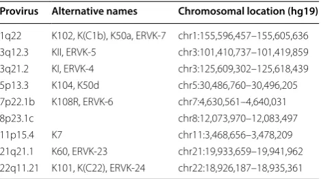
eight proviruses, plus 8p23.1c, a segmental duplication of 11p15.4 [4], were chosen as our loci of interest. The alter-native names and chromosomal locations of these provi-ruses are listed in Table 1.
Phylogenetic analysis of the LTRs from these nine pro-viruses shows that most of them are classified as LTR-HS, the LTR group that contains the youngest proviruses, including ~ 90% of the human-specific integrations (Fig. 1a) [4, 9, 35]. The 5′ LTR sequences from each pro-virus were cloned into pGL4.17[luc2/Neo], a promoter-less firefly luciferase vector, directly upstream of the luc2
gene. The relative promoter activity of these sequences was determined based on luc2 expression and normal-ized against that of an internal control vector, containing a Renilla luciferase gene (Rluc) driven by an SV40 pro-moter (Fig. 1b). A panel of eighteen human cell lines was transiently co-transfected with these vectors. The panel comprised of two immortalized HMEC cell lines, fifteen tumorigenic breast cancer cell lines (representing all three molecular subtypes), and one teratocarcinoma cell line known to produce HML-2 transcripts and retroviral-like particles (RVLPs) at high levels [9, 36]. Characteriza-tion of the cell lines used is shown in Table 2.
Although minimal promoter activity was detected in immortalized HMECs transfected with any of the HML-2 LTR reporter constructs, significant upregula-tion of expression driven by one or more LTRs was seen in 73% (11/15) of the tumorigenic breast cancer cell lines (Fig. 1c). This expression pattern is consistent with previ-ous reports that suggest up to 85% of breast cancer sam-ples have a significant increase in HML-2 activity [29,
31, 37]. Overall, each LTR was significantly expressed in at least one cell line tested but showed differential expression across the panel. Two proviruses (3q12.3 and 11p15.4) were significantly upregulated in nearly all neo-plastic cell lines investigated, whereas others were only upregulated in a select few (Fig. 1d). The highest level
of combined HML-2 expression in a breast cell line was exhibited by T47D (Fig. 1c), a tumorigenic breast cancer cell line known to produce RVLPs under hormonally-stimulated conditions [3, 38, 39]. However, this activity level was only about half that seen in the Tera-1 cells, consistent with our previous report that Tera-1 cells pro-duce markedly higher numbers of HML-2 transcripts than breast cancer cell lines [32].
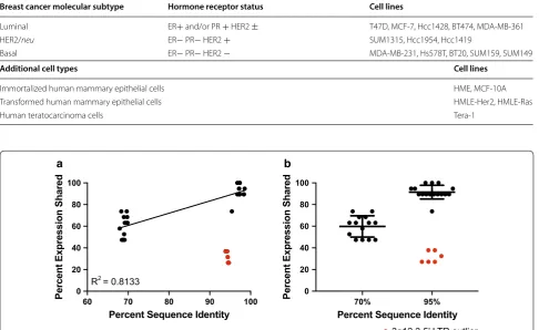
We next sought to determine if LTRs of similar sequence share similar patterns of promoter activity. For this purpose, we created a percent sequence identity matrix, by multiple sequence alignment using Clustal Omega [40], and an HML-2 percent expression similar-ity matrix, determined through pairwise comparisons of significant promoter activity within each cell line tested (Additional file 2). We found the two values to be corre-lated, suggesting that LTRs with high sequence similar-ity are more likely to exhibit significant promoter activsimilar-ity under the transcriptional environment of the same cell line (Fig. 2a). Overall, LTRs with ~ 70% sequence simi-larity shared promoter expression patterns ~ 60% of the time, whereas LTRs with ~ 95% sequence identity shared promoter expression patterns ~ 90% of the time (Fig. 2b). With the exception of the 5′ LTR of 3q12.3 (Fig. 2, red), the sequences clustered into two observable groups. The expression pattern of the 3q12.3 5′ LTR was not similar to any other LTR and instead exhibited unusually high promoter activity levels, with significant promoter expression seen in almost every transformed cell line investigated (Fig. 1d).
Identification of transcription factor binding sites critical for HML‑2 promoter activity during neoplasia
The association between LTR sequence and cell line-spe-cific expression suggests that certain sequence-speline-spe-cific elements, such as transcription factor binding sites, play a large role in determining differential promoter activ-ity. Increased HML-2 expression is largely seen during tumorigenesis and our recent results indicate that LTR-driven transcription does not occur until post-transfor-mation [32]. The following experiments were performed to further investigate the relationship between malignant transformation and expression and to elucidate the spe-cific LTR sequences required.
For this purpose, we focused on three cell lines: HME, HMLE-Her2, and HMLE-Ras. These cells were all derived from the same HMEC population and are therefore iso-genic, differing only by oncogene overexpression. HME cells are non-transformed but immortalized through hTERT (human telomerase reverse transcriptase) overex-pression. The HMLE cells, in addition to being hTERT-immortalized, are transformed through the introduction of SV40 large and small T antigens. HMLE-Her2 and
Table 1 HML-2 proviruses with alternative names
and genomic coordinates
From Subramanian et al. [4] and Montesion et al. [32]
Provirus Alternative names Chromosomal location (hg19)
[image:3.595.57.292.584.715.2]a d
b
c
[image:4.595.59.544.84.578.2]HMLE-Ras differ from one another by their oncogene overexpression, ERBB2 (also known as HER2/neu) and
HRAS, respectively. These cell lines provided the oppor-tunity to investigate how specific differences in the transcriptional environment of the cell can affect LTR expression.
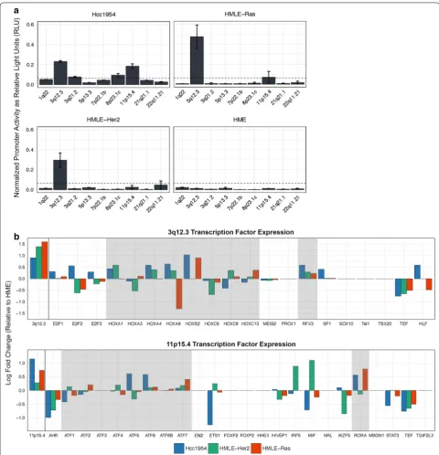
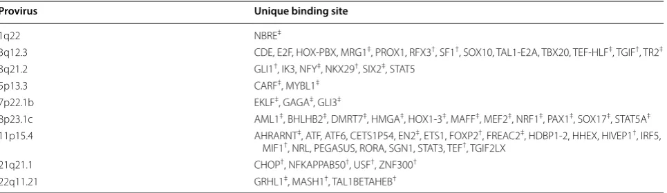
We detected increased promoter activity from 3q12.3 and 11p15.4 in HMLE-Ras cells as well as increased activ-ity from 3q12.3 in HMLE-Her2 cells. The significance of this expression was determined as compared to the HME cell line (Fig. 3a). In effort to explain this pattern, we sought to identify transcription factor binding sites that are unique to each LTR and therefore may be responsible for the selective activation seen of one LTR over another. Using MatInspector, a transcription factor binding site prediction software by Genomatix [41], we found a total of 63 unique sites among the nine LTRs in this study. Of those, 13 were unique to 3q12.3 and 20 were unique to 11p15.4 (Table 3).
The same software was used to create a list of transcrip-tion factors predicted to bind to the unique sites on the 5′ LTRs of 3q12.3 and 11p15.4. In a previous study [32] the expressed RNAs of the HMLE-Ras, HMLE-Her2, and HME cell lines were sequenced, alongside the established human breast cancer cell line Hcc1954, using Illumina MiSeq sequencing. The transcript abundance levels, measured as FPKM, of these transcription factors were compared to assess upregulation of their expression in the tumorigenic cell lines as compared to the non-trans-formed HME control, and related to levels of expression of the proviruses at 3q12.3 and 11p15.4. Overall, we saw a significant increase in expression of transcription fac-tors known to bind to the HOX-PBX and RFX3 sites on the 3q12.3 5′ LTR as well as a significant increase in those known to bind to the ATF and RORA sites on the 11p15.4 5′ LTR (Fig. 3b), implicating these sites and one or more of the upregulated factors in LTR activation dur-ing neoplasia.
Table 2 Characterization of cell lines used for transfection
Breast cancer molecular subtype Hormone receptor status Cell lines
Luminal ER+ and/or PR + HER2 ± T47D, MCF-7, Hcc1428, BT474, MDA-MB-361
HER2/neu ER− PR− HER2 + SUM1315, Hcc1954, Hcc1419
Basal ER− PR− HER2 − MDA-MB-231, Hs578T, BT20, SUM159, SUM149
Additional cell types Cell lines
Immortalized human mammary epithelial cells HME, MCF-10A
Transformed human mammary epithelial cells HMLE-Her2, HMLE-Ras
Human teratocarcinoma cells Tera-1
a b
Fig. 2 LTR sequence identity is correlated with promoter expression patterns, with the exception of 3q12.3. Scatter plots displaying the correlation
between percent sequence identity and shared percent expression. Raw values are shown in Additional file 2 and are based on pairwise comparisons. Best fit line and its R2 value are shown for (a) (black values only). Error bars depict the mean ± standard deviation in (b) (black values
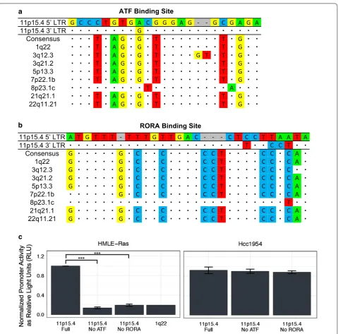
[image:5.595.57.544.103.401.2]Removal of critical binding sites decreases HML‑2 promoter activity in neoplastic cell lines
The functionality of these sites was assessed by mutat-ing each one individually. A multiple sequence alignment was performed using the sequences of all nine 5′ LTRs. From this analysis, we created a consensus sequence for each critical binding site, which we deemed to be the “non-active” version of each site. The full binding site sequence in each 5′ and 3′ LTR of the nine proviruses of interest in this study are provided in Additional file 3, Additional file 4, Additional file 5 and Additional file 6. The 3q12.3 HOX-PBX binding site differed from the con-sensus non-active sequence by a five base pairs, including a duplication of four nucleotides (Fig. 4a). Reversion of these sites significantly decreased LTR promoter activity in both neoplastic cell lines, with activity decreasing by twofold in HMLE-Ras cells (Fig. 4c, left) and by sevenfold in HMLE-Her2 cells (Fig. 4c, middle). The 3q12.3 RFX3 binding site only differed from the consensus sequence by one nucleotide, an A to C transversion (Fig. 4b), and yet removal of this site decreased LTR activity by fivefold in both HMLE-Ras cells (Fig. 4c, left) and HMLE-Her2 cells (Fig. 4c, middle). Activity was decreased to levels comparable to that of 1q22, a proviral LTR with no sig-nificant promoter activity in these cell lines (Fig. 4c). Mutating these sites did not significantly decrease LTR promoter activity in Hcc1954 cells (Fig. 4c, right), which also showed elevated expression of transcription factors known to bind to five unique 3q12.3 sites (E2F, HOX-PBX, RFX3, SF1, TEF-HLF) (Fig. 3b), suggesting that the other active binding sites can compensate for promoter activity when only some of them are removed.
Similar results were seen with the 11p15.4 5′ LTR. The consensus sequence differed from the ATF binding site by nine nucleotides (Fig. 5a) and back mutating the
binding site to match the consensus sequenced decreased promoter activity by sixfold in HMLE-Ras cells (Fig. 5c, left). The RORA binding site differed by eleven nucleo-tides from the consensus sequence (Fig. 5b) and mutating all of these to the consensus bases decreased promoter activity by fivefold in HMLE-Ras cells (Fig. 5c, left). Again, these changes decreased activity to levels compa-rable with 1q22 (Fig. 5c, left). As in the case of 3q12.3, no decrease in promoter activity was seen in the Hcc1954 cell line (Fig. 5c, right), which had elevated expression of transcription factors known to bind to four unique 11p15.4 sites (ATF, HIVEP1, PEGASUS, RORA) (Fig. 3b, bottom).
Most unique HML‑2 transcription factor binding sites were acquired over time following integration and are fixed in the human population
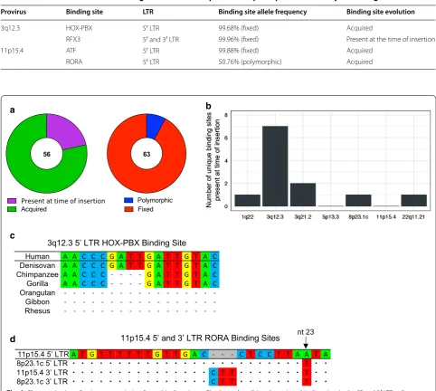
At the time of integration, the 5′ and 3′ LTRs of a pro-virus are almost always identical. Over time, as muta-tions are accumulated, sequence variation between the two LTRs increases. By aligning the 5′ and 3′ LTRs of 3q12.3 and 11p15.4, we were able to determine whether these critical transcription factor binding sites were present at the time of insertion (as evidenced by its presence in both LTRs) or were acquired over time (and found in only one LTR). We determined that one of the sites, RFX3 found in 3q12.3, was present at the time of insertion, but that three of the binding sites were acquired over time (Table 4). We analyzed the remaining unique binding sites in this same man-ner, with the exception of sites found on 7p22.1b and 21q21.1, which do not have full 3′ LTRs. Overall, only 21% (12/56) of the unique sites were present at the time of insertion (Fig. 6a, left), the majority of which (58%, 7/12) were found in the 3q12.3 5′ LTR (Fig. 6b). Table 3 Unique transcription factor binding sites found in HML-2 5′ LTRs of interest
Only sites unique to each 5′ LTR, as compared to the other eight 5′ LTRs, are shown † Present only in other HML-2 solo LTR(s)
‡ Present in other HML-2 full length provirus(es) and solo LTR(s)
Provirus Unique binding site
1q22 NBRE‡
3q12.3 CDE, E2F, HOX-PBX, MRG1‡, PROX1, RFX3†, SF1†, SOX10, TAL1-E2A, TBX20, TEF-HLF‡, TGIF†, TR2‡
3q21.2 GLI1†, IK3, NFY‡, NKX29†, SIX2‡, STAT5
5p13.3 CARF‡, MYBL1‡
7p22.1b EKLF‡, GAGA ‡, GLI3‡
8p23.1c AML1‡, BHLHB2‡, DMRT7‡, HMGA‡, HOX1-3‡, MAFF‡, MEF2‡, NRF1‡, PAX1‡, SOX17‡, STAT5A‡
11p15.4 AHRARNT‡, ATF, ATF6, CETS1P54, EN2‡, ETS1, FOXP2†, FREAC2‡, HDBP1-2, HHEX, HIVEP1†, IRF5,
MIF1†, NRL, PEGASUS, RORA, SGN1, STAT3, TEF†, TGIF2LX
21q21.1 CHOP†, NFKAPPAB50†, USF†, ZNF300†
[image:7.595.58.541.101.241.2]To determine the distribution of these sites within the human population, we analyzed the VCF (Variant Call Format) files of 2504 individuals, as supplied by phase 3 of the 1000 Genomes Project [42]. Of the four binding sites that we found to be critical for HML-2 promoter expression during neoplasia, three had allele frequencies > 99% and are therefore fixed in the popu-lation. The RORA binding site, found in the 11p15.4 5′
LTR, was found to be polymorphic with an allele fre-quency of 50.76% (Table 4). Overall, only 8% (5/63) of the unique sites that we identified were polymorphic in the human population (Fig. 6a, right).
Evolution of the HML‑2 HOX‑PBX and RORA binding sites Alignment of the 5′ and 3′ LTRs of the 3q12.3 provirus revealed a 4 bp insertion, found in the middle of the HOX-PBX site, resulting from duplication of a GATT sequence (Fig. 4a). This provirus is estimated to have integrated ~ 10 million years ago and is present in goril-las, chimpanzees, and bonobos, as well as humans [4]. Using the UCSC Genome Browser, we examined this LTR in several non-human primate reference genomes. We found that despite the conservation of the 3q12.3
provirus across multiple hominoid species, the 4 bp insertion, and consequently the HOX-PBX binding site, is only present in humans and Denisovans (Fig. 6c). These results suggest that this binding site was acquired some-time after the human-chimpanzee evolutionary split and has been stably integrated in the human genome ever since.
The RORA binding site on 11p15.4 was one of the only polymorphic unique binding sites that we identi-fied. This polymorphism is due to a single nucleotide change, where 51% of alleles in the human population contains an A at the 23rd base pair in the site (and therefore an intact RORA site) and 49% of the popula-tion contains a T. This provirus is of particular interest because 11p15.4 is a segmental duplication of 8p23.1c, which is estimated to have integrated ~ 20 million years ago. Although the proviral sequence is quite old, the duplication occurred after the human-chimpanzee split, and the 11p15.4 sequence is human-specific [4]. We aligned the 5′ and 3′ LTRs of these two proviruses and compared their sequences at the RORA binding site. We found that although both of the 3′ LTRs at this site are identical, the 5′ LTRs differ by one nucleotide, a
c
b
[image:8.595.53.542.87.339.2]the same 23rd nucleotide that is responsible for the RORA polymorphism (Fig. 6d). The 5′ LTR also dif-fered from the 3′ by deletion of 3 bp, which must have predated the segmental duplication of this provirus, as it was also found in the 8p23.1c 5′ LTR. Based on
these observations, it appears as though the provirus at 11p15.4 in half of the human population has evolved away from the ancestral 8p23.1c sequence, resulting in a functional RORA binding site.
a
b
c
[image:9.595.60.540.84.557.2]Discussion
Post-integration, retroviral sequences are transcribed and translated like any other cellular gene and are subject to the same selective pressures. Germline sequences with neutral or advantageous effects can become fixed in the population, resulting in endogenization [2, 25, 43]. These sequences provide unique opportunities to study the
evolutionary relationship between host and pathogen, including adaptations for assimilation within the host genome.
The full biological significance of HERVs remains to be uncovered. Repetitive mobile sequences are often cred-ited with contributing to genome plasticity and HERVs, equipped with multiple splice junctions, promoter/ Table 4 Characterization of LTR binding sites critical for 3q12.3 and 11p15.4 promoter activity in tumorigenic cells
Provirus Binding site LTR Binding site allele frequency Binding site evolution
3q12.3 HOX-PBX 5′ LTR 99.68% (fixed) Acquired
RFX3 5′ and 3′ LTR 99.96% (fixed) Present at the time of insertion
11p15.4 ATF 5′ LTR 99.88% (fixed) Acquired
RORA 5′ LTR 50.76% (polymorphic) Acquired
a b
c
d
[image:10.595.56.539.96.528.2] [image:10.595.59.541.101.173.2]enhancer sites, and polyadenylation signals, are abun-dantly capable of altering host gene expression [2, 8, 43]. A number of endogenous retroviruses, including the mouse mammary tumor virus, murine leukemia virus, and Jaagsiekte sheep retrovirus, exhibit both endogenous and exogenous transmission and are capable of inducing carcinogenesis. Since the pathogenicity of these viruses is generally due to LTR activity and integration site, which can result in the alteration of expression of nearby proto-oncogenes [43, 44], endogenous viral sequences are often silenced through epigenetic and chromatin modifications such as CpG methylation [12–14].
Our group recently characterized the HML-2 tran-scriptome during HMEC transformation and found that the site of proviral integration is often crucial for expres-sion, with the majority of expressed proviruses being transcribed by non-LTR-driven mechanisms such as read-through from adjacent promoters. When it was pre-sent, LTR-driven transcription was detected only in tum-origenic cells, suggesting that the altered transcriptional milieu of a transformed cell is critical for LTR promoter activation [32]. The goal of this study was to investigate the interplay between LTR sequence variation and cellu-lar environment and to look for evidence of evolutionary adaptations that could result in increased activity during neoplasia.
LTR hypomethylation, commonly seen in malig-nant cells, is well documented to result in increased ERV expression [13, 29, 45]. To eliminate this issue, we decided to investigate the relationship between LTR sequence similarity and differential expression patterns using reporter construct assays, where methylation sta-tus is not a factor. We chose to study nine HML-2 pro-viruses, shown by single-genome sequencing to be highly transcribed across a number of breast cancer cell lines (Additional file 1). Phylogenetic analysis of these 5′
LTRs classified most of them as LTR-HS, the LTR group that includes the youngest proviruses and most human-specific integrations [4, 9]. Of these, only one provi-rus (3q12.3) is known to not be human-specific, as it is present in gorillas and chimpanzees as well (Fig. 1a). All proviruses in this study are fixed in the human popula-tion, although one provirus (7p22.1b) is considered to be allelically polymorphic. It is present as either a solo LTR, formed through the recombination of the 5′ and 3′ LTRs and excision of the internal proviral sequence, or a full (“2-LTR”) provirus [4, 7]. However, in either case, the 5′
LTR of interest is fixed and as such, for the purpose of this study, we do not consider any of these LTRs to be insertionally polymorphic.
Overall, we found significant HML-2 promoter activ-ity in 73% (11/15) of tumorigenic HME cell lines (Fig. 1c), consistent with previous reports of increased HML-2
expression in up to 85% of breast cancer samples [30, 31,
37]. Molecular subtype, as denoted by hormone receptor status, of the cell lines was noted (Table 2), but no signifi-cant correlation with HML-2 promoter expression was observed (Additional file 7). Pairwise comparisons of 5′
LTR sequence identity and promoter expression in our luciferase panel revealed a positive correlation between the results of the two assays (Fig. 2). These results suggest that LTRs with similar sequences share similar promoter expression patterns, most likely due to conservation of the same transcription factor binding sites and core pro-moter elements.
To further investigate the importance of sequence vari-ation on LTR promoter activity, we used MatInspector, a transcription factor binding site prediction software, to generate a list of all binding sites unique to each of the nine LTRs used in this study (Table 3). We considered unique sites to be candidates for sequence variation that may explain why one LTR would be activated under a certain cellular condition instead of another. Two pro-viruses, 3q12.3 and 11p15.4, exhibited the highest levels of promoter activity across our luciferase panel (Fig. 1d). We used the MatInspector data, alongside RNA-Seq results from a previously published experiment by our group [32], to identify upregulated transcription factors known to bind to the unique binding sites on these two LTRs. These results provided us with two candidate sites per 5′ LTR for the promoter activation we saw during neoplasia: the HOX-PBX and RFX3 sites on 3q12.3 and the ATF and RORA sites on 11p15.4 (Fig. 3b). Removal of these sites individually decreased LTR promoter activity in HMLE-Ras and HMLE-Her2 cells by two to sevenfold (Figs. 4c, 5c).
is evolutionarily conserved amongst several non-human primate species, the HOX-PBX binding site is human-specific. Although HOX proteins are widely expressed during development, aberrant expression has been doc-umented during malignancy and increased HOX gene expression is being investigated as a potential breast can-cer biomarker [50].
Alignments between the 5′ and 3′ LTR of proviruses shed light on the evolution of unique transcription fac-tor binding sites. We were able to determine if sites were present at the time of insertion (present in both LTRs) or acquired over time (present in only one LTR). Only 21% of the unique binding sites that we identified were pre-sent at the time of insertion (Fig. 6a, left), implying that the expression patterns observed for these proviruses would not have reflected those of the ancestral virus that gave rise to them. Furthermore, the majority of unique sites were in the 3q12.3 5′ LTR (Fig. 6b). This distribu-tion is consistent with the greater genetic distance and greater age of this provirus from the rest of the LTR-HS group (Fig. 1a). The high degree of unique sites present at the time of insertion may also explain why this particular provirus had an expression pattern widely different from the other LTRs in this study (Fig. 2).
Due to their possible role in pathogenicity, it is essen-tial to study the genetic differences of HML-2 elements among individuals. Most often, such studies focus on whole proviruses, studying insertional polymorphism and its possible contribution to disease. Thus far, how-ever, no polymorphic proviruses have been found to play a role in the genesis of cancer [34, 51]. To our knowl-edge, ours is the first study to investigate genetic differ-ences at the single nucleotide level, by examining SNPs within LTRs. Of the 63 binding sites unique to one of the expressed LTRs that we identified, only five of them were found to be polymorphic within the 2504 genomes mined (Fig. 6a, right). These allele frequencies were further bro-ken down by super-population, showing only slightly higher prevalence of these binding sites in the African population (Additional file 8).
The RORA binding site, harbored on the 5′ LTR of the 11p15.4 provirus, was the only site critical for HML-2 activation during neoplasia that was also polymorphic (Table 4). This provirus is of particular interest because it is a segmental duplication of 8p23.1c [4], which showed no LTR activity during tumorigenesis. After examining the RORA binding sites on both of these LTRs, we found that 51% of the population contains an active RORA site whereas the other half of the population contains an inactive RORA site, identical to the ancestral 8p23.1c 5′
LTR. Thus, more than half of the human population has evolved away from the ancestral sequence and towards a more active LTR version (Fig. 6d).
Conclusions
The role, if any, of HERV activity during tumorigenesis is unknown. It is currently unclear if HML-2 expression is an ancillary consequence of transformation or if it some-how aids in the event; although recent work ssome-hows that Env protein expression may increase the ability of tumor cells to evade immune surveillance during some can-cers [52] or even participate directly in the transforma-tion process by interacting with cellular proto-oncogenes [53]. Although no provirus of interest in our study is believed to have a viable open reading frame for any viral gene, protein production in these cell lines as well as any sample used in future investigations, should be exam-ined. Our results show that HML-2 promoter activity is present in the majority (73%) of breast cancer cell lines tested and that LTR sequence similarity is correlated with promoter expression patterns. From there, we were able to map binding sites seemingly crucial for HML-2 pro-moter expression during neoplasia, many of which were acquired over evolutionary time. The polymorphism of certain sites provides another dimension in regards to what causes differential expression of ERVs between indi-viduals. These data may shed light on adaptive co-evolu-tion of ERVs within their host cells.
In recent years, there have been numerous reports of co-option of endogenous proviral sequences to dispa-rate features of normal human and vertebdispa-rate biology, including protection against infection by related exog-enous viruses [54], formation of the placental syncy-tiotrophoblast layer [10], expression of salivary amylase [55], stimulation of innate immunity [56], stimulation of neurological synapses promoting long-term memory [57], among others. It is particularly noteworthy that transcription of the two most highly expressed provi-ruses in our panel of ex vivo transformed cancer cell lines was facilitated through binding sites that were cre-ated by mutations in the 5′ LTRs that arose and spread in the human population following integration, imply-ing that the expression patterns observed do not reflect those of the ancestral virus. It is tempting to speculate that responsiveness of the mutant proviruses to com-mon, development-specific transcription factors might have given them some beneficial property along the lines of the ones listed above, thereby providing a selective advantage to the individuals carrying them and promot-ing their rapid fixation in the population.
Methods
Cell culture
all other cell lines were obtained from ATCC (Manassas, VA, USA). All cell lines were grown as per ATCC’s rec-ommendations and detailed information regarding their origin and culture conditions can be found in Additional file 9.
Single‑genome sequencing
ZR-75-1, MCF-7, T47D, SK-BR-3, Hcc1954, BT20, Hs578T, and MDA-MB-231 breast cancer cells were grown to 90% confluency. RNA was extracted and puri-fied using the RNeasy Mini Kit (Qiagen, Valencia, CA, USA, Cat. No. 74104) and all DNA contamination was removed through DNase treatment (Turbo DNA-free Kit, Ambion, Foster City, CA, USA, Cat. No. AM1907). RT reactions were set up as recommended by the manufac-turer’s protocol using an oligo(dT) primer (SuperScript III One-Step RT-PCR System, Invitrogen, Carlsbad, CA, USA, Cat. No. 12574-018). The resulting cDNA was seri-ally diluted down to an average of 1/3 genome per sample and amplified using Taq DNA polymerase (Invitrogen, Cat. No. 10342-020). Two forward primers (5′-TTC CTT TAC AAA GTT GCG TAA AGC -3′, 5′-GTT GCG TAA AGC CCC CTT AT-3′) and one reverse primer (5′-CAC AGA CAC AGT AAC AAT CTG-3′), all targeting the HML-2 env
region, were used in the reaction. The amplified prod-ucts were gel extracted with the QIAquick Gel Extraction Kit (Qiagen, Cat. No. 28704) and purified samples were sent out for sequencing. The primers used for sequenc-ing were 5′-GAC TCC CAG ACT ATA ACC TGTC-3′ and 5′-CGA AGC ATC AAA AGC CCA -3′. Sequencing results were BLAT searched in the UCSC Genome Browser [58] to identify expressed proviruses.
Phylogenetic analysis
The 5′ and 3′ LTR sequence of each provirus of interest was obtained from the UCSC Genome Browser’s Repeat-Masker Track [58, 59] and imported as FASTA files into the Molecular Evolutionary Genetics Analysis (MEGA, v6.06) program for alignment using Multiple Sequence Comparison by Log-Expectation (MUSCLE) [60, 61]. Phylogeny of aligned sequences was determined by sequence dissimilarity and a neighbor-joining tree was constructed using a p-distance algorithm. Bootstrap val-ues were determined using 1000 replicate tests.
Dual‑luciferase assay
Primers for LTR amplification were selected using the Primer3 program [62]. Restriction enzyme cleavage sites were appended to the 5′ end of the primer sequences for proper vector ligation. The primers created are listed in Additional file 10. The LTR sequences were PCR-amplified using Taq DNA polymerase. Template DNA
was purified from Tera-1 cells using the DNeasy Blood and Tissue Kit (Qiagen, Cat. No. 69504). The ampli-fied sequences were cloned using basic molecular biol-ogy techniques and ligated into the multiple cloning region of the pGL4.17[luc2/Neo] promoter-less firefly luciferase vector (Promega, Madison, WI, USA, Cat. No. E6721). All constructs were sequenced to check for PCR-induced mutations before transfection. All cell cul-tures were seeded in triplicate at 100,000 cells/well in a 24-well plate for transfection. Cultures were co-trans-fected with the pGL4 vector alongside a pRL-SV40 inter-nal control Renilla luciferase vector (Promega, Cat. No. E2231) at a 30:1 ratio using Opti-MEM reduced serum media (Gibco, Cat. No. 31985-070) and Lipofectamine 2000 (Thermo Fisher Technologies, Cat. No. 11668-019), as recommended by the manufacturer’s protocol. Post-transfection, cells were incubated at 37 °C for 48 h before lysis and analysis. Luminescence was measured via the dual-luciferase assay system (Promega, Cat. No. E1910) and quantified as relative light units (RLU) on a BioTek Synergy HT plate reader using Gen5 Data Analysis Soft-ware (BioTek Instruments, Winooski, VT, USA). Empty vectors as well as non-transfected cells were measured as a control to determine any cell-specific background sig-nal. LTR promoter activity was calculated as luc2 activity normalized against that of the internal Renilla luciferase control signal.
HML‑2 similarity matrices
The sequence of each “full length” (i.e., not solo LTR) HML-2 provirus annotated within the human refer-ence genome (hg19 build) was obtained from the UCSC Genome Browser [58]. These sequences were input into the Clustal Omega program (The European Bioinfor-matics Institute (EMBL-EBI), Hinxton, Cambridge, UK) [40] to create a multiple sequence alignment using the HHalign algorithm [63] and to create a percent sequence identity matrix. The HML-2 percent expression similar-ity matrix was created by making pairwise comparisons of significant promoter expression in each of the eighteen cell lines used in our dual-luciferase analysis.
Transcription factor binding site analysis
the Hcc1954, HMLE-Ras, HMLE-Her2, and HME cell lines were determined by Cuffdiff analysis of our previous RNA-Seq results. A full description of the study used to obtain these values is detailed in our previous publication [32] and the RNA-Seq data are deposited in the NCBI Gene Expression Omnibus database under Accession Number GSE84275.
Consensus sequences of the HOX-PBX, RFX3, ATF, and RORA binding sites were determined through a separate MEGA alignment. New reporter constructs containing the consensus (non-active) sites were cre-ated through IDT’s gBlocks® Gene Fragments synthesis service (Integrated DNA Technologies, Inc., Coralville, IA, USA). These fragments were directly cloned into the pGL4[luc2/Neo] firefly luciferase vector and transfected into cell lines as previously described in the Dual-Lucif-erase Assay section of the Materials and Methods.
The 5′ and 3′ LTRs of each of the nine proviruses of interest were analyzed in an additional MEGA alignment. All unique transcription factor binding sites found in only one LTR were regarded as being “acquired” and any unique binding sites found in both LTRs were character-ized as “present at time of insertion”. Sites located in the 7p22.1b and 21q21.1 proviruses were excluded from the analysis since they no longer possess intact 3′ LTRs [4].
The allele frequencies of each unique binding site were calculated from the VCF (Variant Call Format) files of 2504 individuals, as supplied by phase 3 of the 1000 Genomes Project [42]. VCF files were analyzed compu-tationally using VCFtools, by specifying the genomic coordinates (hg19 build) of each site of interest. All sites with an allele frequency of at least 89% were considered to be fixed in the human population. All sites that were classified as polymorphic within the population had allele frequencies of 52% or less. No binding site that we iden-tified had an allele frequency intermediate of those two thresholds, i.e. calculated to be greater than 52% but less than 89%.
The HOX-PBX binding site was further analyzed in several non-human primate reference genomes as sup-plied by the UCSC Genome Browser [58]. The Denisovan reference genome sequence was obtained from the Den-isova High-Coverage Sequence Reads of the DenDen-isova Seq Track. The chimpanzee, gorilla, orangutan, gibbon, and rhesus reference genome sequences were obtained from the Vertebrate Multiz Alignment & Conservation Track.
Additional files
Additional file 1: Table S1. HML-2 transcript levels detected through single-genome sequencing in breast cancer cell lines of varying molecular subtype.
Additional file 2: Table S2. HML-2 similarity matrices.
Additional file 3: Table S3. HOX-PBX binding site sequences and genomic coordinates (hg19).
Additional file 4: Table S4. RFX3 binding site sequences and genomic coordinates (hg19).
Additional file 5: Table S5. ATF binding site sequences and genomic coordinates (hg19).
Additional file 6: Table S6. RORA binding site sequences and genomic coordinates (hg19).
Additional file 7: Figure S1. HML-2 promoter activity is not breast cancer subtype-specific. Total relative 5′ LTR promoter activity levels of fifteen tumorigenic breast cancer cell lines broken down by molecular subtype (luminal, HER2+, and basal-like) as compared to two immortalized HME cell lines. Hormone receptor status and cell lines identified as being each molecular subtype are shown in detail in Table 2. All experiments were conducted in triplicate and data display the mean ± standard deviation. Additional file 8: Figure S2. Allele frequencies of polymorphic HML-2 5′ LTR transcription factor binding sites within each superpopulation. Allele frequencies were determined for the proviruses shown from 2504 individuals from the 1000 Genomes Project and broken down by super-population (EAS = East Asian; AMR = Ad Mixed American; AFR = African; EUR = European; SAS = South Asian). The name of the transcription factor binding site as well as the provirus of interest are shown at the top of each graph.
Additional file 9: Table S7. Culture methods for cell lines used. Additional file 10: Table S8. Primers used to amplify 5′ LTRs of trans-fected HML-2 proviruses.
Authors’ contributions
MM, CK, and JMC conceived and designed the experiments. MM and RPS per-formed the experiments. MM, ZHW, RPS, and JMC analyzed the data. MM and JMC wrote the paper and all authors read and approved the final manuscript.
Author details
1 Department of Molecular Biology and Microbiology, Tufts University School
of Medicine, Boston, MA, USA. 2 Department of Developmental, Chemical,
and Molecular Biology, Tufts University School of Medicine, Boston, MA, USA.
3 Raymond and Beverly Sackler Convergence Laboratory, Tufts University
School of Medicine, Boston, MA, USA. 4 Present Address: Foundation Medicine,
Inc., Cambridge, MA, USA. 5 Present Address: Excerpta Medica, New York, NY,
USA.
Acknowledgements
We thank the Tufts University Genomics core facility for their RNA-Seq advice and as John Yoon for helpful discussion and editorial advice.
Competing interests
Availability of data and materials
The RNA-Seq data used in this study are deposited in the NCBI Gene Expres-sion Omnibus database under accesExpres-sion number GSE84275 (https ://www. ncbi.nlm.nih.gov/geo/query /acc.cgi?acc=GSE84 275). All other data generated or analyzed during this study are included in this published article and its additional files.
Consent for publication Not applicable.
Ethics approval and consent to participate Not applicable.
Funding
This project was supported by research Grants R37 CA 089441 and R35 CA 200421 from the National Cancer Institute.
Publisher’s Note
Springer Nature remains neutral with regard to jurisdictional claims in pub-lished maps and institutional affiliations.
Received: 26 May 2018 Accepted: 13 August 2018
References
1. Lower R, Lower J, Tondera-Koch C, Kurth R. A general method for the identification of transcribed retrovirus sequences (R-U5 PCR) reveals the expression of the human endogenous retrovirus loci HERV-H and HERV-K in teratocarcinoma cells. Virology. 1993;192:501–11. https ://doi. org/10.1006/viro.1993.1066.
2. Jern P, Coffin JM. Effects of retroviruses on host genome function. Annu Rev Genet. 2008;42:709–32. https ://doi.org/10.1146/annur ev.genet .42.11080 7.09150 1.
3. Ono M, Yasunaga T, Miyata T, Ushikubo H. Nucleotide sequence of human endogenous retrovirus genome related to the mouse mammary tumor virus genome. J Virol. 1986;60:589–98.
4. Subramanian RP, Wildschutte JH, Russo C, Coffin JM. Identification, char-acterization, and comparative genomic distribution of the HERV-K (HML-2) group of human endogenous retroviruses. Retrovirology. 2011;8:90.
https ://doi.org/10.1186/1742-4690-8-90.
5. Armbruester V, Sauter M, Krautkraemer E, Meese E, Kleiman A, Best B, et al. A novel gene from the human endogenous retrovirus K expressed in transformed cells. Clin Cancer Res. 2002;8:1800–7.
6. Gonzalez-Hernandez MJ, Cavalcoli JD, Sartor MA, Contreras-Galindo R, Meng F, Dai M, et al. Regulation of the human endogenous retrovirus K (HML-2) transcriptome by the HIV-1 Tat protein. J Virol. 2014;88:8924–35.
https ://doi.org/10.1128/JVI.00556 -14.
7. Wildschutte JH, Williams ZH, Montesion M, Subramanian RP, Kidd JM, Coffin JM. Discovery of unfixed endogenous retrovirus insertions in diverse human populations. Proc Natl Acad Sci U S A. 2016. https ://doi. org/10.1073/pnas.16023 36113 .
8. Schmitt K, Reichrath J, Roesch A, Meese E, Mayer J. Transcriptional profiling of human endogenous retrovirus group HERV-K(HML-2) loci in melanoma. Genome Biol Evol. 2013;5:307–28. https ://doi.org/10.1093/ gbe/evt01 0.
9. Bhardwaj N, Montesion M, Roy F, Coffin JM. Differential expression of HERV-K (HML-2) proviruses in cells and virions of the teratocarcinoma cell line Tera-1. Viruses. 2015;7:939–68. https ://doi.org/10.3390/v7030 939. 10. Mi S, Lee X, Li X, Veldman GM, Finnerty H, Racie L, et al. Syncytin is a
cap-tive retroviral envelope protein involved in human placental morphogen-esis. Nature. 2000;403:785–9. https ://doi.org/10.1038/35001 608. 11. Reiss D, Zhang Y, Mager DL. Widely variable endogenous retroviral
meth-ylation levels in human placenta. Nucleic Acids Res. 2007;35:4743–54.
https ://doi.org/10.1093/nar/gkm45 5.
12. Gotzinger N, Sauter M, Roemer K, Mueller-Lantzsch N. Regulation of human endogenous retrovirus-K Gag expression in teratocarcinoma cell lines and human tumours. J Gen Virol. 1996;77(Pt 12):2983–90. https :// doi.org/10.1099/0022-1317-77-12-2983.
13. Florl AR, Lower R, Schmitz-Drager BJ, Schulz WA. DNA methylation and expression of LINE-1 and HERV-K provirus sequences in urothelial and renal cell carcinomas. Br J Cancer. 1999;80:1312–21. https ://doi. org/10.1038/sj.bjc.66905 24.
14. Conklin KF, Coffin JM, Robinson HL, Groudine M, Eisenman R. Role of methylation in the induced and spontaneous expression of the avian endogenous virus ev-1: DNA structure and gene products. Mol Cell Biol. 1982;2:638–52.
15. Santoni FA, Guerra J, Luban J. HERV-H RNA is abundant in human embry-onic stem cells and a precise marker for pluripotency. Retrovirology. 2012;9:111. https ://doi.org/10.1186/1742-4690-9-111.
16. Xie W, Schultz MD, Lister R, Hou Z, Rajagopal N, Ray P, et al. Epigenomic analysis of multilineage differentiation of human embryonic stem cells. Cell. 2013;153:1134–48. https ://doi.org/10.1016/j.cell.2013.04.022. 17. Smith ZD, Chan MM, Humm KC, Karnik R, Mekhoubad S, Regev A, et al.
DNA methylation dynamics of the human preimplantation embryo. Nature. 2014;511:611–5. https ://doi.org/10.1038/natur e1358 1. 18. Ohnuki M, Tanabe K, Sutou K, Teramoto I, Sawamura Y, Narita M, et al.
Dynamic regulation of human endogenous retroviruses mediates factor-induced reprogramming and differentiation potential. Proc Natl Acad Sci U S A. 2014;111:12426–31. https ://doi.org/10.1073/pnas.14132 99111 . 19. Grow EJ, Flynn RA, Chavez SL, Bayless NL, Wossidlo M, Wesche DJ, et al.
Intrinsic retroviral reactivation in human preimplantation embryos and pluripotent cells. Nature. 2015. https ://doi.org/10.1038/natur e1430 8. 20. Frank JA, Feschotte C. Co-option of endogenous viral sequences for host
cell function. Curr Opin Virol. 2017;25:81–9. https ://doi.org/10.1016/j.covir o.2017.07.021.
21. Glinsky GV. Activation of endogenous human stem cell-associated retroviruses (SCARs) and therapy-resistant phenotypes of malignant tumors. Cancer Lett. 2016;376:347–59. https ://doi.org/10.1016/j.canle t.2016.04.014.
22. Kudo-Saito C, Yura M, Yamamoto R, Kawakami Y. Induction of immu-noregulatory CD271+ cells by metastatic tumor cells that express human endogenous retrovirus H. Cancer Res. 2014;74:1361–70. https ://doi. org/10.1158/0008-5472.CAN-13-1349.
23. Kreimer U, Schulz WA, Koch A, Niegisch G, Goering W. HERV-K and LINE-1 DNA methylation and reexpression in urothelial carcinoma. Front Oncol. 2013;3:255. https ://doi.org/10.3389/fonc.2013.00255 .
24. Lavie L, Kitova M, Maldener E, Meese E, Mayer J. CpG methylation directly regulates transcriptional activity of the human endogenous retrovirus family HERV-K(HML-2). J Virol. 2005;79:876–83. https ://doi.org/10.1128/ JVI.79.2.876-883.2005.
25. Fuchs NV, Kraft M, Tondera C, Hanschmann KM, Lower J, Lower R. Expres-sion of the human endogenous retrovirus (HERV) group HML-2/HERV-K does not depend on canonical promoter elements but is regulated by transcription factors Sp1 and Sp3. J Virol. 2011;85:3436–48. https ://doi. org/10.1128/JVI.02539 -10.
26. Stengel S, Fiebig U, Kurth R, Denner J. Regulation of human endogenous retrovirus-K expression in melanomas by CpG methylation. Genes Chro-mosomes Cancer. 2010;49:401–11. https ://doi.org/10.1002/gcc.20751 . 27. Knossl M, Lower R, Lower J. Expression of the human endogenous
retrovi-rus HTDV/HERV-K is enhanced by cellular transcription factor YY1. J Virol. 1999;73:1254–61.
28. Ono M, Kawakami M, Ushikubo H. Stimulation of expression of the human endogenous retrovirus genome by female steroid hormones in human breast cancer cell line T47D. J Virol. 1987;61:2059–62.
29. Wang-Johanning F, Frost AR, Johanning GL, Khazaeli MB, LoBuglio AF, Shaw DR, et al. Expression of human endogenous retrovirus k envelope transcripts in human breast cancer. Clin Cancer Res. 2001;7:1553–60. 30. Wang-Johanning F, Frost AR, Jian B, Epp L, Lu DW, Johanning GL.
Quan-titation of HERV-K env gene expression and splicing in human breast cancer. Oncogene. 2003;22:1528–35. https ://doi.org/10.1038/sj.onc.12062 41.
31. Zhao J, Rycaj K, Geng S, Li M, Plummer JB, Yin B, et al. Expression of human endogenous retrovirus type K envelope protein is a novel candidate prognostic marker for human breast cancer. Genes Cancer. 2011;2:914–22. https ://doi.org/10.1177/19476 01911 43184 1.
33. Manghera M, Douville RN. Endogenous retrovirus-K promoter: a landing strip for inflammatory transcription factors? Retrovirology. 2013;10:16.
https ://doi.org/10.1186/1742-4690-10-16.
34. Wildschutte JH, Ram D, Subramanian R, Stevens VL, Coffin JM. The distri-bution of insertionally polymorphic endogenous retroviruses in breast cancer patients and cancer-free controls. Retrovirology. 2014;11:62. https ://doi.org/10.1186/PREAC CEPT-17207 68941 31202 6.
35. Buzdin A, Ustyugova S, Khodosevich K, Mamedov I, Lebedev Y, Hunsmann G, et al. Human-specific subfamilies of HERV-K (HML-2) long terminal repeats: three master genes were active simultaneously during branching of hominoid lineages. Genomics. 2003;81:149–56.
36. Ruprecht K, Ferreira H, Flockerzi A, Wahl S, Sauter M, Mayer J, et al. Human endogenous retrovirus family HERV-K(HML-2) RNA transcripts are selec-tively packaged into retroviral particles produced by the human germ cell tumor line Tera-1 and originate mainly from a provirus on chromo-some 22q11.21. J Virol. 2008;82:10008–16. https ://doi.org/10.1128/ JVI.01016 -08.
37. Wang-Johanning F, Radvanyi L, Rycaj K, Plummer JB, Yan P, Sastry KJ, et al. Human endogenous retrovirus K triggers an antigen-specific immune response in breast cancer patients. Cancer Res. 2008;68:5869–77. https :// doi.org/10.1158/0008-5472.CAN-07-6838.
38. Seifarth W, Baust C, Murr A, Skladny H, Krieg-Schneider F, Blusch J, et al. Proviral structure, chromosomal location, and expression of HERV-K-T47D, a novel human endogenous retrovirus derived from T47D particles. J Virol. 1998;72:8384–91.
39. Keydar I, Ohno T, Nayak R, Sweet R, Simoni F, Weiss F, et al. Properties of retrovirus-like particles produced by a human breast carcinoma cell line: immunological relationship with mouse mammary tumor virus proteins. Proc Natl Acad Sci U S A. 1984;81:4188–92.
40. McWilliam H, Li W, Uludag M, Squizzato S, Park YM, Buso N, et al. Analysis Tool Web Services from the EMBL-EBI. Nucleic Acids Res. 2013;41:W597– 600. https ://doi.org/10.1093/nar/gkt37 6.
41. Cartharius K, Frech K, Grote K, Klocke B, Haltmeier M, Klingenhoff A, et al. MatInspector and beyond: promoter analysis based on transcrip-tion factor binding sites. Bioinformatics. 2005;21:2933–42. https ://doi. org/10.1093/bioin forma tics/bti47 3.
42. Genomes Project C, Auton A, Brooks LD, Durbin RM, Garrison EP, Kang HM, et al. A global reference for human genetic variation. Nature. 2015;526:68–74. https ://doi.org/10.1038/natur e1539 3.
43. Lower R, Lower J, Kurth R. The viruses in all of us: characteristics and biological significance of human endogenous retrovirus sequences. Proc Natl Acad Sci U S A. 1996;93:5177–84.
44. Wang-Johanning F, Liu J, Rycaj K, Huang M, Tsai K, Rosen DG, et al. Expres-sion of multiple human endogenous retrovirus surface envelope proteins in ovarian cancer. Int J Cancer. 2007;120:81–90. https ://doi.org/10.1002/ ijc.22256 .
45. Fanning T, Alves G. A family of repetitive DNA sequences in Old World primates. Gene. 1997;199:279–82.
46. Shah N, Sukumar S. The Hox genes and their roles in oncogenesis. Nat Rev Cancer. 2010;10:361–71. https ://doi.org/10.1038/nrc28 26. 47. Tammimies K, Bieder A, Lauter G, Sugiaman-Trapman D, Torchet R,
Hok-kanen ME, et al. Ciliary dyslexia candidate genes DYX1C1 and DCDC2 are regulated by regulatory factor (RF) X transcription factors through X-box promoter motifs. FASEB J. 2016. https ://doi.org/10.1096/fj.20150 0124R R.
48. Jiang S, Zhang E, Zhang R, Li X. Altered activity patterns of transcrip-tion factors induced by endoplasmic reticulum stress. BMC Biochem. 2016;17:8. https ://doi.org/10.1186/s1285 8-016-0060-2.
49. Cook DN, Kang HS, Jetten AM. Retinoic acid-related orphan recep-tors (RORs): regulatory functions in immunity, development, circadian rhythm, and metabolism. Nucl Recept Res. 2015. https ://doi.org/10.11131 /2015/10118 5.
50. Morgan R, Boxall A, Harrington KJ, Simpson GR, Gillett C, Michael A, et al. Targeting the HOX/PBX dimer in breast cancer. Breast Cancer Res Treat. 2012;136:389–98. https ://doi.org/10.1007/s1054 9-012-2259-2. 51. Burmeister T, Ebert AD, Pritze W, Loddenkemper C, Schwartz S, Thiel E.
Insertional polymorphisms of endogenous HERV-K113 and HERV-K115 retroviruses in breast cancer patients and age-matched controls. AIDS Res Hum Retrovir. 2004;20:1223–9. https ://doi.org/10.1089/08892 22042 54508 1.
52. Serafino A, Balestrieri E, Pierimarchi P, Matteucci C, Moroni G, Oricchio E, et al. The activation of human endogenous retrovirus K (HERV-K) is implicated in melanoma cell malignant transformation. Exp Cell Res. 2009;315:849–62. https ://doi.org/10.1016/j.yexcr .2008.12.023. 53. Lemaitre C, Tsang J, Bireau C, Heidmann T, Dewannieux M. A human
endogenous retrovirus-derived gene that can contribute to onco-genesis by activating the ERK pathway and inducing migration and invasion. PLoS Pathog. 2017;13:e1006451. https ://doi.org/10.1371/journ al.ppat.10064 51.
54. Blanco-Melo D, Gifford RJ, Bieniasz PD. Co-option of an endogenous ret-rovirus envelope for host defense in hominid ancestors. Elife. 2017. https ://doi.org/10.7554/elife .22519 .
55. Samuelson LC, Wiebauer K, Gumucio DL, Meisler MH. Expression of the human amylase genes: recent origin of a salivary amylase promoter from an actin pseudogene. Nucleic Acids Res. 1988;16:8261–76.
56. Hurst TP, Magiorkinis G. Activation of the innate immune response by endogenous retroviruses. J Gen Virol. 2015;96:1207–18. https ://doi. org/10.1099/jgv.0.00001 7.
57. Pastuzyn ED, Day CE, Kearns RB, Kyrke-Smith M, Taibi AV, McCormick J, et al. The neuronal gene arc encodes a repurposed retrotransposon gag protein that mediates intercellular RNA transfer. Cell. 2018;173:275. https ://doi.org/10.1016/j.cell.2018.03.024.
58. Karolchik D, Hinrichs AS, Furey TS, Roskin KM, Sugnet CW, Haussler D, et al. The UCSC table browser data retrieval tool. Nucleic Acids Res. 2004;32:D493–6. https ://doi.org/10.1093/nar/gkh10 3.
59. Kent WJ, Sugnet CW, Furey TS, Roskin KM, Pringle TH, Zahler AM, et al. The human genome browser at UCSC. Genome Res. 2002;2002(12):996–1006.
https ://doi.org/10.1101/gr.22910 2.
60. Tamura K, Stecher G, Peterson D, Filipski A, Kumar S. MEGA6: molecular evolutionary genetics analysis version 6.0. Mol Biol Evol. 2013;30:2725–9.
https ://doi.org/10.1093/molbe v/mst19 7.
61. Edgar RC. MUSCLE: multiple sequence alignment with high accuracy and high throughput. Nucleic Acids Res. 2004;32:1792–7. https ://doi. org/10.1093/nar/gkh34 0.
62. Untergasser A, Cutcutache I, Koressaar T, Ye J, Faircloth BC, Remm M, et al. Primer3: new capabilities and interfaces. Nucleic Acids Res. 2012;40:e115.
https ://doi.org/10.1093/nar/gks59 6.