Non-hematopoietic essential
functions of bone marrow cells:
a review of scientific and
clini-cal literature and rationale for
treating bone defects
David B. Harrell,1Eugenio Caradonna,2
Laura Mazzucco,3Rosmarie Gudenus,4
Berthold Amann,5Vaclav Prochazka,6
Peter V. Giannoudis,7
Christian Hendrich,8Marcus Jäger,9
Rüdiger Krauspe,10Philippe Hernigou11
1Harrell BioScience Consulting LLC,
Quincy, MA, USA; 2Department of
Cardiovascular Disease, Fondazione de Ricerca e Cura Giovanni e Paolo II,
Campbasso, Italy; 3Blood Component and
Regenerative Medicine Laboratory,
Alessandria Hospital, Italy; 4Granzer
Regulatory Consulting and Services,
München, Germany; 5ASKLEPIOS
Westklinikum Hamburg, Germany;
6Interventional Neuroradiology and
Angiology, University of Ostrava, Czech
Republic; 7Trauma and Orthopedic
Surgery School of Medicine, University of
Leeds, UK; 8Orthopaedishe Krankenhaus
Schloss Werneck, Werneck Bayern,
Germany; 9Universitätklinikum Essen,
Germany; 10Klinik Orthopaedische,
University of Düsseldorf, Germany;
11Hôspital Henri Mondor, Creteil, France
Abstract
Hematopoiesis as the only essential func-tion of bone marrow cells has been challenged for several decades through basic science (in vitroand in vivo) and clinical data. Such work has shed light on two other essential functions of bone marrow cells: osteopoiesis and angio-genesis/vasculogenesis. Clinical utility of autologous concentrated bone marrow aspirate (CBMA) has demonstrated both safety and efficacy in treating bone defects. Moreover, CBMA has been shown to be comparable to the gold standardof iliac crest bone graft (ICBG), or autograft, with regard to being osteogenic and osteoinductive. ICBG is not considered an advanced therapy medicinal product (ATMP), but CBMA may become regulated as an ATMP. The European Medicines Agency Committee for Advanced Therapies (EMA:CAT) has issued a reflection paper (20 June 2014) in which reversal of the 2013 ruling that CBMA is a non-ATMP has been proposed. We review bone mar-row cell involvement in osteopoiesis and angiogenesis/vasculogenesis to examine
EMA:CAT 2013 decision to use CBMA for treat-ment of osteonecrosis (e.g, of the femoral head) should be considered a non-ATMP. This paper is intended to provide discussion on the 20 June 2014 reflection paper by reviewing two non-hematopoietic essential functions of bone marrow cells. Additionally, we provide clinical and scientific rationale for treating osteonecrosis with CBMA.
Introduction
According to section 2.2.3. number 2 of the EMA:CAT 20 June 2014 reflection paper, mar-row cells that are aspirated, centrifuged, and re-administered to fulfill their same essential function will generally be regarded as homolo-gous use. In case no substantial manipulation of the cells takes place, the classification is based on the essential function of the cells. Such non-substantially manipulated cells used for the same essential function are not consid-ered ATMPs.1 However, in section 2.3.1 it is
stated that products once considered a non-ATMP have been classified as an non-ATMP. As an example, it is specifically listed that injection of concentrated bone marrow at the site of bone injury with the aim of healing a bone lesion can be considered as non-homologous use.1
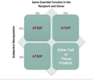
According to EU Regulation 1394/2007 a tissue engineered product (an ATMP) is defined as one that consists of cells or tissues that have been subject to i) substantial manipulation, or ii) are not intended for the same essential function(s) in the recipient and the donor. Since, bone marrow aspirate is centrifuged to produce concentrated bone marrow aspirate (CBMA), it is not substantial manipulation (Annex I of EU Regulation 1394/2007). The issue under discussion is whether or not autol-ogous bone marrow cells active in healing a bone lesion constitute an essential function, and consequently, homologous use of the cells. The hematopoietic stem cell (HSC) is the most characterized somatic stem cell in the adult and has been utilized in treatment of patients following myeloablation therapy.2
Multipotent stem cells from adult bone marrow have been used for decades in these patients because HSCs contribute to long-term and complete haematopoiesis.3,4 Bone marrow is
primarily a hematopoietic organ; however, it has long been recognized that hematopoiesis is not the only essential function of bone mar-row cells.5-8In addition to blood cell
replace-ment, bone marrow cells are active in replac-ing and buildreplac-ing bone and endothelial cells.9-12
Moreover, iliac crest bone graft (ICBG), a gold standard therapy used in orthopedic surgery, is not considered an ATMP and is not used for hematopoietic reconstitution. Rather, ICBG is used clinically for osteopoiesis and
vasculog-nesis obtained from the osteoblasts on the graft surface as well as the bone marrow stem and progenitor cells contained within the graft material.6,13,14
In this paper, we review data demonstrating that bone marrow cells, in addition to hematopoiesis, have the essential functions of osteopoiesis and vasculogenesis/angiogene-sis. As essential functions, to utilize bone mar-row cells for treating bone defects represents a homologous use of the cells. With autologous CBMA, the donor and the recipient are the same individual. The homologous use of the cells is preserved by aspirating bone marrow in order that the acquired bone marrow stem and progenitor cells will form the native tissue of the environment in which they are placed via the normal physiological functions of autocrine and paracrine activity.15,16
Autologous ICBG and CBMA function in the same manner for the treatment of bone defects and consequently, they should both be consid-ered a non-ATMP (Figure 1).
Two essential
non-hematopoi-etic functions of bone marrow
cells
Hematopoiesis is a crucial function of bone marrow cells. The bone marrow hematopoietic compartment produces approximately 500 bil-lion cells per day that use the bone marrow vasculature as a conduit to the systemic circu-lation.17Such high cellular turnover demands Correspondence: David B. Harrell, Harrell BioScience Consulting LLC, 101 Monroe Road, Quincy, MA 02169, USA.
Tel.: +1.617.800.9072. E-mail: [email protected]
Key words: Concentrated bone marrow aspirate; hematopoiesis; osteopoiesis; angiogenesis; osteonecrosis.
Contributions: the authors contributed equally.
Conflict of interest: the authors declare no poten-tial conflict of interest.
Received for publication: 25 October 2014. Accepted for publication: 20 October 2015.
This work is licensed under a Creative Commons Attribution NonCommercial 3.0 License (CC BY-NC 3.0).
upon the bone marrow hematopoietic system certainly demonstrate the importance of the function of hematopoiesis of bone marrow. However, hematopoiesis is not the sole func-tion of bone marrow cells.
It is well recognized and described in the lit-erature that bone marrow is responsible for tissue/bone repair as well as enhancing neo-vascularization. In 1997, it was first demon-strated that CD34+cells (human
hematopoiet-ic stem and progenitor cells are both CD34+
cells) are the precursors to osteoblasts.18
Recently, it was found that a single bone mar-row cell contributed to hematopoietic reconsti-tution and drives robust osteopoiesis.19In this
same study, it was found that long-term repop-ulating HSCs yield progeny that differentiate into osteoblasts following bone marrow trans-plantation (BMT). Therefore, a means for hematopoietic and osteopoietic maintenance was found involving cells historically thought active in hematopoiesis but not osteopoiesis. Moreover, bone marrow is home to mesenchy-mal stromesenchy-mal cells (MSCs) that do not directly contribute to blood cell production. Limiting the essential function of bone marrow cells to hematopoiesis alone does not acknowledge the functions of MSCs.
Osteopoietic potential
Within bone marrow, there is significant heterogeneity. Three main regenerative cell types exist in bone marrow: HSCs, MSCs, and endothelial progenitor cells (EPCs).20 MSCs,
also called skeletal stem/progenitor cells or medicinal signaling cells,21-25 do not directly
contribute to hematopoietic reconstitution.26
CBMA MSC population placed into osteogenic media demonstrated osteoblastic differentia-tion in the same manner as commercially available MSCs (Figure 2).
In 1869, it was first documented that bone marrow cells have osteogenic potential.5It is
well known that donor-derived osteopoiesis in both mice and humans occurs following BMT, even from a single bone marrow cell. This sug-gests that osteopoiesis may be an essential function of bone marrow cells and confirms the earlier findings of osteogenicity of bone marrow.5,6,19
ICBG, a non-ATMP, is considered a gold standard in orthopaedic surgery because it is i) osteogenic (contains cells that contribute to osteopoiesis), ii) osteoinductive (contains growth factors, cytokines, and chemokines active in osteopoiesis), and iii) osteoconduc-tive (provides a three dimensional matrix for cells to provide osteopoiesis). Like ICBG, CBMA is also osteogenic and osteoinductive; however, it is not osteoconductive.13,16,27Within
bone marrow, there are at least two compart-ments of stem and progenitor cells that provide osteogenic cells,28-31with more mature
pre-oes-teoblastic cells localizing to the marrow space
adjacent to trabecular bone.13
Bone repair via cells only of bone marrow mesenchymal origin has also been questioned. That is, a single marrow cell can have both hematopoietic and osteopoietic progeny, depending upon environmental clues.26,31-33
Bone marrow non-MSCs were shown to have a 10-fold greater bone repopulating activity than MSCs in situations of stress and trauma.10,26
CD34+cells have been shown to differentiate
into functional osteoblasts and fibroblasts in vitro18,34,35and an in vivo study revealed that
fibroblasts are of hematopoietic origins.36To
be sure that the cell investigated in vivois a hematopoietic stem cell and not a progenitor, secondary BMT assays are used. Via secondary BMT assays, it was found that long-term HSCs
contribute to hematopoietic reconstitution and drive osteopoiesis, revealing that HSCs have hematopoiesis and osteopoiesis as essential functions. In bone defects, bone formation was shown to be dependent upon the number of bone marrow cells present because differenti-ation of stem and progenitor cells toward osteogenesis requires high cell-cell interac-tion.7CBMA provides a method to produce and
ensure high concentrations of bone marrow cells for treating bone defects.
Bones are dynamic organs in which replace-ment of cells occurs through recruited precur-sor cells. Bone remodeling takes place in the endosteum region, the site that contains HSC niches.37,38Marrow-derived stem and
[image:2.595.224.552.278.363.2]progeni-tor cells enter the circulation as part of host
Table 1. Colony forming units-hematopoietic data by donor and sample type.
Donor Sample Total colonies per 5×10⁴ CFU-E CFU-GM CFU-GEMM
1 Mean 235 109 101 21 Std Dev 21.7 23.9 28.5 7.8 2 Mean 194 63 117 15 Std Dev 29.6 11.6 20.7 3.4 3 Mean 153 60 86 7
Std Dev 22.7 7.1 16.9 3.8
Colony counts per 5×10⁴ nucleated cells plated. Each donor was analyzed 4 times for a total of 12 samples. Colony forming
units-hematopoi-etic (CFU-h); colony forming units-erythroid (CFU-E); colony forming units-granulopoiunits-hematopoi-etic (CFU-GM); colony forming units-granulocyte, ery-throid, macrophage, megakaryocyte (CFU-GEMM). Analysis performed by Kevy, Jacobsen (Harvard Medical School) and Mandle (BioSciences Research Associates).
[image:2.595.233.537.415.677.2]defense, homeostasis, as well as repair and regeneration of injured tissue.12The majority
of osteoblasts in the human skeleton are found on endocortical surfaces or in intracortical remodeling sites, far from red marrow and tra-becular surfaces.10This location raised
ques-tions as to how the marrow stromal cells can be a source of osteoblasts until it was understood that bone and bone marrow are anatomically and functionally contiguous. Via human cell therapy donor osteoblast studies, it was found that engraftment occurred in transplantation of undifferentiated bone marrow,39confirming
the reports of osteopoiesis following BMT and that marrow cells are a natural source of osteopoiesis.
Bone and bone marrow are hosts to cells that are interrelated functionally.7,40,41The
stro-ma system within the bone stro-marrow is not directly involved in hematopoiesis; however, the hematopoietic system and stroma system of the marrow are intertwined.40 The idea of
bone, bone marrow, and blood being inter-twined can be dated to at least 1763,8and was
expanded with the Chronheim hypothesis of 1867 which stated that the bloodstream, and consequently the bone marrow, was the source of the cells involved in healing, including bone.16 Further evidence of the interrelated
functionality of these systems and cells was obtained from data demonstrating that osteoblasts produce many factors required for HSC survival, renewal, and maturation.37,40
Additionally, many bone disorders are hematopoietic disorders or disorders of the relationship of hematopoietic and stromal cells (e.g., myelofibrosis with osteosclerosis, Paget’s disease).28,30
Bone marrow cells in the body, ICBG, and CBMA are involved in the essential function of osteopoiesis. To limit the only essential func-tion of bone marrow cells to hematopoiesis does not take into account the non-hematopoi-etic cells found within bone marrow nor the osteopoietic role of HSCs found within bone marrow, ICBG, and CBMA. Additionally, such a limitation on bone marrow cells does not allow for the intertwining relationship of bone, bone marrow, and blood. Finally, a restrictive view on the essential functions of bone marrow cells does not address the scientific data sup-porting clinical use of ICBG in orthopedics, which is for treatment of bone defects and not for hematopoietic reconstitution.42
Angiogenic and vasculogenic
potential
In addition to hematopoiesis and osteopoiesis, bone marrow cells are also active in supporting the vasculature and differentiat-ing into endothelial cells.43-47 Bone marrow
cells respond to wound healing via various sig-naling pathways that lead to mobilization of bone marrow EPCs and other cells involved in
[image:3.595.213.540.39.420.2]Figure 2. Comparison of harvest bone marrow aspirate cells (BMAC) to commercially available mesenchymal stromal cells (MSCs). A,B) BMC sample culture compared with control hMSC’s from known hMSC sample obtained from commercial lab; C,D) Cell morphology changed rapidly when transferred to osteogenic differentiation medium (ODM). Deposition of mineral evident by light microscopy confirmed cells retained potential for osteoblastic differentiation; E,F) Mineral deposition is an indicator of dif-ferentiation from the plural potent MSC toward osteogenic cells. Slides of MSC control and BMC cells after 10 days in ODM and stained with Von Kossa silver stain.
[image:3.595.214.538.565.692.2]the healing cascade. The typical course of response to injury involves posttraumatic ischemia and edema, local inflammation and removal of damaged tissue via phagocytosis, activation of cellular precursors, revasculariza-tion of the traumatized region, extracellular matrix as a substrate, increased numbers of regenerating cells, differentiation of the regenerating tissue, morphogenesis of regen-erating tissue, and finally, functional restora-tion.48 These naturally occurring processes
involve bone marrow cells.
Bone marrow contains various progenitor and endothelial cells that are incorporated into sites of reduced vascularity to stimulate angio-genesis and vasculoangio-genesis.43-46,49,50Within the
injury site, vascularization occurs through local factors stimulating adjacent cells (angio-genesis) and through bone marrow recruited EPCs (vasculogenesis).51 Consequently,
enhancing angiogenesis and vasculogenesis is another essential function of bone marrow cells.48,52To focus upon bone, fracture healing
relies upon angiogenesis and vasculogenesis to facilitate callus formation and remodeling; the harmatoma is inherently angiogenic in composition (Figure 3).53 These processes
involve cells of the bone marrow and it is known that circulating bone marrow hematopoietic stem and progenitor cells differ-entiate into endothelial cells at sites of vascu-lar remodeling.43-45
All bone maintenance, formation, and heal-ing is linked to vascular supply. Osteoblasts and osteocytes must be within 0.1 mm of a vas-cular structure to survive and function.54
Additionally, trabecular bone formation rates have a positive correlation with blood vessel area. Moreover, vascularity and bone are tight-ly linked in the processes of endochondral ossification, callus formation, and bone regen-eration.55Bone formation and repair develops
heavily upon vascular growth, involving endothelial cells and bone marrow EPCs.56,57
Human CD34+ cells originating from the
bone marrow are involved in angiogenesis and vasculogenesis. In a study investigating bone marrow mononuclear cells (BMMNCs) and the CD34+ cell fraction of the BMMNCs, it was
found that CD34+cells significantly enhanced
neovascularization and bone repopulation, confirming work that BMMNCs and CBMA demonstrate enhanced neovascularization and functional restoration of induced localized ischemic injury.58,59 Blood vessel formation
occurs commonly in adults through bone mar-row cells and is a major factor in regulating bone healing and several other processes.43
The essential functions of bone marrow cells must therefore also include enhanced angio-genesis and vasculoangio-genesis.
Rationale for treating osteonecrosis
of the femoral head with
concen-trated bone marrow aspirate
Treating bone healing disorders with bone marrow is not a new therapy,60 nor is the
osteogenic nature of bone marrow new knowl-edge.5Phemister presented the idea of
creep-ing substitutionin 1930 to describe old bone being gradually absorbed and replaced by new bone.61 Phemister demonstrated that when
there are limited osteogenic elements surviv-ing about the periphery of the necrotic bone, bone absorption may be greatly in excess of bone formation resulting in cavitation. Additionally, Phemister recognized the impor-tance of vessels for the bone and demonstrated that vessel injury may be the cause of some cases of osteonecrosis of the femoral head (ONFH). Vessel injury interferes with blood supply, leading to massive osteonecrosis. Treatment of ONFH has included core decom-pression since the 1960s when core biopsies were found to immediately reduce pain.62
Newer approaches may also include vascular-ized grafting as a means of introducing vascu-larity.63These clinical approaches suggest that
improved vascularity may improve core decom-pression outcomes.
Work relating to Phemister from 342 patients with early stages of ONFH found that common features of ONFH include i) reduction in osteogenic progenitors, ii) increased apop-tosis, and iii) altered intramedullary vasculari-ty.64 The anatomical changes and cellular
mechanisms of note are increased apoptosis of osteocytes and inadequate vessels.65
Non-trau-matic ONFH continues to represent a signifi-cant challenge in orthopedic surgery and cli-maxes through the final common pathway of decreased blood flow to the femoral head that leads to ischemia and death of the bone.66It
was concluded that core decompression should be supplemented by CBMA to overcome these cellular and vascular issues.64
The understanding of the onset of nontrau-matic ONFH to be of cellular origin has been formed gradually.67-71Histologic examinations
of bone marrow at the site of ONFH demon-strated the pathogenesis of necrosis of hematopoietic cells, endothelial cells, and lipocytes; osteocytes atrophy and die, and a subsequent increase in fatty marrow water content is detectable by magnetic resonance imaging.66ONFH represents a gradual
degra-dation of bone marrow; red marrow converts to fatty marrow in the proximal femur and all cell types of bone and marrow are affected.71 In
patients with ONFH, revascularization can occur naturally after the bone dies.66,72Bone
marrow cells induced efficient neovasculariza-tion in rabbit femoral bone defects and enhanced regeneration of the bone defects.50,58
Utilizing CBMA therapy, patients with ONFH have been treated safely and efficacious-ly.60,64,69,73-82 Bone marrow cells are naturally
engaged in the formation of new blood
ves-sels13,43-46,49,53,56,83,84 so that use of CBMA to
address reduced vascularity in ONFH also has clinical utility.
Discussion
CBMA is an autologous cell composition that contains the nucleated cells found in bone marrow. Because the process to produce CBMA does not select any subpopulation of bone mar-row cells, nor involves any culture expansion steps, it is a total nucleated cell (TNC) compo-sition. As such, CBMA retains its hematopoiet-ic potential (Table 1).
ICBG has three significant disadvantages compared with CBMA.13First, the process of
ICBG harvesting adds operative time, pain, and blood loss; carries an increased risk of infec-tion, cutaneous nerve damage, and local frac-ture. Second, the amount of bone available is limited and may be insufficient in many set-tings. Finally, ICBG has biologic limitation as a cellular graft because the metabolic demands of the graft site often exceed the capacity of the graft delivered since all cells greater than 1 to 2 mm of the graft surface die during trans-plantation.85CBMA overcomes the cellular
lim-itation of ICBG.27,86
CBMA results in significant reduction in patient morbidity compared to ICBG because CBMA is obtained through a simple aspiration process rather than surgical resection of the iliac crest to obtain bone and bone marrow and to produce morsels for packing into a bone defect.13,27,60,87In contrast, CBMA requires only
bone marrow aspiration and centrifugation.88
It has been shown that CBMA and ICBG are both osteogenic and osteoinductive.13 Both
ICBG and CBMA have been used clinically for the non-hematopoietic essential functions of the cells delivered.
To be regarded as an ATMP requires that cells/tissues be substantially manipulated and/or used for a purpose other than their nat-ural essential function. Osteopoiesis and vas-culogenesis have been shown to be natural essential functions of bone marrow cells. Therefore, in accordance with EU regulation 1394/2007, CBMA should be classified and reg-ulated as a non-ATMP.
Conclusions
Since bone marrow cells have the natural function of being involved in neovasculariza-tion, it begs the question as to why in addition to core decompression CBMA should be added for treating ONFH. There are two important reasons.
proximal femur has converted to fatty marrow, unable to sustain the creeping substitutionof bone noted by Phemister and others. Because CBMA is the cellular component of ICBG, CBMA addresses the acellularity of the necrot-ic zone.64
Moreover, there is a decrease in blood perfu-sion in ONFH patients which seems to be the major factor in the disease.89 It has been
shown that CBMA significantly improves per-fusion in a murine model.59Administration of
CBMA provides an autologous cellular aug-mentation of the potential beneficial effects of core decompression and involves whole bone marrow as a composite of osteoprogenitor, endothelial, and hematopoietic progenitor cells in an environment supporting them in normal physiologic conditions.73
The essential function and the mechanism of action of the cell population in CBMA includes more than hematopoietic reconstitu-tion. Osteopoiesis is a critically important essential function of bone marrow cells. The scientific and clinical literature support the conclusion that bone marrow cells naturally involved in hematopoietic reconstitution are also involved in osteopoiesis and angiogene-sis/vasculogenesis. Additionally, in 2013, EMA:CAT concluded that CBMA was a non-ATMP for the treatment of osteonecrosis because of three key points: i) non-mesenchy-mal bone marrow stem/progenitor cells are active in osteoblast formation, ii) bone marrow hematopoietic stem/progenitor cells differenti-ate in both hematopoietic and osteocytic path-ways, and iii) certain stromal cells do not con-tribute to hematopoietic reconstitution.90
CMBA is the cellular compartment of ICBG and should be regulated in the same manner as ICBG. As such, CBMA should remain classified as a non-ATMP for the treatment of osteonecrosis.
References
1. European Medicines Agency. Reflection paper on classification of advanced thera-py medicinal products. 2014. Available from: http://www.ema.europa. eu/docs/en_GB/document_library/Scientifi c_guideline/2014/06/WC500169466.pdf. 2. Daley GQ. Hematopoietic stem cells.
Essentials of stem cell biology. 2 ed. San Diego: Elsevier; 2009. pp 211-215. 3. Armitage JO. Bone marrow
transplanta-tion. N Engl J Med 1994;330:827-38. 4. Tabbara IA, Zimmerman K, Morgan C,
Nahleh Z. Allogeneic hematopoietic stem cell transplantation: complications and results. Arch Intern Med 2002;162:1558-66. 5. Goujon E. Researches experimentals sur les proprietes phsiologigues de al moelle
des os. Journal de l’Anatomie et de Physiologie Normales et Pathologiques de l’Homme et des Animaux 1869;6:399. 6. Burwell RG. Studies in the transplantation
of bone. Vii. The fresh composite homo-graft-autograft of cancellous bone; an analysis of factors leading to osteogenesis in marrow transplants and in marrow-con-taining bone grafts. J Bone Joint Surg Br 1964;46:110-40.
7. Friedenstein AJ, Piatetzky S, II, Petrakova KV. Osteogenesis in transplants of bone marrow cells. J Embryol Exp Morphol 1966;16:381-90.
8. Wohlrab F, Henoch U. [The life and work of Carl Weigert (1845-1904) in Leipzig 1878-1885]. Zentralbl Allg Pathol 1988;134:743-51. [Article in German].
9. Karsenty G. Cell differentiation in the skeleton. Essentials of Stem cell biology. 2 ed. San Diego: Elsevier; 2009. pp 223-226. 10. Modder UI, Khosla S. Skeletal
stem/osteo-progenitor cells: current concepts, alter-nate hypotheses, and relationship to the bone remodeling compartment. J Cell Biochem 2008;103:393-400.
11. Dar A, Gerecht-Nir S, Itskovitz-Eldor J. Human vascular progenitor cells. Stem Cell Biology. 2 ed. San Diego: Elsevier; 2009. pp 227-232.
12. Dar A, Kollet O, Lapidot T. Interactions between hematopoietic stem/progenitor cells and the bone marrow: the biology of stem cell homing and mobilization. In: Hoffman R, Benz EJJ, Shattil SJ, et al., eds. Hematology: basic principles and practice. 5th ed. Philadelphia: Churchill Livingstone Elsevier; 2009. pp 245-252.
13. Siemionow KB, Muschler GF. Principles of bone fusion. In: Herkowitz HN, Garfin SR, Eismont FJ, et al, eds. Rothman-Simeone the spine. 6 ed. Philadelphia: Elsevier; 2011. pp 1130-1158.
14. Mirabet V, Roig RJ, Solves P. Viable hematopoietic progenitor cells in frozen femoral heads from living donors for orthopedic surgery. Transfusion 2011;51:443-4.
15. Abkowitz JL, Robinson AE, Kale S, et al. Mobilization of hematopoietic stem cells during homeostasis and after cytokine exposure. Blood 2003;102:1249-53. 16. Marx RE, Harrell DB. Translational
research: The CD34+ cell is crucial for large-volume bone regeneration from the milieu of bone marrow progenitor cells in craniomandibular reconstruction. Oral Craniofac Tissue Eng 2012;2:263-71. 17. Vunjak-Novakovic G, Tandon N, Godier A,
et al. Challenges in cardiac tissue engi-neering. Tissue Eng Part B Rev 2010;16:169-87.
18. Chen JL, Hunt P, McElvain M, et al. Osteoblast precursor cells are found in
CD34+ cells from human bone marrow. Stem Cells 1997;15:368-77.
19. Hofmann TJ, Otsuru S, Marino R, et al. Transplanted Murine long-term repopulat-ing hematopoietic cells can differentiate to osteoblasts in the marrow stem cell niche. Mol Ther 2013;21:1224-31. 20. Rubin R, Strayer DS. Rubin’s patholog :
clinicopathologic foundations of medicine. 5th ed. Philadelphia: Wolters Kluwer/Lippincott Williams & Wilkins; 2008.
21. Caplan AI. What’s in a name? Tissue Eng Part A 2010;16:2415-7.
22. Caplan AI. Bone development and repair. BioEssays 1987;6:171-5.
23. Caplan AI. Meschymal stem cells: the past, the present, the future. Cartilage 2010;1:6-9.
24. Kucia M, Ratajczak J, Ratajczak MZ. Nonhematopoietic stem cells originating within the bone marrow. In: Hoffman R, Benz EJJ, Shattil SJ, et al., eds. Hematology: basic principles and practice. 5th ed. Philadelphia: Churchill Livingston Elsevier; 2009. pp 231-243.
25. Grcevic D, Pejda S, Matthews BG, et al. In vivo fate mapping identifies mesenchymal progenitor cells. Stem Cells 2012;30:187-96.
26. Dominici M, Pritchard C, Garlits JE, et al. Hematopoietic cells and osteoblasts are derived from a common marrow progeni-tor after bone marrow transplantation. Proc Natl Acad Sci USA 2004;101:11761-6. 27. Muschler GF, Boehm C, Easley K.
Aspiration to obtain osteoblast progenitor cells from human bone marrow: the influ-ence of aspiration volume. J Bone Joint Surg Am 1997;79:1699-709.
28. Owen M. The origin of bone cells in the postnatal organism. Arthritis Rheum 1980;23:1073-80.
29. Friedenstein AJ, Chailakhjan RK, Lalykina KS. The development of fibroblast colonies in monolayer cultures of guinea-pig bone marrow and spleen cells. Cell Tissue Kinet 1970;3:393-403.
30. Vaughan J. Osteogenesis and haematopoiesis. Lancet 1981;2:133-6. 31. Olmsted-Davis EA, Gugala Z, Camargo F, et
al. Primitive adult hematopoietic stem cells can function as osteoblast precursors. Proc Natl Acad Sci USA 2003;100:15877-82. 32. Quesenberry PJ, Abedi M, Aliotta J, et al. Stem cell plasticity: an overview. Blood Cells Mol Dis 2004;32:1-4.
33. Quesenberry PJ, Dooner MS, Aliotta JM. Stem cell plasticity revisited: the continu-um marrow model and phenotypic changes mediated by microvesicles. Exp Hematol 2010;38:581-92.
stimu-lating factor-mobilized CD34-positive pro-genitor cells using bioscaffold for modality of unhealing bone fracture. Stem Cells 2008;26:1395-405.
35. Ebihara Y, Masuya M, Larue AC, et al. Hematopoietic origins of fibroblasts: II. In vitro studies of fibroblasts, CFU-F, and fibrocytes. Exp Hematol 2006;34:219-29. 36. LaRue AC, Masuya M, Ebihara Y, et al.
Hematopoietic origins of fibroblasts: I. In vivo studies of fibroblasts associated with solid tumors. Exp Hematol 2006;34:208-18. 37. Hsu YC, Fuchs E. A family business: stem cell progeny join the niche to regulate homeostasis. Nat Rev Mol Cell Biol 2012;13:103-14.
38. Li L, Clevers H. Coexistence of quiescent and active adult stem cells in mammals. Science 2010;327:542-5.
39. Horwitz EM, Prockop DJ, Fitzpatrick LA, et al. Transplantability and therapeutic effects of bone marrow-derived mesenchy-mal cells in children with osteogenesis imperfecta. Nat Med 1999;5:309-13. 40. Taichman RS. Blood and bone: two tissues
whose fates are intertwined to create the hematopoietic stem-cell niche. Blood 2005;105:2631-9.
41. Friedenstein A. Stromal-hematopoietic interrelationships: Maximov’s ideas and modern models. Haematol Blood Transfus 1989;32:159-67.
42. Kevy SV, Jacobson MS, Mandle RJ. Point of care concentration of bone marrow. 2006.
Available from:
http://www.ors.org/Transactions/52/1738.p df
43. Asahara T, Masuda H, Takahashi T, et al. Bone marrow origin of endothelial progen-itor cells responsible for postnatal vasculo-genesis in physiological and pathological neovascularization. Circ Res 1999;85:221-8.
44. Matsumoto T, Kawamoto A, Kuroda R, et al. Therapeutic potential of vasculogenesis and osteogenesis promoted by peripheral blood CD34-positive cells for functional bone healing. Am J Pathol 2006;169:1440-57.
45. Matsumoto T, Kuroda R, Mifune Y, et al. Circulating endothelial/skeletal progenitor cells for bone regeneration and healing. Bone 2008;43:434-9.
46. Matsumoto T, Mifune Y, Kawamoto A, et al. Fracture induced mobilization and incor-poration of bone marrow-derived endothe-lial progenitor cells for bone healing. J Cell Physiol 2008;215:234-42.
47. Murayama T, Tepper OM, Silver M, et al. Determination of bone marrow-derived endothelial progenitor cell significance in angiogenic growth factor-induced neovas-cularization in vivo. Exp Hematol 2002;30:967-72.
48. Carlson BM. Principles of Regenerative biology. Burlington: Academic Press/Elsevier; 2007.
49. Asahara T, Murohara T, Sullivan A, et al. Isolation of putative progenitor endothe-lial cells for angiogenesis. Science 1997;275:964-7.
50. Hisatome T, Yasunaga Y, Yanada S, et al. Neovascularization and bone regeneration by implantation of autologous bone mar-row mononuclear cells. Biomaterials 2005;26:4550-6.
51. Velazquez OC. Angiogenesis and vasculo-genesis: inducing the growth of new blood vessels and wound healing by stimulation of bone marrow-derived progenitor cell mobilization and homing. J Vasc Surg 2007;45:A39-47.
52. Trueta J. The role of the vessels in osteo-genesis. J Bone Joint Surg Br 1963;45B:402-18.
53. Street J, Bao M, deGuzman L, et al. Vascular endothelial growth factor stimu-lates bone repair by promoting angiogene-sis and bone turnover. Proc Natl Acad Sci USA 2002;99:9656-61.
54. Oest ME. Dual osteogenic and angiogenic growth factor delivery as a treatment for segmental bone defects. PhD diserrata-tion. Georgia Institute of Technology: Georgia Institute of Technology; 2007. 55. Barou O, Mekraldi S, Vico L, et al.
Relationships between trabecular bone remodeling and bone vascularization: a quantitative study. Bone 2002;30:604-12. 56. Ceradini DJ, Gurtner GC. Homing to
hypoxia: HIF-1 as a mediator of progenitor cell recruitment to injured tissue. Trends Cardiovasc Med 2005;15:57-63.
57. Rabbany SY, Heissig B, Hattori K, Rafii S. Molecular pathways regulating mobiliza-tion of marrow-derived stem cells for tis-sue revascularization. Trends Mol Med 2003;9:109-17.
58. Yasuhara S, Yasunaga Y, Hisatome T, et al. Efficacy of bone marrow mononuclear cells to promote bone regeneration compared with isolated CD34+ cells from the same volume of aspirate. Artif Organs 2010;34:594-9.
59. Hermann PC, Huber SL, Herrler T, et al. Concentration of bone marrow total nucle-ated cells by a point-of-care device pro-vides a high yield and preserves their functional activity. Cell Transplant 2008;16:1059-69.
60. Hendrich C, Engelmaier F, Waertel G, et al. Safety of autologous bone marrow aspira-tion concentrate transplantaaspira-tion: initial experieinces in 101 patients. Orthop Rev 2009;1:99-103.
61. Phemister DB. Repair of bone in the pres-ence of aseptic necrosis resulting from fractures, transplantations, and vascular
obstruction. J Bone Joint Surg 1930;12:769-87.
62. Steinberg M. The advantages of core decompression for treating avascular necrosis. Univ Pennsylvania Orthop J 2000;13:84-8.
63. Marker DR, Seyler TM, Ulrich SD, et al. Do modern techniques improve core decom-pression outcomes for hip osteonecrosis? Clin Orthop Relat Res 2008;466:1093-103. 64. Hernigou P, Poignard A, Zilber S, Rouard
H. Cell therapy of hip osteonecrosis with autologous bone marrow grafting. Indian J Orthop 2009;43:40-5.
65. Fessel J. There are many potential medical therapies for atraumatic osteonecrosis. Rheumatology 2013;52:235-41.
66. Seamon J, Keller T, Saleh J, Cui Q. The pathogenesis of nontraumatic osteonecro-sis. Arthritis 2012;2012:601763.
67. Gao YS, Zhang CQ. Cytotherapy of osteonecrosis of the femoral head: a mini review. Int Orthop 2010;34:779-82. 68. Abdallah BM, Kassem M. Human
mes-enchymal stem cells: from basic biology to clinical applications. Gene Ther 2008;15: 109-16.
69. Gangji V, Hauzeur JP. Cellular-based thera-py for osteonecrosis. Orthop Clin North Am 2009;40:213-21.
70. Gangji V, Hauzeur JP, Schoutens A, et al. Abnormalities in the replicative capacity of osteoblastic cells in the proximal femur of patients with osteonecrosis of the femoral head. J Rheumatol 2003;30:348-51. 71. Lafforgue P. Pathophysiology and natural
history of avascular necrosis of bone. Joint, Bone Spine : 2006;73:500-7. 72. Mankin HJ. Nontraumatic Necrosis of
bone (osteonecrosis). N Engl J Med 1992; 326:1473-9.
73. Edgar C, Einhorn TA. Treatment of avascu-lar necrosis of the femoral head with drilling and injection of concentrated autologous bone marrow. Tech Orthop 2011;26:2-8.
74. Hernigou P, Beaujean F. Treatment of osteonecrosis with autologous bone mar-row grafting. Clin Orthop Relat Res 2002:14-23.
75. Hernigou P, Daltro G, Filippini P, et al. Percutaneous implantation of autologous bone marrow osteoprogenitor cells as treatment of bone avascular necrosis relat-ed to sickle cell disease. Open Orthop J 2008;2:62-5.
76. Hernigou P, Mathieu G, Poignard A, et al. Percutaneous autologous bone-marrow grafting for nonunions. Surgical tech-nique. J Bone Joint Surg Am 2006;88:322-7.
of the number and concentration of pro-genitor cells. J Bone Joint Surg Am 2005;87:1430-7.
78. Hernigou P, Poignard A, Manicom O, et al. The use of percutaneous autologous bone marrow transplantation in nonunion and avascular necrosis of bone. J Bone Joint Surg Br 2005;87:896-902.
79. Gangji V, De Maertelaer V, Hauzeur JP. Autologous bone marrow cell implantation in the treatment of non-traumatic osteonecrosis of the femoral head: five year follow-up of a prospective controlled study. Bone 2011;49:1005-9.
80. Gangji V, Hauzeur JP. Treatment of osteonecrosis of the femoral head with implantation of autologous bone-marrow cells. Surgical technique. J Bone Joint Surg Am 2005;87:106-12.
81. Gangji V, Hauzeur JP, Matos C, et al. Treatment of osteonecrosis of the femoral head with implantation of autologous bone-marrow cells. A pilot study. J Bone
Joint Surg Am 2004;86A:1153-60. 82. Jager M, Hernigou P, Zilkens C, et al. Cell
therapy in bone healing disorders. Orthop Rev (Pavia) 2010;2:79-87.
83. Carano RA, Filvaroff EH. Angiogenesis and bone repair. Drug Discovery Today 2003;8:980-9.
84. Hristov M, Erl W, Weber PC. Endothelial progenitor cells: mobilization, differentia-tion, and homing. Arterioscler Thromb Vasc Biol 2003;23:1185-9.
85. Muschler GF, Nakamoto C, Griffith LG. Engineering principles of clinical cell-based tissue engineering. J Bone Joint Surg Am 2004;86A:1541-58.
86. Muschler GF, Midura RJ. Connective tissue progenitors: practical concepts for clinical applications. Clin Orthop Relat Res 2002:66-80.
87. Muschler GF, Nakamoto C, Rozic R, et al. Comparison of bone marrow aspiration and bone core biopsy as methods for har-vest and assay of human connective tissue
progenitor. Paper 41 presented at the 58th Association of Bone and Joint Surgeons; 2006. Buenos Aires, Argentina.
88. McLain RF, Fleming JE, Boehm CA, Muschler GF. Aspiration of osteoprogenitor cells for augmenting spinal fusion: com-parison of progenitor cell concentrations from the vertebral body and iliac crest. J Bone Joint Surg Am 2005;87:2655-61. 89. Jin H, Xia B, Yu N, et al. The effects of
autologous bone marrow mesenchymal stem cell arterial perfusion on vascular repair and angiogenesis in osteonecrosis of the femoral head in dogs. Int Orthop 2012;36:2589-96.