This is a repository copy of
Effect of 50 Hz Electromagnetic Fields on the Induction of
Heat-Shock Protein Gene Expression in Human Leukocytes
.
White Rose Research Online URL for this paper:
http://eprints.whiterose.ac.uk/519/
Article:
Coulton, L.A., Harris, P.A., Barker, A.T. et al. (1 more author) (2004) Effect of 50 Hz
Electromagnetic Fields on the Induction of Heat-Shock Protein Gene Expression in Human
Leukocytes. Radiation Research, 161 (4). pp. 430-434. ISSN 0033-7587
[email protected]
https://eprints.whiterose.ac.uk/
Reuse
Unless indicated otherwise, fulltext items are protected by copyright with all rights reserved. The copyright
exception in section 29 of the Copyright, Designs and Patents Act 1988 allows the making of a single copy
solely for the purpose of non-commercial research or private study within the limits of fair dealing. The
publisher or other rights-holder may allow further reproduction and re-use of this version - refer to the White
Rose Research Online record for this item. Where records identify the publisher as the copyright holder,
users can verify any specific terms of use on the publisher’s website.
Takedown
If you consider content in White Rose Research Online to be in breach of UK law, please notify us by
430
q2004 by Radiation Research Society. All rights of reproduction in any form reserved.
Effect of 50 Hz Electromagnetic Fields on the Induction of Heat-Shock
Protein Gene Expression in Human Leukocytes
Les A. Coulton,
a,1Paul A. Harris,
aAnthony T. Barker
band A. Graham Pockley
aaDivision of Clinical Sciences (North), University of Sheffield, andbDepartment of Medical Physics & Clinical Engineering,
Royal Hallamshire Hospital, Sheffield, United Kingdom
Coulton, L. A., Harris, P. A., Barker, A. T. and Pockley,
A. G. Effect of 50 Hz Electromagnetic Fields on the Induction
of Heat-Shock Protein Gene Expression in Human
Leuko-cytes. Radiat. Res. 161, 430–434 (2004).
Although evidence is controversial, exposure to
environ-mental power-frequency magnetic fields is of public concern.
Cells respond to some abnormal physiological conditions by
producing cytoprotective heat-shock (or stress) proteins. In
this study, we determined whether exposure to
power-fre-quency magnetic fields in the range 0–100
m
T rms either alone
or concomitant with mild heating induced heat-shock protein
gene expression in human leukocytes, and we compared this
response to that induced by heat alone. Samples of human
peripheral blood were simultaneously exposed to a range of
magnetic-field amplitudes using a regimen that was designed
to allow field effects to be distinguished from possible artifacts
due to the position of the samples in the exposure system.
Power-frequency magnetic-field exposure for 4 h at 37
8
C had
no detectable effect on expression of the genes encoding
HSP27, HSP70A or HSP70B, as determined using reverse
transcriptase-PCR, whereas 2 h at 42
8
C elicited 10-, 5- and
12-fold increases, respectively, in the expression of these
genes. Gene expression in cells exposed to power-frequency
magnetic fields at 40
8
C was not increased compared to cells
incubated at 40
8
C without field exposure. These findings and
the extant literature suggest that power-frequency
electro-magnetic fields are not a universal stressor, in contrast to
physical agents such as heat.
q2004 by Radiation Research SocietyINTRODUCTION
Environmental exposure to power-frequency
electromag-netic fields continues to be of public concern.
Epidemio-logical studies provide no firm evidence of a carcinogenic
hazard; however, a possible increased risk of childhood
leu-kemia associated with magnetic-field amplitude greater
than 0.4
m
T and prolonged exposure has been highlighted
(1). The experimental evidence from animal and cellular
studies is controversial, and no consensus regarding the
bi-1Address for correspondence: Division of Clinical Sciences (North),
(University of Sheffield), Northern General Hospital, Herries Road, Shef-field, S5 7AU, UK: e-mail: [email protected].
ological models and exposure conditions that can elicit
con-sistent measurable effects has yet emerged. One way in
which the biological effects of power-frequency
magnetic-field exposure and its significance can be evaluated is by
comparing the effects of power-frequency magnetic-field
exposure to those induced by physical stimuli known to
cause a cellular response.
Cells respond to a variety of stresses, the precise nature
of the response being determined by the insult encountered.
For several types of stressors, such as ionizing radiation,
heat and some chemicals, the response involves the
pro-duction of stress proteins (heat-shock proteins) (2, 3). These
proteins are highly conserved families of molecules that
serve a range of intracellular functions including protecting
cells against injury. Heat-shock proteins are sensitive
mark-ers of protein damage, and increased levels after exposure
to power-frequency magnetic fields would reflect abnormal
intracellular conditions and a potential for cellular damage.
The regulation of heat-shock protein gene transcription
is mediated by the interaction of heat-shock factor (HSF)
transcription factors with heat-shock elements (HSEs) in
the heat-shock protein gene promoter regions (4, 5) Under
normal circumstances, HSF1 is present in the cytoplasm as
a latent monomeric molecule that is unable to bind to DNA.
On exposure to stress, an intracellular flux of newly
syn-thesized non-native proteins activates HSF1 (5), which is
hyperphosphorylated by members of the mitogen-activated
protein kinase subfamilies (6, 7). HSF1 is converted to
in-ducibly phosphorylated trimers that have the capacity to
bind DNA, and the phosphorylated trimers translocate from
the cytoplasm to the nucleus (8). The consequences of
HSF1 binding to its target, and the events that result in the
ensuing transcription of heat-shock protein genes, have
been reviewed extensively (9, 10).
The evidence that exposure to power-frequency magnetic
fields induces heat-shock protein expression is equivocal,
and as yet there have been few independent confirmatory
replication studies. However, reports that power-frequency
magnetic fields induce heat-shock proteins in a variety of
biological models from bacteria (E. coli) to nematodes (C.
elegans) and chick embryos (11–15) continue. The
431
[image:3.612.332.562.58.450.2]HSP INDUCTION IN HUMAN LEUKOCYTES EXPOSED TO POWER-FREQUENCY MAGNETIC FIELDS
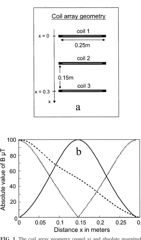
FIG. 1. The coil array geometry (panel a) and absolute magnitude of
magnetic-field strength along the central axis of the coil array for three different configurations of active turns (panel b).
increased at magnetic-field strengths ranging from 8
m
T to
500 mT (15–17). However, other studies have reported that
magnetic-field strengths of a similar range (6.3
m
T–50 mT)
have no effect on heat-shock protein expression in a variety
of biological models (18–21). The physiological
implica-tions of field-induced heat-shock protein expression are
un-known, but they could range from therapeutically beneficial
to detrimental. Hence it is important to establish whether
the reported effects are robust and to define the exposure
limits and the biological conditions of any induced effect.
This study was designed to determine whether exposure
of human peripheral blood to power-frequency magnetic
fields induces heat-shock protein gene expression in
leu-kocytes and to compare this response to that induced by
heat alone. The study also examined whether cells
subject-ed to mild heat (40
8
C) become more sensitive to a
concom-itant exposure to power-frequency magnetic fields.
MATERIALS AND METHODS
Power-Frequency Magnetic-Field Exposure System
A purpose-built three-coil exposure system was used to provide mag-netic-field exposures in the range 0–100mT at 50 Hz. Each coil was 25 cm in diameter and spaced 15 cm apart with the center axis vertical (Fig. 1). The coils were wound with multiple taps connected in series and driven by a single amplifier fed with a 50 Hz sinusoid from a master oscillator. In these studies, six 96-well plate holders were stacked in an open frame, vertically above each other, with gaps of several centimeters between them to ensure good air circulation. This structure occupied the 30-cm vertical span of the coil system. Sixteen samples were placed in a cluster of wells at the center of each plate such that they were all close to the central magnetic axis of the coil system. The number of active turns within each individual coil and their relative polarity were prese-lected before each experiment. This enabled a variety of magnetic-field spatial distributions to be created, and hence different plates within the stack were exposed simultaneously to different field strengths based on their position and the coil configuration.
The field distributions that can be generated by the exposure system include: zero field at the center of the stack with maxima at the top and bottom, maximum field at the center of the stack with zero at top and bottom, and zero at one end of the stack with a maximum at the other (Fig. 1). The finite depth of the blood sample (5 mm) and positioning variances result in a magnetic-field exposure variation of approximately 2mT through the sample at each nominal exposure amplitude. The ability to vary the relative magnetic-field exposure at each plate allows any effects due to magnetic fields to be distinguished from possible artifacts relating to the physical position of the plates. The two field distributions used in this study were maximum (100mT) at the bottom and zero at the top, and maximum at the top and zero at the bottom. These field distributions were alternated between sequential experiments.
The coil system was housed in a wooden incubator (to avoid distortion of the magnetic fields), and temperature control was maintained using a custom-designed, proportional control system. Temperature was logged throughout the course of a 4-h exposure and varied by less than60.18C at any one position. The spatial variation of temperature within the coil exposure system was less than60.28C. The magnetic-field intensity was not logged continuously throughout the experiments due to the intrinsic stability of the technology; however, the coil current was monitored and maintained at a constant value. The field distributions as measured using a Hall Effect probe agreed with theoretical calculations to within 1% of the maximum fields.
Cell and Culture Conditions
Peripheral blood was collected from healthy male volunteers (n53, age range 30–50 years) into Vacutainers containing EDTA anticoagulant (BD Biosciences, UK). Volunteers gave informed consent, and the study complied with the University of Sheffield’s REH Ethics policy on re-search involving human participants, data and tissue; UK National Health Service ethical approval was not required for the study. The rationale for using whole blood for these experiments, rather than leukocytes isolated from whole blood, was to minimize the stress to the cells before exposure. Whole blood was diluted 1:1 with RPMI 1640 growth medium pre-warmed to 378C and divided into aliquots (200 ml/well) into the four center wells of each of the four central strips in 96-well Stripwell plates (Costar). The lids were modified to allow air flow around the wells while in the exposure system. Samples were maintained at 378C for 1 h before the exposure period. Six plates were exposed simultaneously for 4 h, each plate to a different magnetic-field strength. Additional samples were maintained in a separate incubator at 378C for the same period, a subset of which was heat-shocked at 428C for the final 2 h of the 4-h experi-mental period (positive control).
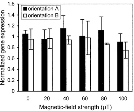
FIG. 2. Expression of the gene encoding HSP27 normalized to control
at 378C for power-frequency magnetic-field orientations A and B (means 6SEM), after 4 h exposure.
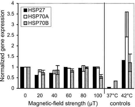
FIG. 3. Gene expression normalized to control at 378C. Data are means 6SEM for six experiments. Power-frequency magnetic-field exposure was 4 h. Heat was applied for the last 2 h of the experimental period for the 428C control samples.
Heat-Shock Protein Gene Expression
Heat-shock protein gene expression was determined by reverse tran-scriptase-polymerase chain reaction (RT-PCR). Messenger RNA was iso-lated from the leukocyte cell pellet using the PolyATract System 1000 according to the manufacturer’s recommended protocol (Promega Cor-poration, technical manual No. 228). Isolated RNA was reverse tran-scribed and the cDNA was amplified using Promega’s single-buffer Ac-cess RT-PCR system and primer pairs for HSP70A, HSP70B and HSP27 (Stressgen Biotechnologies Corp, Canada). The cycling conditions were: initial denaturation 948C for 2 min, denaturation at 948C for 30 s, an-nealing at 548C for 1 min, extension at 728C for 2 min, with the final extension at 688C for 7 min. Preliminary experiments determined the number of cycles that were required to ensure that the PCR products for each of the target genes lay within the linear portion of the amplification curve. RT-PCR products were electrophoresed through agarose gels con-taining ethidium bromide, and the fluorescence intensities of the amplicon bands were referenced to those of the housekeeping geneb-actin using Scion Image analysis software (Scion Corporation, Frederick, MD). Data are expressed as a ratio of these densities. The genes encoding HSP27 and HSP70A are constitutively expressed, and expression is increased after stress, whereas the gene encoding HSP70B is expressed only in stressed cells.
Data Analysis
All experiments were blinded to the individual performing the expo-sures and the assays, and they included negative (maintained at 378C in a separate incubator) and positive (heat-shocked at 428C for 2 h) controls. To compare results between experiments, the HSP/b-actin ratio was nor-malized to that of control samples that were maintained at 378C and not exposed to power-frequency magnetic fields. In the case of the experi-ments in which samples were exposed to power-frequency magnetic fields and heat concomitantly, normalization was made to the 0-mT exposure samples.
The null hypothesis that all populations had identical means was tested using one-way analysis of variance. Based on six experiments and the observed standard deviation, there was a 90% power to detect a 50% change in heat-shock protein gene expression at the P50.05 level. For the concomitant power-frequency magnetic fields and mild heat experi-ments (n53), there was a 90% power to detect a 60% change in gene expression at the P50.05 level.
RESULTS AND DISCUSSION
A total of six experiments were performed. In three, the
maximum magnetic field was at the bottom of the stack
(orientation A); the maximum field was at the top of the
stack in the remainder (orientation B). Samples at
physi-cally different positions could therefore be exposed to
iden-tical field strengths. The effects of six
magnetic-field strengths (0, 20, 40, 60, 80, 100
m
T rms at 50 Hz)
were investigated in each experiment. The vertical position
of the samples within the exposure system had no
discern-ible influence on heat-shock protein gene expression (Fig.
2), which would be anticipated given the small spatial
tem-perature variation (
6,
0.2
8
C) within the coil stack. Given
that there was no difference between the results from the
two magnetic-field orientations, the two data sets were
combined for the subsequent analysis of magnetic-field
ef-fects.
There were no significant differences (at P
5
0.05) in
gene expression at the end of a 4-h exposure for any of the
heat-shock proteins at any of the six magnetic-field
strengths (Fig. 3). The expression of the gene encoding
HSP70B (the inducible form of HSP70) in cells from
con-trol samples indicated that some degree of stress was
oc-curring, despite the fact that experiments were performed
using whole blood rather than isolated leukocyte
popula-tions. Incubation at 42
8
C for 2 h (as a positive control)
induced a 12-fold increase in expression of the gene
en-coding HSP70B, thus demonstrating that cells remained
re-sponsive to appropriate stimulation.
[image:4.612.317.547.67.253.2]433
[image:5.612.64.297.68.252.2]HSP INDUCTION IN HUMAN LEUKOCYTES EXPOSED TO POWER-FREQUENCY MAGNETIC FIELDS
FIG. 4. Gene expression (normalized to value for 0mT) for concom-itant heat (408C) and magnetic-field exposure (4 h). Heat was applied for last 2 h of the experimental period for the 428C control samples. Data are means6SEM for three experiments.
by differential centrifugation might elicit physical stress on
the isolated cells. While the presence of erythrocytes,
plate-lets and their lysis and secreted products in whole blood
might influence the observed responses, we opted to use
whole blood to minimize the stressing of the leukocytes.
We studied the potential influence of a concomitant stress
on the response to power-frequency magnetic fields by
rais-ing the temperature of samples (40
8
C) during exposure to
power-frequency magnetic fields. The elevated temperature
alone increased expression of the genes encoding HSP27,
HSP70A and HSP70B compared to that of cells maintained
at 37
8
C; however, the superimposition of power-frequency
magnetic fields had no additional effect (Fig. 4). Similar
findings have been reported for higher field strengths by
Miyakoshi et al. (19). However, these investigators have
also reported a decrease in HSP70 expression when
HL60RG cells were simultaneously exposed to 50 mT
pow-er-frequency magnetic fields and 40
8
C or 42
8
C for longer
than 5 h. Although the lower levels of heat-shock protein
expression, particularly HSP70B at 100
m
T (Fig. 4), in our
study might be interpreted as an inhibitory trend, the
dif-ferences did not reach statistical significance, and the small
sample size in these experiments does not allow such a
conclusion to be drawn.
Published data on the effects of power-frequency
mag-netic-field exposure on heat-shock protein expression are
equivocal, the reasons for which are currently unclear.
However, the difficulties in interpreting the literature can
be illustrated by studies that have used HL60 (human
pro-myelocytic leukemia) cells to investigate heat-shock protein
expression. Pipkin et al. (17) found that magnetic-field
strength was an important factor: 1 mT would elicit a
heat-shock protein response, whereas 100
m
T was insufficient.
In contrast, Lin et al. (16) found an effect at low intensities
(8
m
T), and Miyakoshi (19) found no effect with intensities
up to 50 mT. Whether cells respond to power-frequency
magnetic fields will be influenced by a number of factors,
including the intensity and duration of the exposure and the
cell type used to assess the response. At present it is not
possible to predict which factor or combination of factors
will elicit a biological response.
Based on the extant literature and our results, it is clear
that power-frequency magnetic-field exposure is not a
uni-versal physical stressor of cells in the same way as heat. If
power-frequency magnetic fields do indeed elicit changes
in the intracellular environment that are sufficient to induce
heat-shock proteins, then factors other than the magnitude
of the magnetic-field strength and exposure time appear to
be involved. The primary aim of our investigation was to
examine the effects of environmental power-frequency
magnetic-field amplitudes that encompass those
encoun-tered by the general public. In that context, our upper limit
of 100
m
T is considerably higher than normal exposure
levels. It is thus perhaps reassuring that normal leukocytes
appear not to be stressed by such fields.
CONCLUSION
In contrast to some studies (11–17) which have reported
on the induction of heat-shock proteins in a range of cell
types after exposure to power-frequency magnetic fields but
in agreement with others (18–21), we have found no
evi-dence that 50 Hz magnetic fields of amplitudes 0–100
m
T
induce expression of genes encoding heat-shock proteins in
human peripheral blood leukocytes. In addition, exposure
to mild heat at 40
8
C did not sensitize the cells to a
con-comitant exposure to power-frequency magnetic fields.
ACKNOWLEDGMENT
The authors wish to thank the EMF Biological Research Trust, UK for supporting this project (Grant No. BRT 00/13).
Received: March 6, 2003; accepted: November 17, 2003
REFERENCES
1. NRPB, ELF Electromagnetic Fields and the Risk of Cancer. Report of an Advisory Group on Non-ionising Radiation, Vol. 12, pp. 1–
179. National Radiological Protection Board, Chilton, 2001.
2. W. J. Welch, How cells respond to stress. Sci. Am. 268, 56–64 (1993). 3. J. Bulmer, A. E. Bolton and A. G. Pockley, Effect of combined heat,
ozonation and ultraviolet irradiation (VasoCare) on heat shock pro-tein expression by peripheral blood leukocyte populations. J. Biol.
Regul. Homeost. Agents 11, 104–110 (1997).
4. R. Voellmy, Transduction of the stress signal and mechanisms of
transcriptional regulation of heat shock/stress protein gene expression in higher eukaryotes. Crit. Rev. Eukaryot. Gene Expr. 4, 357–401 (1994).
5. R. I. Morimoto, D. A. Jurivich, P. E. Kroger, S. K. Mathur, S. P.
Murphy, A. Nakai, A. K. Sarge, K. Abravaya and L. T. Sistonen, Regulation of heat shock gene transcription by a family of heat shock factors. In The Biology of Heat Shock Proteins and Molecular
Chap-erones (R. I. Morimoto, A. Tissie`res and C. Georgopoulos, Eds.), pp.
6. U. Knauf, E. M. Newton, J. Kyriakis and R. E. Kingston, Repression
of human heat shock factor 1 activity at control temperature by phos-phorylation. Genes Dev. 10, 2782–2793 (1996).
7. J. Kim, A. Nueda, Y. H. Meng, W. S. Dynan and N. F. Mivechi,
Analysis of the phosphorylation of human heat shock transcription factor-1 by MAP kinase family members. J. Cell Biochem. 67, 43– 54 (1997).
8. R. I. Morimoto, Regulation of the heat shock transcriptional
re-sponse: cross talk between a family of heat shock factors, molecular chaperones, and negative regulators. Genes Dev. 12, 3788–3796 (1998).
9. C. Wu, Heat shock transcription factors: Structure and regulation. Annu. Rev. Cell Dev. Biol. 11, 441–469 (1995).
10. E. A. Nollen and R. I. Morimoto, Chaperoning signaling pathways:
Molecular chaperones as stress-sensing ‘heat shock’ proteins. J. Cell
Sci. 115, 2809–2816 (2002)
11. K. C. Chow and W. L. Tung, Magnetic field exposure stimulates
transposition through the induction of DnaK/J synthesis. Biochem.
Biophys. Res. Commun. 270, 745–748 (2000).
12. S. Carmody, X. L. Wu, H. Lin, M. Blank, H. Skopicki and R.
Good-man, Cytoprotection by electromagnetic field-induced hsp70: A mod-el for clinical application. J. Cmod-ell Biochem. 79, 453–459 (2000).
13. S. H. Li and K. C. Chow, Magnetic field exposure induces DNA
degradation. Biochem. Biophys. Res. Commun. 280, 1385–1388 (2001).
14. A. Di Carlo, N. White, F. Guo, P. Garrett and T. Litovitz, Chronic
electromagnetic field exposure decreases HSP70 levels and lowers cytoprotection. J. Cell Biochem. 84, 447–454 (2002).
15. T. Miyakawa, S. Yamada, S-I. Harada, T. Ishimori, H. Yamamoto and
R. Hosono, Exposure of Caenorhabditis elegans to extremely low frequency high magnetic fields induces stress responses.
Bioelectro-magnetics 22, 333–339 (2001).
16. H. Lin, M. Opler, M. Head, M. Blank and R. Goodman,
Electro-magnetic field exposure induces rapid, transitory heat shock factor activation in human cells. J. Cell Biochem. 66, 482–488 (1997).
17. J. L. Pipkin, W. G. Hinson, J. F. Young, K. L. Rowland, J. G.
Shad-dock, W. H. Tolleson, P. H. Duffy and D. A. Casciano, Induction of stress proteins by electromagnetic fields in cultured HL-60 cells.
Bioelectromagnetics 20, 347–357 (1999).
18. K. I. Kang, I. Bouhouche, D. Fortin, E. E. Baulieu and M.
Grazia-Catelli, Luciferase activity and synthesis of Hsp70 and Hsp90 are insensitive to 50 Hz electromagnetic fields. Life Sci. 63, 489–497 (1998).
19. J. Miyakoshi, Y. Mori, H. Yaguchi, G. Ding and A. Fujimori,
Sup-pression of heat-induced hsp-70 by simultaneous exposure to 50 mT magnetic field. Life Sci. 66, 1187–1196 (2000).
20. C. A. Morehouse and R. D. Owen, Exposure to low-frequency
elec-tromagnetic fields does not alter HSP70 expression or HSF-HSE binding in HL60 cells. Radiat. Res. 153, 658–662 (2000).
21. B. Shi, B. Farboud, R. Nuccitelli and R. R. Isseroff, Power-line
fre-quency electromagnetic fields do not induce changes in phosphory-lation, localization, or expression of the 27-kilodalton heat shock protein in human keratinocytes. Environ. Health Perspect. 111, 281– 287 (2003).
22. B. Junkersdorf, H. Bauer and H. O. Gutzelt, Electromagnetic fields
enhance the stress response at elevated temperatures in the nematode Caenorhabditis elegans. Bioelectromagnetics 21, 100–106 (2000).
23. H. O. Gutzeit, Interaction of stressors and the limits of cellular