by
Jillian Mary Packham, B. Sc. Hons., (U. C. N. W., Bangor)
Submitted in fulfilment of the requirements
for the degree of
Doctor
~fPhilosophy
This thesis is dedicated to the memory of my father, the late Brian James Packham B. Sc., who instilled in me a great love of natural history.
This thesis contains no material which has been accepted for the award of any other degree
or diploma in any tertiary institution and, to the best of my knowledge and belief, contains no
material previously published or written by another person, except where due reference is
made in the text of the thesis.
This thesis may be made available for loan and limited copying in accordance with the Copyright Act 1968
J. M. Packham
ABSTRACT
Nothofagus cunninghamii, or myrtle, is the dominant tree species in many Tasmanian and
Victorian cool temperate rainforest communities. The main cause of myrtle death in undisturbed stands is the disease myrtle wilt, which is caused by the pathogenic hyphomycete Chalara australis. Early literature, aerial surveys and aerial photography indicated that myrtle wilt was endemic in at least part (and possibly most) of the range of myrtle, but that in some areas, disease levels may have increased in the recent past.
In Tasmania, measured mortality rates were found to be variable but not escalating, with no apparent overall trend. A new estimate of annual mortality due to myrtle wilt was calculated to be 0.61 % p.a. Logging, thinning and reading of myrtle-dominated rainforest led to increased myrtle wilt incidence. For some disturbed areas, there was evidence that after an average of nine years, elevated myrtle wilt mortality levels declined to background levels. The spread of myrtle wilt into areas adjacent to disturbances was clearly detectable up to 180 m from the disturbance, although not all sites were affected.
Small, experimental stem wounds on myrtle saplings provided suitable infection courts for C. australis spores, with most infections occurring within 14 days. Functional root grafts
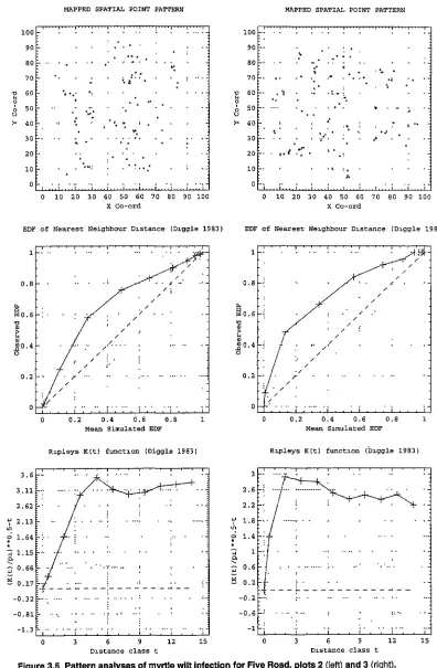
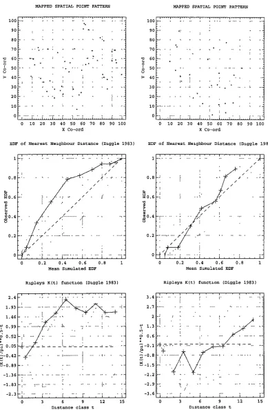
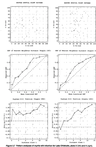
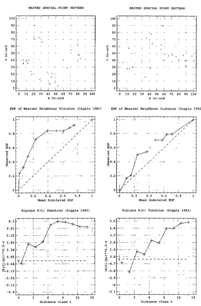
commonly occurred in young myrtles, and experimental inoculation, root excavation and sectioning strongly indicated underground, tree to tree transfer of C. australis. Re-isolations of C. australis were made from these trees, and characterisation of these isolates verified spread via root grafts. Root grafting probably predisposes stands to epidemics, plays a major role in the spread of myrtle wilt, and causes the clumped pattern of infected trees. Clumping occurred on a scale of 2.5-14 m and gave rise to patches of dead and diseased myrtles, often resulting in large gaps in the forest canopy.
Floristic studies showed that there was no distinct set of species which characterised myrtle wilt gaps, but that there were generally more myrtle seedlings than in control forest. Data from one site suggested that the vegetation composition in large, old gaps was reverting to that of the surrounding forest, and that myrtle was self replacing in such gaps. The probable
long-term effects of myrtle wilt on Tasmanian myrtles were investigated using a simple population model.
ACKNOWLEDGMENTS
This work has been jointly supervised by Humphrey Elliott, Glen Kile and Bob Hill. I am indebted to Humphrey for his advice, help and unfailing encouragement and enthusiasm; to Glen Kile for his consistent support of, and suggestions for, this work, and his constructive criticisms; and to Bob for guidance on botanical studies and thesis construction, and his administrative backup.
Many thanks also to John Hickey for administering the original project. Mick Brown suggested the aerial transect method and helped with design and analysis of botanical surveys. Ralph Cruickshank provided invaluable assistance with gel electrophoresis work, and Leanne Sherriff's reading thesis was most instructive. Unpublished data were kindly made available for my use by Humphrey Elliott, Bob Ellis, Glen Kile, John- Hickey and Sue Jennings. I am grateful to Phil Barker, Roger Beaver, Bob Ellis, John Hickey, Debbie Kent Lawrence Kirkendall, John Madden, Bob Mesibov and Tim Wardlaw for helpful comments.
Sue Jennings, Peter Duckworth and Sean Blake were of great help in providing information and organising field assistance, and John Traill allowed me to hijack his helicopter for an aerial survey. Thanks also to Gordon Hosking, Glenn Stewart and Rob Allen for their interest in this project and their hospitality while I was in New Zealand. Similarly, thanks to David Cameron, Jeff Jugovic and Ian Roberts for organising the Victorian surveys.
vi
The following people were brave enough to assist with field work and I would like to take this opportunity to apologise for the weather and for at least one case of mild hypothermia: T. Barber, M. Barker, R. Bashford, S. Candy, N. Cannon, S. Casey,
K.
Casten, S. Cook, M. Davies, G. Davis, H. Elliott, J. Fitch, G. Haig, G. Hall, M. Hall, J. Harries, J. Hickey, R. Hill (and students), S. Jennings, G. Kile, J. Lyn~h, M. Mahoney, S. McArthur, N. McCormick, R. Murray, N. Ramsden, R. Robinson, S. Rosa, J. Sargison, S. Scott, G. Todd, D. Wittie, A. Wallis and J. Weller.Many thanks to Malcolm Hall for much valued technical help and advice, and for the
provision and maintenance of fungal cultures. Thanks also to Dick Bashford for his technical help and advice, also for his sense of humour (particularly in the rain) and an endless supply of measuring tapes. Richie Robinson and Steve Casey were of invaluable assistance both in the field and in the laboratory and contributed much to this project. Greg Jordan, Yvonne Menadue and Tim Wardlaw assisted with photography, and Jane Heath did a marvellous job producing prints. I am also indebted to Kristen Williams, Mick Brown, Jean Jarman,
T
I am grateful to Des Hankin and Ian Woolley for tree felling, to Brian Denton and Leigh Johnson for their assistance in the workshop, and to Peter Bobbi for his help with constant temperature facilities. I would also like to acknowledge the late Charlie Pearson for his help in the workshop and with equipment design. Thanks to Bob Menary for the loan of his sledge microtome, to Leigh Johnson for repairs, and to Kate Clark for sharpening the blades. Barry O'Brady assisted with the calibration of equipment.
Steve Candy, Glen MacPherson, David Ratkowsky and Brad Potts provided invaluable statistical help and advice. I would particularly like to thank Steve Candy for writing the computer program represented by Figures 4.5-4.9, and for allowing me to use the program represented by Figures 3.3-3.8. John Donaldson, Nuno Borralho and Stuart Young advised on the conversion of the myrtle population model to the EXCEL format shown in Appendix 29.
I would like to thank Tim Wardlaw, Steve Candy, Yvette Brown, Jean Jarman and Mirranie Barker for their help with computer systems; Judy Deans, Judy Broadby and Carol Foyle for typing; Yvette Brown for data entry; Jenny Gray and Tony Rainbird for Figure 1.5; and Bill Brown for help with graphics. My thanks are due also to Mike Brouder, John Harris and Tony Rainbird who helped so much with maps, photo interpretation and aerial photographs, respectively.
Many thanks to Humphrey Elliott, Glen Kile and Bob Hill for their extensive and constructive comments on the manuscript, also to Mick Brown, Ralph Cruickshank, D_avid Ratkowsky, John Madden and Ken Old for their criticism of different sections of the thesis. Steve Candy and John Hickey made comments on earlier drafts, and Tony Mount suggested the title for Chapter 7. Margaret Aldridge, Debbie Ploughman and Andrew Wilson kindly proof read the work, while Judy Sprent did a wonderful job of locating relevant literature.
Work in remote sites was partially funded by the World Heritage Area Programme and the New Zealand trip by a Maxwell Ralph Jacobs Award. The original myrtle wilt project, and the Victorian surveys were jointly funded by State and Commonwealth Governments under the National Rainforest Conservation Program. Continuation of the work was made possible by a OPIE Forestry Postgraduate Studentship. I am most grateful to Forestry Tasmania, for allowing me to participate in the assisted study scheme, and for granting me extended leave to complete the thesis.
viii
CONTENTS
ABSTRACT ...
v
ACKNOWLEDGMENTS ... vi
1. INTRODUCTION ... ~ ... 1
2. MYRTLE WILT IN SPACE AND TIME ... 24
2.1 INTRODUCTION ... 24
2.2 MATERIALS AND METHODS ... 29
2.3 RESULTS ... 36
2.4 DISCUSSION ... 41
2.5 SUMMARY ... 51
3. INCIDENCE LEVELS AND MORTALITY RATES ... 53
3.1 INTRODUCTION ... 53
3.2 MATERIALS AND METHODS ... 54
3.3 RESULTS ... 60
3.4 DISCUSSION ... 66
3.5 SUMMARY ... 72
4. DISTURBANCE AND MYRTLE WILT ... 73
4.1 INTRODUCTION ... 73
4.2 MATERIALS AND METHODS ... 74
4.3 RESULTS ... 81
4.4 DISCUSSION ... 86
4.5 SUMMARY ... 90
5. WOUNDS, ROOT GRAFTS AND MYRTLE WIL T.. ... 91
5.1 INTRODUCTION ... 91
5.2 WOUNDS AS INFECTION COURTS FOR CHALARA AUSTRALIS ... 94
5.2.1 BACKGROUND AND AIMS ... 94
5.2.2 MATERIALS AND METHODS ... 97
5.2.3 RESULTS AND CONCLUSIONS ... 101
5.3 ROOT GRAFTING IN NOTHOFAGUS CUNNINGHAM/1 ... 110
5.3.1 BACKGROUND AND AIMS ... 110
5.3.2 MATERIALS AND METHODS ... 111
5.3.3 RESULTS AND CONCLUSIONS ... 114
5.4 CHALARA AUSTRALIS ISOLATIONS FROM ROOT GRAFTS ... 119
5.4.1 BACKGROUND AND AIMS ... 119
5.4.2 MATERIALS AND METHODS ... 121
5.5 DISCUSSION ... 139
5.6 SUMMARY ... 153
6. MYRTLE WILT AND RAINFOREST FLORISTICS ... 156
6.1 INTRODUCTION ... 156
6.2 MATERIALS AND METHODS ... 159
6.3 RESULTS ... 165
6.4 DISCUSSION ... 181
6.5 SUMMARY ... 190
7. THE DECLINE AND FALL OF THE HOLEY RAINFOREST EMPIRE? ... 192
7.1 INTRODUCTION ... 192
7.2 MATERIALS AND METHODS ... 192
7.3 RESULTS ... 196
7 .4 DISCUSSION ... 197
7.5 SUMMARY ... 200
REFERENCES ... 201
1. INTRODUCTION
AN INTRODUCTION TO MYRTLE WILT
Nothofagus cunninghamii (Hook.) Oerst. or myrtle, is the dominant tree species in many of the Tasmanian and Victorian cool temperate rainforest communities. The main cause of myrtle death in undisturbed Tasmanian stands is the disease myrtle wilt (Elliott et al. 1987).
The phenomenon of myrtle wilt, or myrtle dieback as it was initially called, was first noted by Howard (1973a) in both disturbed and undisturbed forests in north-west Tasmania. The deaths of large groups of mature myrtles were associated with accelerated attack by an ambrosia beetle, thought to be a vector of a fungal pathogen. The beetle was identified as
Platypus subgranosus Schedl, the mountain pinhole borer (E. C. Zimmerman, unpublished
data).
The pathogen was found to be a previously unrecorded hyphomycete, Chalara australis Walker and Kile. A vascular stain disease rather than a true vascular wilt, C. australis produces dark brown radial streaks in the wood of infected myrtles. Symptoms are wilting, followed by leaf death; the dead leaves being retained on the trees, giving them an
orange/brown appearance (Kile and Walker 1987; Kile et al. 1989).
1
Kile and Hall (1988) showed that C. austra/is was not dependent on P. subgranosus for its spread or entry into trees, and that infection actually occurred prior to attack by these
beetles. Wounding of trees was shown to produce a direct infection site for C. austra/is (Kile and Walker 1987). The tunnels and frass of P. subgranosus were found to be a good early indicator of the disease, but not its cause (Kile and Hall 1988).
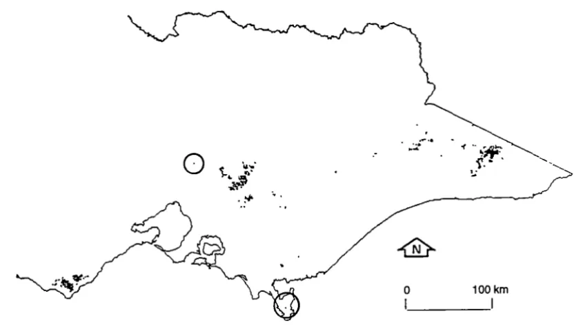
-Figure 1.1 The distribution of cool temperate rainforest in Tasmania. (From Hickey et al. 1993.)
0
[image:11.564.180.449.69.370.2]··,h..~··· ~.-...,.
Figure 1.2 The distribution of cool temperate rainforest in Victoria.
(From Cameron 1992.)
o 100 km
[image:11.564.99.515.452.689.2]-2
SPECIES NOMENCLATURE
Species nomenclature follows: Buchanan et al. (1989) for the Tasmanian flora; Hnatiuk (1990) for the mainland Australian flora; Hill and Read (1991) for Nothofagus species; Schedl (1972) and Roberts (1979, 1986) for Platypus species; Nag Raj and Kendrick (1975) and Kile and Walker (1987) for Chalara species; and Seifert et al. (1993a) for Ceratocystis and Ophiostoma species. Other references are cited in the text.
NOTHOFAGUS
The genus Nothofagus (Fagaceae) is considered to be an important Gondwanic relict, with extant species being distributed in the southern cool temperate zone; in Argentina and Chile (nine species), south-eastern Australia (three species) and New Zealand (five species); also occurring in the cooler tropical highlands of New Guinea (13 species) and New Caledonia (five species)(Wardle 1984; Hill 1990; Hill and Read 1991). Its distribution does not overlap those of the other genera in the Fagaceae (Hutchinson 1973). The genus is divided into four subgenera: Nothofagus; Lophozonia; Brassospora and Fuscospora (Hill and Jordan 1993).
Of the three Australian species, N. cunninghamii (myrtle) is the most widely distributed, being the dominant tree in many of the Tasmanian and Victorian cool temperate rainforest communities (Figures 1.1 and 1.2). N. moorei (antarctic beech) is found in the cool temperate rainforests of northern New South Wales and southern Queensland, while N. gunnii (fagus), is a deciduous Tasmanian native 1, restricted to high altitude areas but co-occurring with myrtle in parts of its range (Jarman et al. 1984, 1991; Robertson and Duncan 1991 ). N. cunninghamii and N. moorei belong to the subgenus Lophozonia, while N. gunnii belongs to the subgenus Fuscospora (Hill and Jordan 1993).
Myrtle occurs in pure rainforest, as an understorey to Eucalyptus species in mixed forest, and in many swamp, riparian and scrub forest communities (Jarman et al. 1984, 1991; Kirkpatrick et al. 1988; Cameron 1992; Pannell 1992). Tasmanian rainforest vegetation has been divided into broad groups on the basis of community structure and floristics. Three of these groups contain myrtle as a dominant species:
• callidendrous rainforest has tall, well formed trees and an open understorey;
• thamnic rainforest has well formed trees of moderate height, and a distinct shrub layer;
1
• implicate rainforest has shorter trees, a tangled understorey and an uneven broken canopy (Jarman et al. 1984, 1991 ).
Typically callidendrous forest occupies more fertile sites than thamnic or implicate forest. In Tasmania, myrtle-dominated rainforest is very extensive and occurs in large and often continuous areas in the west/south-west, while in the east it is usually confined to small, remnant patches (Jarman et al. 1984; Neyland 1991; Neyland and Brown 1994). The distribution of mixed forest (with a mature myrtle understorey) is similar but extends further to the north and east, into drier and more fire-prone sites (Hickey and Sawa 1992). Swamp forests containing myrtle are largely restricted to north-west Tasmania (Pannell 1992).
In Victoria the definition of cool temperate rainforest is broader and includes some
secondary rainforest communities. Myrtle is generally restricted to small patches (often gully communities), and its distribution is limited to sites in the Central Highlands, the Otway Ranges and the Strzelecki Ranges, with some isolated occurrences on Wilsons Promontory (Cameron 1992). Victorian primary rainforest which contains myrtle, broadly resembles Tasmanian callidendrous rainforest.
THE CERATOCYST/S COMPLEX AND CHALARA
The form genus Chalara belongs to the Fungi lmperfecti (Deuteromycotina:
Hyphomycetidae). Where teleomorphs (perfect forms) of Chalara are known, they are Ceratocystis species (Ascomycotina).
The genera Ceratocystis, Ophiostoma, Ceratocystiopsis and Sphaeronaemella together form what has been termed 'the Ceratocystis complex' (De Hoag and Scheffer 1984). The taxonomy of this group has been revised by De Hoag (1974), De Hoag and Scheffer (1984), and Harrington (1987). A synopsis of the taxonomic revisions of Ceratocystis is given by Perry (1991 ). Wolfaardt et al. (1992) produced a synoptic key and computer database for identification of species of Ceratocystis sensu lato. Subsequent revisions have been made by Hausner et al. {1993a, 1993b).
The Ceratocystis complex
Species within the Ceratocystis complex may have several anamorphs belonging to different form genera (of which Chalara is only one). Upadhyay and Kendrick (1975) listed 16
possible conidial anamorphs: Hya/odendron; Sporothrix; Hyalorhinocladiel/a; Verticicladiella; Pesotum; Hyalpesotum; Pachnodium; Leptographium; Graphium; Graphilbum; Acremonium;
and Kendrick (1975) considered Chalaropsis and Thielaviopsis to be synonyms of Cha/ara, while Upadhyay (1981) included Gabardnaudia, Graphioc/adiella and Phialographium. Wingfield et al. (1991) considered Phialographium and Pesotum to be synonyms of Graphium, although this has been disputed by Upadhyay (1993a).
4
While Ophiostoma was at one stage regarded as a synonym for Ceratocystis (Upadhyay and Kendrick 1975), more recently, differences between the genera have become apparent. De Hoag and Scheffer (1984) proposed the separation of Ceratocystis Ellis and Haist. sensu Jato into Ceratocystis sensu sticto, which was restricted to species with Chalara anamorphs;
and Ophiostoma, which was composed of species which lacked Chalara anamorphs, had rhamnose in their cell walls, and were resistant to cycloheximide. Upadhyay (1993b) considered that the absence of information on the cell walls of some species, rendered the taxonomic application of such information premature. However, Nag Raj and Kendrick (1993) suggested that Chalara anamorphs of Ceratocystis species were likely to be of monophyletic origin. Samuels (1993) considered that Ceratocystis, Ophiostoma and Ceratocystiopsis were distinct genera, whose anamorphs indicated different lines of
derivation. The Ophiostomataceae were retained as a distinct family of the Xylariales.
Phylogenetic analysis of partial ribosomal DNA sequences supported the separation of Ceratocystis and Ophiostoma, and it was suggested that the teleomorphic similarities of the
two genera were due to convergence, possibly due to the independent evolution of insect dependant ascospore dispersal mechanisms. It was suggested that Ophiostoma should remain the sole genus of the Ophiostomataceae, which should be the sole family within the Ophiostomatales, whereas Ceratocystis would be best disposed within the Microascales (Hausner et al. 1992, 1993a, 1993c, 1993d). Ceratocystiopsis was reduced to synonymy with Ophiostoma (Hausner et al. 1993b).
Using similar methods, the results of Spatafora and Blackwell (1994) supported the
separation of Ceratocystis and Ophiostoma. Ceratocystis and Sphaeronaemella were more closely related, and were proposed as sharing a recent common ancestor with members of the Microascales.
Some species in the Ceratocystis complex e.g. Ophiostoma u/mi (Buism.) Nannf., are heterothallic, requiring two mating types to produce the teleomorph. Others, e.g.
not been found, implying that the second mating type is either absent or at a very low frequency in the population (C. M. Brasier, personal communication).
The Ceratocystis complex, with its imperfect forms, has a worldwide distribution (Upadhyay 1981) and includes a number of economically and ecologically important species. Diseases caused by these organisms occur on diverse hosts (mainly angiosperms), and include vascular wilts, vascular stain diseases, canker diseases, root and stem rots and rots of fruits, tubers, seed pods, leaves and buds. Major methods of dispersal of pathogenic species are via soil, wind and water, root-grafts, insect or animal vectors, insect frass or via pruning and other tools (Kile 1993).
Ceratocystis species are some of the primary fungal colonisers of timber, and are able to
invade both moribund and living tissue (Dowding 1984). Many are wood-staining fungi, both saprophytes and pathogens (Seifert 1993). The majority of the saprophytic wood-staining fungi are found in bark beetle galleries (Hutchison and Reid 1988).
In general, Ceratocystis species are most frequently reported from habitats other than beetle galleries (Malloch and Blackwell 1993). However, some Ceratocystis and Ophiostoma species exhibit complex symbiotic relationships with insects. While Ophiostoma species are generally identified with bark beetles, Ceratocystis species may be associated with ambrosia beetles, but tend to form less specialised arthropod host relationships (Spatafora and Blackwell 1994). Notable pathogens include 0. ulmi and 0. novo-ulmi Brasier, causal agents of Dutch elm disease, and Ceratocystis fagacearum (anamorph Chalara quercina), causal agent of oak wilt.
Dutch elm disease
Ophiostoma u/mi and 0. novo-ulmi are vectored by bark beetles (Webber 1990). Both
conidiospores (asexually produced) and ascospores (sexually produced) are carried by the beetles, although conidiospores are probably the most important. Trees become infected via both the feeding groves and the breeding tunnels made by the insects (Webber and Brasier 1984). Root grafting is known to be an important mechanism for local disease spread (Neely and Himelick 1963).
6
recognised as a separate species, 0. novo-u/mi (Brasier 1991 ), which has two distinct races. The NAN race is thought to have originated in North America and is now spreading
eastwards through Europe. The EAN race probably originated in Central Europe and is spreading westward. The NAN and EAN races of
0.
novo-ulmi are considered jointly responsible for the recent outbreaks of Dutch elm disease in Western Europe, and are progressively replacing the less aggressive 0. u/mi (Brasier 1979).The current Dutch elm disease pandemic has encompassed most of Europe, eastern North America and south-west Asia. During the 1970s the disease killed 20 million elms (Ulmus Linn. species) in Great Britain alone. There is evidence that the disease will not only attack mature elms, but also the seedlings and suckers that arise to replace them (Webber and Brasier 1984). Thus over the next 40 years the disease is predicted to reduce European field elms to an understorey or scrub population, with occasional escapes in mountain valleys (Brasier 1983). Dutch elm disease has also been proposed as a possible cause of the Neolithic elm decline in north-west Europe (Perry and Moore 1987; Turner and Hodgson 1991 ). In Italy, a recent survey identified only about 100 large (bole diameter greater than 1 m) elms in the country (Mittempergher 1989).
Cha Iara
The form genus Chalara comprises over 80 known species, of which about ten are known plant pathogens, a number of these having considerable economic importance (Kile and Walker 1987): C. thielavioides is associated with root and graft rot in walnut, carrot and lupin; C. neocaledoniae causes a vascular stain disease in coffee and guava and C. popu/i causes a canker disease in poplar and willow (Kile 1993).
It is difficult to assess the distribution of Chalara in Australasia and South America. Nag Raj and Kendrick (1975) listed 70 species, of which 28 were recorded from New Zealand and one from Australia. Later workers have added to this list (e.g. Kile and Walker 1987; Old et al. 1991) but the distribution of known species still probably reflects that of mycologists,
rather than that of Chalara!
Kile and Walker (1987) listed Chalara species recorded on plants in the family Fagaceae. Most were saprophytic, with some causing timber spoilage. There were only three parasitic species: the Cha/ara anamorph of Ceratocystis fimbriata on Fagus and Quercus species; Cha/ara quercina causing oak wilt of fagaceous species in North America; and C. australis
-one third of the known species of Chalara recorded on them, may have some significance as hosts for the genus.
Oak wilt
Chalara quercina has affinities with C. australis, both in terms of its morphology (Nag Raj
.
and Kendrick 1975) and in causing a vascular infection of a Fagaceous host (Kile and Walker 1987). Oak wilt was described from Quercus species by Henry et al. (1944), and was at that stage widespread in much of Wisconsin, and also present in Minnesota, Iowa and Illinois (USA). The anamorph was described by Henry (1944) and the teleomorph ( Ceratocystis fagacearum), discovered in culture, was described by Bretz (1952).
The disease is vectored by nitidulid beetles. These beetles carry both conidiospores and ascospores to wounds caused by woodpeckers, which they use for sap feeding (Dowding 1984; Webber and Brasier 1984). Local spread of the disease also occurs via root grafts (Kuntz and Riker 1950, Beckman and Kuntz 1951).
Anderson and Anderson (1963) investigated the rate of spread of oak wilt in southern Wisconsin and south-eastern Minnesota between 1955 and 1959. They monitored both the new infection centres, which became established through relatively long distance spread of the fungal spores, and the increase in size of existing centres, as the fungus spread through root grafts and by local spread of spores. They concluded that there was a slow, steady build up of the disease likely to result in substantial long-term losses. By 1981 the disease had been reported over a wide area from 18 states, and was found to be attacking all native species of oak, regardless of size, age or vigour (Upadhyay 1981 ). However, Tryon et al. (1983) have reported abundant oak regeneration in oak wilt infection centres in West Virginia.
Cha/ara australis and myrtle wilt
8
In myrtles, C. australis causes brown, radial streaks across multiple growth rings, suggesting that it is not a true vascular wilt. Unlike Ceratocystis fagacearum and 0. ulmi, it does not
become systemic in the vascular system, but is generally restricted to the roots and lower to
mid stem (Kile and Walker 1987). Clumping of diseased trees and relationships between
disease incidence and stand density are considered indicative of below ground spread
(Elliott et al. 1987; Kile et al. 1989).
Conidiospores are produced by sporulating black felts which sometimes form on the bark of
infected myrtles or other wood surfaces, mainly in the autumn and winter (Kile et al. 1989).
Two endoconidial forms are produced (Kile and Walker 1987; Kile 1993). lnoculum appears
to be air or water dispersed, and this has been confirmed in rainforest areas by
inoculum-trapping experiments, using fresh myrtle billets and rainwater collection. Infection of wounds
to the trunk, crown or roots, is thought to be the origin of most new disease foci (Kile et al.
1989).
These results contrast strongly with those of Dowding (1984), who found that the sticky
conidiospores of Ceratocystis species were very short lived and susceptible to sunlight and
UV light, although ascospores were longer lived. Since spores were produced under the
bark, he deduced that adaptations for air dispersal were unnecessary, and that spore
stickiness could not be an adaptation to dispersal by rain splash since the site of spore
production and the infection court were usually protected from heavy rain. He concluded
that the spores were adapted for transfer by arthropods.
Kile and Walker (1987) reported myrtle wilt in the Otway Ranges of Victoria, while Elliott et
al. (1987) showed the disease to be widespread in undisturbed Tasmanian myrtle forest. A
number of workers have reported the presence of myrtle wilt in remote Tasmanian sites
(Jarman et al. 1984; Working Group for Nature Conservation 1987, Kile et al. 1987).
The closest known relative of Chalara australis is a new species of Chalara, which has been
isolated from wounds in Eucalyptus sieberi and E. obliqua in Victoria (Old et al. 1991 ). The
distributions of both species are known to adjoin or overlap that of myrtle in Victoria (Hogan
BARK AND AMBROSIA BEETLES AND PLATYPUS
The Coleopteran subfamilies Platypodinae and Scolytinae (Curculionidae) contain a number of genera which attack trees and timber (Lawrence and Britton 1991 ), and are arguably responsible for killing more trees than any other natural cause (Wood 1982). The Scolytinae have a world-wide distribution whereas the Platypodinae are predominantly tropical. The group can be divided on the basis of feeding habits; the phloeophagus scolytids are known as bark beetles, and the xylomycetophagus platypodids and scolytids as ambrosia beetles (Batra 1963; Francke-Grosmann 1967; Cooke 1977). P. subgranosus is thus an ambrosia beetle.
Bark and ambrosia beetles are usually the primary colonists of recently injured, standing trees and newly felled logs (Wood 1982). Bark beetles are the most important invertebrate colonists and their activities facilitate the entry of other organisms. These beetles may become vectors of sticky-spared Ceratocystis species, when emerging adults come into contact with fungal inoculum (Dowding 1984; Carpenter et al. 1988).
The host specificity of bark beetles has been shown to be lower in tropical than in temperate regions. This was thought to be due to the greater species diversity and heterogeneity of tropical forests; in such an environment polyphagy can reduce the problems of host-finding. In contrast ambrosia beetles (which are relatively more important in the tropics), show low host specificity in both regions. However, these beetles feed on the same ectosymbiotic ambrosia fungi, regardless of the host tree species, and it is really the fungi that are polyphagous, although it is the beetle that selects the host (Beaver 1979).
Whilst ambrosia beetles generally have an extremely wide host range, they usually occur on diseased or dead trees (Francke-Grosmann 1967). Thus, in general, with ambrosia beetles, hosts tend to be selected on the basis of their health status rather than their species.
Bark beetles
species (Rane and Tatar 1987), or they may be pathogenic, e.g. 0. ulmi and 0. novo-ulmi, vectored by Scolytus scolytus (F.) and S. multistriatus (Marsham), on Ulmus species (Webber 1990). Beetles may also be instrumental in bringing together the two mating strains in heterothallic species e.g. C. fagacearum (Upadhyay 1981 ).
Ambrosia beetles
10
Ambrosia beetles are not primarily responsible for the death of trees, and inhabit dead or dying wood, often heartwood. They are wood borers but not wood feeders, and have a symbiotic relationship with the ambrosial fungi which are cultured in their tunnels and used as a food source. They are attracted to unhealthy trees, or to those suffering from a
temporary reduction in vitality as a result of environmental conditions, and require wood with a relatively high moisture content (Fisher et al. 1953; Cooke 1977).
Ambrosia fungi are transported by adult scolytids in specialised depressions or invaginations of the body surface, known as mycangia (Batra 1963) or mycetangia (Francke-Grosmann 1967). In platypodids the mycetangia, if present, have a much simpler form, being only pits or notches in the integument. The taxonomy of ambrosial fungi has been reviewed by several authors, and most are hyphomycetes (Batra 1963; Francke-Grosmann 1967). It is uncertain whether ambrosia fungi are specific to beetle species, groups of beetles or to the host plant (Fisher et al. 1953).
Ambrosia beetles may also be important disseminators of saprophytic wood staining fungi (Fisher et al. 1953). Blue-stain fungi, including Ceratocystis species, are often associated with beetle tunnels, although in relation to the ambrosia fungi these are 'weed' fungi (Cooke 1977; F,rancke-Grosmann 1967).
While bark beetles are commonly vectors of pathogens, ambrosia beetles have not been shown, conclusively, to transmit pathogenic fungi, and attack by native ambrosia beetles on healthy, living trees in natural forest is unusual. Specifically, P. subgranosus is a secondary factor in myrtle wilt, the beetles attacking trees already infected by Chalara australis (Kile and Hall 1988).
Platypodid ambrosia beetles
suffered from drought, fire, floods, gales, lightning, sudden exposure and sunburn, volcanic eruptions and similar calamities (Kalshoven 1960).
Host selectivity in ambrosia beetles generally (Beaver 1979), and in platypodids specifically (Kalshoven 1960), is thought to be low. However, work by Browne (1958) in the humid tropics of south-east Asia indicates that this may be an oversimplification. He found that 30 percent of the known ambrosia beetle species were highly host selective, with selectivity occurring throughout particular gene~a. and with closely related species tending to select the same hosts. Hosts were normally selected at the level of botanical family, with very few families being affected. Highly evolved groups were more selective than primitive groups. It is possible that the specificity of the beetles for certain trees is the result of the specific requirements of their particular fungus (Fisher et al. 1953).
Browne (1958) found that the Fagaceae were attractive to ambrosia beetles in general, but also provided the hosts of many selective species. Notably, Platypus species in the section
Platypi spinulosi were typically associated with Fagaceae. He also found that Platypus were
borers of large timber, and did not record any species attacking timber less than 7.5 cm in diameter.
The predominantly tropical distribution of the platypodids is reflected in the number of species recorded by Schedl (1972) from Australasia and South America: New Guinea (123 species); New Caledonia (one species); Australia (30 species); New Zealand (three species) and Argentina (14 species).
Platypus subgranosus and myrtle
wilt
Platypus subgranosus was first described by Schedl (1936), and is in the section Platypi
semiopaci of the genus (Schedl 1972). It has been identified from Tasmania, Victoria and
Queensland.
The blue-stain fungus Leptographium lundbergii Lagerberg and Malin has been constantly isolated from the tunnels of P. subgranosus and was thought to be the ambrosia fungus (Webb 1945). However, Batra (1963) noted that in relation to the beetles, this was a non-specific fungus, which neither formed the characteristic ambrosia phase, nor was actually observed to serve as their food source.
12
records were of dead and dying myrtles (Howard 1973a), but it has subsequently been found in the dead logs of other rainforest species: Atherosperma moschatum; Eucryphia lucida;
Pittosporum bicolor; Phyllocladus aspleniifolius; Anodopetalum biglandulosum (Elliott 1978)
and Lagarostrobos franklinii (H. J. Elliott, unpublished data). It has also been recorded from
unhealthy specimens of the introduced Pinus radiata D. Don (Elliott and De Little 1984). It is widely distributed throughout the state, in rainforest and mixed forest (Elliott et al. 1982; 1987).
In Victoria it was recorded from fire-killed timber in the Central Highlands, following the devastating bush fires of 1939. Eucalyptus regnans, E. delegatensis, E. goniocalyx, E.
obliqua and myrtle were all attacked. P. subgranosus attacked only unhealthy or damaged
green trees, or dead trees which had not become too dry. Standing, fire-killed trees and those in dry log dumps were not badly attacked; this was in contrast to those in water-sprayed dumps where sprays provided ineffective water coverage. Although a number of other Eucalyptus species were involved, E. goniocalyx and myrtle appeared to be most susceptible (Hogan 1944; 1948).
In Queensland it has been recorded from the Dividing Range (Hogan 1948), Palen Creek, Mt Glorious and Brisbane, from Scolopia brownii (fallen log), Brachychiton populneum (sawn timber), B. acerifolium, Eucalyptus maculata and from Pterocymbium beccarii K. Schum., introduced from New Guinea (Schedl 1979).
In Tasmania P. subgranosus attack on standing myrtles is highly indicative of myrtle wilt. The comprehensive survey of Elliott et al. {1987) found very few live myrtles with evidence of having survived P. subgranosus attack (which would indicate they were never infected with
C. australis, but were attractive to the beetle). Although P. subgranosus will attack wounded
or burnt myrtles, Kile et al. (1992) showed t~at only myrtles infected with C. australis suffer sustained attack. Hogan (1944) indicated that standing, fire-killed trees were unlikely to be attacked, probably due to the relatively low moisture content found in dead standing trees. Ethanol (produced from fermenting wood) is known to act as a primary attractant and boring stimulant for P. subgranosus and it is thought that volatiles produced from diseased trees attract the beetles (Elliott et al. 1982, 1983; Kile et al. 1992).
Elliott et al. (1987) found that there was a very low proportion (1.2%) of standing myrtles which were dead or dying, but not attacked by P. subgranosus. This would indicate either that trees dying from causes other than myrtle wilt are not attractive to the beetle, or, that not
would only lead to a slight underestimate of C. austra/is infected trees. Thus P. subgranosus attack on standing myrtles is a relatively accurate indicator of myrtle wilt.
Kile and Hall (1988) undertook a series of studies to assess P. subgranosus as a vector of
C. australis and this work has been extended by Candy (1990). P. subgranosus was found
to lack mycetangia and is therefore considered to be fairly primitive. C. australis could not be isolated from any of the beetles emerging from infected trees or billets, or from those trapped in flight. This was attributed to the fact that most of a P. subgranosus brood emerges from an attacked tree the second summer after infection. These beetles are unlikely to become contaminated (and therefore unlikely to be vectors) because after this time the survival of C. australis in the above-ground wood is minimal. However, C. australis was successfully isolated from P. subgranosus within infected trees (particularly those trees currently dying or recently dead), so it is possible that the beetles and their larvae promote within-tree spread of the fungus.
For recently wounded trees there was no apparent relationship between P. subgranosus attack and C. australis infection: some trees which were not attacked by P. subgranosus became infected with C. australis, while some attacked trees remained uninfected for nine weeks. Whilst the theory of a direct vectoring role for P. subgranosus was disproved, it was conceded that a small percentage of the beetles may carry and transmit C. australis. Also, Kile et al. (1989) indicated that wind-borne beetle frass (contaminated with C. australis conidia and phialides) could be an inoculum source, particularly during the summer months. If proven, this would imply a degree of mutualism between P. subgranosus and C. australis.
GONDWANIC PARALLELS?
In brief, the distributions of Nothofagus, Chalara!Ceratocystis and Platypus are, respectively, Gondwanic, worldwide and largely tropical, with both Chalara/Ceratocystis and Platypus occurring on a wide range of host species. This being the case, numerous, phylogenetic parallels with myrtle wilt appear unlikely, although both Chalara (Kile and Walker 1987) and Platypus (Browne 1958) have been shown to exhibit a degree of specificity for the
Fagaceae.
However, since the distribution of Nothofagus does not overlap those of other members of the family (Hutchinson 1973), an attempt has been made to collate the accessible
information relating to Chalara, the Ceratocystis complex and Platypus species on
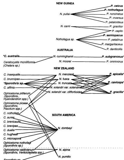
N. carrii --==============~P. graci/ior P. capito
AUSTRALIA
"'C. australis N. cunninghamii---~. subgranosus"'
Ceratocystis
moniliformi~
N. moorei P. omnivorus(Cha/ara sp.)
Ophiostoma piliferum (Sporothrix.,
Hyalodendron spp.) Ophiostoma piceae (Sporothrix, Pesotum spp.)
C. microspo
Ophiostoma nothofa (Sporothrix sp.)
NEW ZEALAND
SOUTH AMERICA
N. dombeyi
[image:24.569.108.537.82.638.2]N. pumilio
Figure 1.3 Associations of NothoftJgus, Ch11/ara, the Csratocystls complex and Platypus.
• Nothofagus species above the dotted line are evergreen, those below 1t are deciduous
• Anamorphs of Ceratocystis and Ophiostoma species shown in parentheses • • Denotes a known fungaVambrosia beetle association
Walker (1987) and Wardle (1984), the following sources were used: Faulds (1973); Milligan (1974); Roberts (1979, 1986, 1987); Browne (1983); Butin and Aquilar (1984); P. Hadlington (unpublished data); H. Peredo (unpublished data, cited in USDA (1993)).
Although there are no relevant records from Nothofagus in New Caledonia, Chalara neocaledoniae is known to occur on other hosts in this region (Kile and Walker 1987). Of
the Chalara species, only C. nothofagi is known from Nothofagus hosts in both New Zealand and in South America. However, it is notable that two Ophiostoma species, 0. picea and 0. pilifera have been recorded from both New Zealand and South American Nothofagus
species. 0. pilifera, 0. picea and 0. valdiviana have been found on both evergreen and deciduous Nothofagus species.
The apparent absence of Platypus species on Nothofagus in South America is thought to be due to two main reasons. Firstly, there are no host records at all for the great majority of South American Platypodinae; secondly there are no records of any Platypodinae in Chile. Particularly in the south of the country (within the range of Nothofagus), enough work has been done to make it likely that the family is actually absent there, although reasons for this absence are not clear (R. A. Beaver, personal communication).
The only known pathogens are C. australis, from Tasmania and Victoria, and an unknown species of Sporothrixfrom New Zealand (Faulds 1973). Except for an unidentified Ceratocystis species isolated from Platypus tunnels in N .fusca (Faulds 1973), these are
also the only species from the Chalara!Ceratocystis complex known to have an association with Platypus species.
Sub-lethal attack by Sporothrix is associated with the formation of pathological wood (Litchwark 1978) in N. fusca and N. menziesii, and Wardle (1984) concluded that there was a relationship between Platypus attack and the formation of core rots in New Zealand Nothofagus species. It is of interest that in New Guinea, P. celcus and P. nothofagus were
recorded from live N. pullei, but only from trees which had heart rot. P. semiopacus was also recorded from live Nothofagus (Roberts 1979). Arentz (1988) assumed that this was because the Nothofagus species were under stress, possibly contributing to the observed Nothofagus dieback. Unfortunately, nothing is known about the cause of the heart rot in this
instance.
15
(Howard 1973a) which is not specific to Nothofagus (Hogan 1948). In New Zealand the system involves five species of Nothofagus: N. fusca (red beech); N. menziesii (silver beech); N. truncata (hard beech); N. solandri var. solandri {black beech) and N. solandri var. cliffortioides (mountain beech). The pathogenic Sporothrix species kills all the New Zealand
Nothofagus species (Milligan 1974). The three species of Platypus involved; P. apicalis, P.
caviceps and P. gracilis each attack the five Nothofagus species, although only P. caviceps
is specific to Nothofagus (Milligan 1979). In both Tasmania and New Zealand Platypus related dieback is generally the most common (proximate) cause of death of mature Nothofagus (Milligan 197 4; Elliott et al. 1987).
In Tasmania the role of P. subgranosus in vectoring C. australis has been specifically investigated and three of the four criteria necessary to prove a vector relationship could not be shown. These systematic studies have yet to be completed in New Zealand (Kile and Hall 1988).
In New Zealand there were early indications that Platypus was not absolutely necessary for the introduction of Sporothrix, since it was isolated from some trees which had been drilled but not inoculated with the fungus (Faulds 1973, 1977). However, Milligan (1972), having observed fungal staining in trees earlier attacked by Platypus, assumed that this pathogenic
'
fungus (or complex) depended on the beetles for transmission. He stated 'in the writer's opinion, Platypus appears to be the vector of a fungus which invades the sapwood ... ' (my
emphasis) In a later publication he stated 'Evidently a pathogenic sapstain fungus is transmitted by the beetles .. .'(Millig§ln 1974). These conclusions were drawn from wide observations, but were not (and were never claimed to be) experimentally proven facts. In fact Faulds (1977) states 'Whether under natural conditions Platypus is a vector of the pathogen or whether the pathogen incidentally invades Platypus wounds is not known. However, as the fungus was recovered from trees into which it had not been inoculated it can obviously invade Platypus-like wounds in the absence of beetles ... .'
In view of this, it is unfortunate that the speculated role of Platypus as a vector of Sporothrix has been accepted as fact by later authors. This aspect needs clarification, as does the identity of the Sporothrix species. Despite this, the New Zealand system clearly represents the closest known parallel to myrtle wilt, at least in terms of species' associations.
was evidence of dark staining around the Platypus holes, and in felled trees black stains were observed in the sapwood only. Samples could not be taken on this occasion, but Platypus attack in N. moorei logs has been previously recorded (K. Fairey, unpublished
data), the only species known being P. omnivorus (P. Hadlington, unpublished data). Ceratocystis/Chalara species have not been recorded from N. moorei.
DISEASE, DISTURBANCE AND DIEBACK IN NOTHOFAGUS FORESTS
Mueller-Dombois (1988a} stated 'From a pathological viewpoint, dieback can always be considered as symptomatic of a disease, even when biotic agents play only a minor role in the causal chain. From an ecological viewpoint, however, it should be possible to distinguish between dieback as the result of disease and dieback as a natural phenomenon.'
Insects and diseases have frequently been implicated in death and dieback in Nothofagus forests (Milligan 1972, 1974; Faulds 1977; Litchwark 1978; Arentz 1983, 1988; Skipworth 1983; Hosking and Kershaw 1985; Hosking 1986; Hosking and Hutcheson 1986; Elliott et al. 1987; Ash 1988), but their true role is not always obvious.
Models of disease, disturbance and dieback
A number of models of disease, disturbance and dieback have been developed, and a consideration of these, with reference to Nothofagus forests, helps to define the questions which need to be answered if we are to understand the ecological significance of myrtle wilt. They may also suggest strategies for disease management (Hosking 1989).
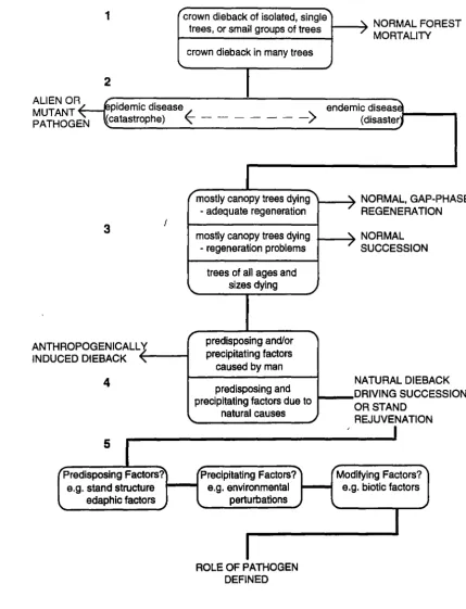
Figure 1.4 combines and adapts a number of these ideas and models in a flow chart, designed to elucidate the role of a known pathogen in a natural forest system. The numbers on the flow chart correspond to those of the relevant paragraphs below.
1. Mueller-Dombois {1988b) defined stand-level dieback (canopy dieback) as being the
1
"crown dieback of isolated, single"'"
NORMALFORES Ttrees, or small groups of trees
crown dieback in many trees \.
2
ALIEN OR MUTANT PATHOGE N
pidemic disease
(catastrophe)
~
- -- - - - ->
r mostly canopy trees dying""""
ANTHROP INDUCED
3
OGENICALL
'J
DIEBACK"
4
I "-r- adequate regeneration
mostly canopy trees dying - regeneration problems
trees of all ages and sizes dying _)
predisposing and/or
-..
precipitating factorscaused by man
predisposing and precipitating factors due to
natural causes
5
Predisposing Factors? Precipitating Factors? e.g. stand structure .._ _ _. e.g. environmental
edaphic factors perturbations
ROLE OF PATHOGEN DEFINED
_)
/
MORTALITY
endemic diseas~ (disasterJ
'\. NORMAL, GAP-PHA
/
REGENERATION SE
'\. NORMAL
/
SUCCESSION
NATURAL DIEBACK DRIVING SUCCESSIO N OR STAND
REJUVENATION
[image:28.569.70.500.84.646.2]Modifying Factors? e.g. biotic factors
2. Diseases have traditionally been classified as epidemic or as endemic (in the sense of being continually present in one place). Van der Plank (1975) viewed epidemic and endemic disease as a continuum, with local or micro-epidemics being inevitable even in a mainly endemic situation, due to variation in local conditions. Harper (1977),
discussing the effect on plant populations, made a distinction between types of large-scale disturbance: a disturbance too infrequent or irregular to exert a sustained selection pressure was termed a catastrophe; one which occurred with sufficient frequency and regularity to exert a selection pressure on the plant population was termed a disaster. Clearly epidemic disease can be catastrophic, while endemic disease is generally merely disastrous in evolutionary terms! In the case of an alien, epidemic disease, stand-level dieback is unlikely to require further explanation (Mueller-Dombois 1988a); the role of endemic disease, however, is not usually so clearly defined.
3. Mueller-Dombois (1988a} emphasised the need to study not only the dying tree population, but the whole community, and cited the three structural classes recognised by Hosking (1986), for the types of stand-level dieback found in New Zealand's
Nothofagus forests. The first two classes were considered to be natural successional
processes; the third class could represent a true decline disease:
• Stands in which mostly old canopy trees are dying but which show adequate regeneration of the canopy species;
• Stands in which mostly old canopy trees are dying but which also have re-establishment problems;
• Stands in which all ages and sizes are affected by dieback.
Read et al. (1990} used similar concepts in a predictive model of Nothofagus dynamics in New Guinea.
4. A three factor theory of forest dieback a!ld disease-induced decline has been developed by a number of workers over the last 30 years and this work has been reviewed by Stewart (1989). Mueller-Dombois (1988a) summarised the decline disease theory and related it to natural dieback. The theory recognises a combination of dieback causes which operate in a chain reaction as: (1) predisposing; (2) precipitating; and (3) modifying factors.
• Precipitating factors are those which synchronise or trigger forest dieback. They are usually fluctuating or recurring environmental perturbations e.g. seismic vibrations, climatic extremes (causing flooding, drought, frost damage, salt spray, storms) or pests and pathogens. Precipitating factors are not always distinct from predisposing factors.
• Modifying factors are those which accelerate or stall dieback and are often biotic factors such as pests and diseases, which attack the already dying stand.
Dieback can be seen as an anthropogenically caused disease if either predisposing or precipitating factors are produced by man. If both have natural origins, stand-level dieback can be viewed as a mechanism driving ecological succession and/or stand rejuvenation (Mueller-Dombois 1988a}.
5. When the predisposing, precipitating and modifying factors are known, the role of a pathogen in stand-level dieback can be defined.
Stand-level dieback in Nothofagus forests
18
There is a good deal of knowledge about the operation of the three factors in New Zealand Nothofagus forests and this has been reviewed by Stewart (1989) and, less specifically, by
Ogden {1985). Species specific regeneration patterns, stand structure and soil stability appear to be important factors predisposing some areas to severe dieback. Ogden {1988) suggested that the synchronised physiological responses to temperature which lead to mast seeding in Nothofagus, may also predispose trees to succumb to subsequent environmental stress.
Nothofagus solandri var. cliffortioides in New Zealand has a relatively high light requirement
In contrast N. menziesii, with a lower light requirement for regeneration, tends towards continuous regeneration and uneven-aged stands on optimal sites, and is less likely to suffer from widespread synchronous dieback (Wardle and Allen 1983), although it can form even-aged stands following landslips (Stewart 1986). Also, in high altitude 'cloud forests' in the Kaimai Ranges, almost continuous soil waterlogging leads to shallow root systems,
predisposing stands to dieback precipitated by drought (Jane and Green 1983). N. menziesii and N. fusca in mixed (and pure) stands usually have gap phase regeneration processes (Stewart and Rose 1990). However, spatial analysis of age classes in one N. fusca/N.
menziesii stand indicated that tree deaths had been clumped in patches up to 40 m across
(Duncan and Stewart 1991 ).
Dieback of N. fusca and N. menziesii in the Northern Kaimanawas (New Zealand) appears to have been precipitated (triggered) by storms, with subsequent build up of Platypus numbers, presumably with the increase in dead and dying material available for breeding sites (Milligan 1972; Hosking 1977). Platypus beetles and Sporothrix infection would in this case have been modifying (accelerating) factors. Drought has also been shown to be a precipitating factor in a number of areas (Skipworth 1983; Hosking 1986; Hosking and Kershaw 1985; Hosking and Hutcheson 1986, 1988) with pests and diseases again acting only as modifying factors.
In New Guinea nutrient deficiencies or drought and frost are suspected of being precipitating factors in Nothofagus stands predisposed to dieback by tree senescence in an even-aged stand structure. In such cases the presence of Platypus beetles in live Nothofagus is merely taken as an indication of the stands being under stress (Arentz 1983, 1988). Ash (1988) implicated a soil pathogen in dieback of mature Nothofagus on Mt Giluwe, with Platypus species again being seen as a secondary factor. In the absence of any other widespread disturbance, such dieback is seen as necessary for Nothofagus regeneration. However, Read et al. (1990) suggested that past volcanic events may be responsible for the
discontinuous size structures found in many Nothofagus populations; in other areas the size structures of the population implied continuous regeneration of Nothofagus.
A number of South American studies are also relevant: Veblen and Ashton (1978) studied the effects of so-called catastrophic influences (mass movement, vulcanism and fire) on the
Nothofagus-dominated vegetation of the Valdivian Andes in Chile. It was apparent that the
20
important force influencing regeneration of N. pumi!b. N. obliqua also depends on partial or complete destruction of old-growth stands for regeneration (Veblen et al. 1979a). Such disturbances would be merely disasters in evolutionary terms (Harper 1977).
In the absence of volcanic disturhRnce, the continuously regenerating N. botuloides is replacing N. pumi~b in high altitude stands in south central Chile (Veblen et al. 1977). At higher elevations, mixtures of N. dombeyi and N. alpina (montane zone), and N. dombeyi and N. pumilio (subalpine zone) are known to regenerate in gaps (Veblen 1985a). In northern Patagonia large-scale disturbances in the form of fire are important for the regeneration of N. dombeyi. However, in the absence of species which could replace it, N. dombeyipersists by gap-phase regeneration (Veblen and Lorenz 1987, Veblen 1989a). N. nitida appears to be continuously regenerating in gaps in N. nitidal Podocarpus forests in southern Chile, and, in contrast to other mixed species Nothofagus forests in Chile, the forest canopy composition may be relatively stable (Innes 1992). Successional stage can also be important; during postglacial succession in Patagonia, moraines were first colonised by N. betuloides, followed by N. antarctica (Armesto et al. 1992).
Competition f ram understorey species strongly influences the regeneration pattern of
Nothofagus. In both mid-elevation and subalpine Nothofagus forests in Chile, Chusquea
bamboos inhibit seedling development of the dominant tree species. At mid-elevations N. dombeyi and N. alpina regeneration is dependant on stand-devastating disturbances; in subalpine forests N. dombeyi, N. betuloides and N. pumilb will also regenerate under canopy
,..
gaps where the less favourable microclimate reduces the relative dominance of bamboo (Veblen et al. 1979b; Veblen 1982).A BRIEF SUMMARY OF THE KNOWN CHARACTERISTICS OF MYRTLE WILT
There are similarities between myrtle wilt and other diseases caused by primary pathogens, such as Dutch elm disease and oak wilt (Howard 1973a, Elliott et al. 1987; Kile and Walker 1987). However, in Nothofagus forests myrtle wilt appears to be unique, being an often severe and sustained stand-level disease caused by a primary pathogen, and apparently unrelated to environmental stress (Kile et al. 1989).
Myrtle wilt - the disease
In summary, both Dutch elm disease and oak wilt are introduced through wounds by insect vectors, and are also spread through root grafts. The causal organisms belong to the Ceratocystis group and both asexual and sexual stages are involved. Both diseases kill
regeneration as well as mature trees, and have increased their distribution in the recent past.
Myrtle wilt, in common with both Dutch elm disease and oak wilt, is caused by a member of the Ceratocystis group, and trees are infected through wound sites. Mortality rates were comparable with those of Dutch elm disease, and like oak wilt, myrtle wilt attacks a fagaceous host (Elliott et al. 1987; Kile and Walker 1987).
However, myrtle wilt appears to be unique in that disease spread occurs without an insect vector by means of air/water borne inoculum, and that only the asexual stage of the fungus is involved (Kile and Hall 1988; Kile and Walker 1987; Kile et al. 1989).
Key aspects which require clarification in this regard are the existence (or otherwise) of root grafts in myrtles, and their importance for disease spread; the importance of wounds as infection sites and the effect of myrtle wilt on myrtle regeneration. Information on the past and present distribution of the disease is also required.
Myrtle wilt - a dieback factor
22
1. Myrtle wilt is an often severe and sustained stand-level disease caused by a primary pathogen (Kile et al. 1989). In Tasmania a recent survey of 20 undisturbed sites showed that on average 24.6% of standing myrtle were dead or dying from the disease, with 1.6% of the live trees currently dying due to wilt. All sites showed evidence of the disease (Elliott et al. 1987).
2. The average mortality rate that was estimated (1.6% pa), is comparable with the early stages of the last Dutch elm disease epidemic in Great Britain (Elliott et al. 1987). Myrtle wilt is known to be present in more remote areas (Jarman et al. 1984) but comparative data are lacking, as is any collated information on the origins and history of the disease.
3. The effect of myrtle wilt on the floristics of rainforest is largely unknown. Myrtle
regeneration (less than 15 cm diameter) has been found beneath canopy gaps caused by wilt (Howard 1973a, 1981; Hickey 1982a), and larger trees are known to have a higher probability of the disease (Elliott et al. 1987). However, canopy and sub canopy trees have a similar disease incidence (Elliott et al. 1987), and myrtle wilt has
occasionally been observed to kill myrtle seedlings (Kile et al. 1989). Gap phase regeneration is known from myrtle forests (Read and Hill 1985a) and it has been postulated that myrtle wilt gaps provide a regeneration niche for myrtle, the disease being in equilibrium with other ecosystem processes (Howard 1981; Jarman et al. 1984, Ellis 1985).
4. Disturbance has been observed to exacerbate the effects of myrtle wilt (Howard 1973a), although there are few replicated experiments. However, damaged trees are known to have a higher probability of the disease (Elliott et al. 1987).
5. The survey of Elliott et al. (1987) indicated relationships between myrtle wilt and a number of stand and environmental variables. Disease incidence:
• was higher in callidendrous than in thamnic-implicate forests;
• increased in mixed forests with both relative and absolute measures of myrtle density;
• decreased with increasing altitude; • was higher for trees of larger diameter;
Clearly, further investigations are necessary in order to assess the significance and ecological role of myrtle wilt, and in order to recommend strategies for forest conservation and management. Mueller-Dombois (1983a) stated that a knowledge gap existed in the area of canopy dieback, because it fell between the realms of three disciplines; forestry, pathology and ecology. This project represents one attempt to bridge that gap.
PROJECT AIMS
The primary objectives of this project are to:
• collate the existing unpublished information on the rate of spread and impact of myrtle wilt;
• establish permanent plots to monitor rate of spread of disease in different rainforest types;
• record the extent of the distribution of myrtle wilt in Victoria;
• undertake disease assessments in Tasmanian areas remote from human disturbance; • test the effects of wound age and chemical treatments on infection rates;
• look for root grafting in myrtles, and if present to assess its effect on disease spread; • investigate the rate of spread of myrtle wilt in relation to the extent and type of
disturbance in rainforest;
• study the impact of myrtle wilt on the floristics of rainforest;
• investigate the role of myrtle wilt in the regeneration process of myrtle.
FIELD SITES
•18 e17 .1e .1e 0 •24
TASMANIA
50 100km
oScottsdale
150012 21. 0
LAUNCESTON •23
•28
&()
'fl
oO~
SITENAllE Adamsen• Pealc Alv•Loop
Bennetts I Espennce unkRoad
Blad<wallr Road 4
~··
Frt• Road 5
mnkland Ring• 5 •8
Ftllldvn.,. Cap 7
-e 20
Frodlhlm1 Pua I
J'*"Oolcmttl 9
l.li8 Cl'llhaim 10
Ml Kllg Wiln 11
Mt.Miiia 12
Mt.Mc:llMI 13
Ml Rolllld Clall 1'
Ml Seel 15
OolWI 11
~Rold 17
Pru.wRold 11
Rlllll9a Rold 19
Scol1I PMll fad 20
Srncrl• Rold 21
Sumac:Rold 22
Tambs-Rold 23
WISldcrllrl F-1 24
Wilen Rold/
Nadl;all Ct9lk 25
WoWfCr91i 211
Rut.aRoad 27 U!f9yFlb 211
Figure 1.5 Location of study sites In Tasmania. {Map produced by Forestry Tasmania.)
•27
2•
{Sites 10, 11, 21, 23 & 25 are those of Elliott et al. 1987.)
2. MYRTLE WILT IN SPACE AND TIME
2.1 INTRODUCTION
A major problem of dealing with a disease which was reported only 20 years ago is the lack of accurate historical data on its occurrence, distribution and abundance, and the resultant lack of perspective with which the disease is viewed. In short we do not know how long it has been present or whether its severity is increasing (Elliott et al. 1982), although both Platypus subgranosus and Cha/ara australis are believed to be indigenous to Australia
(Hogan 1944; Kile and Walker 1987).
Early Tasmanian references to myrtle
The early Tasmanian literature abounds with references to myrtle and myrtle forests, and these bear investigation. Burns and Skemp (1961) in their record of the Van Diemens Land correspondence of J. D. Hooker, cite letters of R. C. Gunn and A. Cunningham, written 1838-49, which note its presence at Black River near Circular Head, along the Hobart Rivulet, at Macquarie Harbour, and in the vicinity of the Franklin River; also a description, by Gunn, of a myrtle forest from north-east Tasmania. Hooker (1860) later summarised that myrtle was 'common in mountainous and western humid districts, forming a large proportion of the forest'.
Burn (1855) recorded the overland journey of Sir John and Lady Franklin from Hobart to Macquarie Harbour in 1842, following the track cut previously by Calder, and his account contains numerous descriptions of myrtle and myrtle forests: south of Mt Cheyne; at
Surprise River, King River, Painters Plains, and at Loddon River; west, south-west of Fatigue Hill; at Bagota Fall and in the Acheron Valley; at Black Forest and in the Franklin Valley.
The reports of G. S. Perrin, the first Tasmanian Conservator of Forests are of particular relevance. He mentioned the use of myrtle on the Mt Bischoff tram line (Perrin 1886b ), and after a trip from Waratah to Macquarie Harbour and the West Coast in 1886, described the quality of the myrtle forests on the summit of the Magnet Range and along the Corinna track south of Corinna. East of Strahan he noted that the two-year-old, gravelled Government road passed through a myrtle forest, and myrtle was also recorded from the Queen River area and the Linda Valley (Perrin 1886a).
25
(Perrin 1887). Myrtle was evidently widespread and was specifically mentioned in the Picton valley. One note is of interest: 'Considerable numbers of dead King William pine are met with of both species, Athrotaxis se/aginoides and A. cupressoides; the former showing their larger trunks - bare, bleached skeletons glistening white in the sun, and shining at intervals with a silvery lustre among the dark green myrtles and scrub; a few live trees are met with, together with stunted specimens of the latter species, but the majority are dead, probably killed by the great frosts of 1837.' In the same year he recorded the export of myrtle to London, from Burnie and Hastings (Perrin 1887).
In 1887 Perrin accompanied the Deputy Surveyor General and Deputy Commissioner of Lands, C. P. Sprent on a trip from Hobart, via Ouse, Lake St. Clair, Mt Arrowsmith,. the Collingwood Valley, Mt Lyell and the Queen River, to Strahan and Macquarie Harbour. The party included
J.
B. Walker and W. V. Legge. From Lake St Clair to the Linda goldfield they followed the new Linda Track, which had been cut by T. B. Moore four years earlier (Perrin 1887; Walker 1993).Perrin (1887) recorded that at Mt Arrowsmith (and along the mountain chain), the eucalypt forests gave way to the myrtle forests of the West Coast, this being coincident with a change in geological characteristics. He also noted: 'The descent from Mt Arrowsmith is tolerably steep, but a good roadway has been cut into the gravel round the sides of the mount.' The party later crossed the newly completed bridge over the Franklin and stayed at the road party's camp near the Upper Collingwood. Myrtle forests were reported from the area of the Surprise, Franklin, Collingwood and Cardigan Rivers. He concluded; 'Myrtle is the prevailing feature of the timbered lands, and exists in immense quantities.'
Walker (1887) noted myrtle scrub below Mt Gell, and myrtle forests on the lower slopes running down into the Franklin River gorge. Legge (1887) specifically mentioned myrtle groves on the western shore of Lake St Clair, near Mt Rufus and at the foot of Mt King William and noted the prominence of myrtle along the rest of the route to the West Coast, and compared this with the solitary specimens seen around Mt Wellington. Of particular note is his description of the Mt King William forest: 'a splendid beech grove, in which I measured a monarch of the forest which was 27 feet in girth.'
On subsequent trips Perrin reported myrtle from the area of Russells Falls River and