Copyright © 2001, American Society for Microbiology. All Rights Reserved.
Structural and Functional Analysis of Interhelical Interactions in
the Human Immunodeficiency Virus Type 1 gp41 Envelope
Glycoprotein by Alanine-Scanning Mutagenesis
MIN LU,1* MARISA O. STOLLER,2SHILONG WANG,1JIE LIU,1MELINDA B. FAGAN,2†
ANDJACK H. NUNBERG2*
Department of Biochemistry, Weill Medical College of Cornell University, New York, New York 10021,1
and Montana Biotechnology Center, The University of Montana, Missoula, Montana 598122
Received 23 May 2001/Accepted 8 August 2001
Membrane fusion by human immunodeficiency virus type 1 (HIV-1) is promoted by the refolding of the viral envelope glycoprotein into a fusion-active conformation. The structure of the gp41 ectodomain core in its fusion-active state is a trimer of hairpins in which three antiparallel carboxyl-terminal helices pack into hydrophobic grooves on the surface of an amino-terminal trimeric coiled coil. In an effort to identify amino acid residues in these grooves that are critical for gp41 activation, we have used alanine-scanning mutagenesis to investigate the importance of individual side chains in determining the biophysical properties of the gp41 core and the membrane fusion activity of the gp120-gp41 complex. Alanine substitutions at Leu-556, Leu-565, Val-570, Gly-572, and Arg-579 positions severely impaired membrane fusion activity in envelope glycoproteins that were for the most part normally expressed. Whereas alanine mutations at Leu-565 and Val-570 destabi-lized the trimer-of-hairpins structure, mutations at Gly-572 and Arg-579 led to the formation of a stable gp41 core. Our results suggest that the Leu-565 and Val-570 residues are important determinants of conserved packing interactions between the amino- and carboxyl-terminal helices of gp41. We propose that the high degree of sequence conservation at Gly-572 and Arg-579 may result from selective pressures imposed by prefusogenic conformations of the HIV-1 envelope glycoprotein. Further analysis of the gp41 activation process may elucidate targets for antiviral intervention.
Infection by human immunodeficiency virus type 1 (HIV-1) is initiated by fusion of the viral and cellular membranes to allow virus entry into the cell. This process is mediated by the viral envelope glycoprotein through interaction with cellular receptors. The HIV-1 envelope glycoprotein is synthesized as a precursor polyprotein, gp160, that is proteolytically processed to generate two subunits, the surface glycoprotein gp120 and the transmembrane glycoprotein gp41 (53). These subunits remain noncovalently associated to form the oligomeric enve-lope glycoprotein spike on the viral membrane. gp120 is re-sponsible for the sequential binding of the envelope glycopro-tein spike to CD4 and a chemokine receptor (typically CCR5 or CXCR4) on the surface of target cells (reviewed in refer-ences 2 and 75). These events trigger gp41 to undergo confor-mational changes that are crucial for activation of HIV-1 mem-brane fusion. By analogy with the pH-induced structural changes in the hemagglutinin (HA) protein of influenza virus, the HIV-1 fusion activation process likely involves substantial conformational changes from a metastable prefusogenic state to an energetically more stable fusogenic conformation (re-viewed in references 14 and 64). The control of these structural
rearrangements is thought to be central to HIV-1 entry and to strategies for intervention.
The structure and mechanism of the gp41 molecule have been extensively studied. Protein dissection studies demon-strated that the N- and C-terminal heptad repeat regions of the gp41 ectodomain associate to form an␣-helical trimer of an-tiparallel dimers (50). X-ray crystallographic analysis con-firmed that this gp41 core is a trimer of hairpins (13, 68, 69). Three N-terminal helices form a trimeric coiled coil, and three C-terminal helices pack in the reverse direction into three hydrophobic grooves on the surface of the coiled coil. Recent evidence indicates that this helical-hairpin structure corre-sponds to the fusion-active conformation of gp41 (13, 36, 50, 68, 69). Because the membrane anchor and the fusion peptide of the gp41 ectodomain are embedded in the viral and target cell membranes, respectively, the formation of the fusogenic hairpin structure results in the colocalization of the two mem-branes and thus overcomes the energy barrier for membrane fusion (27, 32, 69).
Peptides corresponding to the C-terminal heptad repeat re-gion, referred to as C peptides, can specifically inhibit viral entry into target cells at nanomolar concentrations (37, 73). One such peptide (T-20) is in clinical study and has shown antiviral activity in humans (42). T-20 binds to gp41 only after interaction of the envelope glycoprotein complex with the cel-lular receptors (27, 39, 56) and is thought to act in a dominant-negative manner by binding to the N-terminal coiled coil of gp41 during its conformational change to the fusogenic state (15, 50, 72). Considerations of the efficiency of C peptide competition with its cognate sequence suggest a relatively long-* Corresponding author. Mailing address for Jack H. Nunberg:
Montana Biotechnology Center, The University of Montana, Missoula, MT 59812. Phone: (406) 243-6421. Fax: (406) 243-6425. E-mail: [email protected]. Mailing address for Min Lu: Department of Biochemistry, Weill Medical College of Cornell University, New York, NY 10021. Phone: (212) 746-6562. Fax: (212) 746-8875. E-mail: mlu @med.cornell.edu.
† Present address: Department of Philosophy, University of Texas, Austin, TX 78712.
11146
on November 9, 2019 by guest
http://jvi.asm.org/
lived prehairpin intermediate (14, 27, 39, 56). Intermediate states of the gp41 molecule may serve as a target for the development of small-molecule HIV-1 inhibitors (22, 25).
Considerable evidence now implies that packing interactions between the central coiled-coil trimer and the C-terminal helix are important determinants of HIV-1 entry and its inhibition (35, 54, 60, 63). For example, the replacement of a conserved glutamine (Gln-652), buried in this helical interface, by the hydrophobic residue leucine increases HIV-1 infectivity and underlies an enhancement in the antiviral activity of C peptides (8, 63). Biophysical and structural characterization directly demonstrates that this substitution strengthens the interhelix interaction in the fusogenic hairpin structure by providing ad-ditional hydrophobic packing forces (63). Taken together, these results have been interpreted to indicate that the recep-tor-triggered conformational changes of the HIV-1 envelope glycoprotein are thermodynamically controlled and that the process of membrane apposition and lipid bilayer fusion is driven by the currency of energy released from the formation of the fusogenic gp41 core (33, 49, 51, 63, 69).
Therefore, it is of fundamental importance to understand the structural and mechanistic basis for the helical interactions in this core. We show here, using alanine-scanning mutagene-sis, that amino acids Leu-556, Leu-565, Val-570, Gly-572, and Arg-579 ateandgpositions of the N34 coiled coil are essential for envelope glycoprotein function. Whereas Leu-565 and Val-570 contribute to the conserved packing interactions between the N- and C-terminal helices of gp41, Gly-572 and Arg-579 appear to be critical for prefusogenic conformations of the HIV-1 envelope glycoprotein complex.
MATERIALS AND METHODS
Plasmid construction and mutagenesis.High-fidelity XL PCR (rTth and Vent DNA polymerases; PE Applied Biosystems) and oligonucleotide primers envA and envN (29) were used to adapt the HIV-1 HXB2revandenvgenes from the plasmid pIIIenv 3-1 (65). Expression of the HXB2 envelope glycoprotein was within the context of the eukaryotic expression vector pCR-Uni 3.1 (Invitrogen) (45). Alanine mutations were introduced into the HXB2 envelope glycoprotein and the N34(L6)C28 peptide (51) using QuikChange mutagenesis (Stratagene, La Jolla, Calif.) and single-stranded mutagenesis (44), respectively. Mutations were verified by DNA sequencing.
Envelope glycoprotein expression.Simian COS-7 cells were used for transient expression of the envelope glycoprotein (75). Typically, 3g of the HIV-1env expression plasmid and 9l of FuGENE-6 reagent (Roche Molecular Biochemi-cals) were used, according to the manufacturer’s instructions, to transfect 4⫻105
to 8⫻105cells in a 6-cm culture dish. Cultures were washed 18 h later with
physiological buffered saline (PBS) and refed with Dulbecco’s modified Eagle’s medium with 10% fetal bovine serum. Transfection efficiencies were determined in microcultures from the transfected cell cultures. These microcultures were fixed with⫺20°C methanol-acetone (1:1) and stained using biotinylated anti-HIV immunoglobulin (anti-HIVIG) from infected persons, NeutrAvidin-horseradish peroxidase (HRP) (Pierce Chemical Corp., Rockford, Ill.), and diaminobenzi-dine.
Western blot analysis.Two days after transfection, cell culture supernatants were harvested and filtered. gp120 shed from expressing cells was immunopre-cipitated using HIVIG and protein A-Sepharose (Sigma). Cell monolayers were surface biotinylated using EZ-Link NHS-LC-biotin (Pierce Chemical) as previ-ously described (48, 76). Cells were then lysed on ice in 50 mM Tris-HCl (pH 7.5)–150 mM NaCl–1% Triton X-100 containing 1g ml⫺1each of aprotinin,
leupeptin, and pepstatin. Biotinylated surface proteins were isolated by using NeutrAvidin-agarose (Pierce Chemical). Envelope glycoprotein was detected by Western blot analysis using the gp120-specific monoclonal antibody (MAb) Chessie B13 (kindly provided by George Lewis [1]). Western blots were visual-ized by chemifluorescence using ECL-Plus (Amersham Pharmacia Biotech) and quantitated by fluorescence imaging using a Fuji FLA3000G instrument.
Deglycosylation of cell surface Env.Biotinylated envelope glycoprotein was isolated from the cell surface using NeutrAvidin-agarose and was deglycosylated using proteinN-glycanase F (New England Biolabs, Beverly, Mass.). The result-ing polypeptides were analyzed by Western blot usresult-ing MAb Chessie 12 (kindly provided by George Lewis [1]). gp160 from a cleavage-defective envelope gly-coprotein mutant (26) and soluble gp120 from a truncated envelope glygly-coprotein construct were deglycosylated and used as size markers.
Pulse-chase analysis. COS-7 cells expressing envelope glycoprotein were pulse-labeled for 30 min in replicate six-well cluster dishes using 100Ci each of [35S]cysteine and [35S]methionine in 1 ml of cysteine- and methionine-free
me-dium. Cultures were then washed and grown in cysteine- and methionine-con-taining medium for the indicated times. gp120 was isolated from cell culture supernatants by immunoprecipitation. Proteins were separated by sodium dodecyl sulfate-polyacrylamide gel electrophoresis and visualized by phosphorimaging.
Cell-cell fusion assay.The ability of the wild-type and mutant envelope gly-coproteins to mediate cell-cell fusion was determined by coculturing envelope glycoprotein-expressing COS-7 cells with U87 cells expressing CD4 and CXCR4 coreceptor (31, 45, 49). Transfected COS-7 cells were resuspended using PBS with 0.5 mM EDTA, and 5⫻103cells were added to 96-well microcultures
containing 5⫻103U87-CD4-CXCR4 cells. In order to assess the rate and extent
of syncytium formation, cocultures were fixed and immunochemically stained after 6 and 24 h. The number of envelope glycoprotein-expressing cells involved in syncytium formation and the number of nuclei contained within each syncy-tium were determined microscopically.
Peptide production.Mutant N34(L6)C28 peptides were expressed in Esche-richia coliBL21(DE3)/pLysS using the T7 expression system (67). Cells, freshly transformed with an appropriate plasmid, were grown to late log phase. Protein expression was induced by addition of 0.5 mM isopropylthio--D-galactoside.
After another 3 h of growth at 37°C, the bacteria were harvested by centrifuga-tion, and the cells were lysed by glacial acetic acid as described previously (52). Proteins were purified from the soluble fraction to homogeneity by reverse-phase high-pressure liquid chromatography (Waters) with a Vydac C18 preparative
column using a water-acetonitrile gradient in the presence of 0.1% (vol/vol) trifluoroacetic acid.
CD spectroscopy.Circular dichroism (CD) spectra were acquired on an AVIV 62DS CD spectrometer with a thermoelectric sample temperature controller. Samples for wavelength spectra were 10M peptide in 50 mM sodium phos-phate (pH 7.0) and 150 mM NaCl. The cuvette was 0.1 cm in path length. The wavelength dependence of molar ellipticity, [], was monitored at 4°C as the average of five scans, using a 5-s integration time at 1.0-nm wavelength incre-ments. Spectra were baseline corrected against the value for the cuvette with buffer alone. Fractional helix content was calculated from the CD signal by dividing the mean residue ellipticity at 222 nm by the value expected for 100% helix formation by helices of comparable size (⫺33,000° cm2dmol⫺1) (17).
Thermal stability was determined in the same buffer by measuring []222as a
function of temperature. A 1.0-cm path length cell was used with continuous stirring. Thermal melts were monitored in 2°C intervals with a 2-min equilibra-tion at the desired temperature and an integraequilibra-tion time of 30 s. The midpoint of the thermal unfolding transition (Tm) was determined from the maximum of the first derivative, with respect to the reciprocal of the temperature, of the []222
values (7). The error in estimation ofTmis⫾1°C. Protein concentrations were determined by measuring absorbance at 280 nm in 6 M guanidinium chloride (23). Sedimentation equilibrium.Sedimentation equilibrium measurements were performed on a Beckman XL-A Optima analytical ultracentrifuge, using an An-Ti rotor and six-sectored equilibrium centrifugation centerpieces. Protein samples were dialyzed overnight against 50 mM sodium phosphate (pH 7.0)–150 mM NaCl, loaded at initial concentrations of 10, 30, and 100M, and analyzed at rotor speeds of 20 and 23 krpm at 20°C. Data were acquired at two wave-lengths per rotor speed and processed simultaneously with a nonlinear least-squares fitting routine (38). Solvent density and protein partial specific volume were calculated according to solvent and protein composition, respectively (47). Molecular weights were all within 10% of those calculated for an ideal trimer, with no systematic deviation of the residuals.
Crystallization, data collection, and structure determination.The G572A and R579A peptides were crystallized by hanging-drop vapor diffusion at room tem-perature. Initial crystallization conditions were screened by using sparse matrix crystallization kits (Crystal Screen I and II; Hampton Research, Riverside, Calif.) and then optimized. Rhombohedral crystals of G572A were grown from 10 mg ml⫺1of peptide, 0.1 M sodium acetate (pH 5.2), 0.2 M ammonium sulfate, and
10% polyethylene glycol 4000. Rhombohedral crystals of R579A peptide were obtained from 20 mg ml⫺1of peptide, 0.1 M sodium acetate (pH 4.5), 0.2 M
ammonium sulfate, and 29% polyethylene glycol methyl ether 2000. Crystals were transferred to a cryoprotectant solution containing 15% (vol/vol) glycerol in
on November 9, 2019 by guest
http://jvi.asm.org/
the corresponding mother liquor and frozen in liquid nitrogen for data collec-tion. Diffraction data on the G572A peptide trimer were collected at 95 K using an R-axis IV image plate detector mounted on a Rigaku RU200 rotating anode X-ray generator at the X-ray Crystallography Facility at the Weill Medical College of Cornell University. Data on the R579A peptide trimer were collected at 95 K using a Quantum-4 charge-coupled device-based detector at the X12B beamline of the National Synchrotron Light Source. Diffraction intensities were integrated by using Denzo and Scalepack software (59) and reduced to structural factors with the program Truncate from the CCP4 program suite (10).
The structures of the G572A and R579A peptides were determined by mo-lecular replacement with the program AMoRe (57) using the N34(L6)C28 trimer (Protein Data Bank [PDB] file name 1SZT) as a search model. The initial models were built by using conventional (2Fo⫺Fc)Fcalcand (Fo⫺Fc)Fcalcmaps at 3.0
Å. Overall anisotropic B-factor and bulk solvent corrections were applied. Many cycles of torsional angle simulated annealing and grouped B-factor refinement (4) were followed by extensive rebuilding. The models were rebuilt to reflect the sequences of the G572A and R579A peptides by using the (2Fo⫺Fc)Fcalcand
(Fo⫺Fc)Fcalcmaps with the program O (40). The structures were refined by
using positional and B-factor refinements (4). Crystallographic refinement of the structures was done with the program CNS 1.0 (5). Model geometry was analyzed by Procheck (46), with all residues occupying most-preferred regions of the Ramanchandran space. Ser-546, Gly-547, Asn-651, Gln-652, Gln-653, Glu-654, and Lys-655 of the G572A and R579A peptides were left out of the model because of the absence of interpretable electron density for these atoms. Atomic coordinates for G572A peptide (PDB file name 1I5Y) and R579A peptide (PDB file name 1I5X) have been deposited in the PDB.
RESULTS
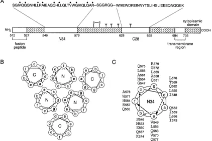
In the trimer-of-hairpins structure, residues at positions a and d of the C28 helix pack against residues at the eand g
positions of the N34 coiled-coil trimer in an antiparallel ori-entation (Fig. 1). Sequence comparisons among HIV-1 isolates and between HIV-1 and simian immunodeficiency virus (SIV) show that the residues at these contact positions are highly conserved (43). This high degree of sequence conservation may result from selective pressure to maintain interhelical packing interactions and, hence, the structural integrity of the trimer of hairpins. To directly test this hypothesis, we changed the individualeandgresidues of N34 to alanine in both the recombinant N34(L6)C28 peptide model and the intact enve-lope glycoprotein (Fig. 1C). Alanine-scanning mutagenesis was chosen because alanine, albeit a good helix-inducing residue, has a relatively low hydrophobicity and contributes little to protein-protein interactions (19). We reasoned that alanine replacements were likely to alter the stability of the trimer-of-hairpins structure but not to disrupt its folding per se. In this analysis, the biophysical properties of mutant N34(L6)C28 peptides were compared with the biological phenotypes of mutant envelope glycoproteins.
Expression of mutant envelope glycoproteins.Alanine mu-tant envelope glycoproteins were expressed by transient trans-fection in COS-7 cells. As determined by Western blot analysis of cell surface-biotinylated proteins, all mutant envelope gly-coproteins were expressed and transported to the cell surface with efficiencies comparable to that of the wild-type glycopro-FIG. 1. Core structure of the HIV-1 gp41 envelope glycoprotein. (A) Schematic representation of gp41. The important functional features of the gp41 ectodomain and the amino acid sequences of the N34 and C28 segments are shown. Amino acids mutated to alanine in this study are indicated by dots above the sequence. The naturally occurring alanine at position 558 was not altered. The disulfide bond and four potential N-glycosylation sites are depicted. The residues are numbered according to their position in gp160. The recombinant N34(L6)C28 peptide model consists of N34 and C28 plus a linker of six hydrophilic residues. (B) Cross-section of helix packing in the gp41 core. Residues at theaandd positions of the N helices form the hydrophobic interface of the trimeric coiled coil, and residues at theaanddpositions of the C helices pack in an antiparallel orientation against residues at theeandgpositions of adjacent N helices. (C) Helical wheel projection of the heptad repeat sequence of the N34 peptide. The view is from the NH2terminus.
on November 9, 2019 by guest
http://jvi.asm.org/
[image:3.587.83.505.77.359.2]tein (Fig. 2A). The predominant species of envelope glycop-rotein on the cell surface was gp160. The incomplete cleavage of the gp160 precursor is likely due to saturation of the cellular furin-like proteases that are responsible for envelope glyco-protein processing (3). gp120 represented⬇10 to 20% of the envelope glycoprotein on the cell surface. This was observed in the wild-type and in all alanine mutant proteins except G572A. In order to quantitate the relative amounts of cell surface gp120 among the mutants, we deglycosylated the cell surface glycoproteins to generate discrete polypeptides (Fig. 2B). Steady-state levels of gp120 in the alanine mutants V549A, Q551A, Q563A, L565A, V570A, and Q577A were similar to that in the wild-type glycoprotein. We conclude that alanine substitutions at Val-549, Gln-551, Gln-563, Leu-565, Val-570, and Gln-577 did not affect envelope glycoprotein transport to the cell surface, gp160 cleavage, or gp120-gp41 association. By contrast, the G572A mutant glycoprotein exhibited a 10-fold reduction in the relative amount of cell surface gp120. Lesser reductions were also noted in the L556A and R579A mutants (two- and fourfold, respectively).
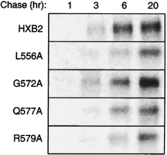
To examine whether the marked decrease in the steady-state level of gp120 in the G572A mutant was due to a reduction in the efficiency of gp160 cleavage, we analyzed the rate of ap-pearance of gp120 in the cell culture medium. Given the high rate of spontaneous gp120 shedding from the envelope glyco-protein complex (55), we reasoned that this assay would
vide a sensitive measure for gp160 cleavage efficiency. If pro-teolytic cleavage were reduced by the mutation, the amount of shed gp120 would likewise be reduced. Figure 3 shows the time course from a pulse-chase labeling experiment. The rate and/or extent of gp120 shedding in G572A was not decreased relative to that of the wild-type glycoprotein. We conclude that the reduction in gp120 accumulation in G572A is not due to a defect in proteolytic cleavage. By extension, the reduction in the steady-state level of gp120 in the G572A mutant may be caused by an increase in gp120 shedding; the Gly-572-to-Ala mutation may affect the stability of the gp120-gp41 association. In the pulse-chase labeling experiments (Fig. 3), the L556A and R579A mutants exhibited a slight reduction in the rate and/or extent of gp120 shedding relative to the wild-type gly-coprotein. The significance of this finding is unclear, as a sim-ilar decrease was observed in Q577A, a mutant in which the gp120 accumulation on the cell surface was unaltered. The mechanism for the two- to fourfold reduction in the steady-state levels of gp120 in L556A and R579A remains undefined. Taken together, our results indicate that the biosynthesis and expression of the V549A, Q551A, Q563A, L565A, V570A, and Q577A envelope glycoproteins were unaffected by alanine substitutions (Table 1). On the other hand, G572A, L556A, and R579A displayed reduced steady-state levels of gp120 on the cell surface.
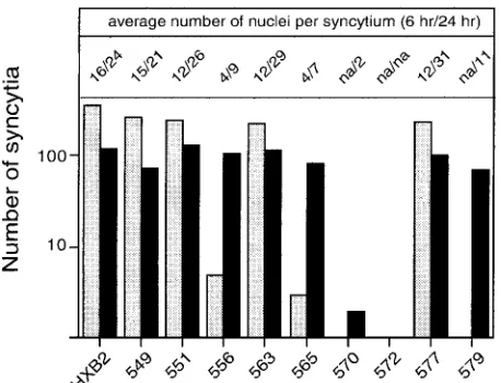
[image:4.587.340.503.73.224.2]Fusogenic potential of mutant envelope glycoproteins.The ability of the alanine mutant envelope glycoproteins to mediate cell-cell fusion was assessed by coculturing transfected COS-7 cells with a fusion partner, U87 cells expressing CD4 and CXCR4. In this assay, syncytium formation proceeds rapidly, and the maximum number of syncytia is typically attained by 6 h of coculture. Thereafter, neighboring syncytia begin to fuse, resulting in a decreased number of syncytia and an in-creased number of nuclei per syncytium. By these criteria, V549A, Q551A, Q563A, and Q577A were indistinguishable FIG. 2. Cell surface expression of mutant envelope glycoproteins.
[image:4.587.45.285.75.252.2]Mutant and wild-type envelope glycoproteins were transiently ex-pressed in COS-7 cells, and cell surface proteins were biotinylated using NHS-LC-biotin. Mutant envelope glycoprotein names are abbre-viated by the position number. (A) Envelope glycoprotein was immu-noprecipitated using HIVIG and detected by Western blot analysis using NeutrAvidin-HRP and ECL-Plus imaging. Representative im-ages obtained from the analysis of comparably transfected cell cultures (40 to 60% transfection efficiency) are shown. (B) Biotinylated enve-lope glycoproteins were deglycosylated and detected by Western blot analysis using the gp120-specific MAb Chessie 12 (1) and ECL-Plus imaging. HXB2 gp160 and gp120 were similarly deglycosylated to provide size markers (p160, 94 kDa; and p120, 55 kDa). Using this method of analysis, we noted a consistent diminution in the ratio of gp120 to gp160. Although the basis for this apparent skewing is not known, the relative amounts of gp120 among the mutants are retained relative to the analysis of the unmodified glycoproteins. A dark image is used to emphasize deglycosylated gp120s.
FIG. 3. Pulse-chase analysis of gp120 shedding by mutant envelope glycoproteins. Cells expressing mutant (L556A, G572A, Q577A, or R579A) and wild-type envelope glycoproteins were pulse-labeled for 30 min using [35S]cysteine and [35S]methionine. Comparable amounts of radioactivity were incorporated in envelope glycoproteins by all cultures (1.2⫻104to 2.2⫻104phosphostimulated luminescence units in gp160). Culture supernatants were harvested at 1, 3, 6, and 20 h, and gp120 was immunoprecipitated using HIVIG. Proteins were resolved by SDS-polyacrylamide gel electrophoresis, and radioactivity was visu-alized by phosphorimaging.
on November 9, 2019 by guest
http://jvi.asm.org/
from the wild-type envelope glycoprotein in fusogenic poten-tial (Fig. 4). In contrast, V570A, G572A, and R579A failed to produce syncytia within 6 h of coculture, and L556A and L565A yielded 100-fold fewer syncytia than the wild-type gly-coprotein. By 20 h of coculture, R579A, L556A, and L565A were able to generate similar numbers of syncytia as the
wild-type envelope glycoprotein, but these syncytia were small and similar to those seen at 6 h with the wild-type glycoprotein. The V570A mutant produced only a few binucleated cells after 20 h of coculture, and G572A was entirely defective.
In summary, V570A, G572A, R579A, L556A, and L565A were severely debilitated in their ability to mediate membrane fusion (Table 1). Because the alanine substitutions in these mutants did not grossly affect envelope glycoprotein biosynthe-sis, our results raise the possibility that the alanine mutations may perturb the interhelical packing interactions in the gp41 core structure, thereby modulating the membrane fusion ac-tivity of the envelope glycoprotein complex.
Biophysical analysis of mutant N34(L6)C29 peptides. Vari-ants of the recombinant N34(L6)C28 peptide with single ala-nine substitutions at theeandgpositions of the N34 coiled coil were produced by bacterial expression and purified by reverse-phase high-performance liquid chromatography. Sedimenta-tion equilibrium measurements indicated that each peptide sedimented as a discrete trimer over a total peptide concen-tration of 10 to 100M at neutral pH (Fig. 5A; Table 2). The helical content and stability of mutant N34(L6)C28 complexes were quantitated by CD. Each peptide was⬎90% helical at 4°C at 10M peptide (Table 2), and each exhibited a cooper-ative thermal unfolding transition (Fig. 5B; Table 2). Under these conditions, the midpoints of the thermal transition (Tm)
for N34(L6)C28 (wild type), V549A, Q551A, L556A, Q563A, L565A, V570A, G572A, Q577A and R579A were 70, 73, 74, 64, 68, 50, 56, 82, 76, and 72°C, respectively (Fig. 5B; Table 2). Therefore, alanine mutations at theeandgresidues of the N34 helix caused only changes in thermal stability, without signifi-cantly influencing the overall topology. In general, introduc-tion of alanine for bulky hydrophobic residues markedly de-stabilized the trimer-of-hairpins structure; the decrease in thermal stability,⌬Tm, between the wild-type and mutant
pep-tides was 6, 20, and 14°C for Leu-556, Leu-565, and Val-570, respectively. On the other hand, the Gly-572-to-Ala substitu-tion led to appreciable stabilizasubstitu-tion of the trimer of hairpins, reinforcing the observation that glycine in␣-helices is generally destabilizing (reviewed in reference 41).
These results indicate that the destabilization of the gp41 ectodomain core by alanine mutations at Leu-565 and Val-570 may underlie the fusion-defective phenotype of the mutant envelope glycoproteins (Tables 1 and 2). In contrast, alanine mutations at Gly-572 and Arg-579 stabilized the gp41 core structure but severely impaired membrane fusion activity. This effect on fusogenicity may reflect the reduced level of mature gp120 in these mutants. The L556A mutant had an interme-diate phenotype, where both the thermal stability of the gp41 core and the level of cell surface gp120 in the envelope glyco-protein complex were slightly reduced. Interestingly, the Gly-572 residue is involved in the formation of hydrophobic cavities on the surface of the N34 trimer that are crucial for interhelical packing interactions (13, 35, 60). The Arg-579 side chain, on the other hand, does not directly participate in the helical interactions (13, 68, 69).
[image:5.587.43.283.93.221.2]Crystal structures of G572A and R579A peptides. The un-expected observation that alanine mutations at Gly-572 and Arg-579 disrupt envelope glycoprotein function while main-taining the thermal stability of the gp41 ectodomain core led us to examine the atomic detail of the respective trimer-of-dimer TABLE 1. Expression and fusion activity of mutant
envelope glycoproteins
Glycoprotein Cell surfaceexpressiona
Cell surface gp120/gp160b
ratio
gp120
sheddingc activityFusiond
Wild type ⫹⫹⫹ ⫹⫹⫹ ⫹⫹⫹ ⫹⫹⫹
V549A ⫹⫹⫹ ⫹⫹⫹ ⫹⫹⫹ ⫹⫹⫹
Q551A ⫹⫹⫹ ⫹⫹⫹ ⫹⫹⫹ ⫹⫹⫹
L556A ⫹⫹⫹ ⫹⫹ ⫹⫹ ⫾
Q563A ⫹⫹⫹ ⫹⫹⫹ ⫹⫹⫹ ⫹⫹⫹
L565A ⫹⫹⫹ ⫹⫹⫹ ⫹⫹⫹ ⫾
V570A ⫹⫹⫹ ⫹⫹⫹ ⫹⫹⫹ ⫾
G572A ⫹⫹⫹ ⫹ ⫹⫹⫹ ⫺
Q577A ⫹⫹⫹ ⫹⫹⫹ ⫹⫹ ⫹⫹⫹
R579A ⫹⫹⫹ ⫹⫹ ⫹⫹ ⫾
a⫹⫹⫹, comparable to wild-type envelope glycoprotein.
b⫹⫹⫹, comparable to wild-type envelope glycoprotein; ⫹⫹, 2- to 4-fold reduction;⫹, 10-fold reduction.
c⫹⫹⫹, comparable to wild-type envelope glycoprotein, as assessed by West-ern blot analysis and/or by pulse-chase measurements;⫹⫹, slight reduction in gp120 shedding.
d⫹⫹⫹, comparable to wild-type envelope glycoprotein;⫾, severe (ⱖ 100-fold) reduction in fusion ability;⫺, complete absence of fusion. Intermediate levels of impairment were not represented in these mutations.
FIG. 4. Cell-cell fusion activity of mutant envelope glycoproteins. Cells expressing mutant and wild-type envelope glycoproteins were cocultured with U87-CD4-CXCR4 cells for either 6 or 24 h. Cultures were fixed with⫺20°C methanol-acetone (1:1), and envelope glycop-rotein-expressing cells were visualized by immunochemical staining. The number of envelope glycoprotein-expressing cells involved in syn-cytium formation and the average number of nuclei contained within each syncytium were determined. Mutant envelope glycoprotein labels are abbreviated by the position number. The graph indicates the num-ber of syncytia at 6 and 24 h (gray and black bars, respectively). The average size of the syncytia (number of nuclei per syncytium at 6 and 24 h) is indicated at the top. na, not applicable. Results depicted are from one experiment and are representative of relative differences among mutants. Note that the number of syncytia is biphasic with time and typically decreases at longer times as syncytia merge.
on November 9, 2019 by guest
http://jvi.asm.org/
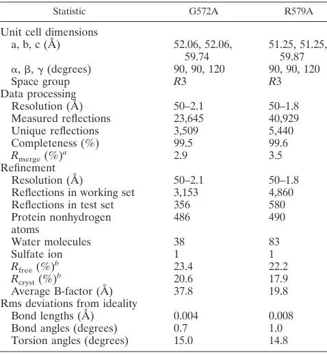
[image:5.587.50.279.411.586.2]structures. The G572A and R579A peptides were crystallized, and their X-ray structures were determined to 2.1 and 1.8 Å, respectively, using molecular replacement methods. The crys-tals of both G572A and R579A belong to the space groupR3 and contain the trimer formed around the crystallographic threefold axis. The electron density maps of G572A and R579A reveal the positions of all of the amino acid residues except a few disordered side chains at the helix termini (see Materials and Methods). Simulated-annealing omit maps were calculated to confirm the assignment of the side chains. The structure of the the G572A peptide was refined to a conven-tionalR-factor of 20.6%, with a freeR-factor of 23.4% over a
resolution range of 50.0 to 2.1 Å, and the final model consists of 61 amino acid residues and incorporates 38 water molecules and a sulfate ion. The structure of the R579A peptide was refined against 50.0 to 1.8 Å resolution data to yield a conven-tionalR-factor of 17.9%, with a freeR-factor of 19.8%, and the final model includes 61 residues, 83 water molecules, and a sulfate ion. Table 3 summarizes the data collection and refine-ment statistics for these two structures.
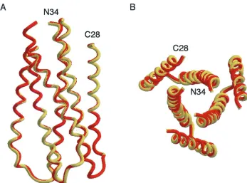
[image:6.587.51.278.75.457.2]As expected, the overall folds of G572A and R579A are very similar to those of the wild-type molecule. In all cases, the interior N34 coiled coil of three parallel helices, wrapped in a gradual left-handed superhelix, is surrounded by a sheath of antiparallel C28 helices (Fig. 6). At the center of the trimer of hairpins, the N34 three-stranded coiled coil displays typical
[image:6.587.300.542.92.213.2]FIG. 5. Biophysical characterization of mutant N34(L6)C28 pep-tides. (A) The L556A (open rhombs), L565A (open circles), and G572A (open triangles) peptides form three-stranded bundles. Sedi-mentation equilibrium data (20 krpm) were collected at 20°C at pH 7.0 for L556A (30M), L565A (100 M), and G572A (10 M). The natural logarithm of the absorbance at 280, 236, and 230 nm is plotted against the square of the radial position. For an ideal single-species system, this plot is linear, with the slope proportional to the molecular weight of the molecule. (B) Thermal melts monitored by CD at 222 nm for L556A (open rhombs), L565A (open circles), V570A (solid circles), G572A (open triangles), and R579A (solid triangles) peptides at 10 M at pH 7.0.
TABLE 2. Alanine mutants of the HIV-1 gp41 ectodomain core form trimer-of-hairpins structures
Mutant ⫺[]222(degrees cm2
dmol⫺1) (°C)Tm Molecular mass(kDa)
N34(L6)C28 31,300 70 24.4
V549A 31,100 73 23.9
Q551A 30,800 74 23.7
L556A 30,500 64 23.2
Q563A 31,200 68 24.2
L565A 31,500 50 22.9
V570A 29,900 56 23.3
G572A 31,400 82 23.5
Q577A 31,000 76 23.6
R579A 31,900 72 24.1
TABLE 3. X-ray data collection and refinement statistics
Statistic G572A R579A
Unit cell dimensions
a, b, c (A˚ ) 52.06, 52.06,
59.74 51.25, 51.25,59.87 ␣,,␥(degrees) 90, 90, 120 90, 90, 120
Space group R3 R3
Data processing
Resolution (Å) 50–2.1 50–1.8
Measured reflections 23,645 40,929
Unique reflections 3,509 5,440
Completeness (%) 99.5 99.6
Rmerge(%)a 2.9 3.5 Refinement
Resolution (Å) 50–2.1 50–1.8
Reflections in working set 3,153 4,860
Reflections in test set 356 580
Protein nonhydrogen
atoms 486 490
Water molecules 38 83
Sulfate ion 1 1
Rfree(%)b 23.4 22.2
Rcryst(%)b 20.6 17.9
Average B-factor (Å) 37.8 19.8
Rms deviations from ideality
Bond lengths (Å) 0.004 0.008
Bond angles (degrees) 0.7 1.0
Torsion angles (degrees) 15.0 14.8
aR
merge⫽ ⌺兩I⫺ 具I典兩/⌺I, whereIis the intensity of an individual measurement
and具I典is the average intensity from multiply recorded reflections. bR
cryst⫽ ⌺兩Fobs⫺Fcalc兩⌺Fobs, whereFobsandFcalcare observed and
calcu-lated structural factors, respectively. No-cutoff was applied.Rfreeis calculated
for a set of reflections that were excluded from refinement.
on November 9, 2019 by guest
http://jvi.asm.org/
[image:6.587.301.540.430.689.2]acute knobs-into-holes packing, where theaanddside chains (knobs) fit into the spaces (holes) between four residues on the neighboring helices (Fig. 7A and B) (18, 30). Residues at thee and gpositions of the N34 helices lie on the outside of the trimeric coiled coil and form three hydrophobic grooves on the surface of this coiled coil. The Gly-572 and Arg-579 residues replaced in G572A and R579A, respectively, are both in g positions of N34. The root-mean-square (rms) deviations be-tween all C␣atoms of the central N34 coiled coil in the wild-type and mutant molecules are 0.27 Å for G572A and 0.30 Å for R579A. The C28 helices in G572A and R579A can also be superimposed upon the wild-type counterpart with rms devia-tions of 0.40 and 0.35 Å, respectively. Thus, the fusion-defec-tive Gly-572-to-Ala and Arg-579-to-Ala mutations do not sig-nificantly alter the trimer-of-hairpins structure.
On the surface of the N34 trimeric coiled coil, there are three prominent, symmetry-related cavities (⬇400 Å3) that
each accommodate three hydrophobic residues from the abut-ting C28 helix: Trp-628, Trp-631, and Ile-635 (13, 68, 69). These conserved coiled-coil cavities have been shown to be critical for HIV-1 entry and its inhibition and provide a poten-tial antiviral drug target (12, 13, 22, 25, 35, 66). The Gly-572 residue, which is involved in forming this cavity, is invariant in all 251 fully sequenced M group HIV-1 isolates (43). The substituted Ala-572 side chains point into the triangular inter-helical space between two N34 helices and a buttressing C28 helix (Fig. 7C). The favorable van der Waals interactions be-tween the Ala-572 and Trp-631 side chains can strengthen interhelical packing and stabilize the trimer-of-hairpins struc-ture, as suggested by the large increase inTmof G572A relative
to that of N34(L6)C28. In addition, the Gly-572-to-Ala
substi-tution also leads to local structural rearrangements: the side chains of Trp-628 and Ile-635 deviated substantially. Overall, the methyl groups of the Ala-572 residues in G572A pack efficiently into the hydrophobic cavity of N34 and make a good C28 interaction (Fig. 7C).
The conserved grooves along the N34 coiled-coil surface are lined with predominantly hydrophobic residues (Fig. 1C). In-terestingly, the Arg-579 residue at agposition is completely conserved in 245 of the 251 sequenced HIV-1 strains. Of the remaining six isolates with different residues at this position, three possess a conservative lysine substitution (43). In gp41 core crystal structures (13, 68, 69), the Arg-579 side chain is poorly ordered, as suggested by very high individual atomic B factors. While electron density for Ala-579 in the R579A pep-tide is unambiguous, this substituted residue contributes little to the interhelical packing interactions. It would therefore ap-pear that the slight stabilization of the trimer-of-hairpins struc-ture associated with the alanine substitution at Arg-579 reflects the favorable helix propensity of alanine relative to arginine in the wild-type molecule.
DISCUSSION
[image:7.587.119.466.69.326.2]The HIV-1 envelope glycoprotein complex controls the key process of viral entry. The current model of HIV-1 membrane fusion implies that, as the trimer of hairpins is assembled, a ready source of energy can be made available for overcoming the activation energy needed for lipid bilayer fusion. Because the native and prehairpin intermediate conformations of gp41 are not known, the basic property of gp41 activation during HIV-1 membrane fusion is not well understood. Nevertheless, FIG. 6. Overall views of the mutant G572A and R579A peptide six-helix bundles. (A) Side view. The amino termini of N34 and the carboxyl termini of C28 are at the top of the figure. Helices of G572A (yellow) and R579A (pink) peptides were used for the superposition. (B) View from the top, looking down the threefold axis of the trimer of hairpins. Figures were generated with the program Setor (24).
on November 9, 2019 by guest
http://jvi.asm.org/
as has been established for the case of influenza virus HA protein (6, 9, 74), it is widely believed that the native gp41 structure is substantially different from the fusogenic trimer-of-hairpins structure. In the simplest model, the N-terminal heptad repeat region of gp41 exists in an non-coiled-coil con-formation in the native state but forms a coiled-coil trimer in the prehairpin intermediate and fusogenic hairpin conforma-tions (16, 21, 34, 51, 70, 73). The N-terminal heptad repeat sequence is one of the most highly conserved regions within the primate immunodeficiency virus envelope glycoproteins (11, 20, 28). Residues at theaanddpositions form a hydro-phobic interface at the interior of the N-terminal trimeric coiled coil (62). There is only one nonconservative substitution at anaposition between HIV-1 and SIV (Ile-573 in HIV-1 and Thr in the corresponding position of SIV). Mutations at this position affect the folding and stability of the gp41 ectodomain core and, coordinately, the ability of the mutant envelope
gly-coproteins to mediate membrane fusion (21, 49, 51). Forma-tion of the N-terminal three-stranded coiled coil appears to occur as an early event in the gp41 refolding process.
[image:8.587.49.442.68.461.2]The N-terminal coiled-coil surface contains three hydropho-bic grooves that are the sites for the C-terminal helix interac-tions (13, 68, 69). Theeand gresidues lining these grooves pack against residues at theaanddpositions of the C-terminal helices. The high sequence conservation at these contact po-sitions has led to the proposal that interactions between the N-and C-terminal helices are important for the resolution of the prehairpin intermediate into the hairpin structure during membrane fusion (13, 68, 69). Indeed, mutagenesis studies demonstrate that mutations at these interhelical interface po-sitions often abolish infectivity and membrane fusion (70, 71). This proposal is also consistent with a large body of data on the inhibition of HIV-1 infection and syncytium formation by de-rivatives of the peptides that make up the gp41 core (12, 35, 49, FIG. 7. Conserved interhelical interactions in the crystal structures of the G572A and R579A trimers. The final 2Fo⫺Fcelectron density maps
of the G572A and R579A peptides are shown with the refined model superimposed (panels A and B, respectively). Oxygen, nitrogen, and carbon atoms are colored red, blue, and yellow, respectively. The maps are contoured at 1.0 standard deviation above the average density. Interactions of the C28 helix with a deep cavity on the surface of the N34 coiled coil of the G572A and R579A peptides are shown (panels C and D, respectively). The C28 helices, represented as ribbons, are drawn against a surface representation of the N34 coiled coil. The side chains of the mutated residues are colored green. Figures were generated with the programs Setor (24) and Grasp (58).
on November 9, 2019 by guest
http://jvi.asm.org/
61). In the present study, we have investigated the role of individual side chains at the nineeandgpositions of the N34 coiled coil in conferring structural specificity and conforma-tional stability to the gp41 core and in determining the fusion potential of the envelope glycoprotein complex.
Our alanine-scanning mutagenesis results show that the Leu-556, Leu-565, Val-570, Gly-572, and Arg-579 residues play a critical role in envelope glycoprotein function, while the Val-549, Gln-551, Gln-563, and Gln-577 side chains per se are not essential for this membrane fusion activity. These findings with regard to L556A, Q563A, V570A, G572A, and Q577A agree with those reported previously by Weiss and colleagues (70, 71). Our biophysical analysis reveals that alanine muta-tions in the fusion-defective L565A and V570A envelope gly-coproteins destabilize the trimer of hairpins by aTmshift of
⬇14 to 20°C. In contrast, theTms of the four mutant gp41 cores
carrying the fusion-competent alanine substitutions are com-parable to or even higher than that of the wild-type molecule. In general, the stability of the trimer-of-hairpins structure modulates the membrane fusion properties of the gp120-gp41 complex. Interestingly, the fusion-defective Gly-572-to-Ala and Arg-579-to-Ala mutations lead instead to enhanced stabi-lization of the gp41 core structure.
The conformational stability of the gp41 core structure is thought to play a critical role in the refolding of the envelope glycoprotein into a fusion-active conformation (33, 34, 49, 51). Although melting temperature has been useful as a qualitative guide to thermodynamic stability, it appears that the depen-dence of HIV-1 fusion potential on the stability of the trimer of hairpins can vary. In contrast to our previous observation that the Ile-573-to-Thr substitution decreases the thermal sta-bility of the gp41 core by 25°C relative to the wild-type mole-cule and has only moderate effects on membrane fusion activ-ity (49), we show here that a destabilization of the trimer of hairpins by aTmshift of 14 to 20°C can markedly reduce the
fusion potential of the envelope glycoprotein. It is possible that theaposition Ile-573-to-Thr mutation affects formation of the N-terminal coiled coil of gp41 after interaction of the envelope glycoprotein complex with cellular receptors, while the alanine mutations at the e and g positions studied here act on the association of the C-terminal helices with the N-terminal coiled coil during the transition from the prehairpin interme-diate to the trimer-of-hairpins structure.
The crystal structures of the wild-type gp41 core and the G572A and R579A mutants can be superimposed, with rms deviations of 0.39 and 0.40 Å, respectively. From the structures we can see that each substituted alanine side chain is readily accommodated in the trimer-of-hairpins structure by using dif-ferent sets of atoms. Our data suggest that local structural perturbations in the gp41 core are unlikely to be a major cause of the fusion-defective phenotype of the G572A and R579A glycoproteins. Instead, both mutant proteins exhibit a reduc-tion in the steady-state level of gp120 on the cell surface. In contrast to an earlier interpretation of the G572A phenotype (70), our results suggest that this reduction is not due to a deficiency in gp160 cleavage but may reflect an increase in gp120 shedding from the envelope glycoprotein complex. We propose that alanine mutations at Gly-572 and Arg-579 affect the association between the gp120 and gp41 subunits in the native envelope glycoprotein. This proposal is consistent with
previous disulfide cross-linking studies that demonstrated the physical proximity of the Thr-605 residue of the gp41 ectodo-main and the C-terminal region of gp120 (3). Therefore, we suggest that Gly-572 and Arg-579 are required for maintaining the structural integrity of the native gp120-gp41 complex. The defect in membrane fusion may reflect this instability and the resultant reduction in gp120 on the cell surface. Further stud-ies of the G572A and R579A mutant glycoproteins may open new perspectives in the search for effective approaches to sta-bilizing the native envelope glycoprotein complex for vaccine development.
Several approaches towards antiviral intervention have re-cently converged to highlight the critical importance of transiently exposed conformations of the HIV-1 envelope glyco-protein. Synthetic peptides targeting the prehairpin interme-diate of gp41 are now in clinical study (42). Small-molecule inhibitors of the formation of the trimer-of-hairpins structure have also been developed (22, 25, 66). Mutations in the enve-lope glycoprotein that specifically arrest the fusion process at critical steps may allow the accumulation of intermediates for vaccine development. Further analysis of protein structure and conformation will also provide insights into the complex biol-ogy of HIV-1 entry.
ACKNOWLEDGMENTS
This work was supported by National Institutes of Health grants AI48385 (M.L.) and AI44669 (J.H.N.). J.H.N. is grateful to the J. B. Pendleton Charitable Trustfor enabling the purchase of key instru-mentation.
We thank Meg Trahey, Scott Larson, and Amanda Wilhelm for technical help in this project and George Lewis (Institute of Human Virology, University of Maryland) for providing Chessie B13 and 12 monoclonal antibodies.
REFERENCES
1.Abacioglu, Y. H., T. R. Fouts, J. D. Laman, E. Claassen, S. H. Pincus, J. P. Moore, C. A. Roby, R. Kamin-Lewis, and G. K. Lewis.1994. Epitope map-ping and topology of baculovirus-expressed HIV-1 gp160 determined with a panel of murine monoclonal antibodies. AIDS Res. Hum. Retrovir.10:371– 381.
2.Berger, E. A., P. M. Murphy, and J. M. Farber.1999. Chemokine receptors as HIV-1 coreceptors: roles in viral entry, tropism, and disease. Annu. Rev. Immunol.17:657–700.
3.Binley, J. M., R. W. Sanders, B. Clas, N. Schuelke, A. Master, Y. Guo, F. Kajumo, P. J. Maddon, W. C. Olson, and J. P. Moore.2000. A recombinant human immunodeficiency virus type 1 envelope glycoprotein complex stabi-lized by an intermolecular disulfide bond between gp120 and gp41 subunits is an antigenic mimic of the trimeric virion-associated structure. J. Virol. 74:627–643.
4.Bru¨nger, A. T.1992. XPLOR version 3.1: a system for X-ray crystallography and NMR. Yale University Press, New Haven, Conn.
5.Bru¨nger, A. T., P. D. Adams, G. M. Clore, W. L. DeLano, P. Gros, R. W. Grosse-Kunstleve, J.-S. Jiang, J. Kuszewski, M. Nilges, N. S. Pannu, R. J. Read, L. M. Rice, T. Simonson, and G. L. Warren.1998. Crystallography & NMR system: a new software suite for macromolecular structure determi-nation. Acta Crystallogr.D54:905–921.
6.Bullough, P. A., F. M. Hughson, J. J. Skehel, and D. C. Wiley.1994. Structure of influenza haemagglutinin at the pH of membrane fusion. Nature371:37– 43.
7.Cantor, C., and P. Schimmel.1980. Biophysical chemistry, part III. W. H. Freeman and Company, New York, N.Y.
8.Cao, J., L. Bergeron, E. Helseth, M. Thali, H. Repke, and J. Sodroski.1993. Effects of amino acid changes in the extracellular domain of the human immunodeficiency virus type 1 gp41 envelope glycoprotein. J. Virol.67:2747– 2755.
9.Carr, C. M., and P. S. Kim.1993. A spring-loaded mechanism for the conformational change of influenza hemagglutinin. Cell73:823–832. 10.CCP4.1994. Collaborative computational project number 4. Acta
Crystal-logr.D50:760–763.
11.Chambers, P., C. R. Pringle, and A. J. Easton.1990. Heptad repeat se-quences are located adjacent to hydrophobic regions in several types of virus
on November 9, 2019 by guest
http://jvi.asm.org/
fusion glycoproteins. J. Gen. Virol.71:3075–3080.
12.Chan, D. C., C. T. Chutkowski, and P. S. Kim.1998. Evidence that a prominent cavity in the coiled coil of HIV type 1 gp41 is an attractive drug target. Proc. Natl. Acad. Sci. USA95:15613–15617.
13.Chan, D. C., D. Fass, J. M. Berger, and P. S. Kim.1997. Core structure of gp41 from the HIV envelope glycoprotein. Cell89:263–273.
14.Chan, D. C., and P. S. Kim.1998. HIV entry and its inhibition. Cell93:681– 684.
15.Chen, C. H., T. J. Matthews, C. B. McDanal, D. P. Bolognesi, and M. L. Greenberg.1995. A molecular clasp in the human immunodeficiency virus (HIV) type 1 TM protein determines the anti-HIV activity of gp41 deriva-tives: implication for viral fusion. J. Virol.69:3771–3777.
16.Chen, S. S.-L., C.-N. Lee, W.-R. Lee, K. McIntosh, and T.-H. Lee.1993. Mutational analysis of the leucine zipper-like motif of the human immuno-deficiency virus type 1 envelope transmembrane protein. J. Virol.67:3615– 3619.
17.Chen, Y.-H., J. T. Yang, and K. H. Chau.1974. Determination of the helix andform of proteins in aqueous solution by circular dichroism. Biochem-istry13:3350–3359.
18.Crick, F. H. C.1953. The packing of␣-helices: simple coiled coils. Acta Crystallogr.6:689–697.
19.Cunningham, B. C., and J. A. Wells.1989. High-resolution epitope mapping of hGH-receptor interactions by alanine-scanning mutagenesis. Science244: 1081–1085.
20.Delwart, E. J., G. Mosialos, and T. Gilmore.1990. Retroviral envelope glycoproteins contain a “leucine zipper”-like repeat. AIDS Res. Hum. Ret-rovir.6:703–706.
21.Dubay, J. W., S. J. Roberts, B. Brody, and E. Hunter.1992. Mutations in the leucine zipper of the human immunodeficiency virus type 1 transmembrane glycoprotein affect fusion and infectivity. J. Virol.66:4748–4756. 22.Eckert, D. M., V. N. Malashkevich, L. H. Hong, P. A. Carr, and P. S. Kim.
1999. Inhibiting HIV-1 entry: discovery of D-peptide inhibitors that target the gp41 coiled-coil pocket. Cell99:103–115.
23.Edelhoch, H.1967. Spectroscopic determination of tryptophan and tyrosine in proteins. Biochemistry6:1948–1954.
24.Evans, S. V.1993. SETOR: hardware-lighted three-dimensional solid model representations of macromolecules. J. Mol. Graph.11:134–138.
25.Ferrer, M., T. M. Kapoor, T. Strassmaier, W. Weissenhorn, J. J. Skehel, D. Oprian, S. L. Schreiber, D. C. Wiley, and S. C. Harrison.1999. Selection of gp41-mediated HIV-1 cell entry inhibitors from biased combinatorial librar-ies of nonnatural binding elements. Nat. Struct. Biol.6:953–960. 26.Freed, E. O., D. J. Myers, and R. Risser.1989. Mutational analysis of the
cleavage sequence of the human immunodeficiency virus type 1 envelope glycoprotein precursor gp160. J. Virol.63:4670–4675.
27.Furuta, R. A., C. T. Wild, Y. Weng, and C. D. Weiss.1998. Capture of an early fusion-active conformation of HIV-1 gp41. Nat. Struct. Biol.5:276–279. 28.Gallaher, W. R., J. M. Ball, R. F. Garry, M. C. Griffin, and R. C. Montelaro. 1989. A general model for the transmembrane proteins of HIV and other retroviruses. AIDS Res. Hum. Retrovir.5:431–440.
29.Gao, F., S. G. Morrison, D. L. Robertson, C. L. Thornton, S. Craig, G. Karlsson, J. Sodroski, M. Morgado, B. Galvao-Castro, H. von Briesen, S. Beddows, J. Weber, P. M. Sharp, G. M. Shaw, B. H. Hahn, and WHO and NIAID Networks for HIV Isolation and Characterization.1996. Molecular cloning and analysis of functional envelope genes from human immunode-ficiency virus type 1 sequence subtypes A through G. J. Virol.70:1651–1667. 30.Harbury, P. B., P. S. Kim, and T. Alber. 1994. Crystal structure of an
isoleucine-zipper trimer. Nature371:80–83.
31.Hill, C. M., H. Deng, D. Unutmaz, V. N. Kewalramani, L. Bastiani, M. K. Gorny, S. Zolla-Pazner, and D. R. Littman.1997. Envelope glycoproteins from human immunodeficiency virus types 1 and 2 and simian immunode-ficiency virus can use human CCR5 as a coreceptor for viral entry and make direct CD4-dependent interactions with this chemokine receptor. J. Virol. 71:6296–6304.
32.Hughson, F. M.1997. Enveloped viruses: a common mode of membrane fusion? Curr. Biol.7:R565–R569.
33.Jelesarov, I., and M. Lu.2001. Thermodynamics of trimer-of-hairpins for-mation by the SIV gp41 envelope protein. J. Mol. Biol.307:637–656. 34.Ji, H., C. Bracken, and M. Lu.2000. Buried polar interactions and
confor-mational stability in the simian immunodeficiency virus (SIV) gp41 core. Biochemistry39:676–685.
35.Ji, H., W. Shu, F. T. Burling, S. Jiang, and M. Lu.1999. Inhibition of human immunodeficiency virus type 1 infectivity by the gp41 core: role of a con-served hydrophobic cavity in membrane fusion. J. Virol.73:8578–8586. 36.Jiang, S., K. Lin, and M. Lu.1998. A conformation-specific monoclonal
antibody reacting with fusion-active gp41 from the human immunodeficiency virus type 1 envelope glycoprotein. J. Virol.72:10213–10217.
37.Jiang, S., K. Lin, N. Strick, and A. R. Neurath.1993. HIV-1 inhibition by a peptide. Nature365:113.
38.Johnson, M. L., J. J. Correia, D. A. Yphantis, and H. R. Halvorson.1981. Analysis of data from the analytical ultracentrifuge by nonlinear least-squares techniques. Biophys. J.36:575–588.
39.Jones, P. L., T. Korte, and R. Blumenthal.1998. Conformational changes in
cell surface HIV-1 envelope glycoproteins are triggered by cooperation be-tween cell surface CD4 and coreceptors. J. Biol. Chem.273:404–409. 40.Jones, T. A., J. W. Zou, S. W. Cowan, and M. Kjelgaard.1991. Improved
methods for building protein models in electron density maps and the loca-tion of errors in these models. Acta Crystallogr.D47:110–119.
41.Kallenbach, N. R., P. Lyu, and H. Zhou.1996. CD spectroscopy and the helix-coil transition in peptides and polypeptides, p. 201–259. InG. D. Fasman (ed.), Circular dichroism and the conformational analysis of biomol-ecules. Plenum Press, New York, N.Y.
42.Kilby, J. M., S. Hopkins, T. M. Venetta, B. DiMassimo, G. A. Cloud, J. Y. Lee, L. Alldredge, E. Hunter, D. Lambert, D. Bolognesi, T. Matthews, M. R. Johnson, M. A. Nowak, G. M. Shaw, and M. S. Saag.1998. Potent suppres-sion of HIV-1 replication in humans by T-20, a peptide inhibitor of gp41-mediated virus entry. Nat. Med.4:1302–1307.
43.Kuiken, C., B. Foley, B. Hahn, P. Marx, F. McCutchan, J. W. Mellors, J. Mullins, S. Wolinsky, and B. Korber.2000. Human retroviruses and AIDS: a compilation and analysis of nucleic acid and amino acid sequences. The-oretical Biology and Biophysics Group, Los Alamos National Laboratory, Los Alamos, N.Mex.
44.Kunkel, T. A., J. D. Roberts, and R. A. Zakour.1987. Rapid and efficient site-specific mutagenesis without phenotypic selection. Methods Enzymol. 154:367–382.
45.LaCasse, R. A., K. E. Follis, T. Moudgil, M. Trahey, J. M. Binley, V. Planelles, S. Zolla-Pazner, and J. H. Nunberg.1998. Coreceptor utilization by human immunodeficiency virus type 1 is not a primary determinant of neutralization sensitivity. J. Virol.72:2491–2495.
46.Laskowski, R. A., M. V. MacArthur, D. D. Moss, and J. M. Thornton.1993. PROCHECK: a program to check the stereochemical quality of protein structures. J. Appl. Crystallogr.26:283–291.
47.Laue, T. M., B. D. Shah, T. M. Ridgeway, and S. L. Pelletier.1992. Com-puter-aided interpretation of analytical sedimentation data for proteins, p. 90–125.InS. E. Harding, A. J. Rowe, and J. C. Horton (ed.), Analytical ultracentrifugation in biochemistry and polymer science. Royal Society of Chemistry, Cambridge, England.
48.Lisanti, M. P., M. Sargiacomo, L. Graeve, A. R. Saltiel, and E. Rodriguez-Boulan.1988. Polarized apical distribution of glycosyl-phosphatidylinositol-anchored proteins in a renal epithelial cell line. Proc. Natl. Acad. Sci. USA 85:9557–9561.
49.Liu, J., W. Shu, M. B. Fagan, J. H. Nunberg, and M. Lu.2001. Structural and functional analysis of the HIV-1 gp41 core containing an Ile573 to Thr substitution: implications for membrane fusion. Biochemistry40:2797–2807. 50.Lu, M., S. C. Blacklow, and P. S. Kim.1995. A trimeric structural domain of
the HIV-1 transmembrane glycoprotein. Nat. Struct. Biol.2:1075–1082. 51.Lu, M., H. Ji, and S. Shen.1999. Subdomain folding and biological activity
of the core structure from human immunodeficiency virus type 1 gp41: implications for viral membrane fusion. J. Virol.73:4433–4438.
52.Lu, M., and P. S. Kim.1997. A trimeric structural subdomain of the HIV-1 transmembrane glycoprotein. J. Biomol. Struct. Dyn.15:465–471. 53.Luciw, P. A.1996. Human immunodeficiency viruses and their replication, p.
1881–1952.InB. N. Fields, D. M. Knipe, P. M. Howley, R. M. Chanock, J. L. Melinick, T. P. Monath, B. Roizman, and S. E. Straus (ed.), Fields virology. Lippincott-Raven, Philadelphia, Pa.
54.Malashkevich, V. N., D. C. Chan, C. T. Chutkowski, and P. S. Kim.1998. Crystal structure of the simian immunodeficiency virus (SIV) gp41 core: conserved helical interactions underlie the broad inhibitory activity of gp41 peptides. Proc. Natl. Acad. Sci. USA95:9134–9139.
55.McKeating, J. A., A. McKnight, and J. P. Moore.1991. Differential loss of envelope glycoprotein gp120 from virions of human immunodeficiency virus type 1 isolates: effects on infectivity and neutralization. J. Virol.65:852–860. 56.Munoz-Barroso, I., S. Durell, K. Sakaguchi, E. Appella, and R. Blumenthal. 1998. Dilation of the human immunodeficiency virus-1 envelope glycopro-tein fusion pore revealed by the inhibitory action of a synthetic peptide from gp41. J. Cell Biol.140:315–323.
57.Navaza, J.1994. AMoRe: an automated package for molecular replacement. Acta Crystallogr.A50:157–163.
58.Nichols, A., K. Sharp, and B. Honig.1991. Protein folding and association: insights from the interfacial and thermodynamic properties of hydrocarbons. Proteins Struct. Funct. Genet.11:281–296.
59.Otwinowski, Z., and W. Minor.1996. Processing X-ray diffraction data col-lected in oscillation mode. Methods Enzymol.276:307–326.
60.Rimsky, L. T., D. C. Shugars, and T. J. Matthews.1998. Determinants of human immunodeficiency virus type 1 resistance to gp41-derived inhibitory peptides. J. Virol.72:986–993.
61.Root, M. J., M. S. Kay, and P. S. Kim.2001. Protein design of an HIV-1 entry inhibitor. Science291:884–888.
62.Shu, W., H. Ji, and M. Lu.1999. Trimerization specificity in HIV-1 gp41: analysis with a GCN4 leucine zipper model. Biochemistry38:5378–5385. 63.Shu, W., J. Liu, H. Ji, L. Radigen, S. Jiang, and M. Lu.2000. Helical
interactions in the HIV-1 gp41 core reveal structural basis for the inhibitory activity of gp41 peptides. Biochemistry39:1634–1642.
64.Skehel, J. J., and D. C. Wiley.1998. Coiled coils in both intracellular vesicle and viral membrane fusion. Cell95:871–874.
on November 9, 2019 by guest
http://jvi.asm.org/
65.Sodroski, J., W. C. Goh, C. Rosen, and W. A. Haseltine.1986. Role of the HTLVIII/LAV envelope in syncytium formation and cytopathicity. Nature 322:470–474.
66.Sodroski, J. G.1999. HIV-1 entry inhibitors in the side pocket. Cell99:243–246. 67.Studier, F. W., A. H. Rosenberg, J. J. Dunn, and J. W. Dubendorff.1990. Use of T7 RNA polymerase to direct expression of cloned genes. Methods En-zymol.185:60–89.
68.Tan, K., J. Liu, J. Wang, D. Shen, and M. Lu.1997. Atomic structure of a thermostable subdomain of HIV-1 gp41. Proc. Natl. Acad. Sci. USA94: 12303–12308.
69.Weissenhorn, W., A. Dessen, S. C. Harrison, J. J. Skehel, and D. C. Wiley. 1997. Atomic structure of the ectodomain from HIV gp41. Nature387:426– 430.
70.Weng, Y., and C. D. Weiss.1998. Mutational analysis of residues in the coiled-coil domain of human immunodeficiency virus type 1 transmembrane protein gp41. J. Virol.72:9676–9682.
71.Weng, Y., Z. Yang, and C. D. Weiss.2000. Structure-function studies of the self-assembly domain of the human immunodeficiency virus type 1
trans-membrane protein gp41. J. Virol.74:5368–5372.
72.Wild, C., T. Greenwell, D. Shugars, L. Rimsky-Clarke, and T. Matthews. 1995. The inhibitory activity of an HIV type 1 peptide correlates with its ability to interact with a leucine zipper structure. AIDS Res. Hum. Retrovir. 11:323–325.
73.Wild, C. T., D. C. Shugars, T. K. Greenwell, C. B. McDanal, and T. J. Matthews.1994. Peptides corresponding to a predictive a-helical domain of human immunodeficiency virus type 1 gp41 are potent inhibitors of virus infection. Proc. Natl. Acad. Sci. USA91:9770–9774.
74.Wilson, I. A., J. J. Skehel, and D. C. Wilson.1981. Structure of the haem-agglutinin membrane glycoprotein of influenza virus at 3 Å resolution. Na-ture289:366–375.
75.Wyatt, R., and J. Sodroski.1998. The HIV-1 envelope glycoproteins: fuso-gens, antifuso-gens, and immunogens. Science280:1884–1888.
76.York, J., K. E. Follis, M. Trahey, P. N. Nyambi, S. Zolla-Pazner, and J. H. Nunberg.2001. Antibody binding and neutralization of primary and T-cell line-adapted isolates of human immunodeficiency virus type 1. J. Virol. 75:2741–2752.