0022-538X/91/116015-09$02.00/0
CopyrightC1991, American Society for Microbiology
Mechanisms of
Receptor-Mediated Rhinovirus
Neutralization
Defined by
Two
Soluble
Forms
of ICAM-1
JEFFREYM. GREVE,* CARLAP. FORTE, CHRISTOPHERW. MARLOR, ANNM. MEYER,
HELANA HOOVER-LITTY, DAVID WUNDERLICH, AND ALAN McCLELLAND
Molecular Therapeutics, Inc., MilesResearch Center, 400 Morgan Lane, West Haven, Connecticut 06516 Received23 May 1991/Accepted 15August 1991
The majority of human rhinoviruses use intercellular adhesion molecule 1 (ICAM-1) as a cell surface
receptor. Twosolubleforms of ICAM-1, onecorresponding tothe entire extracellularportion[tICAM(453)]
and one corresponding to the two N-terminal immunoglobulin-like domains [tICAM(185)], have been
produced,and theireffectsonvirus-receptorbinding, virusinfectivity, and virusintegrity have been examined.
Results fromcompetitive binding experiments indicatethat thevirus binding siteis largely contained within the twoN-terminaldomains ofICAM-1. Virus infectivity studies indicate that tICAM(185)preventsinfectionby
directcompetition forreceptor binding sitesonvirus, while tICAM(453) preventsinfectionatconcentrations 10-fold lower than that needed to inhibit binding and apparently acts at the entry or uncoating steps.
Neutralizationby both forms of solubleICAM-1requires continualpresenceofICAM-1during theinfection
and is largely reversible. Both forms ofsoluble ICAM-1canalter rhinovirus toyield subviral noninfectious particles lackingthe viral subunit VP4 and the RNAgenome,thus mimicking virus uncoatingin vivo, although thisirreversible modification ofrhinovirus isnotthemajor mechanism of virus neutralization.
Themajority ofhuman rhinoviruses, the major causative
agent of the common cold, utilize intercellular adhesion
molecule 1 (ICAM-1) as a receptor on host cells (11).
ICAM-1 is an integral membrane protein with a large
N-ter-minalextracellularportion, a transmembrane anchor, and a
short C-terminal cytoplasmic domain. The normal
physio-logical function ofICAM-1istoserve as amembrane-bound
ligand of the leukocyte integrin lymphocyte
function-associ-ated antigen 1 (LFA-1) and mediate intercellular adhesion
betweenleukocytes and avariety of cell types (17, 18, 33).
The protein has sequence homology with members of the
immunoglobulin supergenefamily, and its extracellular
do-maincan be dividedinto five immunoglobulin-like domains
(32, 35). Electron microscopy (34) has indicated that
ICAM-1 isahighly elongated molecule.
The 3-dimensional structure oftwo rhinovirus serotypes
have now been determined to atomic resolution by
Ross-mann and colleagues (15, 25). The virion is composed ofa
protein capsid of60protomeric units, consistingof thefour
protein subunits (VP1 to VP4), surrounding an RNA
ge-nome. Eachofthe 60protomeric unitspossesses arecessed
canyonthat islikelytocontainthe receptorbindingsite(for
a review, see reference 24). The dimensions of the canyon
aresuch that itistoosmalltoadmit thecombiningsite ofan
antibody but is apparently large enough to admit the virus
binding site of ICAM-1. The
precise
extent of the virusbinding siteonICAM-1remainstobe
determined, although
resultsfrom mouse-human chimerasandsite-directed
muta-genesis
indicate that the N-terminal domainplays
amajor
role in virusbinding(20,34),andamolecular model has beendeveloped for the interaction ofthe N-terminal domain of
ICAM-1 with the rhinovirus canyon (9).
Detergent-solubi-lized transmembrane ICAM-1 bindstorhinovirus insolution
(11), and a truncated form of ICAM-1
consisting
of theextracellulardomainbindstoandneutralizes rhinovirus
(19).
In an effortto further understand the molecular basis of
*
Corresponding
author.rhinovirus-ICAM-1 interaction and to determine the
mech-anism by which solubleICAM-1 neutralizes rhinovirus, we have produced two truncated soluble forms of ICAM-1. In this report, thepropertiesof these proteins are described and their abilities to inhibit rhinovirus-receptor binding and infectivity are compared. These data indicate that there are three distinct mechanisms by which soluble ICAM-1 pre-ventsvirus infection and have implications for the roleof the receptorin virusuncoating within host cells.
MATERIALSAND METHODS
cDNA constructions. Modified forms oftheICAM-1cDNA werecreated by polymerase chainreaction(29)by using the
full-length ICAM-1cDNApHRR-2 (11)astemplate. Plasmid
DNAwasdigestedwith EcoRl to excisetheICAM-1 insert
and treated with alkalinephosphatasetoprevent
recircular-izationofthe vectorin subsequentligation steps. Template
DNA(10ng)was subjectedto10cyclesofpolymerase chain
reaction amplification with the 5' oligonucleotide
primer
GGAATTCAAGCTTCTCAGCCTCGCTATGGCTCCCAG
CAGCCCCCGGCCC and the following 3'
oligonucleotide
primers:
GGAATTCCTGCAGTCACTCATACCGGGGGG
AGAGCACATT for tICAM(453), TTCTAGAGGATCCTC
AAAAGCTGTAGATGGTCACTGTCTG for
tICAM(284),
TTCTAGAGGATCCTCAAAAGGTCTGGAGCTGGTAGG
GGGfortICAM(185), andTTCTAGAGGATCCTCACCGT
TCTGGAGTCCAGTACACGG
fortICAM(88).
Thepoly-merase chain reaction
products
weredigested
with either EcoRl [tICAM(453)] or EcoRl and BamHl[tICAM(284),
tICAM(185), and
tICAM(88)]
andcloned into thepolylinker
site ofBluescript SK+
(Stratagene).
Clonescontaining
thedesired insertswereverified
by
restrictionanalysis
and DNAsequencing. The inserts were excised
by
digestion
withHindlll and XbaI and inserted into the
expression
vectorCDM8 (30).
Transfections and analysis ofsecreted
proteins.
For tran-sientexpression,
COS cellsweretransfectedby
the DEAE-dextran method(16)and labeled 72 h after transfection with6015
on November 10, 2019 by guest
http://jvi.asm.org/
[35S]cysteine in cysteine-free Dulbecco's modified essential medium
(DMEM)-2%
fetal calf serum for 18 h; culture supernatants were then immunoprecipitated with the anti-ICAM-1 monoclonal antibody c78.4 immunoglobulin G (IgG)-Sepharose and analyzed by sodium dodecylsulfate-polyacrylamide gel electrophoresis (SDS-PAGE) as
de-scribed previously (11). Stable CHO transfectants were
obtained by cotransfection of ICAM-1 cDNAs with
pSV2-dihydrofolate reductase into dihydrofolate
reductase-defi-cient CHO cells by the calcium phosphate method or by
electroporation(3). Transfected cellswere cloned, and
indi-vidual clones secreting ICAM-1 protein were identified by radioimmunoassay (RIA) ofculture supernatants. Cell lines secreting tICAM(453) (CT.2A) and tICAM(185) (CD12.1A) were selectedfor further study and were subjected to gene amplification inmethotrexate-containing media(3). A clone derivedfrom CT.2A (resistant to 100 nM methotrexate) and aclonederived from CD12.1A (resish,,ntto1FxM methotrex-ate) wereused forpurification of solubletruncatedproteins.
RIA. Twomonoclonalantibodies,c92.5(whichrecognizes
the same epitope as c78.4) and c78.5, define two distinct
conformational epitopes on ICAM-1 (20). These two
anti-bodies were utilized in an RIA for soluble ICAM-1. c92.5 IgG was absorbed onto Immulon-1 (Dynatech, Inc.) micro-titer plates, the plates were blocked by treatment with a solution of 10 mg ofbovine serum albumin (BSA) per ml-N
buffer (10 mM HEPES
[N-2-hydroxyethylpiperazine-N'-2-ethanesulfonic acid]-200 mM NaCI-1 mM CaCl2-1 mM MgCl2 [pH7.5]),and the plates wereincubated with ICAM-1-containing samples. The plates were then washed with N
buffer-0.05% Tween 20 and incubated with 1251-c78.5 IgG
(labeled with 125I-Bolton-Hunter reagent), and the bound
radioactivity was determined after the plates were washed and the radioactivity was solubilized with 1% SDS. The concentration of truncated ICAM-1 was determined by
comparison with a standard curveof purified ICAM-1.
Proteinpurification. ICAM-1 was prepared from detergent lysates ofHElcells, and tICAM(453) and tICAM(185) were purified from culture supernatants of their respective CHO transfectant cell lines by monoclonal antibody affinity chro-matography asdescribed previously (11) and then by either ion-exchange chromatography on Mono-Q for tICAM(453) with absorption in 10 mM Tris (pH 6.0) and elution with a 0 to 0.5 M NaCl gradient or gel filtration on Superose-12 columns (Pharmacia) for tICAM(185). Protein concentration wasquantitated by amino acid analysis and by RIA. Amino acid analysis was performed on an Applied Biosystems model 420A amino acid analyzer.
Hydrodynamic properties. Thefexplfovalues fortruncated ICAM-1 proteins were determined from the apparent
Stokes' radii
(Rs)
determined by gel filtration on aSuper-ose-12 column calibrated with protein standards (ferritin, 61.0 nm; catalase, 52.2 nm; bovine serum albumin, 35.5 nm; ovalbumin, 30.5 nm; and RNase A, 16.4 nm) and the calculated molecular weights of the core-glycosylated form of the proteins (determined by SDS-PAGE synthesized in
the
presence of swainsonin), as follows:
fexp
=6rqRs andfo
=(v2
+&v10/v2)113
fmin (frictional coefficient of solvated sphere), where fmin =6Trq(3Mv2/4rTNO)
13 (frictionalcoeffi-cient of an unsolvated sphere). The following values are
assumed: v2 = 0.73
cm3/g
(partial specific volume ofpro-tein),
v10
=1.0cm3/g
(partial specific volume of solvent), 8=0.35 gof
H20
per gof protein (solvation of protein),ar
= 0.01g/(cm- s)(viscosityof solvent), and
v10
= 1.0cm3/g
(specificvolume of the solvent). The molecular weights of tICAM
(453) and tICAM(185)wereassumed to be 64,100 and 27,200,
respectively.
CD. Circular dichroism(CD) spectrawererecorded on an
AVIV model 62DS spectrometer. Protein solutions at
ap-proximately0.5mg/mlin the indicated bufferswerescanned
at 20°C in a cell witha 0.1-cm path length. Five repetitive scans (1-nm interval, 1.5-nm bandwidth) were averaged,
buffer-subtracted, and then smoothed for each spectrum.
Molar ellipticitywascalculatedby using protein
concentra-tions determined by amino acid analysis. The spectra for c92.5 IgG, tICAM(453), and tICAM(185) were collected in 20mMsodiumphosphatebuffer(pH 7.5), and the spectrum
for ICAM-1 wascollected in0.1% octylglucoside-150 mM
NaCl-10 mMsodium phosphate (pH 7.5).
Virus binding assay. ICAM-1 (100 ng), purified as
de-scribed previously (11) in the presence of 0.1%
beta-octyl-glucoside,wasabsorbedtoImmulon-4(Dynatech)microtiter
platesby 10-folddilution into Nbuffer, and theplates were
incubatedovernightat4°C. TheICAM-1-coatedplateswere
washed, blockedwitha solution of 10 mg of BSA-N buffer
per ml, and then washed extensively with 0.1% Triton X-100-N buffer-1 mg BSA per ml. The absorbed ICAM-1
was stably bound undertheseconditions andsupportedthe
binding of 35S-HRV3. 35S-HRV3 (2 x 10⁵ cpm/ml) was
preincubated with various amounts of ICAM-1proteinsfor
30 minat34°C. A0.1-mI
portion
ofthesesampleswasadded to thewells of the ICAM-1-coated microtiterdish and thenincubated for 3 h at 34°C. The plates were washed
exten-sively,andtheboundradioactivitywassolubilized with 1%
SDS and
quantitated by
scintillation counting; maximumbinding rangedbetween 20and 25% ofinputvirus.
Virusgrowthandinfectivityassays.Humanrhinovirus type
3 (HRV3) was used as a prototype of a major receptor
rhinovirus throughout this study because it has a
higher
affinity toward receptor than the more commonly used
serotype, HRV14 (10). HRV3(obtained fromthe American
TypeCulture
Collection)
and[35S]methionine-labeled
HRV3werepropagatedin HeLaS3 cells andpurifiedasdescribed
previously (11).
[3H]uridine-labeled
HRV3 was preparedinthesame manner as
[35S]methionine-labeled
HRV3, exceptthat infected cells were labeled in DMEM-2% fetal calf
serum containing 1
pLg
ofactinomycin C1 and 100 ,uCi of[3H]uridine
perml (Amersham). All infectivity assayswereperformed with HeLa S3 cells in DMEM-2% fetal calf
serum. Viral plaque assays were performed by incubating
various dilutions ofHRV3withmonolayersof HeLa cells in
35-mm-diameter cluster wells(Costar) for 30 minat 34WCin a volume of1 ml. The monolayers were then washed and
overlayed with 0.5% agarose (SeaPlaque; FMC Corp.) in
DMEM-2%fetal calf serum,andplaques werescoredafter
2 to 3 days of incubation at 34°C following staining with
crystal violet (21). For measurement of infectivity at high
multiplicityofinfection (MOI)(seeFig. 3C), 0.1 ml of HRV3
at107 PFU/mlwaspreincubatedwithvarious concentrations
ofsoluble ICAM-1 for 30 minat34WCand then added to wells
of a 96-well microtiter dish containing 104 HeLa cells per well. The cultures were then scored after a single cycle of virus replication by staining with crystal violet (22) at 24h
postinfection
anddeterminingthe opticaldensity at 550nmon aplatereader(MolecularDevices). All experiments were
performedintriplicate,and the results were expressed as the
concentration of ICAM-1 needed to reduce the optical
density at 550 nm by 50%. Specific infectivity (PFU/cpm)
was determinedby plaque assay from the peak fractions of
clearly resolved 149S, 135S,and 80Sspecies.
Sedimentation analysis of rhinovirus. Samples in 0.1 ml
on November 10, 2019 by guest
http://jvi.asm.org/
weresedimented through 5 ml of 5 to 25% sucrose gradients
(in N buffer-1 mg of BSA per ml) at 225,000 x g in an SW50.1 rotor for 45min at 4°C. Gradients were fractionated from the bottom into approximately 20 fractions. Apparent S values were determined by the rate of sedimentation relative to standards (catalase, 11.3S; glutamate dehydrogenase, 22.6S; and rhinovirus, 149S).
Dot blot analysis of viral RNA. Peak fractions from a sucrose gradient were precipitated with 7% polyethylene
glycol-0.6MNaCl (21), and the pellets were resuspended in
0.1 mlof1% SDS. RNase-free glycogen (Boehringer Mann-heim) was added as a carrier to a concentration of 200 ,ug/ml, and RNA was extracted essentially as described by Rueckert
and Pallansch (27) from equal amounts of [35S]methionine radioactivity (2,500 cpm, or approximately 10 ng of HRV3). Samples were applied to Gene Screen Plus filters (NEN) in dot blot apparatus as described previously (31). Filters were then prehybridized with 2x SSC (lx SSC is 0.15 M NaCl
plus 0.015 M sodium citrate)-5x Denhardt's solution-0.5%
SDS-0.2mgof salmon sperm DNA per ml for 1 h at 37°C and
then hybridized with a -32P-labeled oligonucleotide probe
for nucleotides 455 to 471 of the positive (+) strand of
HRV14 (5'-GCATTCAGGGGCCGGAG-3'; final
concentra-tion of2ng/ml)for 18 h at 37°C. The blot was washed with
2x SSC-1% SDS twice at 20°C and twice at 42°C before
autoradiography.
RESULTS
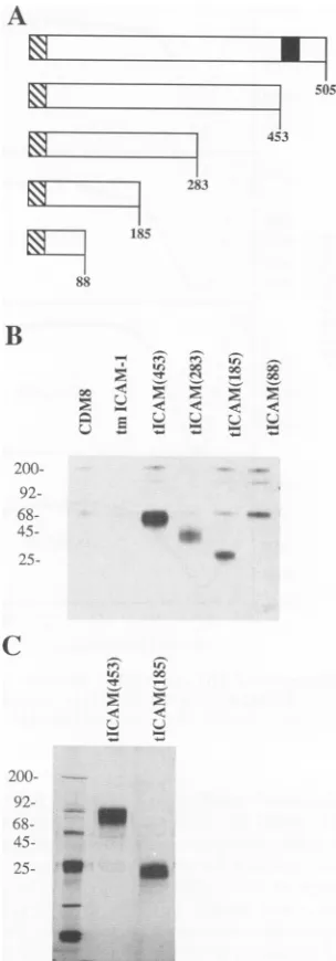
Expression of soluble truncated forms of ICAM-1. ICAM-1 cDNAs encoding soluble proteins were constructed by
in-sertingstopcodonswithin the reading frame of the ICAM-1
polypeptide. Thus, stop codons were inserted immediately
beforethe first residue of the transmembrane domain, at the
predictedendsofdomains 1, 2,and 3,domains 1and 2, and
domain 1 to produce a series of progressively truncated
proteins (Fig. 1A). ThecDNAs wereclonedinto the
expres-sionvector CDM8, transfected into COS cells for transient
expression, and cotransfected with pSV2-dihydrofolate
re-ductaseintoCHOcells for establishmentof stable celllines.
In theexperimentforwhichthe resultsareshown inFig.1B,
the secretion of various forms of truncated ICAM-1 in
transiently transfected COS cellswasexamined. The results indicated that the entire extracellular domain, tICAM(453),
and the two N-terminal domains, tICAM(185), were
effi-ciently secreted from transfected COS cells as species of
80,000Da and43,000 Da,respectively(Fig. 1B). Thelevelof
expression of domains 1, 2,and 3 [tICAM(283)]was
approx-imately 10-fold lower than those of the above-mentioned
fragments,and the secretedproteinwasmoreheterogeneous
[image:3.612.362.514.79.514.2]in mobility on SDS-PAGE. Expression of domain 1
[tICAM(88)] could notbe detectedin COS cells, and
alter-native constructs in which the stop codon was shifted to several sites N or C terminal to residue 88 also failed to
producedetectable amounts of
protein.
In order to obtainsufficient
quantities
ofprotein
for functional and structuralstudies, CHO cell transfectants were prepared, cloned, and
subjected to stepwise geneamplification in increasing
con-centrations of methotrexate. This resulted in thederivation
of cell lines secreting 1.5 ,ug of
tICAM(453)
and 1.0 ,ug oftICAM(185)perml. Astablecell line
expressing tICAM(283)
was notobtained, perhapsbecause the lowlevelofsecretion was atthe limit ofsensitivityof theimmunoassay.Thecells
wereadaptedtoserum-freemedia,and thesecreted ICAM-1
proteinswere
purified
tohomogeneity
from culturesuperna-tants (Fig. 1C).
A
505
453
uZiZ
283
v
18588
B
200- 92- 68-
45-
25-ir, x x
Xc
*, 'r c ~c _
z <-e < < <
U,W
C
2200- 92- 68-
45-
25
-a
FIG. 1. Secretion of soluble ICAM-1 proteins. (A) Diagramof progressively truncated forms of ICAM-1 used in transfection experiments. The crosshatched section indicates signal sequence, and the filled section indicates the transmembrane region. (B) Fluorograph of[35S]cysteine-labeled productssecretedbyCOScells analyzed by SDS-PAGE. The loading of lanes containing tICAM(283) and tICAM(88) is 10-fold higher than the loading of other lanes. (C) Silver-stained gel of purified tICAM(453) and tICAM(185)produced by CHOcelltransfectants.InpanelsBandC, molecularweight markers(in thousands)areindicated ontheleft.
Several of the physical properties of tICAM(453) and
tICAM(185) were examined. Both
proteins
werequantita-tively
immunoprecipitated by
two monoclonalantibodies,
c78.4andc78.5,directed
against
twodistinctconformation-dependentepitopesonICAM-1
(data
nowshown),
indicating
that these epitopes were contained within the first two
domains and
providing
evidence that thepurified proteins
were correctly folded. The Stokes radii of
tICAM(453)
andtICAM(185) were 5.3 and 3.9 nm,
respectively,
and thefrictional ratio,
f/fo
(the ratio between the observed andon November 10, 2019 by guest
http://jvi.asm.org/
-1000.
-2000 -3000
c)
0
'0
(D 2000
0 -2000 4000 6000 -8000
-2000
4000
-6000
A A
A & IgG
A
:
*
. . .
I- a A ICAM-1
A {17
A~~A
I-6000
2000 -2000 -6000
A
A A
A A
A tICAM(453)
A A
. a
a*:
LA
r tICAM(185)
Iu
195 215 235 255 275 295
WAVELENGTH(nm)
FIG. 2. CDspectraofIgG and ICAM-1proteins.(A)c92.5IgG; (B)ICAM-1; (C) tICAM(453); (D)tICAM(185).Datawerecollected andanalyzedasdescribedinMaterials and Methods.
calculated frictional coefficients), was 1.9 for both tICAM
(453) and tICAM(185), indicating that both fragments of
ICAM-1arehighlyasymmetricandelongatedmolecules. CD spectra were obtained for the soluble forms of ICAM-1. A
singleminimumat210to220nmis indicative ofthe presence
of ,B structure and should be seen in proteins containing
immunoglobulin-like domains because of the extensive
amount ofP structure in theimmunoglobulin fold (38). As
internalstandards, spectra werecollected fortwomembers
oftheimmunoglobulinsupergenefamilyof known
3-dimen-sional structure, ,-2-microglobulin (4) and a murine
mono-clonalIgG (1). As expected, the spectra of
P-2-microglobulin
(not shown) and IgG (Fig. 2A) possessed single minima at 215 nm and 217 nm, respectively. The spectra ofthe two truncatedproteins, tICAM(453)andtICAM(185),were sim-ilartoeach other and to that ofICAM-1(Fig. 2B, C, and D), with minima at 216 to 217 nm and a broad shoulder at 225 to 230 nm, although the shoulder was more pronounced in
tICAM(185)than intICAM(453). These CD spectra provide
additional evidence that the soluble ICAM-1 proteins are
properlyfoldedand thatthey possess significant amounts of ,Bstructure.
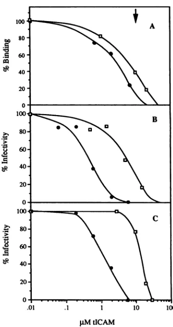
Inhibition of virus binding by solubleICAM-1. To compare
theeffectsof the soluble ICAM-1 proteins on virus-receptor
binding,acompetitionbinding assay was employed. Various
concentrations of soluble competitor ICAM-1 proteins were
incubated with
35S-HRV3,
andbinding to ICAM-1immobi-lizedonmicrotiter dishes was determined. As can be seen in
Fig. 3A, both truncated forms of ICAM-1 inhibit HRV3
binding
atsimilar concentrations:tICAM(453)andtICAM(185) [image:4.612.351.528.75.409.2]pM
tICAMFIG. 3. Inhibition of virusbindingand virusinfectivity by solu-ble formsofICAM-1. (A)35S-HRV3waspreincubated with various concentrationsof soluble ICAM-1 for 30minat34°C, and binding to immobilized ICAM-1wasdeterminedasdescribed in Materialsand Methods.(B) Reduction in virustiterbysoluble ICAM-1. Approx-imately 2 x 10² PFU of HRV3 was preincubated with soluble ICAM-1 in 1 ml, and virus titerwasdeterminedby plaque assayas described in Materials and Methods. (C) Inhibition of virus infec-tivity under conditions of the virus-binding assay. HRV3 at 107 PFU/ml was preincubated with various concentrations of soluble
ICAM-1 used to infect HeLa cells in a single-cycle, high-MOI
infection (MOI, 100). Infectivity was determined after 24 h as described in Materials and Methods. tICAM(453), solid circles; tICAM(185), open squares. Arrow indicates the concentration of ICAM-1atwhichthe data shown inFig.4wereobtained.
atconcentrations of 3.1 + 1.8 ,uM(n = 3) and 9.7 + 3.2 ,uM
(n=3),respectively, inhibit thebindingof35S-HRV3 by50%.
Inhibition of virusinfectivitybysolubleICAM-1. The effect of soluble ICAM-1 on rhinovirus
infectivity
was examined under several differentconditions (Fig. 3B and C;Table 1).Intheexperimentfor which the results are shown inFig. 3B
and Table 1 (experiment I), HRV3 was preincubated with various concentrations of soluble ICAM-1, and virus titer was determined byaplaque assay in which soluble ICAM-1 waspresentduringtheincubation of the virus with the HeLa
cell monolayers. The concentrationoftICAM(185) required
toreduceinfectivityby50%
(IC50)
inthis experimentwas5.3 ,uM, similar to the concentration needed to inhibit virusbinding by 50%. In contrast, the IC50 for infectivity of
tICAM(453) was 0.4 ,uM, eightfold lower than its IC50 for
e& 4
on November 10, 2019 by guest
http://jvi.asm.org/
[image:4.612.103.261.79.376.2]TABLE 1. Neutralizationofrhinovirusbysoluble ICAM-1
Presence of ICAM-1 IC50
Experimenta during: (p.M)b
Preincubation Infection tICAM(185) tICAM(453)
I + + 5.3 0.4
II + + 13.2 1.2
III + >20 >20
aDataforexperimentI areshown in Fig. 3B, data forexperimentII(high
MOI)areshown in Fig. 3C, anddata forexperimentIII aredescribed inthe
text.
bSee Materials and Methods foradescriptionofeach assay.
virus binding. The IC50 for infectivity of tICAM(453) was
13-fold lower than that of tICAM(185). Even though the
binding experiments were performed in molar excess of
ICAM-1 overreceptorbinding sites onrhinovirus (the
con-centrationof HRV3 in these experiments was approximately
17 pM, and theconcentration of potential receptor binding
siteswas60-fold higher, or 1 nM), another infectivity
exper-iment was performed in which the concentration of HRV3
was identical to that in the binding experiments.
Conse-quently, HeLa cells were infected at a high MOI in the
presenceofsolubleICAM-1 in a single-cycle infection (Fig.
3C and Table1[experimentII]).AlthoughtheIC50values for
tICAM(453) and tICAM(185) are approximately 3-fold higherinthehigh-MOI experiment than in the plaque assay,
the relative difference in the
IC50
values between the twoICAM-1 species was approximately 11-fold, or essentially
the same as in the low-MOI experiment. The difference in
absolute IC50 valuesbetween these two experiments may be
duetoa nonlinearrelationship betweeninfectious particles
and infected cellsathighMOIs.Todetermineif the
neutral-izationofHRV3 wasreversible,virus waspreincubatedwith
soluble ICAM-1 at20 ,uM (aconcentrationknowntoreduce
infectivity >99%; see Fig. 3B), and the mixture was then
diluted tonegligible ICAM-1 concentrationsfor infectionof
HeLa cells. The titers of tICAM(453)- and
tICAM(185)-treatedviruswere1.9 x
105
PFU/mland 3.0 x 10⁵ PFU/ml,respectively, compared with 3.6 x
105
PFU/ml for controlvirus (Table 1, experiment III). Only marginal reduction of
virus titer
(<50%)
wasobserved at20 ,uM solubleICAM-1,indicatingthat theICAM-1-mediated neutralization of
rhino-virus is largely reversible; this reversibility is presumably
due to simple dissociation of the virus-receptor complex
upondilution.
Thus, the neutralizing activity of tICAM(185) is directly
correlated with itsability toinhibit virus-receptor binding,
whiletICAM(453) neutralizes rhinovirus at aconcentration
considerablylower than that necessarytoinhibitbindingand
is
presumably
actingby amechanism in addition to directcompetitionfor
receptor-binding
sitesonthe virus.Neutral-ization of rhinovirus by both forms of ICAM-1 is
largely
reversible.
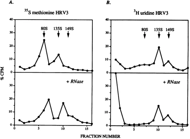
Effect of solubleICAM-1 onrhinovirus integrity. Samples
of 35S-HRV3 incubated with
tICAM(453)
andtICAM(185)
underconditions similartothosein thebindingand
infectiv-ity experiments were
analyzed
by sedimentationthrough
sucrosegradients. HRV3incubatedwith 10,uM
tICAM(453)
separatedinto threepeaks, 149S
(cosedimenting
withnativevirus), 135S, and 80S (Fig. 4A). The 149S and 135S
peaks
were infectious, with
PFU/cpm
ratios of 200 and 267com-pared to a value of 200 for native HRV3. The
specific
infectivity of the 80S
species
wasdramatically
reducedtoaA.
FRACTION
NUMBERB.
tICAM(185),
24hr3000
2000
1000-0 2 4 6 8 10 12 14 16 18
FRACTION
NUMBERFIG. 4. Sedimentation analysis ofHRV3incubatedwithsoluble ICAM-1. 35S-HRV3 (105
cpm)
was incubated with 10puM
tICAM (453) (A)ortICAM(185) (B) inNbuffer-1mgofBSAperml(pH7.5)
for 0.5 and24hat
34°C.
The mixtureswerethensedimentedthrough sucrosegradients asdescribed in MaterialsandMethods, andthe radioactivityin thefractionswasdeterminedbyscintillation count-ing. Fractionsarenumberedfromtoptobottomofthegradient.on November 10, 2019 by guest
http://jvi.asm.org/
[image:5.612.321.549.109.634.2]_
W -VP2+V1`3
-VP4
FIG. 5. Capsid protein composition of solubleICAM-1-modified forms ofHRV3. Fractions from control 35S-HRV3 and149S, 135S, and80S [from3"S-HRV3 incubated with 10,uMtICAM(453)] were subjected to SDS-PAGE followed by fluorography. Densitometry (normalized to the VP1 band) indicated that the VP4 contentofthe 80S sample was9%of control HRV3,149S, or135Sspecies.
PFU/cpmratio of15; the residualinfectivityis probably due
to slight contamination from the 149S and 135S peaks since the gradients were fractionated from the bottom. This 80S species was also generated upon incubation with tICAM(185), although less efficiently (Fig. 4B). The fraction of HRV3 in the80S peak in the sample treated with 10 FM tICAM(453) was 18% after 30 min and61% after 24 h. The rate of conversion to the 80S species is highly temperature dependent and is greater at 37°C than at 34°C, the optimal temperature for rhinovirus growth (10). Since this concen-tration of tICAM(453) reduced infectivity to<0.2%(Fig. 3B) and since the reduction of infectivity is largely reversible, theconversion ofrhinovirus to the noninfectious 80S species does not play a major role in rhinovirus neutralization by
soluble ICAM-1.
A. B.
35S
methionineHRV3The 149S, 135S, and 80S peaks were characterized with respect to their viralcapsid composition and RNA content.
Analysis of peakfractions by SDS-PAGErevealed that the
149S and 135S peaks possessed all four capsid proteins,
while the 80S peak had dramatically reduced amounts of VP4 (Fig. 5). To assess the RNA contentofthe three
peaks,
preparations of [35S]methionine- and [3H]uridine-labeled
HRV3incubatedwith 10 ,uMtICAM(453)wereseparatedon
sucrose gradients (Fig. 6). The radioactivity from the
[35S]methionine-labeled
virus formed a clear 80S peak,which was insensitive to RNase A. The radioactivity from
the [3H]uridine-labeled virus formed 149S and 135S peaks
but no 80S peak. When RNase A was included in the
incubation,the149S and 135Speakswere notaltered,while
theremainder oftheradioactivity distributedacrossthetop
halfof the gradient shifted to the topof thegradient. These data indicate that the 80S peakdoes not contain RNA and that the 149S,135S, and80SpeaksareinsensitivetoRNase Adigestion. In asecond experiment,the viral RNA content was determined directly by extraction ofRNA from equal
amountsofradioactivity from 149S, 135S, and80Speaks of
[35S]methionine-labeled
HRV3 and thenbydotblotanalysis
withan oligonucleotide probe forthepositive (+) strandof
rhinovirus (Fig. 7). This experiment showed thatthe 149S
and 135Speaks but not the80S peak containedviral RNA. Thus, the 80Speak appears to be an emptycapsid, lacking
bothRNAandVP4. The 135S peakcontains bothVP4and
RNAand is infectious. Althoughthe natureofthe135Speak
is unclear at present, it islikely that it is a virion withaltered
hydrodynamic propertiesdue tointeraction with ICAM-1.
DISCUSSION
In this report, we have described the production and
characterization oftwosoluble forms ofICAM-1,themajor
3H
uridineHRV3I
FRACTION
NUMBERFIG. 6. ViralRNA content of solubleICAM-1-modified forms of HRV3. Sedimentation analysis of[35S]methionine-(A) and [3H]uridine-labeled(B) HRV3 after incubation with 10 ,uMtICAM(453) for 30minat 37°C are shown. Whereindicated, RNase A (10,ug/ml)was included in the reactionmixtures. Data are plotted as the percentage of total radioactivity on each gradient (approximately 75,000 cpm and 60,000 cpm for
[35S]methionine-
and[3H]uridine-labeled HRV3, respectively). Fractions are numbered from top to bottom of the gradient.on November 10, 2019 by guest
http://jvi.asm.org/
[image:6.612.131.241.76.188.2] [image:6.612.150.484.444.689.2]1IR%3RNA
A149S
135S
80S
FIG. 7. Detection of viralRNAby hybridization withan
oligo-nucleotide probe. RNA from the 149S, 135S, and 80S peaks of
[35S]methionine-labeled HRV3 incubated with tICAM(453) was
probedwitha32P-labeled oligonucleotide probe for the positive(+)
strand of rhinovirus. The amount of material loaded from each
sample was normalized tothe amountof [35S]methionine
radioac-tivity extracted, as described in Materials and Methods. HRV3
RNA (50 ng) and RNA from 8ng(of protein) fromthe149S, 135S,
and80Sspecies wereappliedtothefilter.
human rhinovirus receptor, and their effects on
virus-recep-torbinding, virus infectivity,andvirusintegrity. Theresults
fromthese experiments allowustodistinguish three distinct mechanisms by which soluble ICAM-1 blocks virus growth andto identify the regions of ICAM-1 responsible forthese activities.In addition, theresults presented show that recep-tor protein can uncoat the rhinovirus particle in vitro and, thus, have implications for the mechanism of virus uncoating
in vivo.
ICAM-1 has a domain structure that is related to the immunoglobulin supergene family, and its extracellular
por-tion can be divided into five immunoglobulin-like domains
(32, 35). Twosolubleforms of ICAM-1 have beenproduced, one corresponding to the entire extracellular domain [tICAM(453); domains 1 through 5] and one corresponding
to the two N-terminal immunoglobulin-like domains [tICAM(185); domains 1 and 2]. Constructs coding for do-mains 1 through 3were expressed poorly, anddomain 1was
not expressed at all in mammalian cells. These data are
similar tothoseobtained with CD4, arelated memberof the
immunoglobulin supergene family (2). A likely explanation
for these findings is that an intimate interaction exists
between certain domains, particularly between domains 1 and 2, which is required forproperfoldingorsolubilityof the
polypeptide; the close packing between domains 1 and 2 of CD4 revealed by its crystal structurehasprovided evidence in favor of this interpretation (28, 37). Physical characteri-zation of tICAM(453)andtICAM(185) indicate that bothare
asymmetric molecules, consistentwith the dataofStaunton et al. (34) with regard to a soluble form ofICAM-1 that is
essentially the same as tICAM(453). Proteins belonging to.
the immunoglobulin supergenefamily wouldbe expectedto have domains with the immunoglobulinfoldmotif, which is basically two apposed sheets ofantiparallel
0i
strands with connecting loops (38). CD indicates that both tICAM(453) and tICAM(185) have significant amounts of P structure,consistent withtheir sequencehomologiesto immunoglobu-lin supergene family members, and provides evidence that
the proteins areproperly folded. However, the CD spectra dohavefeaturesthataresignificantlydifferentfromthoseof
classical members of theimmunoglobulin supergenefamily;
the shoulder at 225 to 230 nm present in the two ICAM-1 species [particularly in tICAM(185)] is not found in the
spectra of
P-2-microglobulin
and IgG. These differencessuggestthepresence ofnovelsecondary structuralfeatures,
particularlywithindomains1and2. Indeed, thehomologyof
many ofthe domains ofICAM-1 to immunoglobulin super-gene family members determined by the ALIGN program
(5),whilesignificant, isnothigh(datanotshown). Domain 1,
in particular, has a number of unusual features for an
immunoglobulin-like domain, such asa relatively short
dis-tance (44residues)betweenintradomain disulfide bondsand fourinstead of two cysteines in the B and F
P
strands.Thus,structural predictions based on the immunoglobulin fold
motifshould be made withcaution.
Competitivebindingstudies indicatethat the binding site
for rhinovirus is largely contained within the first two
domains. This conclusion is consistent with studies on
transmembrane ICAM-1, in that human-mouse chimera
studies by Staunton et al. (34) indicate that the rhinovirus
binding site is located within domains 1 and 2. Similar
studies byMcClellandet al. (20)indicatethat therhinovirus
binding site islocated within domain 1. The reason for the
small butsignificantly (threefold) greater inhibitoryactivity
oftICAM(453) relative to that of tICAM(185) is unclear,
althoughtwo possibilities are thefactthat(i)tICAM(185)is
less stably folded thantICAM(453) or (ii) the large size of
tICAM(453) creates additionalstericinterference[relativeto
tICAM(185)] for a subsaturated virus particle binding to
membrane-bound ICAM-1. In studies with transmembrane
forms ofICAM-1, Staunton etal. (34) havereportedthat a shortenedformof ICAM-1
containing
only domains1 and2bindsrhinovirusatapproximately 1/10the levelof
full-length
transmembrane ICAM-1, which they propose is due to
inaccessibility to the
receptor-binding
site on the virusbecause ofthe short distance of the virus
binding
site ofICAM-1from theplane ofthe membrane. Direct
analysis
ofthestoichiometry ofsolubleICAM-1-virus
binding
mayhelp
to resolve this issue.
The effects of tICAM(453) and
tICAM(185)
on virusinfectivity are clearly distinguishable.
tICAM(185)
inhibitsvirus infectivity at essentially the same concentration at which it inhibits virus-receptor binding, indicating that its mode of
aption
is bycompetitive inhibition ofvirusbinding
to
cellular
receptor.
tICAM(453), however, inhibitsinfectiv-ity at a concentration 10-fold lower than that
required
forinhibition of binding,
indicating
a second mechanism forneutralizing; virus dependent upon functions encoded
by
domains 3, 4, and 5. Although the nature of this second
mechanism is unclearatpresent, it is reasonabletoconclude
that entry or intracellular
uncoating
steps areinvolved. Onepossibility
is that thelarge size oftICAM(453)
relativetothatof tICAM(185) creates steric
problems
for subsaturatedvirus-receptor complexes
during
the entry oruncoating
steps; another
possibility
is thatadditionalcontacts withthe virus or with adjacent tICAM(453) molecules on the virionmediated by domains 3, 4, or 5 are
responsible
for theenhanced neutralizing
activity
oftICAM(453).
Marlin et al.(19) reported a similar
disparity
between theability
of asoluble ICAM-1 molecule similar to
tICAM(453)
to inhibitvirus-receptor binding and virus
infectivity,
although
thiswas attributedtodifferences in the
experimental
conditionsofthetwoassays. In the
binding
andinfectivity
experiments
reported here, soluble ICAM-1 is in considerable molar
excess abovethatof virus orviral receptor
binding
sites and we have performed ourbinding
andinfectivity
studies at identical rhinovirus concentrations. The resultspresented
here indicate that the differences in the
binding
andneutral-ization activities are significant.
Both tICAM(453) and
tICAM(185)
have theability
toirreversibly inactivate rhinovirus
by
causing
the loss of theviral subunit VP4 and the RNA genome; this constitutes a
third mechanismof virus neutralization
by
solublereceptor.
The subviral
particles
resulting
from this alteration aresimilar insomerespects tothe
eclipse
products
described foron November 10, 2019 by guest
http://jvi.asm.org/
poliovirus andotherpicornaviruses generated during infec-tion of cells(7, 8, 13), which are thoughtto be productsof
the uncoating process (26). It has been demonstrated that
poliovirus can be conformationally altered in cell-free
sys-temsbymembranescontaining poliovirusreceptor(6, 12)or
detergent-solubilized poliovirus receptor (14)to a135S form
lackingVP4butstillcontainingRNA(inanRNase-sensitive
state). However, release of RNA requiresfurther treatment of the 135S species withhigh concentrations of salt(14) or SDS (6, 12), and generation of 80S empty capsids requires
live cells (8). We have demonstrated here that truncated
soluble ICAM-1 can alter rhinovirus to an 80S species
lackingboth VP4 and RNAand, thus, essentiallyuncoatsthe
virus.We have alsoidentified an intermediate 135Sparticle
which results fromICAM-1-rhinovirusinteraction;this
par-ticle differs from the135Spoliovirusalteredparticle inthatit
is infectious and contains VP4. These differences between
rhinovirus and polioviruswith respect to theproducts of in
vitrovirus-receptor interactionmayreflect differences in the
rate-limiting steps for' uncoating between rhinovirus and
poliovirus or may reflect the different experimental
condi-tions under which the experiments were performed. The
alteration of rhinovirusbysoluble ICAM-1clearlyindicates
that receptor can completely uncoat rhinovirus in the ab-senceofother cellular components, suggestingthat
destabi-lization of the rhinovirus by receptor plays a role in the
uncoatingprocess in vivo.This may be ageneral
phenome-non, as it has recently been reported
that
soluble CD4induces the release ofgpl20 from the human
immunodefi-ciency virus virion, and it has been hypothesized that this
releaseofgpl20 from virionsattachedto'the cell surface may expose
regions
ofgp4l moleculesthatcouldpromotefusion of the virion and cell membranes (22, 23), which occurs atneutralpH at the cellsurface (36). However, the
physiolog-ical significance of ICAM-1-mediated uncoating in vivo is
unclear sincethere is alsoa requirement fora
chloroquine-sensitivelow-pHstepinsidethe cellfor rhinovirus infection
(10).A moredetailed description ofthe in vitroalteration of
rhinovirus by ICAM-1 andits rolein virus uncoatinginvivo
will be presented elsewhere. However, it is clearfrom the
datapresentedhere thatuncoatingdoes notplayamajorrole
in soluble ICAM-1-mediated neutralization of rhinovirus
undertheconditions ofoptimal rhinovirus growth,since the
inhibitory
activity of soluble ICAM-1towardvirusinfectionis largely reversible andbecause the conversion to the 80S
form canonly accountfor asmall fraction ofthis activity.
In conclusion, we have demonstrated that two forms of
solubleICAM-1, tICAM(453)andtICAM(185), inhibit virus-receptorbindingand virus infectivity.Differentialeffects of
these two proteins on these processes have defined three
distinctmechanisms of virus neutralization. The first
mech-anismappears tobeasimplecompetitionof soluble receptor
for receptor binding sites on the virus, and determinants within domains 1and 2 of ICAM-1 are responsible for this
activity.
The second mechanism is a reversibleneutraliza-tion in which virus is apparently blocked at an entry or
uncoatingstepand involvescontributions from domains 3, 4,
and 5.Thethird mechanismisan irreversible inactivation of
the virus characterizedby the loss of the virus subunit VP4
and theRNAgenome.
ACKNOWLEDGMENTS
We thankJudy Dziuba for tissueculture and fermentation, Tom Buckholz and Gary Davis foramino acid analysis, Craig Rice and George Mitra (Cutter Laboratories)for fermentation and purifica-tion ofsomeof thetICAM(453)usedinthesestudies, SuzyPafkafor
photography, DavidOstermanfor instructionanddiscussions in the useofcirculardichroism,Mark Cochranforhelpful discussions,and Mike Kamarckforacriticalreadingof themanuscript.
REFERENCES
1. Amzel,L.M.,and R.J.Poijak. 1979. Three-dimensional struc-tureofimmunoglobulins.Annu. Rev. Biochem. 48:961-967. 2. Arthos,J.,K.C.Deen,M. A.Chaikin, J.A.Fornwald,G.Sathe,
Q. J.Sattentau,P.R.Clapham,R. A.Weiss, J.S.McDougal,C. Pietropaolo,R.Axel,A.Truneh,P.J. Maddon,and R. W. Sweet. 1989.Identification of the residuesinhuman CD4 critical for the binding ofHIV. Cell 57:469-481.
3. Bebbington,C.R.,and C. C. Hentschel. 1987. Theuseof vectors basedongeneamplificationfor theexpressionof clonedgenes in mammaliancells, p. 163-188. In D. M. Glover(ed.),DNA cloning-a practical approach,vol. 3. IRLPress,Oxford. 4. Becker, J. W., and G. N. Reeke. 1985. Three dimensional
structure of P2-microglobulin. Proc. Natl. Acad. Sci. USA
82:4225-4229.
5. Dayhoff,M.O.,W. C.Barker,and L. T. Hunt. 1983. Establish-ing homologies in protein sequences. Methods Enzymol. 91: 524-545.
6. DeSena, J., and B. Mandel. 1977. Studies on the in vitro uncoating ofpoliovirus. II. Characteristics of the membrane-modifiedparticle.Virology78:554-566.
7. Fenwick, M. L., and P. D. Cooper. 1962. Early interactions betweenpoliovirus andERK cells: some observations on the
nature andsignificance ofrejected particles. Virology
18:212-223.
8. Fricks, C.E., andJ. M. Hogle. 1990. Cell-induced conforma-tionalchangein poliovirus:externalization oftheamino termi-nus of VP1 is responsible for liposome binding. J. Virol. 64:1934-1945.
9. Giranda,V. L., M. S.Chapman, and M. G. Rossmann. 1990. Modeling of thehuman intercellular adhesion molecule-1, the humanmajorgroup receptor.Proteins 7:227-233.
10. Greve, J.M. Unpublisheddata.
11. Greve, J. M., G.Davis, A. M.Meyer,C. P.Forte, S. C.Yost, C. W. Marlor, M. E. Kamarck, and A. McClelland. 1989. The majorhumanrhinovirusreceptoris ICAM-1. Cell 56:839-847. 12. Guttman, N., and D. Baltimore. 1977. A plasma membrane
componentable tobind and alter virions ofpoliovirustype 1: studiesoncell-free alterationusingasimplifiedassay. Virology 82:25-36.
13. Joklik,W.K.,andJ.E.Darnell. 1961. Theadsorptionandearly fate ofpurified poliovirusinHeLacells. Virology13:439-447. 14. Kaplan, G., M. S. Freistadt,and V. R.Racaniello. 1990.
Neu-tralization ofpoliovirus by cell receptorsexpressed in insect cells. J.Virol. 64:4697-4702.
15. Kim, S.,T.J.Smith,M. M.Chapman,M.G.Rossmann,D. C. Pevear, F. J. Dutko, P. J. Felock, G. D. Diana, and M. A. McKinlay.1990.Crystalstructureofhumanrhinovirusserotype 1A(HRV1A).J. Mol.Biol.210:91-111.
16. Kingston, R. E. 1987. Introduction of DNA into mammalian cells,p.901-906.InF. M.Ausubel,R.Brent,R. E. Kingston, D. D.Moore,J.G. Seidman,J. A. Smith, and K.Struhl(ed.), Current protocols in molecular biology. John Wiley & Sons, Inc., NewYork.
17. Kishimoto,T.K.,R.S.Larson,A. L.Corbi,M. L.Dustin,D. E. Staunton, and T. A. Springer. 1989. The leukocyte integrins. Adv. Immunol. 46:149-182.
18. Marlin, S.D., and T. A. Springer. 1987. Purified intercellular adhesionmolecule-1(ICAM-1)isaligandforlymphocyte func-tion-associatedantigen (LFA-1). Cell51:813-819.
19. Marlin,S.D.,D. E.Staunton,T. A.Springer, C. Stratowa, W. Sommergruber, and V. J. Merluzzi. 1990. A soluble form of intercellular adhesion molecule-1 inhibits rhinovirus infection. Nature(London)344:70-72.
20. McClelland, A, J. deBear, S. Connonly Yost, A. M. Meyer, C. W.Marlor, andJ. M. Greve. 1991. Identification of mono-clonal antibody epitopes and critical residues for rhinovirus
binding in domain 1 of ICAM-1. Proc. Natl. Acad. Sci. USA
88:7993-7997.
on November 10, 2019 by guest
http://jvi.asm.org/
21. Minor, P. D. 1985. Growth, assay, and purification of picorna-viruses, p. 25-40. In B. W. J. Mahy (ed.), Virology: a practical approach. IRL Press, Oxford.
22. Moore, J. P., J. A. McKeating, W. A. Norton, and Q. J. Sattentau. 1991.Directmeasurement of soluble CD4 binding to humanimmunodeficiency virus type 1 virions: gpl20 dissocia-tionand its implications for virus-cell binding and fusion reac-tions andtheir neutralization by soluble CD4. J. Virol. 65:1133-1140.
23. Moore, J. P., J. A. McKeating, R. Weiss, and Q. J. Sattentau. 1990. Dissociation of gp120 from HIV-1 virions induced by solubleCD4. Science 250:1139-1142.
24. Rossmann, M. G. 1989. The canyon hypothesis. Hiding the host cell receptor attachment site on a viral surface from immune surveillance.J. Biol. Chem. 264:14587-14590.
25. Rossmann, M. G., E. Arnold, J. W. Erikson, E. W. Franken-berger, P. J. Griffith, H. Hecht, J. E. Johnson, G. Kamer, M. Luo, A. G. Mosser, R. R. Rueckert, B. Sherry, and G. Vriend. 1985.Structure ofacommoncold virusand functional relation-shiptoother picornaviruses. Nature (London) 317:145-153. 26. Rueckert, R. R. 1990. Picornaviridae and their replication, p.
507-548. In B. N. Fields et al. (ed.). Virology. Raven Press, NewYork.
27. Rueckert, R. R., and M. A. Pallansch. 1981. Preparation and characterization ofencephalomyocarditis (EMC) virus. Meth-odsEnzymol. 78:315-326.
28. Ryu, S., P. D.Kwong, A. Truneh, T. G. Porter, J. Arthos, M. Rosenberg, X. Dai, N. Xuong, R. Axel, R. W. Sweet, and W. A. Hendrickson. 1990. CrystalstructureofanHIV-binding recom-binantfragment ofhumanCD4.Nature(London) 348:419-426. 29. Saiki, R. K., S.Scharf, F. Faloona, K. B. Mullis, G. T. Horn, H. A.Erlich, and N. Arnheim. 1988.Primer-directedenzymatic amplification ofDNA with athermostable DNA polymerase.
Science239:487-491.
30. Sambrook, J., E. F. Fritsch, and T. Maniatis. 1989. Molecular cloning, a laboratory manual. Cold Spring Laboratory Press, ColdSpring Harbor,N.Y.
31. Seed, B. 1987. An LFA-3 cDNA encodes aphospholipid-linked membrane protein homologous to its receptor CD2. Nature (London) 239:840-842.
32. Simmons, D., M. W. Makgoba, and B. Seed. 1988. ICAM, an adhesion ligand of LFA-1, is homologous to the neural cell adhesion moleculeNCAM.Nature(London) 331:624-627. 33. Springer, T. A. 1990.Adhesionreceptorsof theimmune system.
Nature (London)346:425-434.
34. Staunton, D. E., M. L. Dustin, H. P. Erickson, and T. A. Springer. 1990. The arrangement of the immunoglobulin-like domains of ICAM-1 and the binding sites for LFA-1 and rhinovirus. Cell61:243-254.
35. Staunton, D. E., S. D. Marlin, C. Stratowa, M. L. Dustin, and T. A.Springer. 1988.Primarystructureof intercellularadhesion molecule 1 (ICAM-1) demonstrates interaction between the immunoglobulin and integrin supergene families. Cell 52:925-933.
36. Stein,B.S., S. D. Gowda, J. D. Lifson, R. C. Penhallow, K. G. Bensch, and E.G. Engleman. 1987. pH-independentHIV entry intoCD4-positiveTcells via virus envelope fusiontotheplasma membrane. Cell 49:659-668.
37. Wang, J., Y. Yan, T. P. J. Garrett, J. Liu, D. W.Rodgers, R. L. Garlick, G. E. Tarr, Y. Husain, E. L. Rheinherz, and S. C. Harrison. 1990.Atomicstructureofafragmentof human CD4 containingtwoimmunoglobulin-likedomains.Nature(London) 348:411-418.
38. Williams,A.F., and A. N. Barclay. 1988. Theimmunoglobulin superfamily-domains for cell surface recognition. Annu. Rev. Immunol.6:381-405.