Gary Z. Wang,a,bStephen P. Goffc,d,e
Integrated Program in Cellular, Molecular and Biophysical Studies, Columbia University, New York, New York, USAa
; Medical Scientist Training Program, Columbia University College of Physicians and Surgeons, New York, New York, USAb
; Department of Biochemistry and Molecular Biophysics, Columbia University, New York, New York, USAc
; Department of Microbiology and Immunology, Columbia University, New York, New York, USAd
; Howard Hughes Medical Institute, Columbia University, New York, New York, USAe
ABSTRACT
Mason-Pfizer monkey virus (M-PMV) is a prototypical betaretrovirus responsible for simian AIDS (SAIDS) in rhesus macaques. It has been shown previously that mouse cells are resistant to infection by HIV-1 and other primate lentiviruses. However, the susceptibility of mouse cells to primate retroviruses such as M-PMV remains unexplored. In the present study, using single-round green fluorescent protein (GFP) reporter viruses, we showed that various mouse cell lines are unable to support the early stages of M-PMV replication. The block to infection occurs postentry and is independent of the viral envelope. Using quantita-tive real-time PCR, we showed that the block to infection occurs after reverse transcription but before formation of circular DNA or proviral DNA. Finally, we showed that the M-PMV block in mouse cells is not attributable to the previously character-ized mouse restriction factorFv1. Overall, these findings suggest that mouse cells exhibit a previously uncharacterized block to M-PMV infection.
IMPORTANCE
Here we document a novel postentry restriction to M-PMV infection in mouse cells. The block occurs after reverse transcription but before the formation of circular or proviral DNA and is independent of the previous characterized mouse restriction factorFv1.
R
etroviruses are capable of infecting diverse vertebrates, and successful infection requires cellular factors to support each phase of the retroviral life cycle. Retroviral tropism is therefore partly dictated by whether the infected cell possesses the necessary cofactors to assist viral replication. For example, human immu-nodeficiency virus type 1 (HIV-1) infects human T cells and mac-rophages efficiently but is unable to infect mouse cells. Multiple steps in the viral life cycle are blocked in mouse cells. First, the mouse homologues of the human T cell receptors and coreceptors CD4, CXCR4, and CCR5 (1,2) cannot bind HIVenv-encoded gp120, preventing HIV-1 from entering the cell. This entry block can be bypassed in engineered mouse cell lines expressing the human T cell receptors (3,4) or by pseudotyping HIV-1 virions with foreign viral envelope proteins such as glycoprotein G of vesicular stomatitis virus (VSV-G). Upon establishment of the provirus in mouse cells, the late phase of the HIV-1 life cycle is hindered by additional cellular blocks. Transcription initiated in the long terminal repeat (LTR) is defective due to the incompati-bility of the HIV-1 Tat transactivator with mouse cyclin T1 (5–8). In addition, viral assembly is defective due to the absence of nec-essary factors required for late stages of the retroviral life cycle, a phenotype that can be rescued by the fusion of uninfected human cells with infected mouse cells (9–11). Study of blocks to HIV-1 replication in mouse cells has led to the identification and charac-terization of critical cellular factors necessary for infection.Species-specific retroviral tropism is also governed by the pres-ence of species-specific restriction factors. For example, human and primates are known to be resistant to N-tropic murine leuke-mia virus (MLV) infection (12,13). The human protein mediating this restriction was identified as TRIM5␣(14). In addition to re-stricting N-tropic MLV, rhesus macaque TRIM5␣ has the re-markable ability to restrict HIV-1 infection (15,16). This obser-vation was later attributed to species-specific variations in the TRIM5␣sequence (17–19), supporting the notion that
species-specific restriction factors are a major determinant of retroviral tropism. Mechanistically, TRIM5␣has been proposed to interact with the viral capsid (CA) (20), leading to premature dissociation of viral cores (21) and terminating infection prior to nuclear entry (16). In mice, the Friend virus susceptibility factor 1 (Fv1) gene functions similarly to TRIM5␣.Fv1 encodes a gag-like protein with homologies to the ERV-L family of endogenous retroviruses (22). There are two major naturally occurring alleles ofFv1. NIH Swiss mice carry theFv1nallele, which restricts the replication of B-tropic MLVs but not N-tropic MLVs. In contrast, BALB/c mice carry theFv1ballele, which confers resistance to N-tropic MLV infection but not B-tropic MLVs (23,24). A few laboratory mice and some wild mice carry a thirdFv1allele,Fv1nr, which restricts the replication of B-tropic MLVs and a subset of N-tropic MLVs (25,26).
Mason-Pfizer monkey virus (M-PMV) is a prototypical betaretrovirus capable of causing simian AIDS (SAIDS) in rhesus macaques (27). Though mouse cells are known to be resistant to lentiviral infections, it was not known whether they would also be resistant to PMV. In the present study, we showed that M-PMV-based vectors are unable to transduce various mouse cell lines. Mouse resistance to M-PMV infection is envelope indepen-dent, with the strongest block occurring after reverse transcription
Received17 October 2014Accepted16 December 2014
Accepted manuscript posted online24 December 2014
CitationWang GZ, Goff SP. 2015. Postentry restriction of Mason-Pfizer monkey
virus in mouse cells. J Virol 89:2813–2819.doi:10.1128/JVI.03051-14.
Editor:K. L. Beemon
Address correspondence to Stephen P. Goff, [email protected]. Copyright © 2015, American Society for Microbiology. All Rights Reserved. doi:10.1128/JVI.03051-14
on November 7, 2019 by guest
http://jvi.asm.org/
but before the formation of circular or proviral DNAs. The block to M-PMV infection is not attributable to theFv1gene, and addi-tional tests ruled out the possibility of transcripaddi-tional silencing of the M-PMV LTR in mouse cells. Taken together, the results dem-onstrate that infection by M-PMV is strongly blocked in mouse cells during the early phase of the life cycle, most likely at the time of import of the viral DNA into the nucleus.
MATERIALS AND METHODS
Cell culture.HEK-293T, HeLa, COS-7, L929, NIH 3T3, and RAW264.7 cells, mouse embryonic fibroblasts (MEFs), andMus dunnitail fibroblasts (MDTFs) were cultured in Dulbecco’s modified Eagle’s medium (DMEM) supplemented with 10% heat-inactivated fetal bovine serum (FBS), 2 mM glutamine, 1,000 U/ml of penicillin, and 100 mg/ml of strep-tomycin. Ba/F3 cells were cultured in RPMI medium supplemented with 10% heat-inactivated FBS and 1.6 ng/ml of recombinant interleukin 3 (IL-3). All cells were maintained in a 37°C incubator with 5% CO2.
Plasmids.Plasmids pCIG3-B and pCMV-intron expressgagandpol from B-tropic (28) and NB-tropic (29) MLVs, respectively. pMD.G ex-presses the vesicular stomatitis virus (VSV) envelope glycoprotein. pNCA-GFP is a replication-defective single-round MLV vector described previously (30). pNCA-Luc is a firefly luciferase reporter virus in which the green fluorescent protein (GFP) cassette in pNCA-GFP is replaced with the firefly luciferase gene via BamHI/XhoI restriction sites. pSARM-EGFP is a replication-defective single-round M-PMV vector in which the envgene has been replaced with the gene for enhanced GFP (EGFP) as described previously (31). pSARM-Luc is a firefly luciferase reporter virus in which the GFP cassette in pSARM-GFP has been replaced with the firefly luciferase gene via XhoI/BlpI restriction sites. pCI-Fv1nand pCI-Fv1bexpress thenandballeles of the mouseFv1gene as described previ-ously (22). pHA-mCAT is a plasmid encoding the mouse cationic amino acid transporter (mCAT), which serves as a receptor for MLV ecotropic envelope (gift from Walther Mothes, Yale University School of Medicine). MLV-LTR-Luc and MPMV-LTR-Luc are constructs expressing luciferase under the control of the viral LTRs. The U3-R-U5 regions of the respective viral LTRs were amplified by PCR using the following primer pairs: for MLV, 5=-AGATCTGCGATCTAAGTAAGCTTAATGAAAGACCCCAC CTGTAGGT-3=and 5=-CCAACAGTACCGGAATGCCAAGCTTGGTG GTCCCTGGGCAGGG-3=, and for M-PMV, 5=-AGATCTGCGATCTAA GTAAGCTTTGTCCGGAGCCGTGCTG-3=and 5=-CCAACAGTACCG GAATGCCAAGCTTACTGTCCCGACCCGCGG-3=. The PCR products were cloned into the HindIII site of pGL3-Basic vector (Promega) using sequence- and ligation-independent cloning (32). All constructs were ver-ified by DNA sequencing.
Retroviral transduction assay.M-PMV–GFP reporter viruses were produced by transfection of 293T cells with 8g of pSARM-EGFP and 8 g of pMD.G per 100-mm plate using polyethylenimine (PEI). Reporter viruses were harvested 48 h later, filtered (0.45m), and used directly for transduction assays. Similarly, NB-tropic MLV-GFP reporter viruses were produced by 293T cell transfection with 8g of pNCA-GFP, 4g of pCMV-intron, and 4g of pMD.G DNAs. For transduction assays, the desired cell types were seeded in 12-well plates at a density of 1⫻105cells
per well and infected with GFP reporter viruses for 5 h at 37°C. Forty-eight hours postinfection, cells were trypsinized, diluted using flow cytometry buffer (PBS with 1% BSA), and subjected to flow cytometry using an automated cell analyzer (LSRII; BD Bioscience). ForFv1overexpression experiments, HeLa cells were transfected with 0.5g of pCI-Fv1nor pCI-Fv1btogether with 0.1g of pHA-mCAT for 24 h, followed by transduc-tion experiments outlined above using MLV or M-PMV luciferase re-porter viruses.
Quantitative real-time PCR analysis of viral replication intermedi-ates.At various time points following infection, cells were washed with PBS, and total DNA was isolated using a Qiagen DNeasy kit according to the manufacturer’s instructions. For analysis of early and late reverse tran-scription (RT) intermediates, 80 ng of total DNA was combined with
M-PMV-specific TaqMan primer/probe sets targeting minus-strand strong-stop DNA and GFP, respectively, as described previously (33). For the detection of 2-LTR circles, 80 ng of total DNA was mixed with SYBR green PCR master mix (Roche) containing 15 pmol of both forward (5= -TCCTCCAGGTTCCTACTGTT-3=) and reverse (5=-ACGGAGAAGAAC CAGGAAATAC-3=) primers. PCRs were performed in 96-well plates us-ing a 7900 Fast real-time PCR system (Applied Biosystems) with the following reaction conditions: 10 min at 95°C, followed by 40 cycles of 30 s at 95°C and 1 min at 60°C. For all reactions, relative expression was quantified using the threshold cycle (2⫺⌬⌬CT) method (34) normalized to the human and mouse Tert genes.
Dual-luciferase assay.HeLa cells (1⫻105cells/well) and NIH 3T3
and L929 cells (2⫻105cells/well) were seeded in a 12-well plate 1 day
before transfection. The next day, each well was transfected with 1g of various LTR firefly luciferase constructs and 10 ng of herpes simplex virus thymidine kinase (HSV-TK) Renilla luciferase control plasmid using Lipofectamine 2000 (Invitrogen) at a 1:4 ratio. Thirty-six hours later, cells were lysed in 100l of 1⫻reporter lysis buffer (Promega) and luciferase levels were quantified using a POLARstar Omega multimode plate reader (BMG Labtech) according to the manufacturer’s instructions. Relative luciferase expression was calculated by first dividing the firefly luciferase signal by theRenillaluciferase signal in a given cell, and the resulting ratio was then normalized to the ratio obtained from HeLa cells transfected with the simian virus 40 (SV40)-firefly luciferase construct and HSV-TK Renillaluciferase (set to 1).
RESULTS
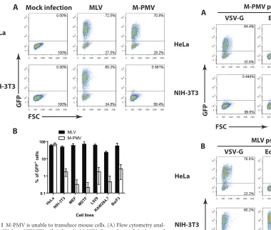
M-PMV is unable to transduce mouse cells. To determine whether mouse cells are susceptible to M-PMV infection, we uti-lized a single-round M-PMV reporter genome in which the viral envgene was replaced with the GFP gene (31). Virions capable of infecting a wide range of cell lines were prepared by collecting culture medium after transfection of 293T cells with the M-PMV reporter genome and a DNA encoding the VSV-G envelope pro-tein. To assess the susceptibility of mouse cells to M-PMV infec-tion, NIH 3T3 cells were infected with VSV-G-pseudotyped M-PMV-GFP, and the efficiency of infection was monitored by flow cytometry analysis 48 h later. HeLa cells, previously shown to be permissive to M-PMV (33), were infected in parallel. As positive controls, we also transduced both cell lines with VSV-G-pseu-dotyped MLV-GFP viruses. While NIH 3T3 and HeLa cells were equally and highly susceptible to MLV infection, NIH 3T3 cells showed a 100-fold reduction in the formation of M-PMV GFP⫹ cells compared to that by HeLa cells (Fig. 1A). Next, we extended our analysis of M-PMV infectivity to other mouse cell lines, in-cluding MEFs, MDTFs, and L929, RAW264.7, and Ba/F3 cells
(Fig. 1B). These cell lines were selected on the basis of differences
in their genetic backgrounds and variations in theFv1allele, as well as cell type (Table 1). Interestingly, while all cell lines sup-ported MLV replication to similar levels, M-PMV transduction was potently blocked in all the mouse cell lines tested (Fig. 1B). Since our reporter viruses were pseudotyped with VSV-G, the observed effect is not likely due to defective viral entry. Moreover, the fact that the cells were susceptible to MLV indicates that they were dividing adequately for infection by viruses that require di-vision. These results suggest that mouse cells broadly exhibit a postentry block to M-PMV infection.
M-PMV replication block in mouse cells is not envelope de-pendent.Different viral envelopes mediate virus entry into the target cell via distinct mechanisms. For example, VSV-G pro-motes viral entry via endocytosis, while the amphotropic (ampho) MLV envelope may facilitate direct fusion with the plasma
mem-Wang and Goff
on November 7, 2019 by guest
http://jvi.asm.org/
brane (35) and has very recently been suggested to promote entry into some cells by macropinocytosis (36). In the case of the eco-tropic (eco) MLV envelope, there exists evidence consistent with both endocytosis (i.e., pH dependence) and direct cell fusion (i.e., pH independence) (37, 38). To evaluate whether the M-PMV block in mouse cells is dependent on the route of entry, we peated the experiment outlined above using M-PMV–GFP re-porter viruses pseudotyped with either MLV ecotropic or ampho-tropic envelopes. As controls, infections with pseudotyped MLV-GFP reporter viruses were carried out in parallel. M-PMV– GFP–VSV-G and M-PMV–GFP–ampho were able to infect the permissive HeLa cells efficiently, with undiluted virus
prepara-tions resulting in 64% and 6.5% GFP⫹cells, respectively (Fig. 2A). The reduced titer of the M-PMV–GFP–ampho virus was presum-ably due to decreased stability of the amphotropic envelope com-pared to that of VSV-G. The M-PMV–GFP– eco virus was unable to infect HeLa cells, consistent with the fact that the MLV eco-tropic glycoprotein cannot bind the human homologue of the mouse ecotropic MLV receptor (mouse cationic amino acid trans-porter [mCAT]) (39) (Fig. 2A). In contrast to HeLa cells, NIH 3T3 cells were resistant to all three pseudotyped M-PMV reporter vi-ruses (Fig. 2A). As expected, pseudotyping MLV with each of the three envelopes conferred infectivity to NIH 3T3 cells (Fig. 2B). Overall, these findings suggest that the M-PMV block in mouse cells occurs in an envelope-independent manner.
Characterizing the M-PMV block in mouse cells.Our single-round GFP reporter viruses score for all the early events of
infec-HeLa
NIH-3T3
FSC
GFP
Mock infection MLV M-PMV
A
HeLa NI
H-3T3 MEF MDTF L929
RAW 264.7 Ba/F3 0.1
1 10 100
MLV M-PMV
Cell lines
% o
f G
F
P
+ c
ells
B
[image:3.585.55.435.64.386.2]FIG 1M-PMV is unable to transduce mouse cells. (A) Flow cytometry anal-ysis of HeLa or NIH 3T3 cells infected with VSV-G-pseudotyped single-round MLV or M-PMV–GFP reporter viruses 2 days postinfection. Thexaxis shows forward scatter (FSC), and theyaxis shows GFP intensity. One representative experiment of five independent experiments is shown. (B) Experiment similar to that in panel A examining the susceptibility of other mouse cell lines to MLV and M-PMV–GFP infection. A logarithmic graph shows the percentage of GFP⫹cells 2 days postinfection. Results shown are means⫾SD from two independent experiments.
TABLE 1Mouse cell lines used in this study
Cell line Mouse strain Fv1genotype Cell type
NIH 3T3 NIH Swiss n/n Fibroblast
MEF C57BL/6 b/b Fibroblast
MDTF Mus dunni ⫺/⫺ Fibroblast
L929 C3H n/n Fibroblast
RAW267.4 BALB/c b/b Macrophage
Ba/F3 C3H n/n B cell
HeLa
NIH-3T3
VSV-G Ecotropic Amphotropic M-PMV pseudotyped with
FSC
GFP
GFP
FSC HeLa
NIH-3T3
MLV pseudotyped with
VSV-G Ecotropic Amphotropic
A
B
FIG 2M-PMV replication block in mouse cells is not envelope dependent. (A) Flow cytometry analysis of HeLa or NIH 3T3 cells infected with M-PMV– GFP reporter virus pseudotyped with either VSV-G or the envelope of eco-tropic or amphoeco-tropic MLV. Results shown are obtained 2 days postinfection (p.i). Thexaxis shows forward scatter (FSC), and theyaxis shows GFP inten-sity. One representative experiment of two independent experiments is shown. (B) Experiment similar to that in panel A, infecting HeLa or NIH 3T3 cells with MLV-GFP reporter viruses pseudotyped with VSV-G, ecotropic MLV, or am-photropic MLV. Results shown are obtained 2 days postinfection. Thexaxis shows forward scatter (FSC), and theyaxis shows GFP intensity. One repre-sentative experiment of two independent experiments is shown.
on November 7, 2019 by guest
http://jvi.asm.org/
[image:3.585.220.537.68.436.2] [image:3.585.42.286.645.723.2]tion, including reverse transcription, nuclear entry, integration, and reporter gene expression. To define the step of M-PMV infec-tion blocked in mouse cells, NIH 3T3 or HeLa cells were infected with M-PMV–GFP, total DNA was harvested at 12 or 24 h postin-fection, and the formation of viral replication intermediates was monitored by quantitative real-time PCR (Fig. 3A). At both 12 and 24 h postinfection, HeLa and NIH 3T3 cells showed compa-rable levels of early (minus-strand strong-stop DNA) and late (GFP) reverse transcription (RT) intermediates, suggesting that reverse transcription occurred normally in NIH 3T3 cells (Fig. 3A, top and middle graphs). Strikingly, at both 12 and 24 h postinfec-tion, NIH 3T3 cells showed a 10-fold reduction in 2-LTR circle formation compared to that of HeLa cells, implying a defect in the formation of viral circular DNA (Fig. 3A, bottom graph). To ex-tend these observations, we performed similar kinetic studies of viral DNA formation using MDTFs and Ba/F3 and L929 cells (Fig. 3B). All cell lines showed comparable and high levels of early and late viral DNAs. For example, MDTFs showed a 2-fold increase in early and late RT products at 12 h postinfection, while L929 and Ba/F3 cells showed a 2- to 3-fold reduction, compared to HeLa cells (Fig. 3B, top and middle graphs). However, as in NIH 3T3 cells, 2-LTR circle formation was profoundly reduced in all three mouse cell lines, albeit to various degrees (8- to 40-fold for MDTFs, 60- to 200-fold for Ba/F3 cells, and 5- to 50-fold for L929 cells) (Fig. 3B, bottom graph). Taken together, our data indicate that although there were subtle variations in the efficiency of M-PMV reverse transcription, a more potent block in 2-LTR circle formation was observed across all the mouse cell lines tested.
We extended the above-described findings by analyzing the extent of viral integration following infection. Human and mouse cell lines were infected with MLV or M-PMV vectors, as outlined
for Fig. 1B, and passaged for 21 days in culture, after which
genomic DNA was extracted and viral DNA was measured by quantitative real-time PCR using GFP-specific primers. MLV-GFP proviral DNA was readily detectable and at comparable levels in all cell lines tested (Fig. 3C), consistent with the flow cytometry data showing similar levels of MLV transduction among these cells
(Fig. 1B). In contrast, while integrated M-PMV–GFP provirus was
readily detected in HeLa cells, no M-PMV proviral DNA was de-tected in any of the mouse cell lines (Fig. 3C). This is consistent with the observed defects in circular DNA formation and sugges-tive of a block to nuclear entry preceding integration (Fig. 3Aand
B, bottom graphs). Finally, to rule out the possibility that the M-PMV LTR is transcriptionally silent in mouse cells, we per-formed luciferase reporter assays with HeLa, NIH 3T3, and L929 cells after transfection with plasmids encoding firefly luciferase under the control of either the SV40 promoter, the MLV LTR, or the M-PMV LTR. A thymidine kinase (TK) promoter-driven Re-nilla luciferase plasmid was cotransfected for normalization. Transfected cells were harvested 36 h later, and lysates were sub-jected to a dual-luciferase assay. The M-PMV LTR directed high-level expression of firefly luciferase in NIH 3T3 and L929 cells, compared to the levels expressed from the SV40 promoter or the MLV LTR (Fig. 3D). This finding suggests that there are no blocks to M-PMV infection in mouse cells at the level of viral gene ex-pression.
Fv1does not contribute to M-PMV restriction in mouse cells.
The timing of the M-PMV block before nuclear entry partially overlaps the timing of the previously describedFv1restriction. Among inbred mouse strains, two major naturally occurringFv1
alleles exist. NIH Swiss mice carry theFv1nallele, which blocks the replication of B-tropic MLVs and not N-tropic MLVs; BALB/c mice carry theFv1ballele, restricting N-tropic MLVs and not B-tropic MLVs. To explore the potential antiviral activities of mouse Fv1against M-PMV, HeLa cells were transfected with plasmids expressing either the N-tropic or B-tropic forms of theFv1protein and the MLV ecotropic receptor mCAT. Twenty-four hours later, cells were challenged with either B-tropic MLV-eco–luciferase or M-PMV-eco–luciferase, and they were scored by luciferase assay 48 h later to monitor the extent of restriction. This experimental design ensures that only cells expressing theFv1allele are infected, thereby minimizing background signals. As expected, no infection of HeLa cells lacking the mCAT-expressing plasmid was observed. Introduction ofFv1n, but notFv1b, reduced B-tropic MLV infec-tion 10-fold (Fig. 4). Overexpression of eitherFv1norFv1bfailed to confer resistance to M-PMV infection. This phenotype is con-sistent with our above-described observations that mouse cells carryingFv1n(NIH 3T3, L929, and Ba/F3 cells) orFv1b(MEFs and RAW264.7 cells), as well asFv1-null MDTFs, all restricted M-PMV (Fig. 1B).
DISCUSSION
Mouse cells do not support the replication of HIV-1 due to mul-tiple blocks acting at the levels of entry (3, 4), gene expression (5–8), and viral assembly (9–11). In this study, we showed that mouse cells are resistant to infection by M-PMV, a prototypical simian betaretrovirus. Resistance to M-PMV appears to be wide-spread, seen among all mouse cell types tested, including hemato-poietic B cells (Ba/F3 cells), macrophages (RAW247.6 cells), and fibroblasts (NIH 3T3 and L929 cells, MEFs, and MDTFs) (Fig. 1). The observation that mouse cells blocked M-PMV reporter vi-ruses pseudotyped with various envelope proteins suggests that the block occurs postentry (Fig. 2). Moreover, the process of re-verse transcription, as measured by the levels of minus-strand strong-stop DNA and the reporter GFP DNA, occurred normally in all the mouse cell lines tested (Fig. 3AandB, top and middle graphs). The levels of 2-LTR circles, however, were drastically re-duced, suggesting a significant block at the time of nuclear entry
(Fig. 3AandB, bottom graphs). This conclusion is further
sup-ported by diminished levels of integrated proviral DNA remaining in mouse cells after long-term passage (Fig. 3C). It is not known if infection of permissive cells by M-PMV requires cell division as is the case for MLVs, but the cells used in all our experiments were dividing, ensuring that this is not the basis for the block. Given that the defect in viral circular DNA formation partially coincides with the timing ofFv1restriction, we hypothesized that theFv1 alleles present in the various mouse lines might account for the block in M-PMV infection. But overexpression of theFv1norFv1b alleles in human cells failed to induce any resistance to M-PMV
(Fig. 4). This observation was consistent with the inability of
M-PMV to transduce mouse cells harboring differentFv1alleles (
Ta-ble 1), makingFv1an unlikely candidate for M-PMV restriction.
Interestingly, it has been reported thatFv1also failed to block primate lentiviruses (40). Of note, although our data suggest a major defect at the approximate time of nuclear entry, we cannot exclude the possibility that additional blocks at the level of inte-grationper semay also exist in mouse cells.
In summary, the present study revealed a potent block to M-PMV infection occurring after reverse transcription in all mouse cells tested. The inability of M-PMV to replicate in mouse cells
Wang and Goff
on November 7, 2019 by guest
http://jvi.asm.org/
HI virus No env 12 h p.i 24 h p.i 0.0 0.5 1.0 1.5 HeLa NIH-3T3 R e la ti v e l a te R T pr oduc ts (G F P /T e rt)
HI virus No env 12 h p.i 24 h p.i 0.0 0.5 1.0 1.5 HeLa NIH-3T3 R el a ti ve ear ly R T p ro d u c ts (ssD N A /T er t)
0 6 12 24 48
0 4 8 12
Time post infection (h)
R e la ti ve ear ly R T p ro d u ct s (s s D N A /T e rt) HeLa MDTF Ba/F3 L929 HeLa MDTF Ba/F3 L929
0 6 12 24 48 72 96
0.0 0.5 1.0 1.5
Time post infection (h)
R el a ti ve vi ra l ci rcu lar D N A (2 -L T R c ir c le s /T e rt)
HI virus No env 12 h p.i 24 h p.i 0.0 0.5 1.0 1.5 2.0 HeLa NIH-3T3 **
** P < 0.001
** R el a ti ve vi ral ci rcu lar D N A (2-L T R ci rcl es/ T e rt )
0 6 12 24 48
0 4 8 12
Time post infection (h)
R e la ti v e l a te R T pr oduc ts (G F P /T e rt) HeLa MDTF Ba/F3 L929
C
D
HeLa NIH-3T3 L929
0 10 20 30 40 SV40-Luc MLV-LTR-Luc M-PMV-LTR-Luc Cell lines R e la ti ve l u ci fe rase expr essi o n HeLa NI
H-3T3 MEF
MDTF L9
29 RAW 264 .7 Ba/F 3 0.1 1 10 MLV M-PMV Cell lines R e la ti ve vi ra l co p y n u m b e r
FIG 3Characterizing the M-PMV block in mouse cells. (A) HeLa or NIH 3T3 cells were infected with VSV-G-pseudotyped M-PMV–GFP reporter virus. To control for potential plasmid DNA carryover in the viral supernatant, heat-inactivated (HI) virus and M-PMV–GFP with no envelope (“No env”) were used in parallel. Total DNA from infected cells was isolated at indicated time points, followed by real-time quantitative PCR to quantify the levels of viral replication intermediates. The graph shows detection of early RT products (minus-strand strong-stop DNA), the middle graph shows detection of late RT products (GFP), and the bottom graph shows detection of viral circular DNA (2-LTR circles). Levels of various viral replication intermediates were first normalized using the 2⫺⌬⌬CTmethod to the value for the human or mouse Tert gene; the obtained values were then normalized to the HeLa cell 12-h time point (set to 1). Results
shown are means⫾SEMs from three independent experiments performed in triplicate. Student’sttest was used for statistical analysis. **,P⬍0.001. (B) Viral kinetic study similar to that in panel A using additional mouse cell lines. (C) Various mouse cell lines were infected with either VSV-G pseudotyped MLV or M-PMV-GFP reporter viruses for 5 h at 37°C. Infected cells were then propagated for 21 days, after which genomic DNA was extracted and real-time quantitative PCR was carried out using primers specific for GFP. The extent of viral integration (relative viral copy number) was quantified using the 2⫺⌬⌬CTmethod by
normalizing the GFP signal to the human or mouse Tert gene. Results shown are means⫾SD from two independent experiments performed in triplicate. (D) Indicated cell lines were transfected with SV40 promoter-, MLV LTR-, or M-PMV LTR-driven firefly luciferase reporter plasmid for 36 h. Relative luciferase expression was calculated by first dividing the firefly luciferase signal by theRenillaluciferase signal in a given cell, and the resulting ratio was then normalized to the ratio obtained from HeLa cells transfected with the SV40 firefly luciferase construct and HSV-TKRenillaluciferase (set to 1). Results shown are means⫾ SEMs from three independent experiments performed in duplicate.
on November 7, 2019 by guest
http://jvi.asm.org/
[image:5.585.124.455.33.562.2]may be due either to the absence or incompatibility of critical host factors or to the presence of active viral restriction factors exem-plified byFv1. Infection of heterokaryons formed by the fusion between permissive and nonpermissive cells can be used to distin-guish between the two possibilities. In preliminary tests, we found that heterokaryons formed by the fusion of HeLa and NIH 3T3 cells remained resistant to M-PMV infection (data not shown), implying that mouse cells may contain a novel dominant restric-tion factor withFv1-like activity. We cannot rule out the possibil-ity, however, that the fusion process itself causes virus resistance and that the heterokaryons are resistant for other reasons. Re-cently, the effects of human and mouse TRIM proteins on the early and late events of HIV and MLV life cycle identified numer-ous family members with antiviral activities (41). With that in mind, it is possible that a ubiquitously expressed mouse-specific TRIM protein is responsible for the M-PMV resistance. Prelimi-nary tests of a panel of mouse TRIMs expressed in HeLa cells did not reveal any TRIM capable of blocking M-PMV. Identification of the putative dominant mouse restriction factor, if present, re-mains a subject for future investigations. Finally, although mouse cells are nonpermissive to M-PMV transduction, we note that they are susceptible to infection by other betaretroviruses such as mouse mammary tumor virus (MMTV). It remains to be deter-mined whether this is due to MMTV’s ability to escape host re-striction or the presence of compatible cofactors necessary for infection in mouse cells.
ACKNOWLEDGMENTS
This work was supported by NCI grant R01 CA 30488 from the National Cancer Institute. S.P.G. is an investigator of the Howard Hughes Medical Institute.
We thank Eric Hunter (Emory University School of Medicine) and Walther Mothes (Yale University School of Medicine) for their generosity with reagents.
REFERENCES
1.Atchison RE, Gosling J, Monteclaro FS, Franci C, Digilio L, Charo IF, Goldsmith MA. 1996. Multiple extracellular elements of CCR5 and
HIV-1 entry: dissociation from response to chemokines. Science274:
1924 –1926.http://dx.doi.org/10.1126/science.274.5294.1924.
2.Landau NR, Warton M, Littman DR.1988. The envelope glycoprotein of the human immunodeficiency virus binds to the immunoglobulin-like do-main of CD4. Nature334:159 –162.http://dx.doi.org/10.1038/334159a0. 3.Browning J, Horner JW, Pettoello-Mantovani M, Raker C, Yurasov S,
DePinho RA, Goldstein H.1997. Mice transgenic for human CD4 and CCR5 are susceptible to HIV infection. Proc Natl Acad Sci U S A94:
14637–14641.http://dx.doi.org/10.1073/pnas.94.26.14637.
4.Clayton LK, Hussey RE, Steinbrich R, Ramachandran H, Husain Y, Reinherz EL.1988. Substitution of murine for human CD4 residues iden-tifies amino acids critical for HIV-gp120 binding. Nature335:363–366. http://dx.doi.org/10.1038/335363a0.
5.Wei P, Garber ME, Fang SM, Fischer WH, Jones KA.1998. A novel CDK9-associated C-type cyclin interacts directly with HIV-1 Tat and me-diates its high-affinity, loop-specific binding to TAR RNA. Cell92:451–
462.http://dx.doi.org/10.1016/S0092-8674(00)80939-3.
6.Fujinaga K, Taube R, Wimmer J, Cujec TP, Peterlin BM.1999. Inter-actions between human cyclin T, Tat, and the transactivation response element (TAR) are disrupted by a cysteine to tyrosine substitution found in mouse cyclin T. Proc Natl Acad Sci U S A96:1285–1290.http://dx.doi .org/10.1073/pnas.96.4.1285.
7.Garber ME, Wei P, KewalRamani VN, Mayall TP, Herrmann CH, Rice AP, Littman DR, Jones KA.1998. The interaction between HIV-1 Tat and human cyclin T1 requires zinc and a critical cysteine residue that is not conserved in the murine CycT1 protein. Genes Dev12:3512–3527.http: //dx.doi.org/10.1101/gad.12.22.3512.
8.Kwak YT, Ivanov D, Guo J, Nee E, Gaynor RB.1999. Role of the human and murine cyclin T proteins in regulating HIV-1 tat-activation. J Mol Biol288:57– 69.http://dx.doi.org/10.1006/jmbi.1999.2664.
9.Bieniasz PD, Cullen BR.2000. Multiple blocks to human immunodefi-ciency virus type 1 replication in rodent cells. J Virol74:9868 –9877.http: //dx.doi.org/10.1128/JVI.74.21.9868-9877.2000.
10. Mariani R, Rasala BA, Rutter G, Wiegers K, Brandt SM, Krausslich HG, Landau NR.2001. Mouse-human heterokaryons support efficient human immunodeficiency virus type 1 assembly. J Virol75:3141–3151.http://dx .doi.org/10.1128/JVI.75.7.3141-3151.2001.
11. Mariani R, Rutter G, Harris ME, Hope TJ, Krausslich HG, Landau NR.
2000. A block to human immunodeficiency virus type 1 assembly in mu-rine cells. J Virol74:3859 –3870.http://dx.doi.org/10.1128/JVI.74.8.3859 -3870.2000.
12. Besnier C, Ylinen L, Strange B, Lister A, Takeuchi Y, Goff SP, Towers GJ.2003. Characterization of murine leukemia virus restriction in mam-mals. J Virol77:13403–13406.http://dx.doi.org/10.1128/JVI.77.24.13403 -13406.2003.
13. Towers G, Bock M, Martin S, Takeuchi Y, Stoye JP, Danos O.2000. A conserved mechanism of retrovirus restriction in mammals. Proc Natl Acad Sci U S A97:12295–12299.http://dx.doi.org/10.1073/pnas.200286297. 14. Perron MJ, Stremlau M, Song B, Ulm W, Mulligan RC, Sodroski J.
2004. TRIM5alpha mediates the postentry block to N-tropic murine leu-kemia viruses in human cells. Proc Natl Acad Sci U S A101:11827–11832. http://dx.doi.org/10.1073/pnas.0403364101.
15. Yap MW, Nisole S, Lynch C, Stoye JP.2004. Trim5alpha protein restricts both HIV-1 and murine leukemia virus. Proc Natl Acad Sci U S A101:
10786 –10791.http://dx.doi.org/10.1073/pnas.0402876101.
16. Stremlau M, Owens CM, Perron MJ, Kiessling M, Autissier P, Sodroski J.2004. The cytoplasmic body component TRIM5alpha restricts HIV-1 infection in Old World monkeys. Nature427:848 – 853.http://dx.doi.org /10.1038/nature02343.
17. Sawyer SL, Wu LI, Emerman M, Malik HS.2005. Positive selection of primate TRIM5alpha identifies a critical species-specific retroviral restric-tion domain. Proc Natl Acad Sci U S A102:2832–2837.http://dx.doi.org /10.1073/pnas.0409853102.
18. Stremlau M, Perron M, Welikala S, Sodroski J.2005. Species-specific variation in the B30.2(SPRY) domain of TRIM5alpha determines the po-tency of human immunodeficiency virus restriction. J Virol79:3139 – 3145.http://dx.doi.org/10.1128/JVI.79.5.3139-3145.2005.
19. Yap MW, Nisole S, Stoye JP.2005. A single amino acid change in the SPRY domain of human Trim5alpha leads to HIV-1 restriction. Curr Biol
15:73–78.http://dx.doi.org/10.1016/j.cub.2004.12.042.
20. Sebastian S, Luban J.2005. TRIM5alpha selectively binds a restriction-sensitive retroviral capsid. Retrovirology2:40.http://dx.doi.org/10.1186 /1742-4690-2-40.
FIG 4Fv1does not contribute to M-PMV restriction in mouse cells. HeLa cells were transfected with the indicated plasmids for 24 h prior to infection with the indicated luciferase reporter viruses. Infected cells were harvested 48 h postinfection and subjected to luciferase assay to quantify the extent of in-fection. Luciferase units obtained from B-tropic MLV or M-PMV infections were then normalized to values obtained from infection of empty vector (EV) plus mCAT (set to 1). Results shown are means⫾SEMs from three indepen-dent experiments.
Wang and Goff
on November 7, 2019 by guest
http://jvi.asm.org/
[image:6.585.79.251.67.210.2]21. Stremlau M, Perron M, Lee M, Li Y, Song B, Javanbakht H, Diaz-Griffero F, Anderson DJ, Sundquist WI, Sodroski J. 2006. Specific recognition and accelerated uncoating of retroviral capsids by the TRIM5alpha restriction factor. Proc Natl Acad Sci U S A103:5514 –5519. http://dx.doi.org/10.1073/pnas.0509996103.
22. Best S, Le Tissier P, Towers G, Stoye JP.1996. Positional cloning of the mouse retrovirus restriction gene Fv1. Nature382:826 – 829.http://dx.doi .org/10.1038/382826a0.
23. Goff SP.1996. Operating under a Gag order: a block against incoming virus by the Fv1 gene. Cell86:691– 693.http://dx.doi.org/10.1016/S0092 -8674(00)80141-5.
24. Stoye JP.1998. Fv1, the mouse retrovirus resistance gene. Rev Sci Tech
17:269 –277.
25. Pincus T, Hartley JW, Rowe WP.1971. A major genetic locus affecting resistance to infection with murine leukemia viruses. I Tissue culture stud-ies of naturally occurring viruses. J Exp Med133:1219 –1233.
26. Kozak CA.1985. Analysis of wild-derived mice for Fv-1 and Fv-2 murine leukemia-virus restriction loci—a novel wild mouse Fv-1 allele responsi-ble for lack of host range restriction. J Virol55:281–285.
27. Marx PA, Maul DH, Osborn KG, Lerche NW, Moody P, Lowenstine LJ, Henrickson RV, Arthur LO, Gilden RV, Gravell M, London WT, Sever JL, Levy JA, Munn RJ, Gardner MB.1984. Simian AIDS: isolation of a type D retrovirus and transmission of the disease. Science223:1083–1086. http://dx.doi.org/10.1126/science.6695196.
28. Bock M, Bishop KN, Towers G, Stoye JP.2000. Use of a transient assay for studying the genetic determinants of Fv1 restriction. J Virol74:7422– 7430.http://dx.doi.org/10.1128/JVI.74.16.7422-7430.2000.
29. Soneoka Y, Cannon PM, Ramsdale EE, Griffiths JC, Romano G, Kings-man SM, KingsKings-man AJ.1995. A transient three-plasmid expression sys-tem for the production of high titer retroviral vectors. Nucleic Acids Res
23:628 – 633.http://dx.doi.org/10.1093/nar/23.4.628.
30. Ooi SK, Wolf D, Hartung O, Agarwal S, Daley GQ, Goff SP, Bestor TH.
2010. Dynamic instability of genomic methylation patterns in pluripotent stem cells. Epigenetics Chromatin3:17.http://dx.doi.org/10.1186/1756 -8935-3-17.
31. Newman RM, Hall L, Connole M, Chen GL, Sato S, Yuste E, Diehl W, Hunter E, Kaur A, Miller GM, Johnson WE.2006. Balancing selection
and the evolution of functional polymorphism in Old World monkey TRIM5alpha. Proc Natl Acad Sci U S A103:19134 –19139.http://dx.doi .org/10.1073/pnas.0605838103.
32. Li MZ, Elledge SJ.2007. Harnessing homologous recombination in vitro to generate recombinant DNA via SLIC. Nat Methods4:251–256.http: //dx.doi.org/10.1038/nmeth1010.
33. Diehl WE, Stansell E, Kaiser SM, Emerman M, Hunter E.2008. Iden-tification of postentry restrictions to Mason-Pfizer monkey virus infection in New World monkey cells. J Virol82:11140 –11151.http://dx.doi.org/10 .1128/JVI.00269-08.
34. Livak KJ, Schmittgen TD.2001. Analysis of relative gene expression data using real-time quantitative PCR and the 2(⫺Delta Delta C(T)) method. Methods25:402– 408.http://dx.doi.org/10.1006/meth.2001.1262. 35. McClure MO, Sommerfelt MA, Marsh M, Weiss RA.1990. The pH
independence of mammalian retrovirus infection. J Gen Virol71(Part 4):767–773.
36. Rasmussen I, Vilhardt F.26 November 2014. Macropinocytosis is the entry mechanism of amphotropic murine leukemia virus (A-MLV). J
Vi-rolhttp://dx.doi.org/10.1128/JVI.02343-14.
37. Mothes W, Boerger AL, Narayan S, Cunningham JM, Young JA.2000. Retroviral entry mediated by receptor priming and low pH triggering of an envelope glycoprotein. Cell 103:679 – 689. http://dx.doi.org/10.1016 /S0092-8674(00)00170-7.
38. Nussbaum O, Roop A, Anderson WF.1993. Sequences determining the pH dependence of viral entry are distinct from the host range-determining region of the murine ecotropic and amphotropic retrovirus envelope pro-teins. J Virol67:7402–7405.
39. Yoshimoto T, Yoshimoto E, Meruelo D.1993. Identification of amino acid residues critical for infection with ecotropic murine leukemia retro-virus. J Virol67:1310 –1314.
40. Hatziioannou T, Cowan S, Bieniasz PD.2004. Capsid-dependent and -independent postentry restriction of primate lentivirus tropism in rodent cells. J Virol78:1006 –1011.http://dx.doi.org/10.1128/JVI.78.2.1006-1011 .2004.
41. Uchil PD, Quinlan BD, Chan WT, Luna JM, Mothes W.2008. TRIM E3 ligases interfere with early and late stages of the retroviral life cycle. PLoS Pathog4:e16.http://dx.doi.org/10.1371/journal.ppat.0040016.