1966
The fine structure of early embryonic development
of a teleost, Brachydanio rerio
Robert Joseph Thomas
Iowa State University
Follow this and additional works at:https://lib.dr.iastate.edu/rtd Part of theGenetics Commons
This Dissertation is brought to you for free and open access by the Iowa State University Capstones, Theses and Dissertations at Iowa State University Digital Repository. It has been accepted for inclusion in Retrospective Theses and Dissertations by an authorized administrator of Iowa State University Digital Repository. For more information, please [email protected].
Recommended Citation
Thomas, Robert Joseph, "The fine structure of early embryonic development of a teleost, Brachydanio rerio " (1966).Retrospective Theses and Dissertations. 3136.
by
Robert Joseph Thomas
A Dissertation Submitted to the Graduate Faculty in Partial Fulfillment of
The Requirements for the Degree of DOCTOR OF PHILOSOPHY
Major Subject: Cell Biology
Iowa State University Of Science and Technology
Ames, Iowa
THOMAS, Robert Joseph,
1940-THE FINE STRUCTURE OF EARLY EMBRYONIC
DEVELOPMENT OF A TELEOST, BRACHYDANIO
RERIO.
Iowa State University of Science and Technology, Ph,D., 1966 Biology-Genetics
TABLE OF CONTENTS
Page
INTRODUCTION 1
MATERIALS AND METHODS 6
YOLK 12
Observations 13
Discussion 30
MITOSIS 37
Observations 39
Interphase 40
Prophase and Prometaphase ^5
Metaphase 52
Anaphase 52
Telophase 60
Cytokinesis 6?
Discussion 81
Preservation of Spindle Microtubules 81
Location of Spindle Microtubule Formation
and Nuclear Envelope Dissolution 82
Anaphase 'Movements' 86
Chromosomal Vesicles 8?
Cytokinesis 89
EXPERIMENTAL TREATMENTS 94
Observations 9^
Page
Deuterium Oxide 123
Vincristine Sulfate 125
Colchicine and Kinetochores 125
Colchicine and Periblast Mitosis 126
Summary of Experimental Conclusions 128
SUMMARY 129
BIBLIOGRAPHY 133
INTRODUCTION
The period of embryonic development, from fertilization
to self-sustenance, is the most rapid and dynamic period
during the individual existence of an organism. In a rela
tively brief period of the life cycle, a single undifferenti
ated cell with maternally supplied nutrients proceeds by cell
multiplication and cell differentiation to form tissues and
organs necessary for independent, adult existence. The in
terest of the scientific community and they layman has been
centered largely upon the events leading to anomalies of
existence. However, anomalies which occur in early develop
ment prior to the period of tissue and organ formation can
also preclude development into the period of tissue and
organ formation. Hence, early deviations in embryonic
development would not lead to an adult organism and thus
would not affect the interest of the community. _
The period of early development, from the fertilized
one-cell stage to the multicellular embryo, is the base upon
which all remaining development rests. Satisfactory estab
lishment of such a base is necessary for the attainment of the
adult organism.
An organism suitable for study is the developing embryo
of Brachydanio rerio (Hamilton-Buchanan), the Zebraflsh. A
cyprldant, indigenous to the fresh water of India, this
merits include its rapid development from fertilization to
hatching in 72 hours at 26®C (Hisaoka and Battle, 1958),
production of large numbers of closely fertilized viable
eggs (50-500 eggs, Hisaoka and Pirllt, 1962a), and a small
egg size (0.5 mu) suitable for observation with bright-field
(Lewis and Rooscn-Hunge, 19^2), phase-contrast (Hisaoka
et al., 1957), Nomanski interference contrast optics
(Hamano, 1964) or for subsequent electron microscopy
(Thomas, 1964a,b). The suborder teleostei, of which B. rerio
is a member, exhibits more evident characteristics of devel
opment. e.g. gastrulation (Devillers, I96I), than seen in
other commonly used embryonic systems (e.g. chick, amphibia).
Other embryological Justifications for the use of teleostei
and comparisons of teleostei to the embryologically well
studied amphibia are given in the review of Devillers
(1961). For these reasons, the early development of the
vertebrate embryo, B. rerio, was considered a very appropriate
organism of study.
The ovaries of B. rerio are located laterally and
ven-trally to the posterior portion of the gastro-intestinal
tract In the female. Mature oocytes are first evident in the
female at 3-4 months when first breeding is possible (Hisaoka
and Pirllt, 1962a). After a spawn, a period of only 5 days
must elapse before mature eggs are again available. The histo
developmental stages are given by Hlsaoka and Plrllt (1962b).
The localization of nucleic acids (Hlsaoka and Plrllt, 1962b)
and the formation of yolk In the ovary as followed by
hlsto-chemlcal methods (Malone and Hlsaoka, I963) will be subse
quently discussed where appropriate for consideration of
events In the embryo. The oocytes of B. rerlo have been
shown to be dependent upon organs of the adult for production
of materials Included In the yolk of the oocyte (Korfsmeler,
1966). Preliminary observations of the ultrastructure of
B. rerlo oocytes (Thomas, unpublished observations) show an
elaborate Interdlgitatlon of oocyte cell membrane with sur
rounding nurse cells forming a zona radiate around the oocyte.
Between the microvlllar elaborations of the oocyte, an extra
cellular deposit of material Is laid down which becomes the
chorion of the embryo after fertilization. Similar membrane
elaborations and extracellular deposits between oocytes and
nurse cells have been reported In amphibia (Hope et al.,
1963); teleosts (Jollle and Jollle, 1964), mammals (Adams
and Hertlg, 1964), and Invertebrates (Rebhun, I962),
The B. rerlo egg has been used as an advantageous test
system by Hlsaoka (1958a,b), Hlsaoka and Battle (1952), and
Hlsaoka and Hopper (1957) to study the effects of carcinogens
and by Jones and Huffman (1957) to study the effects of various
plant derivatives on embryonic development. The normal
and Battle, 1958) by means of timelapse cinematography with
phase contrast optics (Hlsaoka et al., 1957) aiid correlated to
hematoxylin and eosln stained paraffin sections (Hlsaoka and
Plrllt, i960), Marrable's study (I965) of B. rerio provides
data of mitotic index, cell number, and cell volume during
early development.
Upon fertilization the teleost egg, unlike the amphibian
egg, undergoes a separation of cytoplasm and yolk (Devillers,
1961) that does not end in B. rerlo until several cell
cleavages have occurred (see films by Lewis and Roosen-Eunge,
1942; Hlsaoka et al», 1957)» Roosen-Runge's early work (1938)
on bipolar differentiation, where he notes an egg contraction
or pulsation during the yolk-cytoplasm separation, is the
first use of B. rerlo as a test organism. In further investi
gations, Roosen-Eunge (1939) studied karyokinesls and reported
the occurrence of chromosomal vesicles In telophase, a short
duration of the telophase period and cell cycle in general,
the lack of nucleoli and "the absence of a typical spireme
formation in the prophase" (p. 91). These reports cover the
gametes and the development of the Zebraflsh embryo to the end
of bipolar differentiation.
This study will examine the structural events occurring in
B, rerlo from the completion of bipolar differentiation into
the period of gastrulation and eplbollc movements of the
The transfer, distribution, and fate of yolk and the
structure of the mitotic cell will be examined and discussed
in relation to the progression of a B, rerlo embryo through
MATERIALS AND METHODS
Mature Zebrafish, Brachydanlo rerlo (HamlIton-Buchanan),
were obtained from commercial hatcheries. In the laboratory,
the fish were kept in balanced aquaria at 25°C with a
day-period of fourteen hours (tungsten illumination). Both live
brine shrimp (Artemia salina), hatched in the laboratory from
dry eggs, and dry fish food were fed to the fish daily.
For breeding, a gravid female and several mature males
were confined in a plastic breeding trap in an aquarium with
out sand or vegetation (kept at 26°C). Breeding usually
occurred, if at all, within the first hour of "day" illumina
tion (Hisaoka and Firlit, 1962a). Fifty to three-hundred
closely developing eggs were found on the bottom of the
spawning tank after breeding. Spawning will not occur if the
period from the previous spawn has been less than five days
or longer than forty days for a given female (Hisaoka and
Firlit, 1962a). In the first case, prior to the fifth day
eggs are assumed to be maturing; after ten days, two periods
of maturation,the eggs become overripe, and the females
become eggbound and are difficult to breed.
Eggs were pipetted from the spawning tank into a
medium-sized dish for observation and use. The use of very closely
developing populations of eggs was accomplished by further
selection from a given batch of eggs of sub-spawns chosen by
stage to the next, that is, one-cell to two-cell stage within
a sixty-second period. The developmental stages of B. rerio
enumerated and timed by Hisaoka e^ al., (1957) and Hisaoka
and Battle (1958) for 26°C incubation by timelapse cinema
tography have been used.
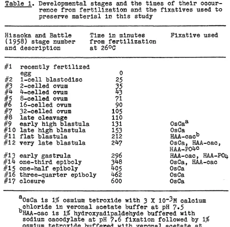
Embryos from early high blastula (stage 8, Hisaoka and
Battle, 1958) to closure of the blastopore (stage 17) were
fixed for studies of normal morphology (Table 1) or exposed
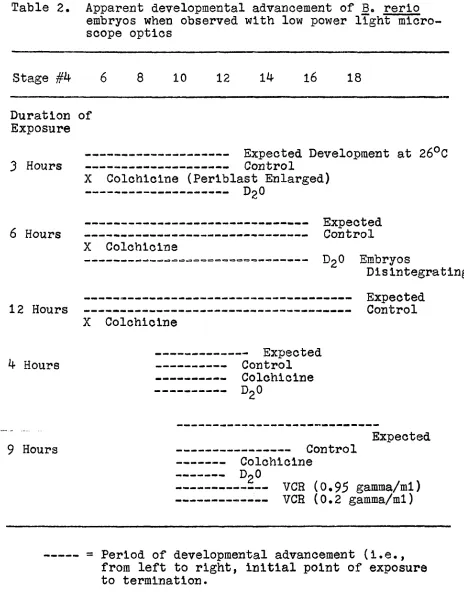
to experimental milieu (Table 2) and subsequently fixed.
Experimental treatments included ^9«5^ deuterium oxide in
aquarial water, 10"^ molar colchicine (Calbiochem, MW=399»^)
or either 0.95 gamma per ml. or 0.2 gamma per ml. of
vincristine (VCR) in aquarial water (Eli Lilly Research
Laboratories, Indianapolis, Indiana). Table 2 is a summary
of eggs exposed, stage of exposure, and stage of controls at
termination of exposure. Parallel sub-spawn samples of eggs
with chorions rented to provide freer access of solutions
were exposed to the abnormal milieu to determine the availa
bility of the milieu to the embryo. For the materials noted,
the chorion did not hinder availability to the embryo. In
all cases, some of the controls were permitted to hatch to
affirm the viability of the spawn.
Fixation of the eggs was carried out in one of three
ways: 1)
1%
osmium tetroxide with either 3 X 10~3 or 10"2Table 1. Developmental stages and the times of their occur rence from fertilization and the fixatives used to preserve material in this study
Hisaoka and Battle Time in minutes Fixative used
(1958) stage number from fertilization
and description at 26*^0
#1 recently fertilized
#2
egg 0
#2
1-cell blastodisc 25#3 2-celled ovum 35
4-oelled ovum 43
8-celled ovum 71
#6 16-celled ovum 90
#7 32-celled ovum 105
#8
late cleavage 110#9
early high blastula 131 OsCa
 late high blastula 153 OsCa
#11 flat blastula 212 HAA-cac"
#12
very late blastula very late blastula 247 OsCa, HAA-cac,HAA-P04C
#13 early gastrula 296 HAA-cac, HAA-POZ(,
#14 one-third epiboly
348
OsCa, HAA-cac#15 one-half epiboly 405 OsCa
#16
three-quarter epiboly462
OsCa#17 closure 600 OsCa
OsCa is 1% osmium tetroxide with 3 X lO'^M calcium
chloride in veronal acetate buffer at pH 7.5 "HAA-cac is 1% hydroxyadipaldehyde buffered with
sodium cacodylate at pH 7,6 fixation followed by 1% osmium tetroxide buffered with veronal acetate at pH 7.6
'^HAA-POlj, is 1% hydroxyadipaldehyde buffered with
Millonig phosphate at pH 7,5 fixation followed by 1%
Table 2. Apparent developmental advancement of B. rerlo embryos when observed with low power light micro scope optics
Stage #4 6 8 10 12 1^ l6 18
Duration of Exposure
Expected Development at 26°C
^ Hours Control
X Colchicine (Periblast Enlarged)
6 Hours
Expected Control X Colchicine
D2O Embryos
Disintegrating
12 Hours
Expected Control X Colchicine
Expected
4 Hours Control
Colchicine DgO 9 Hours Expected Control Colchicine DpO
.—— VCR (0,95 gamma/ml) VCR (0.2 gamma/ml)
= Period of developmental advancement (i.e., from left to right, initial point of exposure to termination.
[image:14.617.72.536.93.686.2]barbltol-sodlum acetate) at pH 7o5 for thirty minutes, 2)
hydroxyadipaldehyde buffered with Millonig phosphate at pH 7.5
for thirty minutes, rinsed in Millonig phosphate buffer at
pH 7.5 for five minutes, and post-fixed in 1% osmium
tetrox-ide buffered with Millonig phosphate at pH 7»5 for thirty
minutes (Sabatini ^ al., 1963), or 3) hydroxyadipalde
hyde buffered with sodium cacodylate at pH 7»6 for thirty
minutes, rinsed in sodium cacodylate buffer at pH 7.6 for
thirty minutes, and post-fixed in 1^ osmium tetroxide
buffered with veronal acetate at pH 7.6 for thirty minutes
(Sabatini et al,, I963). Experimentally treated material
was fixed for thirty minutes only in 1^ osmium tetroxide with
10"2 molar calcium chloride buffered with veronal acetate at
pH 7.5.
Dehydration was achieved by passing the fixed eggs
through an ascending series of graded ethanols. After dehy
dration in absolute ethanol, tissue was passed into propylene
oxide and embedded in Epon resin according to the following
procedure modified from Luft (I96I): instead of preparing
two Epon mixtures (Epon-nadic methyl anhydride (NM) and
Epon-dodecyl succinic anhydride (DDSA)) as Luft suggests, a
suitable mixture was found by using Luft's two mixture pro
cedure, then the total components necessary for the final
mixture were calculated. A representative embedding mixture,
DMP-30 catalyst (Rohm and Haas, Philadelphia) is added, is
thoroughly mixed and used for embedding immediately. This
mixture is the equivalent to Luff s six parts (Epon-DDSA)
and four parts (Epon-NMA). The added convenience of this
modification is the omission of two mixing steps azid the
reliability of using room-temperature anhydrous stock com
ponents. The Epon mixture was polymerized at 60°C for
eighteen hours.
Thick sections were hand-cut with razor blades or with
glass knives on a Reichert ultramicrotome OMU-2 for preliminary
observations and correlation of stage morphology to vivo
observations just prior to fixation. Hisaoka and Firlit's
study (i960) of sectioned paraffin-embedded B, rerio eggs
was used as a basis of comparison. Sections were observed
and photographed under Zeiss Zemicke phase contrast optics or
Zeiss Nomarski interference contrast optics. Selected embryos
were sectioned at ^0-65 mu on an LKB Ultrotome with a Dupont
diamond knife or on a Reiohert ultramicrotome OMU-2 with glass
knives and mounted on 75 x 300-mesh unfilmed copper grids.
All sections for electron microscopy were stained with lead
citrate (Reynolds, 1963) prior to observation on an RCA EMU-3P
electron microscope operated at either 50 or 100 kv. Standard
photographic procedures were used for the processing of nega
YOLK
That soluble and Insoluble nutrients in the form of yolk
platelets, granules, or particles are provided to all eggs is
a generally agreed concept. Of the two types of eggs recog
nized, the determinative egg, by definition, is absolutely
dependent upon all materials provided by the maternal gamete
(Raven, 1958) as shown by cytoplasm and/or cell removal
studies. Organelle distribution and localization of enzymes
have been studied to explain the necessity of all maternal
contributions for development of regulative eggs (Humphreys,
1962, 1964; Raven, 1958; Berg and Long, 1964; Berg and
Humphreys, i960).
Regulative eggs, however, can have, by definition, one or
several cells or a quantity of yolk removed without hindering
normal development (Devillers, I96I). Embryos from such eggs
have been studied by using nuclear transfers (Briggs and King,
1959) and X-irradiation which was designed to stop any nuclear
activity (Neyfakh, 1959) and thus determine the point of
initial regulation of the cytoplasm by the nucleus. Both
Neyfakh (1959) and Briggs and King (1959) agree that nuclear
activity is minimal, if not non-existent, until the initiation
of gastrulation in a regulative egg. During early development
when the nucleus is inactive, mitosis is occurring, and mater
nally provided yolk is assumed to be used by the embryo
Observations
At early high blastula (see Table 1 for stage number and
embryo age), a group of cells, the blastomeres, is situated on
the yolk mass (Figure la). Yolk, as round opaque particles,
is visible at low magnifications with phase contrast optics
and is located at the periphery of all cells (Figure la and
b). A survey electron micrograph (Figure 3) shows a large
yolk particle (4u maximum dimension) in the cytoplasm as well
as yolk particles equal in size or smaller than the 0.5u
mitochondria.
Below the blastomeres in the periblast, cytoplasm that
is contiguous with the yolk mass (Figure lb and M) contains
large closely packed particles of yolk. Between the yolk
particles and in the cytoplasm of the periblast, and the
blastomeres, ribosome-like particles can be seen in high
numbers. Each yolk particle is surrounded by a membrane if
an appropriate cross-section is observed (Figure ^b,m). The
interior of the yolk particle appears uniform under phase con
trast optics (Figures la and lb) and at the electron micro
scopic level of observation except that membranous vesicles
can occasionally be seen internally (Figure 4b,v).
In cells of the late high blastula, yolk particles are
located peripherally to the nucleus or mitotic apparatus
(Figure Ic). The number of yolk particles in cells near the
Figures Ic and Id.
Figures le and If.
particles in the periblast synoitium.
Late high blastula embryos have smaller volume cells. Yolk particles in cells near the periblast are more numerous than in cells away from the periblast. The marginal periblast is now evident, (black arrow, Figure Ic).
Flat blastula embryos have smaller volume cells and the marginal periblast is more evident (white arrow. Figure le). Note several mitotic spindles in Figure If.
Figures 2c and 2d.
Figures 2e and 2f.
Figures 2g and 2h.
phases are frequent.
At early gastrula stage, the blastoderm is lower. Small yolk particles in the periblast and in cells near the peri blast are higher in number than in cells away from the periblast.
One-third epiboly stage embryos still show small yolk particles in the peri blast and in cells near the periblast.
At one-half epiboly stage, the marginal periblast has several prominent nuclei. Small yolk particles in the periblast are absent. Only scattered cells above the periblast contain yolk particles.
cles in the periblast. Osmium tetroxide with calcium fixation; lead citrate stained. X 6300.
At a higher magnification, the membranes (m)^ around the yolk particles are readily apparent. Some yolk particles are within others but
stratum, the surface of the embryo. The periblast contains a
large variety of yolk-particle sizes (Figure Id) as compared
to those of the periblast in early high blastula (Figure lb).
Later, the marginal periblast (arrow, Figure le) is well
developed at flat blastula, whereas it was only suggested at
late high blastula (Figure le, arrow). Yolk particles, that
are more numerous in cells close to the periblast (Figures le
and If), may be seen in the montage of electron micrographs
of a flat blastula (Figure 5)* The periblast contains yolk
particles of varying sizes in late high blastula (Figures If,
5, and 6a).
Yolk particles are found to be more numerous in cells
close to the periblast and to be of various sizes in the
periblast syncitium from late high blastula to the one-third
epiboly stage^ (Figures 2c and 2f). However at one-half
epiboly (Figures 2g and h), only randomly scattered cells
have definite yolk particles. The periblast yolk of the
one-half epiboly embryo (Figure 2h) appears similar to that
of the early high blastula embryo (Figure lb).
Various-sized yolk particles present in the periblast of embryos
epiboly from late high blastula to one-third epiboly stage
are absent at the one-half epiboly stage.
Throughout this entire developmental period, yolk
with sodium oacodylate fixation; lead citrate staised. X 14,600.
particles that were first seen in all cells of the early high
blastula are missing from the cells furthest from the peri
blast until one-half epiboly, when only randomly scattered
cells contain visible yolk particles. In cells of all stages
from late high blastula to one-half epiboly, some yolk
particles {0,5-1•5u in diameter) are seen with various
ratios of yolk and centrally located membranous elements and
ribosome-like particles (Figures 6b-7c), Varying amounts of
yolk surround the membranous elements and ribosome-like
particles, or alternately larger quantities of membranous
elements and rlbosomes can be seen within the membrane-bound
yolk particles of cells in the one-third epiboly embryo
(Figures 6b, 7a and 7b). A cytoplasmic structure (^•7u
average diameter) found in a single cell (Figure 7c) is
suggestive of the ultimate complexity and fate of a yolk
particle. The interior of this complex structure contains
numerous membranous vesicles and ribosome-like particles.
Separated from the cytoplasm by a membrane, a thin layer of
material similar in density and structure to a yolk particle
(arrows) is deliniated from the interior by a second mem
brane. Another micrograph of this complex structure
(Figure 17b) shows its position in a telophase cell and the
to describe the order of yolk utilization. Osmium tetroxlde with calcium fixation; lead citrate stained. X 13,700.
Figure 7b. The yolk particle in the center of the micro graph contains ribosome-llke particles similar to ribosomes of the cytoplasm. Osmium tetroxlde with calcium fixation; lead citrate stained. X 15,000.
Figure 7c. The ultimate fate of a yolk particle may be the structure which dominates this micrograph.
Membrane-bound yolk, similar to yolk in a
Discussion
Regulative eggs by definition may have one or several
cells removed without altering normal development. A
meridional transection of the two-cell stage of teleost
embryos yields within experimental limits separate twin
embryos. The work of Oppenheimer (193^> 1936) and Tung (1955»
see also Devillers, I96I) suggests that an embryo such as
Fundulus. a marine teleost, from early cleavage (two-cell
stage) is more dependent upon the amount of periplast yolk
included in an expiant than is an embryo that is more advanced
prior to explantation. In other words, "if it [the embryo] is
explanted before a so-called critical stage, the blastoderm
turns into a hyperblastula [a non-differentiated mass of cells} ;
if it is explanted after that, it undergoes differentiation.
The critical stage corresponds to 8 b^astomeres in Carassius,
32 in Fundulus, and the young blastula in "Salmo" (Devillers,
1961, p. 391). Devillers, in summary, states that "these
results mean that a substance indispensable for differentiation
exists in the yolk sphere:..." (p. 392). He continues, "On the
other hand, how this hypothetical material may reach the
blastoderm needs to be explained. The base of the blastoderm
is in direct communication with the periblast in the early
stages; later on, the syncitium 'buds' off blastomeres that add
themselves to the embryonic disc" (p. 397)' He then asks the
syncltium can still impose an orientation on the germ? Are
diffusing organizing substances involved?" (p* 397)
The relatively young age of the B. rerio egg at fertili
zation as compared to other species such as Carasslus and
Fundulus has been noted by Devlllers (I96I). The develop
mental events observed in these two species may occur later
in the developmental age of the B. rerio embryo, but what is
stated for Carasslus and Fundulus may be extended to B. rerio.
On the basis of these (Oppenheimer, 193^» 1936; Tung,
1955» Devlllers, I96I) and other studies, the distribution of
yolk and the fate of yolk particles will be discussed In
B. rerio in an effort to find information pertinent to
Devlllers' questions.
Bipolar differentiation, that continues into late
cleavage stages of B. rerio, results in all cells having a
complement of yolk particles. We may assume that the yolk in
any given cell will be utilized almost as readily as in a
neighboring cell. In this study, mitotlcally active cells
with peripherally located yolk were found on the central
periblast of the yolk mass. Marrable's data (1965) showing
that B. rerio has a high mitotic index (83^) and a uniform
distribution of mitosis at blastula stages would suggest,
excluding the occurrence of yolk transfer from the periblast,
that uniform distribution and usage of Intracellular yolk
away from the periblast, and yolk particles are consistently
seen in high concentration in cells near the periblast.
Two possibilities may be considered for the occurrence
of yolk in cells near the periblast. Yolk particles were
never found extracellularly nor was cell-to-cell transfer of
cytoplasm and yolk observed. Therefore, the direct transfer
of yolk from cell to cell is excluded as a possible mechanism.
The alternate explanation is that mitosis from the periblast
contributes blastomeres and causes the small yolk particles
of the periblast to be a natural marker of the periblast
cytoplasm. Mitosis from the periblast has not been observed
but has been assumed as a mechanism of cell contribution from
the periblast (Devillers, I96I). This hypothesis of mitosis
from the periblast though assumed as valid for this study
must be investigated further.
As suggested by Oppenheimer (193^> 1 9 3 6 ) , Tung ( 1 9 5 5 ) ,
and Devillers (I96I), some substance in the yolk of the embryo
carrys information (or material) necessary for the differ
entiation processes. Identification of the components of yolk
should provide important information on the nature of the
'yolk-substance' necessary for differentiation.
Yolk particles as seen in this study in one-third epiboly
cells have been observed initially to have only amorphous yolk
material surrounded by a unit membrane, then to have membra
membrane-bound yolk material, and to appear finally as a
highly complex particle appearing as cytoplasm surrounded by
little yolk and a unit membrane. These yolk particles con
taining various quantities and types of inclusions could be
arranged as suggested above in a sequential pattern of
utilization, Bellairs (1958) has seen a similar, but more
complex, sequential ordering of yolk particles in the develop
ing chick embryo. Strikingly, the final member of the
sequence (not observed or mentioned by Bellairs) appears as an
isolated portion of cytoplasm surrounded by a thin layer of
yolk and an external unit membrane. 'That a supply of maternal
membranous elements and ribosomes be the determining factor
from the periblast necessary for permitting differentiation
at later developmental stages is a highly suggestive
hypothesis.
The possibility of ribosomes Ixi yolk must be considered
by reviewing the formation of yolk during oogenesis in
B. rerio. Hisaoka and Firllt (1962b) suggest the inclusion of
BNA in a masked form in yolk of the mature oocyte. Ribonu
cleic acid content, by methyl green-pyronin-Y staining, is
high (Intense staining) as cell size and yolk material
increase until oocyte stage 5» They state (Hisaoka and
Firllt, 1962b, p. 206): "Throughout oogenesis the
intra-vesicular yolk does not contain RNA. However, HNA is present
(mature oocytes). After fertilization, BNA remains constant
in all cells during cleavage and blastula stages except for
regional decreases during interphase and prophase (Hisaoka and
Firlit, 1961).
In Rana pipiens. Brown and Gaston (I962) report the
binding and masking of exogenous isotope-labeled ribosomes to
yolk in studies of vitro protein synthesis during early
development. Until Shumway stage
21-23
in Eana pipiens,Karasaki (I963) notes the decrease in yolk in the embryo, but
he does not report ribosome-like particles within Rana yolk
platelets. Rounds and Flickinger
(1958)
conclude, however,from their study of neural induction in R. pipiens that yolk
breaks down in chordamesoderm with a subsequent release of
RM. Bellairs
(1958)
reports micro-particles in yolk similarto cytoplasmic micro-particles now known as ribosomes.
Oogenesis in most species is essentially synthesis and
sequestering of material for the period from fertilization to
nuclear activity. Formation of the insect fat body, for the
metamorphosis of larva to pupa, is a similar period. There
fore, the work of Locke and Collins
(1965)
on the structureand formation of protein granules in an insect fat body
deserves critical study. In summary, they found an
intra-cytoplasmic Isolation of mitochondria and rough, endoplasmic
reticulum into cytolysome-like structures. Eventually, the
body are encompassed by a unit membrane and accumulate pro
tein, The membranes and ribosomes within the cytolysome
loose their identity among the accumulated protein and
appear similar to the yolk material within the yolk particles
seen in the periblast of B. rerio. "Protein + RNA granules"
of the insect fat body often contain crystalline protein with
a repeat spacing of 90 to 160 S similar to crystals with an
average spacing of 70 2 found in Rana yolk by Karasaki
(1963). Golgi vesicles, in the fat body, also accumulate
proteinaceous material and appear as "yolk". Locke and
Collins find isolation of membranous material and ribosomes
into membrane-bound, protein-containing granules; in contrast,
B. rerio yolk particles, which appear as the protein granules
of the insect fat body, give rise to membranous material and
ribosome-like particles during breakdown. In the sequester
ing fat body, granules fuse to form larger "fat" particles
within cells; in contrast, in the periblast of B. rerio
during yolk utilization, large yolk particles appear to "pinch
off" into smaller yolk particles.
Yolk; in Rana pipiens embryos, has been reported to
contain RHA. In B. rerio oocytes, RNA appears to be masked
in extravesicular yolk. In an insect fat body, ribosomes
and membranous material are packaged in "protein granules"
similar to yolk particles. Microparticles found in the yolk
developing chick embryo. Therefore, the conclusion is
strongly suggested that membranous material and ribosome-like
particles in the yolk of B. rerio originate maternally and
are necessary for differentiation. This hypothesis might be
further tested by explanting an embryo which should
hyper-blastulate and adding either 1) maternal yolk, or 2)
embryonic or maternal ribosomes (without raessenger-RNA), or
3) embryonic or maternal membranous material, or 4) both
ribosomes and membranous materials to the explanted embryo.
Thus yolk, besides providing soluble nutrients to the
embryonic cells, could be tested for its contribution of the
MITOSIS
The next prominent event after yolk utilization during
early development is the pr-oduotlon of additional cells by
mitosis. The oocyte is a center of synthesis and storage of
material; but after fertilization, an ordering of material by
bipolar differentiation occurs. Some protein synthesis does
occur in developing sea urchin eggs (Gross and Cousineau,
1963), but control of protein synthesis is estranuclear
(Tyler, I966). The non-Involvement of the nucleus in the
physiological activities of the early developing egg is indi
cated also by both the work of Briggs and King (1959) and
Neyfakh (1959)» A high rate of cell division immediately
after fertilization demands that the material to be used
during early development be produced or programmed in the
oocyte. What, then, are the differences between an undiffer
entiated cell and a nucleus-activated, partially differenti
ated cell during mitosis?
The study of mitosis in general has been a focal point
of man's curiosity because of the involvement of mitosis in
all aspects of life (Mazia, I96I). In the last decade, the
study of the mitotic apparatus by the use of electron
microscopy has proceeded from confirmation of the filamen
tous nature of the mitotic apparatus (Roth et al., I96O;
Roth and Daniels, 1962; Harris, 196I, 1962) to general
(Bobbins and Gonatas, 1964; Barnicot and Huxley, 1965) and
to some experimental electron microscope investigations
(vincristine: George et al., 1965; cold, colchicine, urea:
Both, 1964). Some investigators have approached the study
of mitosis with histochemical and isolation-biochemical
techniques. Prom the assumption (Mazia, I96I) that adenosine
triphosphate (ATP) is the energy source for mitosis, attempts
have been made to localize ATPase activity in the mitotic
apparatus (Miki, I963, 1964); or from the suggestion of
Rapkine (1931) that sulphydryl groups may be significantly
involved in the mitotic process, sulfydryl reactions have
been examined by histochemical methods (Kawamura and Dan,
1958; Kawamura, I960). Following the pioneering work of
Mazia et (I96I), many investigators have
physically-isolated the mitotic apparatus of some marine invertebrate
blastomeres, especially from sea urchins. These techniques
have resulted in fruitful but conflicting data. To the
present, investigators state that ATPase activity is both
present or absent in the mitotic apparatus (Miki, 1964;
Stephens, I965, respectively) and state varying values for
the size of mitotic apparatus, spindle protein subunits
(Zimmerman, I96O; Kane, 1965; and Sakai, I966). This work,
however, has centered around a single type of organism, the
sea urchin, although investigators have used different
While the hlstoohemlcal localizations and
isolation-biochemical analysis has provided some information, still
much must be learned of mitosis ^ situ in both vertebrate
and Invertebrate cells. Systems studied to date have either
been invertebrate (protozoan or embryonic invertebrate) or
dedifferentiated tissue culture cells with remnants of the
differentiated state. Zebraflsh blastomeres which are
mltotically active (83^ mitotic Index) and have a short
cell cycle (15-18 minutes, Hoosen-Runge, 1939; Marrable, 1965)
are an ideal source of mitotic cells. Therefore, the morpho
logical characteristics of the undifferentiated early
embryonic, mitotic cell, at a time when little or no nuclear
control is Involved, will be compared to the physiologically
active, nucleus-controlled, mitotic cell. This study is a
considerably more complete examination of mitosis during
early development in B. rerio than those presented earlier
(Thomas, 1964a,b).
Observations
Mitosis has been followed in cells from embryo at early
high blastula to the one-third epiboly stage. The cells in
this period are more favorable for study because of their
smaller size as compared to the large volume of cells found
Interphase
The early embryonic, Interphase cell is undifferentiated
(Figure 8). The nucleus (n) is spherical except that it
contains a cytoplasmic cleft. The nucleoplasm has a uniform,
fibrillar appearance following osmium-tetroxide fixation;
nucleoli are absent. Adjacent to (Figure 8) or completely
surrounding the centriole (Figure 9a) is a 'halo' of fine
material which together with the centriole forms the
centro-some= Golgi material (g) is located directly opposite the
centriole and the nucleus. Spherically shaped mitochondria
are scattered throughout the cytoplasm and concentrated
characteristically next to the nuclear envelope.
Ribosome-like particles are ubiquitous, and yolk particles vary in
quantity and size from cell to cell.
Prior to attenuation of the nuclear envelope during
prophase, spindle microtubules (sp) are seen adjacent to the
centriole (Figure 9b) but do not form a detectable aster
during mitosis. The nucleus surrounding the cleft appears as
finger-like projections toward the centriole (Figures 9a and
c) which is most evident when both centrosome regions are in
the same plane of section (Figure 9c). From the centrosome
(cs), a spindle microtubule (arrow) can be seen leading
the nuclear envelope. A centriole is situated
between the Golgi apparatus (g) and the nucleus.
is at the edge of the nuclear cleft. Osmium tetroxlde with calcium fixation; lead citrate stained, X 32,000
Prior to nuclear envelope interruption, spindle microtubules (sp) radiate from the centrosome. Osmium tetroxlde with calcium fixation; lead citrate stained. X 26,800
During early prophase, the nucleus is
elllpsoidally shaped with few mitochondria
along the nuclear envelope. Two nuclear clefts and two centrosomes are evident. The nucleo plasm is uniform; the chromosomes have not yet condensed. Osmium tetroxlde with calcium
Prophase and Prometaphase
Condensation of chromosome material (c), attenuation of
the nuclear envelope (ne), and the presence of spindle micro
tubules (among the chromosomes and nuclear envelope fragments)
characterizes the transition into prophase (Figure 10b).
Mitochondria are not as concentrated along the nuclear
envelope as was seen in interphase cells. Nuclear envelope
fragments with nuclear pores (np) showing in cross and
tangential section, mitochondria, ribosome-like particles,
chromosomes, and spindle microtubules can be seen inter
mingled at a higher magnification (Figure 11a).
As chromosomes approach alignment on a metaphase plate
(Figure lib), large nuclear envelope fragments persist at
the periphery and within the forming mitotic spindle. The
typical tangential view of spindle microtubules is difficult
to discern because much fine material is close to them.
Yolk particles and mitochondria are excluded from the spin
dle, and Golgi vesicles can be seen at one of the poles. A
cross-section through a similar pro-metaphase figure illus
trates the exclusion of mitochondria and yolk from the
forming mitotic apparatus (Figure 12a). Cross-sections of
spindle microtubules (arrow) are found in groups adjacent
to and removed from the chromosomes but are closely
associ-ated with the nuclear envelope fragments (Figure 12b,c).
edge of the nucleus. Osmium tetroxide with calcium fixation; lead citrate stained. X 38,700
and tangential section where nuclear pores (np) are prominent. Osmium tetroxlde with calcium fixation; lead citrate stained. X 21,800
Figure lib. When chromosomes appear almost aligned at prometaphase, nuclear envelope fragments remain around the formed mitotic apparatus. Golgi apparatuses can be seen at both ends of the mitotic figure. Hydroxyadipaldehyde
buffered with phosphate fixation; lead citrate
buffered with phosphate fixation; lead citrate stained. X 7^00
Figure 12b. Spindle microtubules in groups (arrows) are near either chromosomes or nuclear envelope fragments. Hydroxyadipaldehyde buffered with phosphate fixation; lead citrate stained. X 18,400
spindle microtubules are preserved by osmium-tetroxlde with
calcium or by hydroxyadipaldehyde.
Metaphase
At metaphase (Figure 13), chromosomes are aligned on an
equatorial plate with spindle microtubules extending to and
between chromosomes. At the poles, Golgi vesicles (g) are
present, but astral filaments are absent; the nuclear
envelope fragments are gone (compare with Figure lib).
Microtubules are arranged in groups often ending at a
klnetochore-llke structure (k) at the chromosomes or go
between the chromosomes (arrow. Figure l4a)..
Anaphase
An early anaphase figure in tangential section shows the
interzone (IZ) between daughter chromosome sets (Figure l4b).
The daughter chromosomes are seen at varying separations,
but the poles are at a relatively constant distance of 15u
(Figures 15, l6a). At the initiation of cytokinesis, the
pole-to-pole separation (the chromosomes are at the poles)
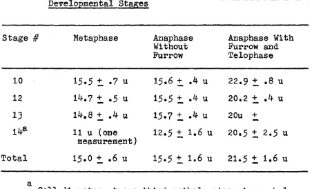
varys up to a maximum of 24u (Figure l6b). Table 3 is a
summary of pole-to-pole distances of the phases of mitosis
from measurements of phase contrast and electron micrographs.
Groups of microtubules extend to the chromosomes, and the
poles and microtubules are found in the interzone (Figure 15);
chromosomes. Osmium tetroxide with calcium fixation; lead citrate stained. X 15,900
Figure l^b. An interzone (IZ) formed by the separation of
daughter chromosomes signals the initiation of anaphase. Spindle microtubules and ribosomes are within the Interzone but membranous
elements but contains continuous spindle microtubules and some small yolk particles. Asters are absent and the cytokinetic
furrow has not yet appeared. Osmium
Table 3* Pole-to-Pole Dlstanoes of Mitotic Figures at Various Developmental Stages
Stage # Metaphase Anaphase Anaphase With
Without Furrow and
Furrow Telophase
10 15.5 t .7 u 15.6 + .4 u 22.9 + .8 u
12 14.7 + .5 u 15.5 + .4 u 20.2 + .4 u
13 14,8 + .4 u 15.7 t .4 u 2 Ou +
14* 11 u (one
measurement)
12.5 + 1.6 u 20.5 t 2.5 u
Total 15«0 + .6 u 15.5 t 1.6 u 21.5 + 1.6 u
[image:64.622.86.538.229.503.2]absent from the interzone (Figures 15, and l6a), are found
in the remainder of the cytoplasm and against the leading
edge of the daughter chromosome sets.
Telophase
Telophase is classically described as the reconstruction
of the Interphase state. The chromosomes, which had membra
nous material on their leading edges during anaphase, are
now completely surrounded with membranous elements (Figures
l6b and 17a), Individual chromosomes or groups of chromo
somes are surrounded with membranous material with nuclear
pores (nuclear envelope) forming either micronuclei which
become interconnected or a single nuclear structure.
In cells of large volume, through early or late high
blastula (Figure 18a), the chromosomes are physically sepa
rated from each other so that each has an envelope around it.
These chromosomal vesicles fuse, enlarge, and form the inter
phase nucleus often with the result that remnants of nuclear
envelope are found within the nucleus (Figure 18a). At the
later stage of very late blastula, cell volume is reduced
and telophase chromosomes are physically together (Figures
15-17b, and 19a). Although each chromosome does have membra
nous elements on its poleward surface, the nucleus does not
form chromosomal vesicles, that is individual chromosomes
surrounded by nuclear envelope. Rather a simple daughter
appears 'clear'. Hydroxyadipaldehyde buffered with phosphate fixation; lead citrate stained. X 3800
ehyde buffered with phosphate fixation; lead citrate stained. X 3850
Figure 17b. The width of the connection between daughter cells decreases as cytokinesis continues and longitudinal elements suggestive of spindle
microtubules appear between the furrow edges. Also in the micrograph, a yolk particle
suggested as the last step in yolk fate is seen near the nucleus. Osmium tetroxide with calcium fixation; lead citrate stained.
Figure 18b. At very late blastula, cell volume is reduced and chromosomes form a single nuclear
structure.
(Figure 18b) or closely associated chromosomes surrounded by
nuclear envelope are seen with the electron microscope
(Figure 19a).
The end of karyokinesis and cytokinesis Is summarized in
Figure 19b. The edge of an Interphase nucleus surrounded by
mitochondria can be seen at the left. A mldbody (mb), the
remnant of the cleavage furrow, has a closely gathered group
of microtubules and is the last connection between daughter
cells.
Cytokinesis
Although the occurrence of a cytoklnetlc furrow is common
at the one-third epiboly stage in B. rerio (Figures 17a,b and
19b), both cytokinesis by the formation of a furrow and by
fusion of a row of vesicles (phragmoplast) has been observed
in the same blastula and after the use of more than one
fixative. A row of vesicles transversing the entire cleavage
plane is the indication of the phragmoplast (Figure 20).
Often at the center of the row an area lacking vesicles
between daughter cells contains material suggestive of
spindle microtubules and the formation of a mldbody (mb,
Figure 21). The indication of the mitotic phase is given by
the nuclear morphology which is similar to a telophase
nucleus. There is no indication of fine material on the
cytoplasmic side of the cell membrane at the end of the
with phosphate fixation; lead citrate stained. X 18,000
furrow almost bisecting two daughter cells is also seen in
the same blastula (Figure 22), Even at low magnification
(Figure 22), a deposit appears along the edge of the furrow
and is not present along the cell membrane that is not in
the furrow area (arrows indicate contrasting areas,
Figure 22). This fine material is a layer about 60-mu thick
and appears as dense fibrillar material (fm) under a
cross-sectioned plasma membrane (Figure 23a). The midbody has
gathered spindle microtubules in a 'neck' between the
daughter cells (Figures 23b-d). The 'neck' of the midbody
has accessory membrane projections (ap) with membranous
vesicles within the bulbous projections extending into the
extracellular space. The center of the midbody 'neck' contains
closely packed microtubules which have a very dense band (db).
Still later at very late blastula, both "Che' formation of
a vesicle-type cleavage (Figure 24) and the formation of a
definitive furrow with fine material along the leading edge
can be found (Figure 17a). However, at the later stage of
one-third epiboly, only cytokinetic furrows with fine material
on the leading edge are observed (Figure 17b). The midbody
formed at one-third epiboly (Figure 19b) resembles closely the
midbody observed in cells in late high blastula (Figures 23
material which is absent from the remainder of the cell membrane (arrows).
furrow in Figure 24. Hydroxyadipaldehyde buffered with sodium cacodylate fixation; lead citrate stained. X 49,000
Figures 23b-d. Serial sections through a midbody of late high blastula cells show a dark band (db) in the center of the midbody 'neck'.
Discussion
Electron microscopic studies of the mitotic portion of
the cell cycle have previously been directed largely toward
the establishment of the fibrillar nature of the mitotic
spindle or the involvement of specific organelles in partial
ly differentiated cells. Prophase events such as the forma
tion of the mitotic apparatus and nuclear envelope dissolu
tion, anaphase spindle elongation and chromosome movements,
telophase nucleus reconstruction, and cytokinesis have been
difficult to study, and little has been reported. A develop
ing embryo such as B. rerio with its high mitotic index and
rapid cell cycle provides a cell population amenable for study
of these events. Therefore, the preservation of spindle micro
tubules, the location of spindle microtubule formation and
nuclear envelope dissolution, anaphase 'movements', chromosomal
vesicles, and cytokinesis as seen in the developing embryo
of B. rerio will be discussed.
Preservation of Spindle Microtubules
The filamentous nature of the mitotic apparatus was
first characterized as a circular profile of about I5 mu by
Roth et (i960) in the mitotic apparatus of amebae and by
Harris (I96I) in the sea urchin embryo. Both Investigators
noted the necessity of including divalent cations such as
(pH 7.4-7.6) as also used In this study. The introduction
of dialdehyde fixation (Sabatini et al., 1963) permitted the
preservation of microtubules of the mitotic spindle at alka
line pH's without divalent cations (Dales, 1963; Roth et al.,
1963; and many others). The dialdehyde of choice for pre
servation of microtubules is considered to be glutaraldehyde,
1,5-pentadial. However, as investigations of microtubules
preserved by glutaraldehyde were reported, microtubule
diameter varied from 15 to 26 mu from laboratory to labora
tory (Dales, 1963; Robbins and Gonatas, 1964; Bamicot and
Huxley, 1965)®
Hydroxyaldehyde (2-hydroxy-l,6-hexenedlal), suggested
by Sabatini et al., (I963) as the preferred fixative for
subsequent localization of adenosine triphosphatase (ATPase)
and other enzymes, was used in an unsuccessful attempt to
localize ATPase in the mitotic apparatus of B. rerlo.
Mitotic spindle microtubules were found well preserved with
diameters of 19-21 mu. Fixation of Zebrafish blastomeres
with osmium tetroxlde with divalent cations resulted in the
preservation of spindle microtubules with the same diameters.
Location of Spindle Microtubule Formation and Nuclear
Envelope Dissolution
The extremely short cell cycle and mitotic period In
B. rerlo make study of rapidly occurring events easy since
period is generally considered to be prophase (Mazia, I96I).
In B, rerlo. Roosen-Runge (1939) reports a prophase period
of 18,5^ of the mitotic period with Interphase time excluded
or 16,5/^ with Interphase time included. Events are occurring
rapidly if the 15-18 minute time period for the cycle is
considered.
Prom light microscope observations, Roosen-Runge (1939)
simply states that chromosomes become visible immediately
before breakdown of the 'nuclear membrane*, and chromosomes
appear at the metaphase plate while still within the nuclear
'membrane'. As observed with the electron microscope in this
study, the interphase nucleus is seen as a large cup near the
small centrosome. The center of the cup is a cleft remaining
from telophase reconstruction of the nucleus. During prophase
and prometaphase, spindle microtubules are seen 1) near the
centrosome at the lip of the cup or cleft prior to nuclear
envelope interruption, 2) in the cleft pointing toward the
center of the cup, and 3) within an interrupted (but rela
tively complete) nuclear envelope. Toward the end of pro
metaphase, large nuclear envelope fragments still surround
the formed mitotic apparatus. The shape of the nuclear
envelope progresses during pro-prometaphase from 1) a sphere
with a single cleft, to 2) an ellipsoid with two clefts, and
then to 3) an interrupted ellipsoidal shape with spindle
fragments. During the period when the shape of nuclear
envelope Is being altered, mitochondria are found 1) in high
numbers along the spherical interphase nucleus, 2) around
the ellipsoidal nucleus (with its condensed chromosomes),
in lower number, to 3) none within the formed mitotic appa
ratus as the nuclear envelope completes dissolution.
The events outlined above agree with the early observa
tions of Roosen-Runge (1939) and some of the observations
agree with other investigations of mitosis in
centriole-containing cells. The occurrence of microtubules at or near
the centrioles (centrosomes) prior to Interruption of the
nuclear envelope has also been documented by Harris and
Mazia (1962) in sea urchin embryos, by Robblns and Gonatas
(1964) in HeLa cells, and by Bamicot and Huxley (1965) ia
tissue culture cells of the newt heart and human fibroblasts.
In general, these studies agree that spindle microtubules
are first seen before the nuclear envelope is interrupted and
always in close proximity to the centrioles (centrosomes).
Harris and Mazia (I962), Robbins and Gonatas (1964), and this
study agree that microtubules are within the nuclear cleft.
The direction of spindle microtubules toward the nucleus
(and the condensed chromosomes) suggests that the considera
tion of the centrioles as a fiber organizing center (Schrader,
1953J Mazia, I96I) is most probable. Jokelainen (I965) in
cells reports that spindle microtubules attach to the
chores rather than microtubules originating from the
kineto-chores. Therefore, it may be concluded that the formation of
the mitotic apparatus originates at the centrosome and pro
ceeds toward the condensed chromosomes.
The occurence of mitochondria around the interphase
nucleus may be only a secondary result of telophase nucleus
expansion. Prom a compact telophase condition, the chromo
somes during their expansion may push the nuclear envelope
outward against the nearby mitochondria. A change in the
nucleus shape, due for example to chromosome condensation
during prophase, would result in mitochondria being separated
from the nuclear envelope. This is in fact what has been
observed in this study. Such a hypothesis would, however,
negate the hypothesis of Barer et al. (1958) that the
mitochondria are involved as producers of enzymes for the
dissolution of the nuclear envelope. It is most probable
that the mitochondria are not involved in production of
enzymes for nuclear envelope dissolution because the nuclear
envelope is intact after the mitochondria are removed.
The nuclear envelope retains most of its integrity
during the formation of the mitotic apparatus. As seen by
Roosen-Eunge (1939) and in this study, the nuclear envelope
(or rather large fragments of the nuclear envelope) remain
of spindle microtubules within the nuclear cleft to prometa
phase when the chromosomes are almost aligned upon the
meta-phase plate, the spindle microtubules appear between nuclear
envelope fragments from the centrosome to the condensed
chromosomes. Jokelainen's (1965) observations and suggestion
of a mechanism of spindle to kinetochore attachment may be
aided by the observations of this study. The restriction of
mitochondria and yolk or particles of similar size from a
mitotic apparatus, as is generally seen (Mazia, I96I), would
be most reasonable by the persistence of the nuclear envelope
fragments until spindle-kinetochore attachment is completed.
In summary, the site of spindle microtubule formation
appears to at the centrioles (centrosomes) before the nuclear
envelope is interrupted. Nuclear envelope fragments persist
until late prometaphase, when the mitotic spindle is formed,
and restrict mitochondria, yolk, and particles of a similar
size from the formed spindle.
Anaphase 'Movements'
During the developmental period of late high blastula to
one-third epiboly, metaphase pole-to-pole distance is a
relatively constant 15 u. Polar separation remains constant
until the daughter chromosome sets have reached their
respective poles. Only after the chromosomes have reached
the poles and the first indications of a cytokinetic furrow
periods of anapha.se chromosome movement, interzonal micro
tubules are present.
Dettlaff (1963) reports that in the first mitotic period
in either sturgeon or trout eggs there is a simultaneous
chromosomal movement to the poles and separation of the
poles. In reviewing the voluminous literature of anaphase
movements, Mazia (I96I, p. 280) concludes that there are two
anaphase "movements: 1) an elongation of the central spindle,
describable as a pushing apart of the poles, and 2) a move
ment of the chromosomes toward the poles. The relative contri
butions of these in the separation of sister chromosomes varies
from one type of cell to another as do their time relations."
The movements of chromosomes during anaphase as
observed in B. rerio are similar to other cells. In addition,
this study shows that both movements occur during anaphase in
this species. The initial movement is the daughter chromo
somal movement of about 7 u from the metaphase plate and then
the poles move apart from 15 u separation to about 21 u.
Chromosomal Vesicles
Chromosomal vesicles, karyomeres, or micronuclei, i.e.,
individual chromosomes or groups of chromosomes surrounded by
nuclear envelope, have been noted as a significant character
istic of early cleavage cells or early embryonic cells in
several species (trout and sturgeon, Dettlaff, 1963; sea
Roosen-Bunge, 1939). They have also been noted In plant cells
with divergent mitosis (Mazia, I96I). Chemical treatments,
such as a short exposure of tissue culture cells to colchicine
(Starr, 1963), also result in the formation of micronuclei by
permitting the dispersal of chromosomes. Starr (19^3) has
used the formation of micronuclei as a test system for the
localization of viruses to specific chromosomes, but
unsuccessfully.
Only in cells with large volume, where chromosomes are
well separated, or in cells where chromosomes are separated
artificially by disruption of the mitotic apparatus do
chromosomal vesicles or micronuclei form. As cell volume is
reduced by repeated mitosis and chromosomes are restricted
within a smaller volume during anaphase movement,, chromosomes
tend to act as a single chromosome mass rather than as indi
vidual entities.
It can be concluded that the formation of chromosomal
vesicles, karyomeres, or micronuclei in cells of large volume
is the result of physical separation of chromosomes during
anaphase movement to the poles. Likewise, the formation of a
single telophase nucleus in a cell of small volume would be
the result of chromosomes acting as a single entity during
the anaphase movement to a pole,, except when chromosomes are
dispersed by artificial means. Therefore, nuclear recon
nucleus appears to be dependent not upon nuclear control but
rather upon physical restrictions of cell volume.
Cytokinesis
Rappaport (I966) with an excellent experimental design
arrives at the conclusion that the studies of Mitchison
(1953) and others on the cleavage of animal cells that occurs
not by a furrow may only be characteristic of a few species.
Cytokinesis in botanical material has been well documented as
the fusion of a row of vesicles (e.g., Whaley ^ aj.., I966).
Animal cells have been characterized as having mainly furrow
cytokinesis (Bobbins and Gonatas, 196^; Buck and Krlshan,
1965); however, vesicle cleavage has also been reported (Buck
and Tlsdale, 1962; Humphreys, 1964). Rappaport's conclusions
(1966) state that either cleavage-type Is mutually exclusive. Zotln (1964) from observations of the first cleavages of
large-celled teleost eggs reports that vesicle-cleavage Is
visible with the light microscope. Mitchison's study (1953)
suggests the occurrence of vesicle-cleavage in some sea
urchin eggs. During this study, early developmental stages
such as late high blastula or very late blastula were char
acterized by the occurrence of both vesicle and furrow
cleavage. Only at later developmental stages such as
one-third eplboly were cleavage furrows alone found.
Vesicle cleavage has been characterized as an ordered
although fusion may not necessarily occur (Humphreys, 196^),
In general; vesicle cleavage occurs in cells with large
volumes (Selman and Waddington, 1955î Zotin, 196^). Furrow
cleavage is characterized initially by a slight constriction
of the cell membrane with a distinct Intracellular layer of
fine material along the furrow area. The furrow edges are
seen closer together until finally the interzonal micro
tubules are gathered to form a midbody. A simple connection
remains between cells until it is assumed broken. The
furrow-type cleavage has been described most frequently in animal
cells which are usually smaller than the first cleavage cells
of teleost or sea urchin eggs. Furrows have even been found
in the minute cells of the blue-green algae (Pankratz and
Bowen, 1963).
It may be concluded from the observation of both
vesicle-and furrow-cleavage in the same biastula of B. rerio that the
two cleavage types are not mutually exclusive as suggested by
Eappaport (I966). The diminution of cell volume (Marrable,
1965) and the concomitant disappearance of vesicle cleavage
in B. rerio suggests strongly that the method of cleavage
used in animal cells is largely dependent upon cell volume.
The mitotic apparatus has often been considered necessary
for the initiation and continuation of cytokinesis (Mazia,
1961).