VARIATIONS IN THE ORIGIN AND COLIC
BRANCHES OF THE SUPERIOR
MESENTERIC ARTERY
Dissertation Submitted to
THE TAMIL NADU DR. M.G.R. MEDICAL UNIVERSITY
CHENNAI
in partial fulfillment of the regulations
for the award of the degree of
M.S. (Anatomy)
BRANCH - V
THE TAMILNADU DR. M.G.R. MEDICAL UNIVERSITY
CHENNAI, INDIA.
C
C
e
e
r
r
t
t
i
i
f
f
i
i
c
c
a
a
t
t
e
e
This is to certify that the dissertation title, ‘Variations in the Origin
and Colic branches of the Superior Mesenteric Artery’ is an original
work done by Dr. M. Nirmaladevi, PG Student, Stanley Medical College,
Chennai-1, under my supervision and guidance.
Dr. Mythili Bhaskaran, M.D., Dr. Sudha Seshayyan, M.S.,
Dean Professor and HOD Stanley Medical College Department of Anatomy Chennai-1 Stanley Medical College Chennai-1
DECLARATION
I solemnly declare that this dissertation "Variations in the Origin and
Colic branches of the Superior Mesenteric Artery" was written by me in
the Department of Anatomy, Govt. Stanley Medical College and Hospital,
Chennai, under the guidance and supervision of Prof. Dr. Sudha Seshayyan,
M.S., Professor and Head of the Department of Anatomy, Govt. Stanley Medical
College, Chennai - 600 001.
This dissertation is submitted to The Tamil Nadu Dr. M.G.R. Medical
University, Chennai in partial fulfillment of the University regulations for the
award of degree of M.S. Anatomy - Branch V examinations to be held in
March 2008.
Place : Chennai.
ACKNOWLEDGEMENT
I have been overwhelmed by the support and guidance that I have
received from a large number of people in completing this study and I
would like to take this opportunity to thank each one of them.
I would like to express my sincere thanks and gratitude to
Dr. Mythili Bhaskaran, M.D. , Dean, Stanley Medical College, Chennai-1, for
granting me permission to utilize the facilities of this Institution, for my
study.
It is my privilege to express my sincere and profound gratitude to
Dr. Sudha Seshayyan, M.S., Professor and HOD, Department of Anatomy,
Stanley Medical College, Chennai-1, for her constant support and guidance
and suggestions to complete my dissertation work.
My heartfelt thanks to Dr. S. Chitra, M.S., Additional Professor,
Department of Anatomy, Stanley Medical College, Chennai-1, for her
I wish to express my sincere thanks to Dr. Shanthakumar, M. D
Professor and HOD, Department of Forensic Medicine, Stanley Medical
College, Chennai-1, for granting me permission to collect specimens.
I wish to express my sincere thanks to Dr. Amrita Priscilla Nalini,
M. D. Director I/c, Department of Obstetrics and Gynaecology, Stanley Medical
College, Chennai-1, for granting me permission to collect foetal specimens.
I would like to extend my thanks to Dr. C. Karunanidhi,
Dr. Mohandas Joe Chandra, Dr. Syed Rafi Ahmad, Dr. N. Rajasekaran,
Dr. V.K. Venkatesan and Dr. T. Vasanthakumar, faculty members in the
Department of Anatomy, Stanley Medical College, Chennai-1, who have
encouraged me in this study.
I would like to express my sincere thanks to Colleagues, technicians
and other workers in the Department of Anatomy, Stanley Medical College,
Chennai-1, who have helped me in this study.
I wish to thank my parents, family members, my daughter and
CONTENTS
S.no Title Page No
1. Aim of the Study 1
2. Anatomical and Embryological Considerations 4
3. Review of Literature 9
4. Materials and Methods 27
5. Observation 32
6. Discussion 45
7. Summary 59
1
AIM OF THE STUDY
Modern abdominal surgical techniques depend partly on the
knowledge of the normal arterial and partly on the anomalous arterial
blood supply. Unlike other anatomical variations, anomalous and variant
blood supply cannot be ignored for the risk of ligating the wrong vessel
or severing an essential artery which may result in ischaemia and
gangrene, and of leaking and bleeding from the sites of repair and at
anastamotic suture lines.
From the anatomical point of view, arterial variations are verifiable
facts of the human constitutions that can be observed time and again.
Variations in the arrangement of arteries that supply the abdominal organs
are very common.
As a guide and a safeguard to the surgeon in ever increasing,
varied and difficult operative procedures like oesophagojejunostomies,
intestine transfers, resections of the small and large intestines and
appendicectomies a descriptive atlas on the variant arterial supply of the
In modern therapeutic techniques like selective arterial chemotherapy
in the treatment of liver cancer, we should know the variations of hepatic
artery to plan the procedure accordingly.
In liver transplantation, with the knowledge of the variational
anatomy of the hepatic pedicle, the extensive damage of the feeding
hepatic artery can be definitely prevented.
The veins, lymphatic vessels and lymph nodes draining a part of
the large intestine converge on the aortic origin of the vessel supplying
that particular part. The extent of bowel resection in carcinoma is thus
determined by the length of bowel supplied by the arterial trunk to the
area involved by the disease. The vessel is divided proximally so that the
resected bowel and mesentery contain the whole of the related lymphatic
apparatus including the proximal lymph node group.
The best way to avoid injury to the blood vessels during any
invasive procedures such as laparoscopic procedures and resection of colon
for cancer or other diseases, is to know them thoroughly and to know
3
It is therefore evident that a knowledge of exceedingly variable
blood supply of the viscera in the abdomen by the superior mesenteric
artery is very important to the operating Surgeon, Radiologist and to the
Anatomists as well.
Responsibility of establishing and disseminating knowledge about
variations, lies with the Anatomist though the consequence of correct and
incorrect informations may depend upon the Surgeon.
Hence, the present study is mainly aimed at
1. Examining the pattern of variations in the origin of the superior
mesenteric artery and its colic branches mainly in south-Indian
population. The concerned data is obtained from cadaver dissections
and from angiographic pictures.
2. Comparing the variation with those obtained by the earlier
workers.
3. And analyzing the cumulative information with reference to
ANATOMICAL AND EMBRYOLOGICAL
CONSIDERATIONS
Superior mesenteric artery arises from the abdominal aorta ( Fig 1) at
a level between 1st lumbar and 2nd lumbar vertebrae, 1 cm below the
origin of the coeliac trunk. It supplies the second part of the duodenum
distal to the major duodenal papilla, the third and fourth part of the
duodenum, a portion of the head and frequently, an extreme area of the
body of the pancreas, the jejunum, ileum and the large intestine up to the
junction of right two third and left one third of transverse colon because it
is the artery of midgut.
From its origin about 1 cm below the coeliac trunk, it leaves the
front of the aorta and is crossed anteriorly at its origin by the splenic vein
and body of the pancreas. It is separated posteriorly from the aorta by the
left renal vein. Proceeding downwards and forwards, it runs anterior to the
pancreatic uncinate process and the horizontal part of the duodenum. It
then descends obliquely in the mesentery near its root to the right iliac
fossa. Accompanied by the superior mesenteric vein to its right, the artery
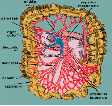
Branches of the Superior mesenteric artery :
Colic branches :
1. Middle Colic Artery: It leaves the superior mesenteric artery just
inferior to the pancreas descending in the transverse mesocolon and
it divides into a right and left branch. The right branch anastamose
with the right colic artery, and the left branch with a branch from
the inferior mesenteric artery. Arches thus formed are 3 to 4 cm
from the transverse colon, which they supply.
2. Right Colic Artery: It arises near the middle of the superior
mesenteric artery and passes to the right behind the parietal
peritoneum. Near the colon it divides into a descending branch
which anastamose with the middle colic artery. These form arches,
from which vessels are distributed to the ascending colon, supplying
6
3. Ileocolic artery: The last branch from the right side of the superior
mesenteric artery, descends to the right under the parietal
peritoneum to the right iliac fossa, where it divides into a superior
branch and inferior branch. Its superior branch anastamose with right
colic artery, the inferior branch with the end of the superior
mesenteric artery.
Its inferior branch approaches the superior border of the
ileocolic junction and branches as a) ascending branch b) anterior
and posterior caecal c) an appendicular artery and d) an ileal
branch.
Other Branches:
4. Inferior pancreaticoduodenal artery: leaves the superior
mesenteric artery, near the superior border of the horizontal part of
the duodenum, usually dividing at once into anterior and posterior
branches. Both branches supply the pancreatic head, its uncinate
5. Jejunal and Ileal branches: arise from the left side of the
Superior mesenteric artery, usually 12-15 branches and are distributed to
the jejunum and ileum.
Embryological Considerations:
The superior mesenteric artery shows many embryological
irregularities in contour, origin and configuration.
The first part of the artery, for an inch or more, may be indented in
spiral form, a vestige of the primitive rotation of the midgut ( Fig 2 ) which
takes place counterclockwise around it, as an axis, to 270 degrees, the
artery itself making a 180 degree rotation.
As a result of dextro rotation of the midgut around the superior
mesenteric artery as its axis and rotation of the latter in the process, the
mode in which the superior mesenteric gives off its Intestinal and colic
branches in the adult is just the reverse of what the order was in the
In the adult, the first branch of superior mesenteric artery, inferior
pancreatico duodenal, a remnant of the primitive condition, the artery
staying on the right side of the primitive unrotated artery.
Thereafter, from the concave side of the artery, arise the middle
colic, the right colic and the ileocolic, the terminal or illeal branch of
ileocolic unites with the terminal end of the superior mesenteric artery.
From the convex left side of the artery arise 12-15 jejunal and
illeal branches, which, in the unrotated gut, arise from the right side of the
superior mesenteric artery and constitute its first branches.
The occurrence of a coeliaco mesenteric trunk has repeatedly been
reported in the literature, its average incidence being about 1%. The
mode of formation of the coeliaco mesenteric trunk can be accounted for
from both an ontogenetic and phylogenetic point of view.
It has been explained (Buhler, 1904) that, in the embryo, the two
9
REVIEW OF LITERATURE
Galen ( 121 – 201 AD ) was among the first to describe arteries
carrying blood and their supply of colon ( Buck 1917, Garrison 1929, Singer
1925 ). This was followed by a period of dark ages when Anatomy
remained dormant for several hundred years.
In 1963 Veslingius reported the superior mesenteric artery in his
work, which was translated by Culpeper.
It was Von Haler ( 1803 ) who between 1759 and 1766 described
the main blood supply to the colon by the superior mesenteric artery and
its branches. Anatomy text books are in agreement with Von Haler’s
description of the superior mesenteric artery ( Gray’s Anatomy 1995, Last’s
Richard Quain published “ The Anatomy of the Human body ” in
1844. In the preface of this classic he wrote :
“The difficulties which have often occurred in the performance of those
surgical operations in which the larger arteries are concerned have arisen
in great point from want of sufficient acquaintance with the differences in
anatomical disposition to which these vessels are subject ” .
Quain in his study of 1040 bodies merely named the colic arteries
without note of the differences in anatomical disposition.
The vascular supply of the small Intestine was investigated by
Cokkin’s (1930) in his study of mesenteric thrombosis. He stated that the
“Collateral circulation stops with the terminal row of arcades in the
mesentery. Beyond this there is absolutely no anastamosis either between
the vasa recta in the mesentery or between the ramifying vessels on the
gut wall ”.
Eisberg ( 1925 ) stated that the vasa recta of the small Intestine
are not end arteries but do anastamose freely with similar arteries of the
11
Noer ( 1943 ), confirmed the findings of Eisberg, with the help of
his liquid latex injected specimens.
Origin of the Superior Mesenteric Artery:
Michels ( 1955 ) reported the site of origin of superior mesenteric
artery from aorta and that of the coeliac trunk varied from 1 to 22 mm
( commonly being 1 to 6 mm ( 60 % ) in 200 bodies. The superior
mesenteric artery normally comes off the front of the abdominal aorta at
the level of the 1st lumbar vertebra about 1.25 cm below the coeliac
trunk.
Anson and Mcvay ( 1936 ) in a study of 100 cadavers found that
in 71% of them, the distance between the coeliac trunk and the superior
mesenteric artery ranged from 1.0 to 2.0 cm.
Wright ( 1959 ) in a case of left sided vermiform appendix found
the superior mesenteric artery to arise from the Aorta 3 cm below the
Kao et al studied 24 superior mesenteric artery angiograms. The
locations of the origin of the coeliac trunk and the superior mesenteric
artery were determined in each case. The superior mesenteric artery arose
at the level of 1st lumbar vertebra in 24 angiograms ( 83 % ), below the
pedicle of 1st lumbar vertebra in 5 cases ( 21% ), none arose below the
1st lumbar - 2nd lumbar inter space.
Michels (1955) reported the causative factors for arterial variations as
1. variations in constitutional inheritance
2. variations of evolution
3. variations in haemodynamic potential
4. variations of race difference
5. ontogenetic developmental peculiarities
He further stated that the pattern of the arteries are determined by
internal and external factors. Developmental peculiarities formed in the
arteries to the supramesocolic organs (coeliac and superior mesenteric
13
a) variations in the degree and the site of gut rotation
b) persistence of differently interrupted sections of the primitive roots of
the omphalomesenteric (vitelline) arteries (10, 11, 12, 13 ventral
segments) and their longitudinal anastamosis
Adachi ( 1928 ) published the book “ Das arteriensystem Der Japanese”
with an extensive study of variational anatomy of arteries.
Von Haler ( Tripod of Haler ) reported that the coeliac trunk may arise
from superior mesenteric artery. The coeliac trunk give rise only to the
splenic and left gastric artery, the hepatic arising from the superior
mesenteric artery or the hepatic and left gastric artery form coeliac, the
splenic from the superior mesenteric artery.
Tandler ( 1904 ) gave the first comprehensive description of the
embryogenesis of the coeliacomesenteric trunk in human beings.
Henle ( 1809 – 1885 ) and later Delannoy ( 1923 ) reported the
Coeliacomesenteric trunk was reported by Lipshutz ( 1917 ), Munger
and Mangoushi ( 1941 ). Michels ( 1955 ) in his study of 200 dissections
described the occurrence to be 1%.
Eaton ( 1917 ) studied 206 bodies and reported the origin of the
hepatic artery from the aorta or superior mesenteric artery, left gastric and
splenic arteries from common trunk and classified this as type 1 coeliac
trunk.
Higashi. N and Hirai. K observed that the hepatic artery arising from
an unusual hepatomesenteric trunk of aorta immediately inferior to the
gastro duodenal trunk was reported as type 2.
Adachi ( 1928 ), Professor of Anatomy at the University of Okayama
and Kyoto, who spent 30 years, studying the arterial and venous variations
in 252 Japanese cadavers observed that
• the hepatic, the splenic and the superior mesenteric artery arise as
a common trunk from the abdominal aorta - 1.2 %
• the left gastric, the splenic, the hepatic and the superior mesenteric
15
Professor Nicholes A. Michels ( 1955 ) of the Daniel Baugh Institute
of Anatomy, Jefferson Medical College, Philadelphia, made a detailed study
of the arterial supply of the supramesocolic organs. He dissected more
than 500 cadavers painstakingly. He statistically analyzed in 200 bodies
regarding the origin and distribution of all the arteries in supramesocolic
region. He observed that,
• in 0.5 % cases the left gastric arises separately at the level of
the coeliac trunk or from the aorta. The hepatic, the splenic and
the superior mesenteric artery from a common trunk.
• In 11.5 % cases the left gastric and the hepatic arise from a
common trunk and the splenic artery arising from the superior
mesenteric artery.
• Coeliacomesenteric trunk ( 2 / 500 cases ). The 4 arteries
( hepatic, splenic, left gastric and the superior mesenteric artery )
Dr. Kalavathy, Director, Institute of Anatomy, Madras Medical
College, carried out a detailed study in 75 cases (1980) and observed that
the superior mesenteric artery with coeliac arising as a common trunk in
3.3 % cases.
Dr. Radhakrishnayya ( 1990 ) reported the distance of origin
between the coeliac trunk and the superior mesenteric artery in 25 cases.
Yamaki et al ( 1995 ) reported a rare case of absence of coeliac
trunk in the dissection of a Japanese female cadaver in 1993. In this case
the left gastric, the splenic, the hepatic and superior mesenteric artery
arose independently in that order from the abdominal aorta.
Middle Colic Artery :
W. Henry Hollinshed, in his book of Anatomy for Surgeons, Vol. 2
states that in 30 to 50 % of cases the common stem which shares middle
19
Waldeyar ( 1989 / 1900 ) describes a colica media ( middle colic
artery ) and a colica media accessoria, but none of these branches were
well defined.
Henle ( 1876 ) reported a case of presence of two middle colic
arteries and several cases where branches of superior and inferior
mesenteric arteries replace the middle colic arteries.
Steward and Rankin ( 1933 ) studied 40 specimens injected with a
celluloid material or injected with an opaque material and x – rays were
taken and they found out the variability of the blood supply to the large
bowel. The observations reported were middle colic artery through a large
branch or an accessory middle colic artery supply the left side of the
transverse colon in 37 % only. Occasionally the middle colic artery
trifurcated or had 4 branches. In 2 cases it was absent ( 2 % ) being
Steward and Rankin also reported an accessory middle colic artery
( 10 % ) from the Superior mesenteric artery above the origin of the middle
colic artery.
Sonneland et al ( 1958 ) studied 600 specimens and demonstrated
the classical pattern of the colic arteries in 23.8 %. They observed not
only anomalies but described 24 patterns of colic arteries. They reported
the absence of middle colic artery in 3.6 % ( 22 bodies in 600 ). Single
middle colic artery was present in 7 %. Two middle colic arteries with two
separate origin was found.
Vandamme and Schuren ( 1956 ) explored and reported single
middle colic artery in 75 % , Two middle colic arteries with separate origins
in 24 % and Three middle colic arteries in 1 % of the cases. In one case,
the middle colic artery was absent.
Benton and Coter observed that the superior mesenteric artery
gave rise to one major trunk, which was divided into ileocolic and right
21
Michels ( 1955 ) reported that
• the middle colic branch of the superior mesenteric artery is very
variant.
• the middle colic artery is often absent and will be replaced by
branches of the right colic or by a left colic reaching the hepatic
flexure.
• the Middle colic artery may arise from the coeliac, common
hepatic or a replaced right hepatic of the superior mesenteric
origin.
• It may arise from the coeliac and this branch gave off the
dorsal pancreatic and its transverse pancreatic branch.
• In some cases the Middle colic, the superior pancreaticoduodenal
and the right gastroepiploic arose from superior mesenteric artery
via a common trunk.
• Middle colic artery was absent in 3 % of cases.
Moynhan ( 1913 ) reported an accessory middle colic artery running
Ridan found an accessory middle colic ( Arc of Riolan ) connecting
superior mesenteric artery with superior left colic artery.
Koizumi. M and Horiguch.,M reported that an accessory colic artery
which arose from the superior mesenteric artery was observed in 32 of 65
subjects (49.2 % ).
Dr. Radhakrishnayya ( 1990 ) reported the absence of middle colic
artery.
Kerofi et al (1995 ) reported an anomalous middle colic artery from
the proximal segment of the splenic artery.
Garcia et al did dissection of superior mesenteric artery in 56
human cadavers and reported the presence of middle colic artery in 55
cases.
23
Right colic artery
Right colic artery is most variable among the colic arteries.
Steward and Rankin observed that No right colic artery in 18 % of
cases. Origins in the 82 % in which they identified the vessel were from
superior mesenteric artery in 40 %, with middle colic in 30 % and with
ileocolic in 12 %.
Waldeyer ( 1899 - 1900 ) stated that the right colic has an
independent origin from the superior mesenteric artery in about one half of
the cases.
Jamieson ( 1909 ) and Dobson found the right colic artery a direct
branch of the superior mesenteric artery in 50 % and of the ileocolic artery
Sonneland et al ( 1958 ) reported 12.6 % of absence of right colic
artery in a series of 600 bodies,
• 78 % - right colic artery arose as a single vessel
• 8.7 % - shows two right colic arteries.
• 0.7 % - had three right colic arteries.
Vandamme and Schuren ( 1976 ) reported the presence of 32 %
right colic arteries in 156 cases and in 1.5 % of the cases two right colic
arteries. In one case it arises from the ileocolic.
Garcia et al ( 1996 ) reported the right colic arteries was emanating
directly from superior mesenteric artery in 6 cases ( 10.7 % ) out of 56
cases.
Michels and coworkers ( 1963 ) failed to identify the right colic
artery in only 2 %. They found an origin from the superior Mesenteric
artery in 38 %, an origin with middle colic in 52 % and one with the
25
Basmajian ( 1955 ) agreed that it arises more commonly with either
the middle colic or the ileocolic.
Dr. Radhakrishnayya ( 1990 ) reported the normal origin of the right
colic artery and its origin from the ileocolic artery.
Ileocolic artery
Vandamme and Schuren ( 1976 ) stated that the ileocolic artery is
the most constant collateral of the superior mesenteric artery.
Michels ( 1955 ) reported the ileocolic artery which divides into three
branches and the site of origin of the appendicular artery is extremely
varied.
Garcia et al ( 1996 ) reported the ileocolic artery to be the constant
branch.
Anson ( 1951 ) reported different types of origin of appendicular
artery :
from end branching point of the ileocolic in 28.5 %
from the anterior caecal - 13.5 %
from ileocolic in 1 %
from right colic in 1.5 %
Communications of the Superior Mesenteric artery
Bertelli et al ( 1991 ) reported the rare occurrence of three cases
( 0.4 % ) and anastamotic arterial trunk between the coeliac trunk and
superior mesenteric artery and its importance for the surgeons in the
procedures upon the pancreas.
Feigl et al ( 1975 ) noted anastamosis between coeliac trunk and
superior mesenteric artery like :
• direct connection
• anastamosis with the hepatic artery
• anastamosis following pre or postnatal stenosis
27
MATERIALS AND METHODS
Materials
A total number of 50 superior mesenteric arteries were studied from
various sources.
Eighteen ( 18 ) superior mesenteric arteries were collected from the
cadavers in the dissection hall, Department of Anatomy .
Twenty two ( 22 ) superior mesenteric arteries were collected from
the Mortuary during Postmortem.
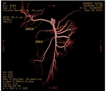
Five ( 5 ) superior mesenteric artery pictures of CT angiogram were
Three ( 3 ) superior mesenteric arteries of Foetuses were obtained
from the Department of Obstetrics & Gynaecology.
Two ( 2 ) clinical cases with history of injury to the superior
mesenteric arteries from the Department of Surgery.
Methods
1. Dissection :
Specimens were collected from the cadavers during routine
dissection programme. Specimens were cleaned by removing the
remains of peritoneum , fat and blood. The specimens were washed
thoroughly in tap water. The origin of the superior mesenteric artery
29
2. Specimens from the Forensic department :
Specimens were collected from the Forensic department and cleaned
well in running water. Specimens were preserved in 10 % formalin and
then taken for dissection. The sex , age and cause of death were noted
in all cases.
3. CT Angiogram :
Patient positioned in supine position, after the test dose, 150 ml of
omnipaque which is an iodinated contrast is injected at a rate of 3 ml
per second by pressure injection. Using Spiral CT scan the scan
performed. The interval time was 30 seconds and the scan time was
15 seconds. After acquiring all slices using maximum intensity projection
technique, the superior mesenteric artery is mapped out.
4. Foetal specimens :
Foetal specimens (Fig 8) collected and were Embalmed routinely and
dissected to find out the origin and Colic branches of the Superior
30
The following findings were observed and noted down from the Dissection,
CT angiogram pictures and Surgeries :
1. The origin of the superior mesenteric artery from the abdominal
aorta with the vertebral level.
2. The distance between the origins of the coeliac trunk and the
superior mesenteric artery from the abdominal aorta was measured
using a thread, marker and scale. The thread was placed on the
origin of the coeliac trunk and the other end was stretched towards
the origin of the superior mesenteric artery and the markings were
made on the thread. The marked distance was measured using a
scale.
3. Branching pattern of the middle colic , the right colic and the
ileocolic arteries.
5. Abnormal origin and branching pattern of the superior mesenteric
artery and its colic branches supplying the neighbouring organs.
Diagrammatic representations were made to compare with the
previous studies mentioned in the Review of Literature. The
observations were analyzed with reference to age , sex , normal and
abnormal patterns. Necessary photographs were taken pertaining to the
observations made.
Data analysis was carried out using relevant statistical tables and
charts.
32
OBSERVATION
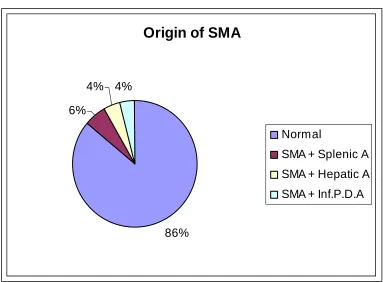
In the present study of 50 superior mesenteric arteries studied, the
origin of the superior mesenteric artery ( Tab 1 & Chart 1 ) was normal in
43 specimens.
No abnormal site of origin was observed in the study.
In three specimens, the superior mesenteric artery and the splenic
artery both arising from the abdominal aorta by a common trunk (Fig 3a).
The coeliac trunk had only the hepatic and the left gastric artery.
In two specimens, the superior mesenteric artery and the hepatic
artery arising from the abdominal aorta by a common trunk (Fig 3b). The
coeliac trunk had only the splenic and the left gastric artery.
In two specimens the superior mesenteric artery and the inferior
pancreatico duodenal artery arising from the abdominal aorta by a common
33
Origin of Superior Mesenteric Artery
Normal 43 SMA + Splenic A 3
[image:43.612.133.516.299.581.2]SMA + Hepatic A 2 SMA + Inf.P.D.A 2
Table 1 Origin of Superior Mesenteric Artery
Chart 1 Origin of Superior Mesenteric Artery
Origin of SMA
86% 6%
4% 4%
Normal
The distance between the origin of the coeliac trunk and the
superior mesenteric artery from the aorta (Tab 2 & Chart 2 ), ranged from
the 2 mm to 20 mm.
The distance between the coeliac trunk and the superior mesenteric
artery, from the aorta was 2 mm in 3 specimens, 3 mm in 3 specimens,
5 mm in 11 specimens, 8 mm in 1 specimen, 10 mm in 20 specimens,
15 mm in 11 specimens and 20 mm in 1 specimen. It was observed that
the prevalent distances were 5 mm (11), 10 mm (20) and 15 mm (11).
Branching Pattern of Superior Mesenteric Artery
Inferior Pancreatico duodenal Artery
Out of 50 superior mesenteric arteries studied, 45 ( 90 % ) had the
normal origin of the inferior pancreatico duodenal artery from the right side
35
Distance of SMA from Coeliac Trunk
Distance (in mm) Male Female Percentage 2 2 1 6 3 3 0 6 5 8 3 22 8 1 0 2 10 14 6 40 15 8 3 22 20 1 0 2 Total 37 13 100
Table . 2 Distance of SMA from Coeliac Trunk
Chart 2 Distance of SMA from Coeliac Trunk
0 2 4 6 8 10 12 14 N o . o f S p e c im e n s
2 3 5 8 10 15 20
Distance (in mm)
Distance of SMA from Coeliac Trunk
In two specimens, the inferior pancreatic duodenal artery, had a
common origin along with the superior mesenteric artery from the
abdominal aorta.
In one specimen, the origin was from the posterior surface of the
superior mesenteric artery.
In two specimens, it was from the first jejunal branch.
In this study it was observed that 48 of the inferior pancreatico
duodenal artery took origin above the level of the first jejunal branch.
Middle Colic Artery
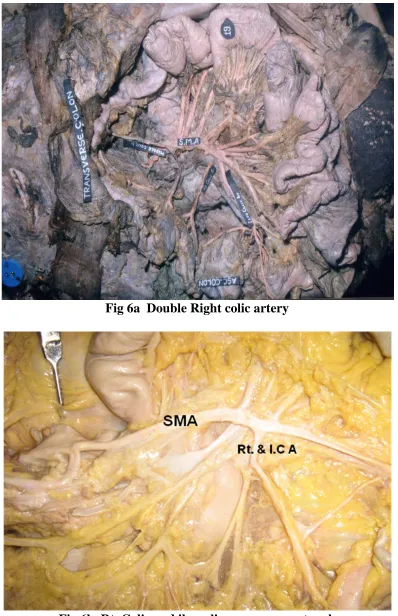
In the present study of 50 superior mesenteric arteries , 48 ( 96 % )
middle colic arteries (Tab 4 & Chart 3 ) were present and in 2 specimens (
4 % ) it was absent (Fig 4a). The specimens in which the middle colic
arteries were absent, a large branch from the left colic artery was
Fig 4a SMA and Inf. Pancreatico duodenal artery as a common trunk
Middle Colic Artery
Present 48
Absent 2
Common with Rt. Colic A
6
Double 4
Table 4 Middle Colic Artery
Fig 5a Middle and Right colic artery as a common trunk
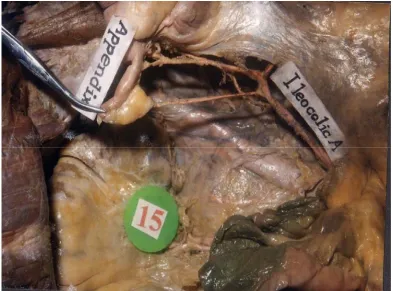
[image:49.612.139.493.403.675.2]In six specimens ( 12 % ) both middle colic artery and right colic
artery arose as a common trunk (Fig 5a) from the superior mesenteric
artery.
Double middle colic arteries (Fig 5b) were observed in 4 specimens (
8 % ). Out of this form, 2 middle colic arteries ( 4 % ) had their common
origin directly from the superior mesenteric artery. In 1 specimen ( 2 % ), 2
middle colic arteries were observed to take origin from the superior
mesenteric artery and the right colic artery had its origin from the ileocolic
artery. In another specimen, 1 middle colic artery ( 2 % ) from the superior
mesenteric artery and the other middle colic artery from the right colic
artery.
Right Colic Artery
In the present study of 50 superior mesenteric arteries , right colic
artery (Tab 5& Chart 4 ) was present in 43 specimens ( 86 % ) and absent
in 7 specimens ( 14 % ).
Out of 43 right colic arteries, 34 ( 68 % ) had normal site of origin
Fig 6a Double Right colic artery
Right Colic Artery
Present 43
Absent 7
Double
6
Common with Ileocolic A
1
From Ileocolic A
2
Table 5 Right Colic Artery
40
The right colic artery was double (Fig 6a) in six specimens ( 12 % )
and both were taking origin from the superior mesenteric artery.
In one specimen ( 2 % ) the right colic artery and the ileocolic artery
had common origin (Fig 6b) from the superior mesenteric artery.
In two specimens ( 4 % ), the right colic artery took origin from the
ileocolic artery.
In the specimens where the right colic artery was absent, it was
replaced by branches from the middle colic artery and ascending branches
of ileocolic artery.
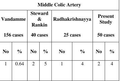
Ileocolic Artery
Ileocolic Artery (Tab 6 & Chart 5 ) was present in all the 50 superior
mesenteric artery. In two specimens ( 4 % ) the right colic artery was given
off from the ileocolic artery. In one specimen ( 2 % ) the ileocolic artery
Ileocolic Artery
Present 50
Common with Right
Colic A
1
From Right Colic A
6
Table 6 Ileocolic Artery
Chart 5 Ileocolic Artery
42
The appendicular branch of the ileocolic artery (Tab 7 & Chart 6 )
varied in its origin. In 31 specimens ( 62 % ) the appendicular branch took
origin from the ileocolic artery before its division.
In nine specimens ( 18 % ) there were 2 appendicular branches, 1
from the ileocolic artery before division and the other from the inferior
division of the ileocolic artery.
In four specimens ( 8 % ) a single branch (Fig 7b) was observed to
arise from the inferior division of the ileocolic artery.
In three specimens ( 6 % ) the appendix was supplied by 2
branches, 1 from the ileocolic artery and another from the posterior caecal
artery.
In two specimens ( 4 % ) dual arterial supply (Fig 7a) was observed
to arise from the ileocolic artery before division and another from the
Fig 7a Double Appendicular Artery
43
Appendicular Artery
Percentage Appendicular Artery
No. of Specimens
(%)
Ileocolic 31 62
Inferior Div. of Ileocolic 4 8
Ileocolic+Inferior Div 9 18
Ileocolic+Anterior Caecal 2 4
Ileocolic+Posterior Caecal 3 6
Nil 1 2
Table 7 Appendicular Artery
44
In one specimen ( 2 % ) appendix was absent may be due to
appendicectomy. In this specimen the branches of the ileocolic artery, the
ileal, anterior and posterior caecal and colic branches were normally
present but the appendicular artery were absent. In the same specimen,
the middle colic artery was also absent.
Abnormal Hepatic Artery
In this study of 50 superior mesenteric arteries , 5 specimens (10%)
had the accessory right hepatic artery arising from the superior mesenteric
45
DISCUSSION
A total of fifty ( 50 ) superior mesenteric arteries were studied in the
different age group in the south Indian population by Dissection , Angiogram
pictures and Surgeries.
The observations have been recorded, summarized and discussed
with reference to their surgical application with special reference to the
intestine transfers , that are becoming popular. The observations of the
normal and abnormal branching pattern gains greater importance in the
specialty of Gastroenterology where these data were of greatest importance
in major abdominal surgeries.
The data obtained in the present study was correlated with the data
Origin of the Superior Mesenteric Artery
Classical description of the superior mesenteric artery origin was
found in 86 % in the present study. This was in confirmity to the normal
description of the text books (Gray’s Anatomy 38th edition, Cunningham’s
manual of practical Anatomy 15th edition, Last’s Anatomy regional and
applied 10th edition).
In three specimens ( 6 % ) of the present study, the common origin
of the superior mesenteric artery and the splenic artery from the abdominal
aorta was observed. Michels ( 1955 ) in his study of 200 specimens stated
that, in 11.5 % of his specimens the common origin was found. Van haler
( Tripod of Haler ) also recorded the common origin.
The distance between the site of origin of the superior mesenteric
artery and the coeliac trunk from the aorta in the present study of 50
specimens ranged from 2 to 20 mm. Michels ( 1955 ) in his study of 200
47
Michels ( 1955 ) study ranged from 1 to 6 mm ( 60 % ). In the present
study it was observed only in seventeen specimens (34 % ) the distance
ranged from 2 to 5 mm.
Anson and Mcvay ( 1936 ) reported in their study that 71 % of the
cases ranged from 1 to 2 cm. In the present study the range was 0.2 to
2cm.
The study of Radhakrishnayya ( 1990 ) revealed that in 18
specimens ( 72 % ) , the distance found to be 12.5 mm ( 1.25 cm ) out of
25 specimens. He had also found the minimum range to be 2 mm and
the maximum to be 12.5 mm in the south Indian population. The variation
range between Radhakrishnayya’s and the present study was minimal.
When compared with the study of Michels ( 1955 ), Anson and
Mcvay ( 1936 ) and Radhakrishnayya ( 1990 ), the observations of the
present study ( 2 to 20mm ) were almost similar to that of Anson and
The variations in the distance observed in the present study
suggests that this range had to be observed during major abdominal
surgical procedures.
Inferior Pancreatico duodenal Artery
The present study indicated that the pattern of the origin of the
inferior pancreatico duodenal artery (Tab 3) was normal in 45 (90%) out of
50 superior mesenteric arteries examined.
Michels ( 1955 ) reported this type of origin in 60 % of his 200
cases studied.
Text books and some workers ( Gray’s Anatomy 38th edition,
Michels etc..) have reported the origin of the inferior pancreatico duodenal
artery from the first jejunal branch to be normal. The same is observed in
the present study in 2 cases ( 4 % ). So in the present study the total
49
Origin of Inferior Pancreatico duodenal Artery
Normal
From
SMA
From 1
stJejunal
Branch
From
Aorta with
SMA
From
posterior
Surface of
SMA
No % No % No % No %
[image:63.612.127.506.255.460.2]45 90 2 4 2 4 1 2
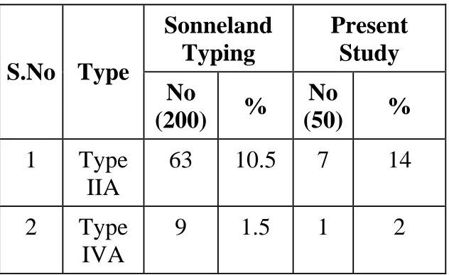
Middle colic Artery
In the present study the Middle colic artery ( Tab 8 ) was absent 2
specimens ( 4 % ).
Vandamme and Schuren ( 1976 ) in the series of 156 specimens
reported the absence of middle colic artery in only one specimen( 0.64 % ).
Steward and Rankin ( 1933 ) in their series of 40 cases of Radiological
studies by injecting celluloid material reported the absence of middle colic
artery in 2 cases ( 5 % ).
The study of Radhakrishnayya ( 1990 ) was also in the south
Indian population with absence of middle colic artery in 4 %.
In the present study, out of 48 middle colic arteries in 6 ( 12 % ) it
was duplicated ( 12 % ). Of the 6, 5 of them had their origin from the
superior mesenteric artery except one pair, in which one was from superior
51
Comparison Table showing the Absence of Middle Colic Artery
Middle Colic Artery
Vandamme
Steward
&
Rankin
Radhakrishnayya
Present
Study
156 cases
40 cases
25 cases
50 cases
No % No % No
% No %
[image:65.612.117.523.227.501.2]1 0.64 2 5
1
4
2 4
Henle ( 1876 ) also reported duplication of the middle colic artery in
his studies.
Steward and Rankin ( 1933 ) reported 7 % of double middle colic
artery in 40 cases. They also reported the presence of duplication of
middle colic artery in 10 % of cases. Radhakrishnayya ( 1990 ) in his
study of south Indian population reported the double middle colic artery in
1 case ( 4 % ).
Sonneland et al ( 1958 ) reported absence of middle colic arteries
which was 10 % less than the present study.
The common stem giving rise to the middle colic and right colic
artery was observed in 6 cases ( 12 % ) in the present study. The same
was observed in the study of Sonneland et al ( 1958 ) in 0.5 % out of
600 cases.
Trifurcation branching pattern reported by Steward and Rankin
53
Right colic Artery
In the present study the right colic artery ( Tab 9 ) was present in 43
specimens ( 86 % ) and absent in 7 cases ( 14 % ). Out of the 43 right
colic arteries studied, 2 of them had abnormal origin ( 4 % ). 6 double right
colic arteries were observed in the present study.
Waldeyer ( 1899 – 1900 ) reported the origin of the right colic artery
from the superior mesenteric artery in 50 % of cases. Steward and
Rankin ( 1933 ) reported the presence of right colic artery in 40 % of
cases.
Sonneland et al ( 1958 ) reported the presence of right colic artery
in 78 % of cases. Radhakrishnayya ( 1990 ) in his study reported the
normal origin of right colic artery in 23 specimens ( 92 % ).
The report of Sonneland et al ( 1958 ) is almost comparable with
the present study. It is interesting to note, that the present study of
south Indian population matched the report by Sonneland et al whose
Classification of Pattern of Right Colic Artery
Sonneland
Typing
Present
Study
S.No Type
No
(200)
%
No
(50)
%
1 Type
IIA
63 10.5 7 14
2 Type
IVA
[image:68.612.138.454.303.497.2]9 1.5 1 2
55
The comparative study between Radhakrishnayya and the present
study were pertaining to the south Indian population but variation in
observation was high as 10 %.
The common origin of right colic and ileocolic artery as a common
trunk ( 2 % ) or right colic artery arising as a branch from the ileocolic
artery ( 2 % ) were observed in the present study.
Radhakrishnayya ( 1990 ) reported the origin of the right colic artery from
the ileocolic artery in 4 % but he found no common origin.
Steward and Rankin ( 1933 ) in their studies reported the origin of
right colic artery from the ileocolic artery in 12 % and from the middle
colic artery in 30 %, which was not observed in the present study.
The report of Sonneland et al ( 1958 ) was the origin of right colic
artery from the ileocolic artery in 9.7 %.
However the pattern of right colic artery arising from the ileocolic
Double right colic artery was observed in 6 specimens ( 12 % ) in
the present study. A similar observation was reported by Sonneland et al
( 1958 ) in 8.2 % out of 600 cases. Vandamme and Schuren ( 1976 )
observed the occurrence of 1.5 % in 156 specimens. Radhakrishnayya did
not report any occurrence of such a double right colic artery.
In the present study, the 6 double right colic arteries were arising
only from the superior mesenteric artery ( 12 % ) whereas Sonneland et al
( 1958 ) observed in their study the second right colic artery to arise from
the Ileocolic in 1.5 % and the same was reported by Vandamme and
Schuren.
Triple right colic artery arising from the ileocolic artery in the
Sonneland et al ( 1958 ) report is 0.7 %, which was not observed in the
present study.
The double right colic arteries of the present study was not present in
the Sonneland et al typing or any other worker. Hence, this pattern is
57
Ileocolic Artery
In the present study the ileocolic artery was observed in all the
specimens.
Though the pattern of origin was normal, the branching pattern
presented few variations. It was observed in 1 specimen that the ileocolic
artery was giving origin to the right colic artery ( 2 % ). The similar
observations was reported by Sonneland et al ( 6.7 % ) and
Radhakrishnayya’s ( 4 % ).
The Triple branching of the Ileocolic artery was reported by Sonneland et
al was not observed in the present study.
Appendicular Artery
The present study of appendicular artery was observed in 49 cases
( 98 % ) and it was absent in 1 case ( 2 % ). In this case appendix was
The appendicular artery arising from the ileocolic artery was
observed in 31 cases ( 62 % ) and from the inferior division of ileocolic
artery in 4 cases ( 8 % ).
The double arterial supply was found in 14 cases in which 9
( 18 % ) derived from the ileocolic and its inferior division. In 3 cases
( 6 % ) the branches were from ileocolic and posterior caecal branch, and
in 2 cases ( 4 % ) the branches were from ileocolic and anterior caecal
branch. These observations were also reported by Shah and Shah
59
SUMMARY
A study was done in 50 superior mesenteric arteries for finding its
different pattern of origin and colic branches.
The observatory findings are more or less coinciding with that of the
observations of the eminent scientists in the field.
The following findings have been observed :
Superior mesenteric artery and Splenic artery arising as a
common trunk from the abdominal aorta.
Superior mesenteric artery and hepatic artery arising as a
common trunk from the abdominal aorta.
Superior mesenteric artery and inferior pancreatico duodenal
artery arising as a common trunk from the abdominal aorta.
Distance between the origin of coeliac trunk and superior
Inferior pancreatico duodenal artery arising from the posterior
surface of superior mesenteric artery and also from the 1st
jejunal branch.
Middle colic artery and right colic artery arising as a common
trunk from superior mesenteric artery.
Double middle colic arteries, both from the superior mesenteric
artery or one from superior mesenteric artery and the other
from the right colic artery.
Double right colic artery.
Right colic artery and ileocolic artery arise as a common
trunk from superior mesenteric artery.
Right colic artery arising from the ileocolic artery.
Ileocolic artery and right colic artery had a common trunk from
superior mesenteric artery.
Ileocolic artery arising from the right colic artery.
Appendicular artery arising from inferior division of ileocolic
61
Double appendicular arteries, one from ileocolic artery before
division and other from inferior division of the ileocolic artery
or one from the ileocolic artery and other from anterior or
posterior caecal artery.
With the knowledge about the aberrant vessels by their origin or by
their branches and termination, the surgeon can take adequate precautions
against the bleeding vessels in the operating field.
The anatomists can stress these variant occurrence and their clinical
importance while teaching and guiding the medical students.
Hereby I hope this analysis of mine about the variational and surgical
anatomy of the superior mesenteric artery will be of definite use and guide
BIBLIOGRAPHY
Adachi B. : Das Arteriensystem der Japaner, Tokyo, Kenkyyusha press,
1928.
Anson., B J., and Mcvay C B.: The topographical positions and the mutual
relations of the visceral branches of the abdominal aorta, Anat. Rec. 67:7,
1936.
Anson B J., and Mcvay C B.: 1951 Surgical anatomy 5th edition, Saunders:
Philadelphia.
Benton and Cotter, An unusual variation of the arterial supply of the
transverse and descending colon. Anatomical record, 1962, vol. 142. page
215.
Bertelli et al – 1991. Various cases of direct connection between the coeliac
Cokkins A. J. Observation on the mesenteric circulation. Journal of Anatomy,
1930, 64 : 200 – 205.
Delannoy, E. : Artere mesenterique superieure double, Bull. Et mem. Soc.
Anat. deParis 93:346, 1923.
Eaton, P. B.: The coeliac axis, Anat. Rec. 13: 369, 1917.
Feigl. W., Firbas W., Sinzinger H., Wicke L. Various forms of the coeliac
trunk and its anastamosis with the superior mesenteric artery, 1975.
Garcia Reiz A, Milson J. W., Ludwid K.A., Marchesa P. – Right colonic
arterial anastamosis. Implications for laparoscopic surgery. Dis – colon,
Rectum. 1996. Aug 39 (8) : 906 – 11.
Gray’s Anatomy, 39th edition. The Anatomical basis of Clinical Practice.
Griffith J D 1961, Extramural and intramural blood supply of the colon.
Br.Med J 1: 323 - 326
mesenteric artery. 1995. Aug. 70 (4) : 338 – 46.
Jamieson, J. and Dobson, J.: The lymphatics of the colon, Ann. Surg. 50 :
1077, 1909.
Kao .GD, Whithington R, Coia L. Anatomy of the coeliac axis and superior
mesenteric artery and its significance in radiation therapy. Int. J. Radial.
Oncol. Biol. Phys. 1993. Ja.25 (1) : 131 – 134.
Koizumi M, Horiguchi M, Accessory arteries supplying the human
transverse colon. Acta Anat ( Basel ), 1990: 137 (3) : 246 – 51.
Lipshutz, B.: A composite study of the coeliac artery, Ann. Surg. 65:159,
1917.
Michels N A. : Illustration with subscripts of the 10 basic types of the blood
supply to the liver, in behrend’s diseases of the gall bladder and allied
structures. Philadelphia, Davis, 1947.
Michels N A.: Variations in the blood supply of the supramesocolic organs,
Michels N A : Variations in blood supply of liver, gall bladder, stomach,
duodenum and pancreas, Yearbook of the Am. Philosophical Soc., 1943,
p.150: J.Internat. Coll. Surgeons & : 502, 1945, and in yearly abstracts thereof
in Anat. Rec. from 1942 to 1954.
Michels N A.: Blood supply and Anatomy of the upper abdominal organs.
Philadelphia. JB Lippincott co 1955 : 1 – 581.
Moynihan, B .: Abdominal Operations, Philadelphia, Saunders, 1913.
Munger, R. S.” Report of an unusual coeliacomesenteric trunk with unique
distribution and anastamosis, Anat. Rec. 80 :55, 1941.
Quain, Richard: The anatomy of the arteries of the Human body, 2 vols.,
London, Taylor and Walton, 1844.
Shah, M. A. and Shah M: The arterial supply of the vermiform appendix,
Sonneland John M. D., Barry J. Anson, PhD. (Med,Sc), Lindsay E.
Reaton,M.D. Surgery. Gynaecology and Obstetrics. 1958 April, Vol 106, no 4,
384 – 398.
Sreward, J. A., and Rankin, F. W.: Blood supply of the Large intestine: its
Surgical considerations, Arch. Surg. 26 : 843,1933.
Tandler, J.: Uber die Varietaten der Arteria Coelica und deren
Entwickelung, Anat. Hefte 25:472, 1904.
Vandamme J. P. J. and G. Van der Schuren.: Re-evaluation of the colic
irrigation from the SMA. Act. Anatomica, 95:578 – 588, 1956.
Wright W. Clarence. : Aberrant blood vessels and nerves in a case of left
sided vermiform appendix, caecum and anomalous colon. Anat. Rec 1959.
page 187 – 201, vol 133.
Yamaki K., Tanaka N., Matsushima T., Miyazaki K., Yoshizuka M.: A rare