A CLINICO – PATHOLOGICAL STUDY OF 300
CASES OF WARTS IN IMMUNOCOMPETENT
AND IMMUNOCOMPROMISED PATIENTS
Dissertation Submitted in partial fulfillment
of university regulations for
M.D. DEGREE IN
DERMATOLOGY, VENEREOLOGY AND LEPROSY
BRANCH XII – A
THE TAMILNADU DR. M.G.R. MEDICAL UNIVERSITY
CHENNAI, TAMIL NADU
CERTIFICATE
This is to certify that this Dissertation entitled “A CLINICO – PATHOLOGICAL STUDY OF 300 CASES OF WARTS IN IMMUNOCOMPETENT AND IMMUNOCOMPROMISED PATIENTS” is a bonafide work done by Dr.SAMUEL JEYARAJ DANIEL, Postgraduate student of Department of Dermatology, Leprosy and Institute of STD, Madras Medical College and Government General Hospital, Chennai – 600 003, for the award of Degree of M.D. (Dermatology, Venereology and Leprosy ) Branch XII – A during the academic year of 2003-2006. This work has not previously formed in the basis for the award of any degree or diploma.
Prof. Dr. B. Parveen, MD., DD.,
Professor & Head,
Dept. of Dermatology and Leprosy,
Madras Medical College &
Govt. General Hospital,
Chennai – 600 003.Prof. Dr. Kalavathy Ponniraivan,
MD.,
Dean
Madras Medical College &
Govt. General Hospital,
Chennai – 600 003.SPECIAL ACKNOWLEDGEMENT
I sincerely thank
Prof. Dr. Kalavathy Ponniraivan, MD.,
Dean,
Madras Medical College & Govt. General Hospital, Chennai – 600 003, for
ACKNOWLEDGEMENT
I sincerely thank Prof.B.Parveen, MD., D.D., Professor and Head of Department of Dermatology for her invaluable guidance and encouragement for the successful completion of this study. I express my heart felt gratitude to Dr.N.Gomathy MD.,DD, former Head of department of Dermatology who was instrumental in the initiation of this project, giving constant guidance throughout my work. I immensely thank Dr.C.Janaki, MD., DD (Mycology) Reader of Dermatology, for her invaluable help in guiding me in all stages of this study.
I earnestly thank Dr. D. Prabavathy MD., DD, Professor and Head of Department of Occupational Dermatology and Contact Dermatitis for her kind support. I am also thankful to Dr. V. Somasundram MD., DD, Additional Professor, Department of Occupational Dermatology and Contact Dermatitis.
I am grateful to Dr.V.S.Durairaj MD., DV, Director, Institute of STD for the immense help and support during my study period. I also thank Dr.N.Usman MD.,DV.,Phd, former Director, Institute of STD and Dr.S.Mohan MD.,DV, Registrar, Institute of STD for their kind support.
I wish to express my grateful thanks to Prof. Dr. Jayaraman, M.D., D.M., Professor of Nephrology, who helped me to carry out the study in the Nephrology wards with necessary assistance.
I render my sincere thanks to Dr.K.Rathinavelu MD.,DD, Professor of Leprosy, Dr.R.Arunadevi, MD., DD, Registrar, Department of Dermatology for their support.
DNB (Paed), and Dr. G.K.Tharini MD (Derm) for their immense help, support and encouragement throughout my study.
I am thankful to
Dr.Hamedullah, M.D., D.D., Dr.Kumaravelu, M.D., D.D. and Dr.J.Manjula, M.D., DNB., Assistant Professors, Department of Occupational Dermatology and Contact Dermatitis for their encouragementI express my sincere thanks to Dr. S.Venkateshwaran MD., DV, Dr.Ilangovan MD., DV, Dr.Thilagavathy MD., DV, Dr.Thirunavukarasu, MD., DV, Dr.Ramachandra Reddy MD., DV, Dr.P.Mohan MD.,DV, and Dr.S.Arunkumar MD., DV, Assistant Professors, Institute of STD for their help and support.
I am also grateful to Dr.G.S.Sentamil Selvi MD., DD. MNAMS., Ph.D., Dr.Manoharan, MD., D.D., Dr.V.Sampath MD., DD., for their continuing guidance and support.
I duly acknowledge the paramedical staff and my colleagues for their help and favour.
CONTENTS
S.No.
Title
Page
No.
PART - I
1.
INTRODUCTION
1
2.
REVIEW OF LITERATURE
2
PART - II
3.
AIMS AND OBJECTIVES OF THE STUDY35
4.
METHODS AND MATERIALS
36
5.
OBSERVATION
39
6.
DISCUSSION
56
7.
CONCLUSION
62
APPENDICES
BIBLIOGRAPHY
STUDY PROTOCOL
INTRODUCTION
Among all the common cutaneous viral infections, warts are the commonest. Warts form an interesting study because of their frequent occurrence, varied morphological appearance, relatively asymptomatic behavior and their unpredictable course. Warts are mostly self-limiting and benign conditions. But the possibility of malignant in some types like genital warts, epidermodysplasia verruciformis and the heterogeneity in serotypes have additional importance in their study1,2.
The possibility of increase in both oral and anogenital pathologic conditions due to HPV in patients infected with HIV is of concern and is the focus of numerous current research studies. HIV-infected women are at a higher risk for cervical HPV infection with high oncogenic types of HPV leading to cervical intraepithelial neoplasms. HIV-infected men are at increased risk for anal HPV infection with high oncogenic types of HPV with persisting type of infection. Recent studies have shown an increased risk of oral warts in HIV-infected individuals despite treatment with highly active antiretroviral therapy (HAART)78.
It appears that transplant recipients are susceptible to infections with diverse HPV types including rare types, that multiple infections are possible and that the site specificity of certain HPV types differs from normal population and is often recalcitrant, painful and deeply affects the patient’s quality of life63.
REVIEW OF LITERATURE
History:
Warts were well known since 400 BC to the physicians of Greece and Rome. The term Verruca was first introduced by Sennertus first meaning a steep place or height appearing as little hills. Condyloma is a Greek word used by Martiales (40-102 AD). In Roman – Hellenistic cultures the genital warts were known as ficus and thymion. Celsus in the first century AD described 3 types of warts: Achrochordon in children, Thymion or genital warts and Myrmecia or plantar warts3.
Less attention was however paid to the cause of warts. Romans were well acquainted about the sexual transmission of genital warts. Daniel Turner (1712) mentioned it as ‘Nutritious juices’ seeping from damaged nerve terminals.
Joseph Payne (1891) first described the infectious nature of the warts. The viral etiology of warts was established in 190749 by inoculation of wart filtrates into skin, inducing
papillomas at the injection site. In 1950 Strauss and his coworkers demonstrated the virus under the electron microscope.
In 1962 Melnich coined the acronym PAPOVA. They noted the striking similarity of wart virus with those of Polyoma virus of mice and the vacuolating virus of monkey (SV40) and coined the name ‘PAPOVA’ for these groups of viruses. The first detailed study about the Papova virus was done by Klug et al4.
Warts occur worldwide with more or less the same incidence. The viability of HPV in the environment is unknown. The virus has not been cultured for a long time. The incubation period varies from few weeks to more than a year. The virus is inactivated by heating to temperature about 55o C 5 and by 0.4% formalin for 72 hours at 4oC. The important
predisposing factors for the spread of warts are trauma, friction and pressure6. Koebner’s
phenomenon is due to trauma at or near the wart region and consequent spread. The home, school, barracks Swimming pools7 and the instruments of chiropodists and barbers, towels of
bathrooms provide opportunities for spread.
Contagion of HPV probably depends on several factors, including the location of lesions, the quantity of infectious virus present, the degree and nature of the contact and the general and HPV-specific immunological status of the exposed individual. Patients with impaired cellular immunity are particularly susceptible to HPV infection, and warts occur in the majority of renal transplant patients on immunosuppressive therapy.
An increased incidence of warts in the community over the decades may be due to increased awareness and referral rates. An increased incidence of anogenital warts suggests changing cultural attitude towards sex.
Infectivity is very high in the case of genital warts. Nearly two thirds of sexual contacts of patients with genital warts develop or have sub clinical warts8. The infectivity of the other
types of warts is significantly lower than genital warts.
Modes of transmission:
HPV affects only humans. It is transmitted by contact directly or indirectly9. Disruption
source of infection is usually a clinical or sub clinical case of HPV infection as well as virions, which may be present in the environment. Plantar warts spread by the use of common bathrooms in the hostels and army barracks where injury to an infected foot will cause virions to be discharged into the environment from where they enter the moist skin of the other contacts. Common warts may spread to different locations on the same person by habits like finger sucking, nail biting and scratching, suggesting that autoinoculation may be an important mode of spread. Certain occupations in which the individuals are prone to develop injury and come into contact with flesh of animals like butchers, fish and poultry workers have a high incidence of warts. Shaving can cause the spread of warts to different parts of the face.
Iatrogenic transmission can occur in gynecology clinics due to use of common instruments for examination like specula, which can spread the virus from one person to another10. Dermatology clinics using liquid nitrogen to treat warts may spread the virus if a
common flask is used to dip cotton swabs for different patients11. Viral particles have been
isolated from the smoke that emerges on treating a wart by laser or electrosurgery. The operator in this case is at risk of acquiring laryngeal warts due to inhalation of the smoke12.
Using sensitive PCR detection methods, it is becoming increasingly evident that normal skin in immunocompetent individuals can harbor HPVs, including novel types and those found in Epidermodysplasia verruciformis.
Sexual modes of Spread:
significant percentage of these are caused by HPV types isolated from nongenital lesions13,14,15.
Penile lesions occur frequently in the sexual contacts of women with cervical intraepithelial neoplasia.
Perinatal Transmission:
Warts in infants and children are usually acquired from the mother during their passage via the birth canal16,17. Warts acquired this way persist well into childhood. Vertical
transmission of HPV form mother to child in utero can rarely result in laryngeal papillomatosis. Genital warts in infants and children commonly result from virus inoculation at birth or from incidental spread from cutaneous warts.
Age and Sex incidence:
Warts can affect all ages and both sexes. In general, a higher incidence of warts is noted in adolescents and young adults than in other age groups.
Plane warts are common in childhood and among females than in males. Genital warts were found more commonly in the age group of 20 – 29 years. There was a slight male preponderance.
Social class:
The highest prevalence of verruca vulgaris is seen in the lower socioeconomic class, especially those having manual occupations. The reason for this is the increased exposure in these people due to over crowding and occupational trauma.
Human Papilloma virus belongs to the papovavirus family of DNA viruses. It is spherical in shape measuring 55nm in diameter. They contain a circular dsDNA molecule of 7.2-8.0 kbp. The geneome is placed inside a non-enveloped icosahedral capsid composed of 72 pentamers, as a covalently closed circle of super coiled DNA. Their lack of an envelope renders these viruses relatively resistant to heating and to organic solvents. It has a molecular weight of 5 x 10⁶ Daltons. Each genome is composed of 8000 nucleotide base pairs and codes for viral
protein classified as early ‘E’ and late ‘L’ proteins. The E proteins, most of which participate in viral DNA replication, are expressed before the L proteins and are not incorporated in the infectious viral particle. Because E genes do not encode a DNA polymerase or thymidine kinase, papilloma viruses are not susceptible to inhibition by acyclovir. The L1 L2 genes encode the structural proteins that form the outer protein shell, called the capsid of the viral particle, which is called the virions. The spherical virion surrounds the viral DNA.
HPV infects the stratum basale of the epidermis. Viral replication however takes place only in the upper terminally differentiated cell layers like the stratum spinosum and stratum granulosum. There are about 100 different HPV serotypes24 identified by recombinant DNA
technology. They are generally organized by degrees of relatedness in DNA hybridization. Those that share less than 50% homology in hybridization with a known HPV is considered a new HPV type25,26. With the advent of polymerase chain reaction (PCR), which has a high
sensitivity and specificity it is now possible to amplify and sequence any viral DNA. Hence HPV types are classified by DNA sequence homology. PCR can be used to classify DNA which are decades old, like those obtained from old histopathological specimens1,10. Besides
clinical lesions, HPV have been now produced in human keratinocytes grown under renal capsule of thymic nude mice27,28 and the raft culture system29,30.
Although more than 80 HPV types have been completely or partially sequenced, more than 220 HPV types have been identified so far, and 91 types have been characterized49. Each
type tends to be associated with particular tissue specificity, pathology and oncogenic risk. Currently, the relatedness of their DNA sequence distinguishes between HPV types. The ease with which PCR, which has both high specificity and sensitivity, can be used to amplify and sequence any viral DNA isolate has now led to HPV types being defined by DNA sequence homology. Two HPV isolates are assumed to be of the same type if the sequences of their L1 genes are at least 90 percent identical. It is likely a new HPV type, after the viral genome has been molecularly cloned and its L1 gene sequenced, the sequence is less than 90 percent homologous to the closest known HPV type.
The association of particular type with a specific group of warts, however is not absolute. HPV – 1 has been found in verruca vulgaris and condylomata acuminate, HPV – 2 in condylomata acuminate and HPV – 3 in verruca vulgaris.
CLINICAL LESIONS HPV TYPES
PalmoPlantar warts 1, 2, 4
Common warts 2, 4, 7, 29
Plane wart 3, 10, 41, 65
Epidermodysplasia verruciformis 5, 8, 9, 12, 14, 15, 17, 19-27, 36, 46, 47, 49, 50 Genital warts (low risk malignancy) 6, 11, 30, 34, 40, 42 - 44 Genital warts (high risk malignancy) 16, 18, 31, 33, 35, 39, 45, 51 Oral papillomas ( in HIV infected persons) 72, 73
Common warts in renal allograft recipients 75, 76, 77
HPV infection occurs through inoculation of virus into the viable epidermis through defects in the epithelium. Maceration of the skin is probably an important predisposing factor, as suggested by increased incidence of plantar warts in swimmers who frequent public pools. Although receptors by which cells take up HPV infection has not been identified, cell surface heparin sulfate, which is encoded by proteoglycans and binds to papilloma virus particles with high affinity, is required for HPV infection. To establish chronic infection characteristic of a wart, it is probably necessary to infect an epidermal stem cell. It is believed that a single copy or at most a few copies of viral genome are maintained as an extra chromosomal plasmid within the infected epithelial basal cells. When the basal cells divide the viral genome is also replicated and transported within the daughter cells as they migrate upward to form the differentiating epithelium. Viral RNA expression (transcription) remains low until the upper malpighian layer, just before the granular layer, where viral DNA replication may result in hundreds of copies of viral DNA per cell. The viral capsid proteins (L1 and L2) are synthesized and assembled into virions in the nuclei of the cells at this level. The newly synthesized viral DNA is also incorporated into the virions in the nuclei of these differentiated malpighian cells. A viral protein called E1-E4 may induce collapse of the cytoplasmic keratin filament network. This is postulated to facilitate release the virions from the cross-linked cytoskeleton so that they can be inoculated into another site or desquamated into the environment. Papillomaviruses are not enveloped, because they do not bud from the nuclear or plasma membrane.
High risk HPV 16 or HPV 18 can alter benign cell proliferation towards dysplasia by means of viral genes E5,E6 and E7. High risk viruses rapidly induce chromosomal instability and as aneuploid karyotype, whereas low risk viruses fail to do so59. These genes are
the cellular proteins p53 results in rapid degradation of the later. The degradation is apparently the main contributor to the induction of chromosomal instability and aneuploidy60. The E7
protein of the same types forms a complex with the cellular tumor suppressor protein RB, thereby releasing the cellular transcription factor E2F from binding to RB and activating the cell cycle.
In anogenital cancers, HPV is often integrated into the chromosome, which results in dysregulation and increased transcriptition of E6 and E7. This leads to increased chromosomal instability and proliferation of cells, contributing to a setting where other potential carcinogens, such as those in tobacco smoke, can lead to development of invasive carcinoma77.
Clinical features :
Verruca vulgaris or Common warts
They are well – circumscribed, rough, firm papules or nodules with a verrucous surface that can be found anywhere on the skin or mucosa. Their size may vary from a few millimeters to 2 – 3 cms. They occur commonly as single or grouped papules on the back of hands and fingers. They are usually flesh coloured but may be erythematous or pigmented. The common warts may remain single or spread to form large numbers slowly over a period of time.
Warts may spread to new sites of trauma a process known as Koebner’s isomorphic phenomenon. Warts are generally asymptomatic but tenderness is present on certain sites like nail beds, palmar aspect of fingers or when there are fissures or infection in a wart. In general, warts lack dermatoglyphics on their surface. Warts in the nail bed may affect nail growth and those on the eyelid may cause conjunctivitis or keratitis31. Majority (67%) of warts resolve
Filiform warts which are variants of verruca vulgaris, Show finger like projections arising from a horny base. Their common locations are scalp, face and neck. Immunocompromised patients have an increased number of common warts33.
Flat Warts – Verruca Plana
These are 2 – 4 mm slight elevated flat topped papules occurring most frequently on face, hands and feet. They are commonly associated with HPV 3 and 1034. The lesions are
rounded or polygonal in shape and their surface is smooth with no evidence of hyperkeratosis or scaling. Their number may vary from a few to some times even a hundred. They may be flesh coloured or hyperpigmented. They occur commonly as discrete lesions, though some of them may coalesce to form a large lesion. Koebner’s phenomenon was seen along lines of trauma in some lesions.
Signs of inflammation such as itching, erythema, and swelling can accompany the spontaneous resolution of the plane warts. The resolution is usually complete within a month. Depigmented halos may be seen around the lesion36. The resolution or regression is usually the
result of cell mediated immune mechanisms37,38. They differ from epidermodysplasia
verruciformis by the absence of tinea versicolor like patches and lack of malignant transformation of sun – exposed lesions.
Palmo plantar Warts
Traditionally the are classified into:
1. Superficial (mosaic) warts
2. Deep warts (myrmecia – anthill) formed by coalescence of several lesions.
3. Other rare variants39 include
A. a nodular form
B. a pigmented verrucous variant
C. a whitish punctuate keratotic type usually multiple
HPV types 1,2,4 or 57 usually cause these warts. The lesions are initially seen as small papules, which soon grow to form well defined, rounded lesions having a rough keratotic surface. These lesions are surrounded by a smooth collar of thickened horny layer, which on paring becomes more prominent; further paring results in small bleeding points seen at the tips of dermal papilla40. Lesions may be single or multiple. Occasionally, multiple small warts of
varying size may develop surrounding a large central wart. These are known as satellite or weed warts.
Although mosaic warts are painless, a majority of lesions do have pain varying from mild to severe. Spontaneous resolution can occur, generally earlier in children than adults. The resolution time may vary from a few months to a year, and is independent of number of lesions. Mosaic warts may persist for a longer time. Regression is usually seen as drying and gradual separation. Occasionally signs of inflammation are seen41.
epidermal ridges, whereas warts lack dermatoglyphics.
Anogenital warts
This is also referred to as condyloma acuminata or veneral warts. The term Condyloma acuminate literally stands for condyloma = knuckle like; accuminatum = pointed, which describes the morphology of the lesion. They are fleshy, exophytic, verrucous , papules and plaques which coalesce to form cauliflower like masses in the anogenital region. They are usually sexually transmitted and spread rapidly. They are usually asymptomatic but some of the large lesions may cause discomfort, discharge42 or even bleeding. The most common sites of
occurrence of these warts are usually the most frequent sites of friction (Usually coital)43. This
includes the frenulum, corona and glans on the male genitalia and posterior fourchette and perianal region in females.
Besides the classical acuminate type, a flat type is also seen especially on the penile shaft, pubis, perianal region ant the groins. Occasionally common warts may be seen on the on the penile shaft (1to 2%) which may be due to auto inoculation from a lesion elsewhere on the same patient or from the sexual partner. The incubation period may vary but is usually around 2 to 3 months. HPV 6 and HPV 11 are the most common HPV types identified. Malignant progression is associated with HPV type 16, 18, 31, 33 and 51.
The duration of anogenital warts varies from a few weeks to many years. Recurrences can occur in nearly a third of the cases. This has been attributed to the persistence of HPV DNA in the dermis44. Patients having genital warts also have a higher incidence of other genital
Epidermodysplasia Verruciformis
This is a rare entity first described by Lewandowski and Lutz, characterized by chronic extensive HPV induced warts. They occur more commonly in infants and children than adults45. The common presentation is that of extensive wide spread lesions resembling planar
warts ( caused by HPV 3 and 10) or reddish brown macules resembling pityriasis versicolor ( HPV 5 and 8). Most patients have more than one HPV type. The diagnosis is made by presence of wide spread lesions or failure to resolve despite adequate treatment. The male to female ratio is 1:1. Nearly half the cases of EDV have a family history and are inherited as an autosomal recessive pattern with history of parental consanguinity. An X – linked46 inheritance
has also been reported.
Occasionally EDV, instead of being widespread may remain localized to one extremity. The recurrence of EDV after treatment indicates absence of effective immunity47. The immune
defect is usually that of cell-mediated immunity, specifically natural killer cell activity. Patients however do not show an increased risk to bacterial or other viral infections. Their association with malignant skin tumors in the immunosuppressed is well established48. An increased
incidence of cutaneous squamous cell carcinoma has been noted. These tumors rarely metastasize. Their occurrence in sun exposed lesions suggests a contributory role for ultraviolet light in carcinogenesis. These tumors occur in patients with pityriasis versicolor like lesions. The lesions of individuals with EDV caused by Non - EDV type of HPV do not have any risk of becoming malignant.
Diagnosis :
in skin and mucosa. Sub clinical disease on the mucosal surfaces are demonstrated by application of 3-5% dilute acetic acid and examined often with the aid of magnification. External lesions on the skin are examined using 6x or 10x lens, while coloposcopic examination of vagina and cervix may be necessary. However aceto white staining may also be positive in other inflammatory conditions like lichen planus, atopic dermatitis, psoriasis, dermatophytosis, intra epithelial neoplasia and irritant contact dermatitis. Biopsy for histopathological examination is necessary in doubtful cases to confirm diagnosis.
Latent HPV is presence of HPV DNA in clinically and histologically normal skin and mucosa and can be identified by DNA hybridization or polymerase chain reaction that demonstrates in normal mucosa adjacent to treated warts, in immunocompetent and immunosuppressive individuals. For clinical studies or epidemiological purposes the presence of visible cutaneous warts is the definition of the disease.
Cytology
Cytology is very useful in the diagnosis and management of HPV infection and neoplasia of anogenital epithelium. The procedure is noninvasive, inexpensive but useful only for epithelial sites where exfoliated cells can be collected. Cytological diagnosis of HPV infection has been found to limit sensitivity when compared with nucleic acid hybridization method, greater when compared to polymerase chain reaction. This can be overcome by repeated cytological staining. They have an important role in the management of cervical neoplasia.
This involves the detection of HPV DNA or RNA by using complimentary nucleic acid probes, which permits direct identification of the viral genome in tissue samples and has a high degree of specificity even in the absence of clinical and histological findings. Some popular nucleic acid hybridization procedures are southern blot hybridization and in situ hybridization.
Polymerase chain reaction is a special method in which target sequence of DNA are amplified thousands of times and this increases the sensitivity of false positivity in its results, especially when contamination is present.
Serology
The development of serological diagnosis has been limited by the inability to culture HPV. Recombinant viral proteins have been developed from genital types and appear to be type specific.
Electron Microscopic Examination
Viral particles of various types of HPV appear similar on electron microscopic examination. The amount may vary with each type. Old lesions may show a total absence of viral particles on electron microscopic examination. Hence their absence does not excludes the
presence of HPV.
Polymerase chain reaction – using degenerate consensus primers
DIFFERENTIAL DIAGNOSIS :
Common warts
-The most common skin conditions which are mistaken for warts are achrochordon, solar keratosis, lichen planus, seborrhoeic keratosis, filiform warts, acrokerratosis verruciformis, and squamous cell carcinoma.
Plane wart
-Freckles and lichen planus resemble plane or flat warts
Plantar warts -
These are very difficult to diagnosis and they have to be differentiated from corns, callosities and digital fibrokeratomas.
Genital warts
HISTOPATHOLOGY
Common warts
Shows hyperplasia of all the layers of the epidermis. This includes marked hyperkeratosis, acanthosis and papillomatosis. Parakeratosis occurs in columns or tiers over papillomatous projections, just above a focus of vacuolated cells. No granular cells are seen above the papillomatous crests, while they are increased in number in the intervening areas. The granular cells contain coarse keratohyaline granules in clumps seen as eosinophilic inclusion bodies. Koilocytes are large vacuolated kerationocytes, which are seen in the upper layers of stratum malpighii especially in the granular layer.
They have a small eccentric pyknotic nucleus surrounded by a clear – perinuclear halo.
The koilocytes, which are very characteristic of HPV papillomas are not seen in older lesions. Elongated rete ridges often point toward the center of the wart especially at periphery of the lesion. This is called arborization. The nuclei may be vacuolated and may contain viral inclusion bodies seen as basophilic inclusions. The dermis shows few changes like dilated capillaries in the papillomatous projection or a lymphocyte infiltrate. Coagulated blood in stratum corneum appears as minute black dots and small bleeding punctae, which are nothing but transected dermal capillaries seen on paring the wart.
Palmoplantar Wart
whose histopathology is similar to that of verruca vulgaris and deep palmoplantar wart also known as myrmecia – “anthill” or inclusion warts. They have abundant keratohyaline granules, which are deeply eosinophilic. Numerous eosinophilic granules are present in the cytoplasm of many cells. They are present from the lower epidermis and enlarge in the upper stratum malpighii where they coalesce to form large irregularly shaped, homogenous “inclusion bodies”. These surround the nucleus, which may be vacuolated or may be separated from it by perinuclear vacuolization. The granular layer is usually absent. Cells in the stratum corneum have nuclei, which are seen as round deeply basophilic structure surrounded by a clear zone. There are also small round intranuclear eosinophilic inclusion bodies in some of the cells of upper stratum malpighii.
Although numerous similarities with common wart have been observed a major portion of the lesion lies deep to plane of the skin surface and extends well into the dermis. There is prominent hyperkeratosis, although acanthosis is also present with mild papillomatosis.
Verruca Plana
Verruca Plana or flat warts have features like those of verruca vulgaris but of a lesser intensity. It is characterized by hyperkeratosis, hypergranulosis, acanthosis and vacuolated cells in the upper layers of stratum malpighii. The hyperkeratosis and hypergranulosis are less when compared to common wart. Flat wart however does not have parakeratosis and papillomatosis. Koilocytes or vacuolated cells which are seen in the upper layers are large or loose lamellar pattern of hyperkeratosis.
Death of a single epidermal cell may take place by apoptosis with more than one lymphocyte being in contact with degenerating cell. Degenerated epidermal cells are shrunken with eosinophilic cytoplasm and pyknotic nuclear remnants.
Anogenital warts
There is marked acanthosis with papillomatosis and hyperkeratosis. Stratum corneum shows little thickening. Lesions on mucosal surfaces show parakeratosis. Stratum malpighii shows papillomatosis with thickening and elongation of the rete ridges. Rete ridges form thick, round bands extending into the vascular dermis and have a well defined border. The most diagnostic histological picture is the presence of epithelial cells showing distinct perinuclear vacuolization in the granular and spinous layers, especially in the dells between knuckle like papillations.
These vacuolated epithelial cells are larger in size and contain round hyperchromatic nuclei. Vacuolization is not as prominent as in other warts. Since vacuolization occurs normally in the upper layers of the mucosal surfaces. They are significant only when they extend into lower portion of stratum maplighii. There is oedema of the connective tissue, which shows an increased number of capillaries, which are dilated and tortuous. Coarse keratohyaline granules may also be present. Langerhans cell are sometimes prominent. Mitotic figures may be present.
Epidermodysplasia Verruciformis
The nucleoplasm is clear and the cytoplasm contains keratohayline granules of various sizes and shapes. The keratinocyte are vacuolated and appear pale blue-grey on staining with hematoxylin and eosin. This blue-gray pallor is most pronounced in the granular layer of the epidermis and the vacuolated keratinocytes are typically arranged in clusters or columns. As the lesions progress to atypia, the nuclei of the keratinocytes become larger, more hyperchromatic, and cellular maturation is disordered. The stratum corneum has a basket weave appearance. A few dyskeratotic cells are seen in the lower part of the epidermis. Some of the nuclei are pyknotic while others are round and empty due to peripheral accumulation of chromatin. Changes of bowen’s disease or squamous cell carcinoma may ultimately supervene.
Warts in HIV + patients
HPV infections occur commonly during the course of HIV disease with extensive disease often are found to have CD4+ cell numbers lower than 500 cells/mm3. The nature of the
link between HIV and HPV infections seems to be behavioral and immunologic because both infections are sexually transmitted; it is obvious that they share some f the same behavioral risk factors. HIV infection depresses the immune defenses that control HPV infections. Accordingly, the more severe the HIV infection the higher the risk of finding high risk HPV DNA in the anogenital tract, condyloma acuminata or preinvasive squamous cell carcinoma in the cervix and anus of men and women50-57. Due to HIV-induced immunosupression HPV
economic and can detect past or present HPV-infection independently of an anatomical region. HIV- infected patients may also be candidates for prophylactic HPV-vaccination strategies, since these individuals a humoral immune response to HPV occurs.
Common warts
Patients with HIV infection, especially those who are immunocompromised, may present with multiple verruca vulgaris lesions, especially in periungal locations. Multiple plantar warts, including mosaic warts, may develop that may lead to pain with walking. Extensive flat and filiform warts on the bearded area of the face manifest by small verrucous papules. These seen in HIV-infected individuals may be the first sign of HIV infection58. With
advancing HIV diseasae, verrucae can enlarge, become confluent and become unresponsive to therapy. HPV – 5 can cause an unusual pattern of extensive verruca plana and pityriasis versicolor – like warts, similar to the pattern seen in epidermodysplasia verruciformis. Skin coloured to reddish verrucous hyperkeratotic papules or plaques that may occur on any surface of the skin. Glabrous skin is commonly affected, especially around the fingers and on the extremities, although the face head and neck areas are also frequent sites of involvement. With moderate or advanced immunodeficiency, warts may become much more numerous, confluent, and refractory to usual treatment modalities. Precancerous lesions identical to mucosal lesions [ squamous intraepithelial lesions (SIL)] can occur periungually and on the nail bed epidermis. In some cases invasive SCC can arise at one or multiple sites and invade the underlying fingers and nail bed. These tumors are aggressive and invade the underlying periosteum and bone relatively early due to the proximity of the underlying bony structures. Despite immune restitution with HAART, HPV infections often persist and progress unlike other opportunistic infections.
HPV – 6 and HPV – 11 infect minimally keratinized epithelium such as anogenital sites and oropharynx and causes genital warts. HPV – 16 and HPV – 18 cause precancerous lesions, low grade SIL, high grade SIL and invasive SCC. Genital warts may be large, multifocal, and more prone to recurrence in HIV-positive individuals than in non-HIV infected persons75.
Condyloma acuminatum is usually ecognized as soft, sessile tumors that range from smooth to very rough finger like projections58. A second type of HPV - induced condyloma
known as a flat, keratotic plaque may be more difficult to detect.
These lesions project only slightly above the normal epithelium, have a rough surface, and may be some what pigmented58.
Perianal condylomas are usually roughened and cauliflower like, whereas penile lesions are often smooth and papular. Exuberant cauliflower like plaques of condyloma acuminate that affect the perianal region as well as flexural areas such as the axillae and the angles of the mouth also may be seen58.
Penile condylomata are usually 3 to 5 mm in diameter and often occur in groups of three to four. These lesions usually cause no symptoms, but occasionally patients complain of pruritus, irritation or bleeding as a result of trauma58.
Sub clinical HPV infection as well as intraurethral condylomata may serve as a major reservoir for HPV in the population58.
Vaginal condylomata acuminate are seen in one third of women with vulvar condylomata. Generally multiple lesions are present. Vaginal discharge, pruritus, and postcoital bleeding may occur, although most vaginal condylomata are asymptomatic58.
Cervical condylomata occur in 20% of women with HPV infections in other areas of the genital tract. They usually appear as papillary epithelial projections in the cervical transformation zone and in the squamous epithelium on colposcopy58 .
Condyloma acuminatum presenting as a dorsal tongue lesion79 and bladder lesions are
also reported80.
Bowenoid papulosis
It may appear identical to condyloma acuminatum but usually manifests as small, brown, flat-topped papules that affect the genital and peri genital area.
The condition is seen in both sexes but is seen more common in men. Lesions may be confluent and hyperplastic. In some cases there may be progression to fully developed squamous cell carcinoma. The shaft of the penis is affected more commonly than the glans58.
Epidermodysplasia Verruciformis
Oral warts
Oropharyngeal HPV – induced lesions resemble anogenital condyloma, pink or white in color,. Extensive intraoral condyloma acuminatum presents as multiple large plaques that can transform to verrucous carcinoma. HPV lesions in the oral cavity may appear as solitary or multiple nodules. They may be sessile or pedunculated and appear as multiple, smooth-surface raised masses resembling focal epithelial hyperplasia or as multiple, small papilleferous or cauliflower-like projections.
HPV types 7,13 and 32 were seen in these warts76.
HPV – induced Dysplasia :
Severe immunodeficiency provides the necessary milieu for emergence of HPV induced anogenital neoplasia. The incidence of transformation of SCCIS to invasive SCCC appears to be low. The relative risk of HPV related anal invasive SCC is much higher in HIV – infected than in non – HIV – infected homosexual men and is likely in advanced HIV disease. The most common sites for HPV – induced SIL, SCCIS, and invasive SCC are cervix, anus, perineum, vulva, penis, oropharynx, conjunctiva and nail apparatus. HAART / Immune restoration may not cause regression of these dysplasias.
Mechanisms of interactions between HIV an HPV :
Alterations in the natural history of HPV infection and of HPV related neoplasia among the HIV-seropositive individuals are probably the result of result of general or local HIV induced immune system dysfunction. It is possible that control of HPV is impaired when large numbers of lymphocytes or Langerhans cells in the area are infected with HIV70-72. In vitro
E7, an action that is important in the development of squamous cell carcinoma. In vitro studies have shown that extracellular HIV-1 Tat protein can enter cells and up regulate HPV type 16 E6 and E773,74. The HIV-1 Tat protein enhances E2 dependent HPV type 16 transcription. It is
possible that extracellular Tat migrates from Langerhans cells or other HIV-infected mononuclear cells that abut HPV-infected epithelial cells and up regulates HPV71,73,74.
Warts in renal transplants
The incidence of viral warts in Renal transplants varies from 24% to 53% depending on the level of immunosuppression81-83. They can occur on multiple localities and are mostly
detected on sun-exposed areas66. Warts caused by HPV infection are a common problem in post
renal transplant patients receiving long term immunosuppressive therapy. The number of patients with warts and the number of warts per patient correlate with duration of immunosuppression65. Cyclosporine based regimens are associated with a higher prevalence
of warts. In addition prevalence of warts increases with a longer duration of immune compromise, which up to 80 percent of individuals affected 5 years following transplant surgery. The immunocompromised patients may develop warts either due to an increased susceptibility to HPV infection or because of reactivation of subclincal HPV infection. The HPV disease in renal transplants shows a spectrum of lesions from common warts to carcinoma. Warts are less common in children than adults67. They occur more common in
transplant patients with a history of high sun exposure66.
The commonest types of warts that are identified with renal transplant recipients are HPV 2, 3,4 , with HPV types 1,5,6,8,10,11,16 and 18 occur in less frequently64. In addition low
risk types HPV 6 and 11 and high risk types HPV 16,18,31,35 and 51 also occurs84-86. Unusual
novel variant of HPV type 1 are reported68. More than one HPV type can occur in a single
patient and infections can occur at sites not normally associated with certain HPV types69.
Pathogenesis of HPV in renal transplants:
Pathogenic factors involved in malignant progression of cutaneous viral warts probably include both ultraviolet (UV) radiation – induced immunosuppression and HPV infection. UV radiation causes mutations in cellular immune system, and impairs the cellular immune system93, and may also interact with HPV. The promoter activity of HPV 77 can be stimulated
by UV light via a p53 binding site in the viral noncoding control region94. In addition, recent
findings indicate that the E6 protein of some cutaneous HPV types, such as HPV 5, inhibit apoptosis in response to UV damage by blocking the pro-apoptotic protein in a p53-independent manner. These mechanisms may be important in EV HPV-associated warts of transplant patients95.
Clinical features :
Solar keratosis, Keratoacanthoma and SCC coexist with warts in transplant recipients show little tendency to remit and appear more resistant to treatment than usual.
Renal transplants and EDV:
The development of skin cancers from ‘warty like lesion’ was first detected in patients with epidermodysplasia verruciformis (EV), a rare disease associated with impaired cellular immune functions and characterized by polymorphous warts that convert into malignancies in about 35% of cases within 25 years87-88. EV patients have a genetic predisposition to infection
with a sub group of cutaneous HPV types, so called EV-associated HPV’s which are usually nonpathogenic in the healthy population. In squamous cell carcinoma of EV patients, HPV types 5 and 8 are predominant while the wart like precursor lesions are associated with a different spectrum of different EV- HPV types88.
Nonmelanoma skin cancers are the most frequent type of malignant tumors in renal transplants with 50% of all malignant tumors occurring after transplantation. Compared with general population, the frequency of SCC’s and basal cell carcinomas is up to 150 and 10 fold higher, respectively89. Similar to cutaneous warts, HPV DNA was also detected in the majority
TREATMENT OF VIRAL WARTS
The ideal therapy for warts should be effective, painless, safe and cheap. The existence of a variety of treatment modalities suggests there is no uniformly effective treatment. A majority of warts undergo spontaneous resolution if left on their own109. Hence patients
especially children with very few lesions can be assured as to the benign nature of the lesion and the chances of spontaneous regression.
Effective treatment methods are needed for multiple warts in Renal transplants, because warts persist over years and the spontaneous regression rate is low. The level of immunosuppressive therapy determines the incidence of cutaneous warts; however reducing the regimen can only applied to some patients because of risk of rejection of the transplanted organ110. In view that warts may be a risk factor for development of squamous cell carcinoma,
efforts must be made to reduce sun exposure by appropriate clothing, avoidance of sun bathing, change of out door activities and broad spectrum sun barrier creams.
A variety of therapeutic options exist for the treatment of warts. They are :
Tissue destruction
Physical destruction
Cryotherapy
Chemical destruction
Keratolytics
Salicylic acid (6 – 40%) Retinoic acid (0.025 – 0.05%)
Causticizer
Monochloaceticacid
Trichloroacetic acid (30%) Silver nitrate
Cytotoxics
Flurouracil (0.5 – 5%) Bleomycin (max 4 mgs) Podophyllin (10 – 25%)
Cantharidin
Antivirals
Cidofovir
Formaldehyde (37%) Glutaraldehyde (10%)
Imiquimod (5%) Interferon
Cimetidine
Dinitrochlorobenezene Diphencyprone
Retinoids
Alternative methods
AIMS AND OBJECTIVES OF THE STUDY
1. To study the prevalence of various types of warts among the patients attending the Dermatology department.
2. To study the age and sex distribution of various types of warts
3. To find out the clinical presentations of different morphological types and blood group distribution in the study group
4. To find the clinical presentation and distribution of different morphological types in
- Human Immunodeficiency virus infected
- Renal transplant recipients
MATERIALS AND METHODS
This study was conducted at the Department of Dermatology, Government General Hospital, Madras Medical College and Research Institute, Chennai over a period of two years ( 2004 – 2005 ) . A total of three hundred patients were screened for this study.
A proforma with data like age, sex occupation was filled for each patient. A detailed history was taken which included presenting complaints, duration of the problem, history of trauma, history of drug intake, past history of similar lesions, presence of similar lesions in the family, history of exposure to sexually transmitted diseases and if any treatment has been given to the same.
A detailed clinical (systemic and dermatological) examination was carried out in each patient, sites of involvement, number of lesions, type of the wart and it morphological features were noted down. Koebner’s phenomenon and Dowling’s sign (in Plantar warts) were looked for.
The diagnosis in most cases was clinical and routine investigations including haemogram, total and differential white cell counts, Mantoux, blood grouping and Rh typing were done in all cases. VDRL and ELISA were done in patients with genital warts and profusion of warts. Skin biopsy was done in selected cases.
Blood grouping and Rh typing
Warts in HIV Positive patients
This study was conducted at the Institute of Venereology, Government General Hospital, Madras Medical College and Research Institute, Chennai. A total of three hundred HIV Positive patients were screened for this study.
A proforma with data like age, sex occupation was filled for each patient.
A detailed history was taken which included presenting complaints, duration of the problem, history of trauma, past history of similar lesions, presence of similar lesions in the family, history of exposure to sexually transmitted diseases and if any treatment has been given to the same. A detailed clinical (systemic and dermatological) examination was carried out in each patient, sites of involvement, number of lesions, type of the wart were noted down.
Warts in Renal Transplants
This study was conducted at the Department of Nephrology, Government General Hospital, Madras Medical College and Research Institute, Chennai. A total of three hundred renal transplant patients were screened for this study.
OBSERVATION
Among the three hundred cases included in this study, Verruca vulgaris or common warts was observed in one hundred and seventy five patients. This was followed by plantar warts, which occurred in fifty seven cases. Anogenital warts which occurred in fourty two and Plane warts occurred in twenty cases respectively. In ten cases more than one type of wart was seen (Fig 1). The number of males was one hundred and eighty nine and the number of females was one hundred and eleven (Fig 2).
[image:42.612.57.474.405.644.2]Age and sex distribution of different morphological types is shown in Table 1.
TABLE – 1
AGE AND SEX DISTRIBUTION IN WART PATIENTS
Age Male Female Total Percentage (%)
2- 10 13 12 25 9
11-20 94 47 141 47
21-30 42 30 72 24
31-40 25 14 39 13
41-50 8 5 13 4
51-60 4 2 6 2
61-70 3 1 4 1
Also known as the common wart occurred most commonly in this group. The most common age group affected was 16 – 20 years age group. The youngest patient was five year old male and the oldest was a sixty four year old man. The average age of patient with common warts is 19 years and 6 months. The sex incidence was 113 males (65%) and 62 females (35%).
[image:43.612.72.477.315.556.2]The age and sex incidence is shown in Table – 2
TABLE – 2
AGE AND SEX DISTRIBUTION VERRUCA VULGARIS
Age Male Female Total Percentage (%)
2- 10 10 9 19 11
11-20 65 25 90 51
21-30 19 21 40 23
31-40 13 3 16 9
41-50 3 3 6 3
51-60 2 1 3 2
61-70 1 0 1 1
In two patients where lesions were present in the lips and eyelids that spread to the opposite lip / eyelid giving rise to typical kissing wart appearance. Koebner’s phenomenon ( Fig 3 ) was noted in thirty six cases ( 20 %) , twelve of these patients were employed in jobs involving handling heavy machines and had multiple scars in their hands. The lesions were seen in a linear pattern along the line of injury.
In one patient Verruca vulgaris was found in the shaft of the penis (Fig 4) and in other patient multiple warts around the nail fold were noted (Fig 5 ). The morphology of the common wart in a majority of patients was a well circumscribed, firm, rough surfaced, papules and nodules with a verrucous and papillomatous surface. Most of these were discrete, but grouped. The largest number of lesions was seen in a twenty nine year old male who was HIV positive. The number of lesions was more than one hundred and was seen both in the extremities and the trunk.
Associated skin diseases that were found were impetigo in six cases. Eczema in ten patients and contact dermatitis in 4 cases. There was a case in which verruca vulgaris was seen over chicken pox scars (Fig 6), a case in which verruca vulgaris was seen in the dorsum of both hands with porokeratosis on the neck. (Fig 7), a case in which verruca vulgaris was seen in the dorsum of fore arm in a patient with acromegaly (Fig 8 ) and a case of verruca vulgaris with halo phenomenon was seen (Fig 9).
Hemogram was normal in all cases. Four cases had a raised ESR and three patients had eosinophilia (7-10 cells). Mantoux was positive (>10mm) in forty five cases.
Rete ridges at the edge of the lesion were elongated and inclined towards the centre ( arborization) ( Fig 10).
2. PLANTAR WARTS
These were the second most common type encountered in this study. Fifty seven patients out of the total three hundred cases studied had planter warts. The most common age group affected was 16 – 25 years. The youngest was a eleven years old female and the oldest was a fifty five years old woman. The sex distribution was thirty males (53%) and twenty seven females (47%) respectively.
[image:46.612.47.527.389.646.2]The age and sex distribution is shown in Table - 3
TABLE - 3
AGE AND SEX DISTRIBUTION – PLANTAR WARTS
Age Male Female Total Percentage (%)
2- 10 - - -
-11-20 7 12 19 33
21-30 12 6 18 32
31-40 8 7 15 26
41-50 3 1 4 7
51-60 - 1 1 2
61-70 - - - 0
The most common sites affected were the pressure sites like the heel, the heads of metatarsals and the ball of the great toe. A cluster of small satellite warts were seen around a large wart. A majority of these were of the superficial type or mosaic type constituting thirty six cases (Fig 11). These warts were similar to the common warts present else where in the body. Deep warts or myrmecia were found in fourteen cases ( Fig 12 ). A variant whitish keratotic punctuate type of wart was observed in seven cases. ( Fig 13 ) One HIV positive patient showed palmo plantar warts only.
Associated dermatological disorders were contact dermatitis, traumatic fissure, scabies, eczema and dermatophytosis. In the sole it was often difficult to distinguish between a warts and callosity or corn. While warts were tender on lateral or side-to-side pressure, corns exhibited tenderness over their surface. Paring of warts showed punctuate haemorrhagic bleeding in almost all cases of warts.
Routine investigations were done. Haemogram was normal in all cases. There was eosinophilia in three case and raised ESR in two cases. Mantoux was positive in eight cases. Biopsy was done in cases which resembled callosity. The histopathological examination showed massive hyperkeratosis, acanthosis and papillomatosis. The granular layer was sparse or absent. Numerous eosinophilic granules seen in the cytoplasm of many cells coalesce to form homogenous inclusion bodies (Fig 14).
3. ANOGENITAL WARTS
Of these thirty (72%) were males and twelve (28%) females.
The age and sex distribution is shown in Table - 4
TABLE – 4
AGE AND SEX DISTRIBUTION OF PATIENTS – ANOGENITAL WARTS
Age Male Female Total Percentage (%)
2- 10 1 - 1 2
11-20 19 6 25 60
21-30 4 2 6 15
31-40 2 3 5 12
41-50 2 1 3 7
51-60 1 - 1 2
61-70 1 - 1 2
Twenty eight patients gave a history of multiple unprotected sexual exposures to known and un known individuals. Out of the thirty male patients sixteen gave a history of homosexual behavior. Both the female patients had past history of similar lesions for which they were treated.
The typical anogenital wart is soft, pink elongated and sometimes pedunculated (Fig 15 ) or filiform (Fig 16) . The lesions are usually multiple especially on moist surfaces. A female patient presented with a flat type of warts over the genitalia.
were sufficiently pigmented resembling seborrhoeic keratosis. The lesions were present mostly over the coronal sulcus and the glans penis in men. A male homosexual presented with a large malodorous condyloma acuminate type of lesion. (Fig 17) In female patients posterior fourchette was most commonly involved.
Associated skin disease found in these patients were eczema, intertrigo groin, and dermatophytosis. Haemogram was normal in all cases. VDRL and ELISA were done as a routine in all cases. VDRL was negative in all cases. ELISA was positive in six cases with florid vegetating lesions with candidiasis. Mantoux was positive in five cases.
Histopathological examination showed flaky hyperkeratosis, massive acanthosis and papillomatosis. The stratum corneum was parakeratotic but not significantly thick. The distinctive feature was the vacuolated cells in the granular and spinous layer, which were big and contained round hyperchromatic nuclei.(Fig 18)
4. VERRUCA PLANA
Or flat warts found in twenty cases, out of the total three hundred cases. The most common age group affected was the 16 – 25 years age group. The average age of patients suffering from this lesion was twenty one years. The youngest was a seven year old female and the oldest a sixty three year old woman.
Among these twelve were males (60%) and eight females (40%).
The age and sex distribution is shown in TABLE 5
TABLE – 5
Age Male Female Total Percentage (%)
2- 10 1 2 3 15
11-20 3 4 7 35
21-30 6 - 6 30
31-40 2 1 3 15
41-50 - - -
-51-60 - - -
-61-70 - 1 1 5
None of the patients had a history of similar lesions in the past. A history of trauma was also negative, as was also a history of contact with persons having similar lesions. The most common sites involved were the face, dorsum of hands and dorsum of feet. The duration of these lesions varied between two months and three years. The lesions were seen as flat topped slightly elevated flesh coloured or brown papules, varying in size between few mms to 1 to 2 cms.
An atypical case which resembled a seborrhoeic keratosis with browinish papules on the dorsum of the hand (Fig 19), eventually turned out to be verruca plana on histological examination. One patient had multiple plane warts over the palms only. Histopathological examination showed mild hyperkeratosis, acanthosis and hypergranulosis, numerous koilocytes or vacuolated keratinocytes were seen in the upper layers of stratum malpighii. The rete ridges were not prominent or centripetally inclined.
5. FILIFORM WART
Or digitate warts were found in six cases, out of the total three hundred cases. The most common age group affected was the 10 – 25 years age group (67 %). The youngest age was a seven years old female and the oldest a sixty two year old male. Among these 4 were males (67%) and two were females (32%).
The age and sex distribution is shown in Table - 6
TABLE – 6
AGE AND DISTRIBUTION – FILIFORM WARTS
Age Male Female Total Percentage (%)
2- 10 1 1 2 34
11-20 - - -
-21-30 1 1 2 34
31-40 - - -
-41-50 - - -
-51-60 1 - 1 16
61-70 1 - 1 16
The face and the neck were the commonest site involved .A patient presented with digitate wart and verruca in both eyelids associated with conjunctivitis ( Fig 21).
WARTS IN SPECIAL SITUATIONS
1. PIGMENTED WART
Multiple warts with pigmentation was seen in the soles of a patient (Fig 23)
2. ORAL WARTS
An oral wart was seen over the lower lip (Fig 24).
3. EPIDERMODYSPLASIA VERRUCIFORMIS
A Twenty one years old male patient who was diagnosed with lichen sclerosus et atrophicus like skin lesion over the left knee (Fig 25) turned out to be epidermodysplasia verruciformis on histological examination with hyperkeratosis, acanthosis and vacuolation in the keratinocytes (Fig 26).
BLOOD GROUPING :
Blood grouping and RH typing was done in all three hundred cases. 125 patients belonged to B+ve blood group, 94 were O+ve, and 55 were A1+ve , 23 A1B+ve. Of the remaining three patients 2 were A1-ve , one B-ve.
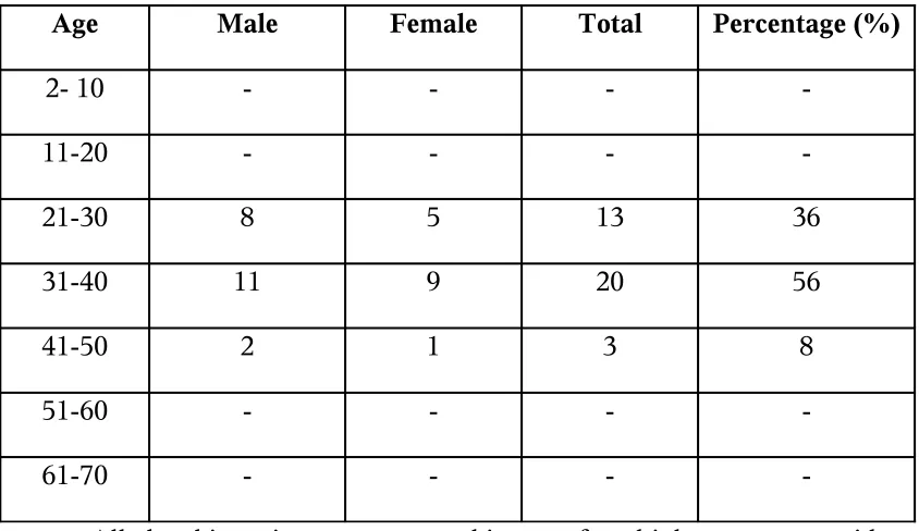
WARTS IN HIV PATIENTS
Among the three hundred HIV + cases in the various spectrum of HIV disease screened in the Well Health clinic thirty six patients had warts. Genital warts were the commonest observed in twenty two cases followed by verruca vulgaris which occurred in eight cases. Perianal wart was seen in five patients and palmoplantar warts in one patient. The commonest age group affected was 25 – 40 age groups. The number of males was twenty one (58%) and the number of females was fifteen (42%).
[image:54.612.51.473.378.622.2]The age and sex distribution is shown in Table - 7
TABLE - 7
AGE AND DISTRIBUTION – WARTS IN HIV PATIENTS
Age Male Female Total Percentage (%)
2- 10 - - -
-11-20 - - -
-21-30 8 5 13 36
31-40 11 9 20 56
41-50 2 1 3 8
51-60 - - -
-61-70 - - -
cases was non reactive.
A male patient presented with a wide spread numerous, confluent verrucae vulgaris involving the face, upper and lower extremities, trunk and palms with characteristic periungual warts also seen. (Fig 27,28,29 )
A female patient presented with a large exuberant cauliflower like plaques of anogenital condyloma that spread to the groins (Fig 30) and histopathological examination showing vacuolated cells in the granular and spinous layer, which were big and contained round hyperchromatic nuclei (Fig 31).
WARTS IN RENAL TRANSPLANT` PATIENTS
Among the three hundred patients screened in renal transplant OP six patients had warts. Verrucae vulgaris was the most common, seen in four cases followed by plane wart, digitate warts, genital wart, one each. All of the six allograft recipients were males of age between 24 to 62 years. All had transplants from related donors. The duration of follow up was 16 to 172 months. All the patients received kidneys from living donors and were kept on immunosupression with daily doses of predisolone, azathioprine and cyclosporine respectively. All the six patients gave a history of sun exposure.
A Twenty eight year old male patient on follow up for 24 monthson immunosuppressants presented with filliform type of genital wart. (Fig 33).
A sixty two year old male patient on follow up for 172 months with immunosuppressants presented with numerous verrucae vulgaris , plane wart and digitate warts in the dorsal aspect of both forearms (Fig 34). There was also associated seborrhoeic keratosis. Histopathological examination was classical of verrucae with no dysplastic changes. (Fig 35).
Other associated dermatological conditions noted were pityriasis versicolor, steroid induced acne, dermatophytosis, mucocutaneous candidiasis and pyogenic bacterial infections.
DISCUSSION
1. VERRUCA VULGARIS
This was the most common type of wart encountered in this study. The most common age group affected was the 16 - 20 age group, a physically active age group. Verruca vulgaris could occur at any age from child hood to old age, from a five-year- old male to sixty four- year-old-male in this study.
Males were more commonly affected than females because of a greater like hood of occupational trauma and environmental exposure. History of trauma was elicited more in men than in women; this may be because the population studied belonged to lower economic class and the men were probably involved in manual work and therefore had a greater chance of trauma. Though warts are highly infectious, a history of contact with persons having similar lesions was elicited in only thirty one patients. This could indicate the presence of sub clinical or latent infections with papilloma virus in the general population, as observed in other studies7.
The occurrence of similar lesions in the past in some of the cases could indicate incomplete resolution of the older lesions or reinfection in the same patient. The common sites affected were the extremities, which indicate trauma, minor or major as the main route of the entry of the virus. It could indicate autoinoculation from the same patient.
Association with porokeratosis, acromegaly, verrucae arising from chickenpox scars, and with halo phenomenon were noted in this study. Immuno compromised patients whether due to disease or drugs showed a profusion of warts33. The histopathological findings
correlated well with that of the morphological features.
These were the second most common type of wart encountered in this study. The most common age group was the 16 - 25 years physically active group like common warts. History of trauma in one fourth of cases suggest trauma as a source or site of infection. The virions may be present in the environment having been discharged from an infected foot, and may spread to new persons through moist skin, a fact which is well documented7. Past history of
similar lesions may suggest an incomplete resolution of a deeper component or a reinfection.
The most common sites involved were pressure points, which are common sites of wear and tear or trauma. Some of the dermatoses found along with plantar warts like contact dermatitis and eczema do not have any relation; hence the association is probably coincidental.
3. ANOGENITAL WART
Was the third most common type of lesion encountered in this study. All the cases were in the age range 16 - 25 years, a sexually active age group. Majority of the patients had sexual intercourse with known and unknown individuals. Other sexually transmitted diseases like trichomoniasis and candidiasis were also present; this was also seen in several studies42.
The common sites of occurrence of the lesions in the male patient were common sites of coital friction and therefore trauma, namely coronal sulcus and glans penis. The morphology of the lesions was classical and biopsy showed typical histopathological features.
4. VERRUCA PLANA