Copyrightq1997, American Society for Microbiology
Characterization of ICP6::
lacZ
Insertion Mutants of the UL15
Gene of Herpes Simplex Virus Type 1 Reveals the Translation
of Two Proteins
DONG YU,1AMY K. SHEAFFER,2DANIEL J. TENNEY,2ANDSANDRA K. WELLER1*
Department of Microbiology, University of Connecticut Health Center, Farmington, Connecticut 06030,1and
Virology Department, Bristol-Myers Squibb, Wallingford, Connecticut 064922
Received 22 October 1996/Accepted 20 December 1996
The herpes simplex virus type 1 (HSV-1) UL15 gene is a spliced gene composed of two exons and is predicted to encode an 81-kDa protein of 735 amino acids (aa). Two UL15 gene products with molecular masses of 75 and 35 kDa have been observed (J. Baines, A. Poon, J. Rovnak, and B. Roizman, J. Virol. 68:8118–8124, 1994); however, it is not clear whether the smaller form represents a proteolytic cleavage product of the larger form or whether it is separately translated. In addition, an HSV-1 temperature-sensitive mutant in the UL15 gene (ts66.4) is defective in both cleavage of viral DNA concatemers into unit-length monomers and packaging of viral DNA into capsids (A. Poon and B. Roizman, J. Virol. 67:4497–4503, 1993; J. Baines et al., J. Virol. 68:8118–8124, 1994). In this study, we detected two UL15 gene products of 81 and 30 kDa in HSV-1-infected cells, using a polyclonal antibody raised against a maltose binding protein fusion construct containing UL15 exon 2. In addition, we report the isolation of two HSV-1 insertion mutants,hr81-1 andhr81-2, which contain an ICP6::lacZ insertion in UL15 exon 1 and exon 2 and thus would be predicted to encode C-terminally truncated peptides of 153 and 509 aa long, respectively.hr81-1 andhr81-2 are defective in DNA cleavage and packaging and accumulate only B capsids. However, both mutants are able to undergo wild-type levels of DNA replication and genomic inversion, suggesting that genomic inversion is a result of DNA replication rather than of DNA cleavage and packaging. We also provide evidence that the 81- and 30-kDa proteins are the products of separate in-frame translation events from the UL15 gene and that the 81-kDa full-length UL15 protein is required for DNA cleavage and packaging.
Most double-stranded DNA (dsDNA) bacteriophages and animal viruses share common features in DNA replication as well as DNA maturation. DNA replication in many dsDNA bacteriophage systems results in the formation of a large head-to-tail concatemeric complex composed of tandem repeats of the viral genome, which is subsequently cleaved into unit-length monomers and packaged into preformed capsid precur-sors, with a concomitant loss of scaffolding proteins (7, 8, 10, 13). Both biochemical and genetic approaches have been ex-ploited extensively for studies of dsDNA bacteriophage sys-tems. Many protein components of the DNA cleavage and packaging machinery have been identified (11, 25, 27). All DNA cleavage and packaging systems identified to date con-tain a two-subunit terminase complex which binds and cleaves viral DNA, a translocase activity which is responsible for trans-location of DNA into the capsid, with the consumption of ATP, and a scaffolding-protein-containing prohead in which portal proteins form a dodecameric ring at a portal vertex through which DNA is taken in (reviewed in reference 10).
Herpes simplex virus type 1 (HSV-1) contains a 152-kb dsDNA genome composed of two covalently linked unique
regions (ULand US), flanked by inverted repeats (45, 50) (Fig.
1). During DNA replication, the two unique regions ULand
US invert with respect to each other, presumably through a
generalized recombination mechanism within the repeated re-gions of the genome, generating four isomeric forms of prog-eny viruses (14, 28, 47, 50). DNA replication is thought to result in the formation of large head-to-tail DNA concatemers
(9, 29, 30), which are cleaved at specific sites within the a
sequences into unit-length monomeric genomes and packaged into preassembled capsids, with loss of scaffolding proteins (18, 38, 54, 56).
Three types of intracellular capsids have been identified in HSV-1-infected cells by sucrose gradient sedimentation: A (empty), B (intermediate), and C (full) (16, 20, 40, 46). A and C capsids have similar protein compositions, but only C capsids contain viral DNA. B capsids were initially thought to be anal-ogous to phage proheads in that B capsids contain a scaffolding protein (designated VP22a, encoded by the UL26.5 gene) which is lost from capsids when DNA is packaged. However, in a cell-free capsid assembly system, Newcomb and colleagues (39) recently found a spherical, unstable capsid intermediate with a protein composition similar to that of B capsids. They proposed that these less angular and more open structures rather than B capsids are authentic procapsid intermediates (39). Although B capsids may be a dead-end product of the capsid maturation process, they represent the most closely related stable structures to procapsids that can be isolated. Thus, procapsids are thought to be precursors to DNA-con-taining C capsids, and empty A capsids are thought to result from abortive attempts at DNA encapsidation (39, 43). Tem-perature-sensitive mutants whose mutations map within six HSV-1 genes, UL6, UL15, UL25, UL28, UL32, and UL33, are defective in DNA cleavage and packaging and in production of A and C capsids (1–3, 6, 44, 51, 52, 58). The involvement of at least six genes suggests that HSV DNA cleavage and packaging is a complex process.
The UL15 gene composed of two exons is one of only a few spliced genes in the HSV-1 genome and is predicted to encode a 735-amino-acid (aa) protein (15, 19). UL15 is highly con-served among the herpesvirus family, and sequence analysis
* Corresponding author. Phone: (860) 2310. Fax: (860) 679-1239. E-mail: [email protected].
2656
on November 9, 2019 by guest
http://jvi.asm.org/
indicates a possible homology between the UL15 protein of HSV-1 and the large subunit (gp17) of the terminase complex of bacteriophage T4 (17). The most significant aspect of this homology is the presence of a putative nucleotide binding fold (Walker boxes A and B) in both proteins (19, 57), suggesting that UL15 may be able to bind and hydrolyze ATP as has been demonstrated for gp17 and other phage terminases (5, 10, 12). A temperature-sensitive mutant (ts66.4) bearing a lesion in the second exon of the UL15 gene has been isolated and shown to be defective in DNA cleavage and packaging (6, 44). How-ever, temperature-sensitive mutants are prone to problems associated with incomplete penetrance (leak), tendency to re-vert to wild type, and potential transdominant interference of wild-type protein function by the presence of a quasi-stable yet nonfunctional mutant protein at the nonpermissive tempera-ture. In this report, we describe the isolation and
characteriza-tion of UL15 insercharacteriza-tional mutants in which an ICP6::lacZ
cas-sette is inserted into either the first or the second exon of the UL15 gene. We show that although two UL15 mutants are able to undergo wild-type levels of DNA replication and genomic inversion, they are defective in DNA cleavage and packaging. Furthermore, we find that two proteins with molecular masses of 81 and 30 kDa are translated separately from the UL15 gene and that the 81-kDa full-length UL15 protein is required for DNA cleavage and packaging.
MATERIALS AND METHODS
Cells and viruses.African green monkey kidney cells (Vero; American Type Culture Collection, Rockville, Md.) were propagated and maintained as de-scribed previously (59). Cell lines M-3, C-2, and G-27, which are permissive for UL15 mutants (see below), were propagated as described above, but with the addition of 100mg of the antibiotic G418 (geneticin, a neomycin analog; GIBCO Laboratories, Grand Island, N.Y.) per ml of medium. The KOS strain of HSV-1 was used as the wild-type virus as well as the parental virus for the isolation of UL15 insertion mutantshr81-1 andhr81-2. A temperature-sensitive mutant
de-fective in the UL15 gene at the nonpermissive temperature (39.58C),ts66.4, a kind gift from B. Roizman (University of Chicago), was propagated at 348C and used to screen candidate UL15-permissive cell lines at 39.58C.
Plasmids and bacteria.Plasmid pUC-H3UL15G was made by cloning the
HindIII J fragment of HSV-1 KOS strain (nucleotides [nt] 28038 to 39849) (37) into theHindIII site of the vector pUC119. ThisHindIII J fragment contains the 59-terminal portion of the UL13, UL14, the first exon of UL15, UL16, UL17, the second exon of UL15, UL18, and the promoterless 39-terminal part of UL19 genes.
UL15E1LacZS, containing the disrupted first exon of UL15, and pUC-UL15E2LacZS, containing the disrupted second exon of UL15, were constructed as follows. pUC-UL15E1 contains theSacI/SphI fragment spanning the first exon of UL15 cloned intoSacI/SphI sites of the pUC119 vector. pUC-UL15E1 was partially digested withXmnI, and the linearized plasmid was gel purified. The 4.3-kbBamHI fragment containing thelacZgene under the control of the ICP6 promoter (ICP6::lacZcassette) (23) was filled in with Klenow enzyme and ligated to the linearized plasmid pUC-UL15E1. The resultant construct, which contains the ICP6::lacZcassette inserted at theXmnI site (corresponding to amino acid residue 153) within the first exon of UL15, was designated pUC-UL15E1LacZS. TheSacI/SphI fragment containing the second exon of UL15 was ligated to the
SacI/SphI sites of pUC119 to generate pUC-UL15E2. pUC-UL15E2LacZS was made by inserting theBamHI fragment of ICP6::lacZcassette into theBamHI site of pUC-UL15E2 at a position corresponding to amino acid residue 509 of UL15. In both pUC-UL15E1LacZS and pUC-UL15E2LacZS, the transcrip-tional orientation of thelacZgene was same as that of the UL15 gene.
Plasmids pT7ApoUL15G, containing the UL15 genomic DNA, and pT7ApoUL15C, containing the UL15 cDNA, were constructed as follows. Plas-mid pUC-H3UL15G was partially digested withApoI. TheApoI fragment con-taining the UL15 locus (nt 28680 to 35009 [37]) was isolated and subcloned into theEcoRI site of pBluescriptIISK1(Stratagene, La Jolla, Calif.) to generate pT7ApoUL15G. To construct the UL15 cDNA, the splice junction of UL15 exons 1 and 2 (15) was generated by PCR, using HSV-1 KOS-infected (13 h, multiplicity of infection [MOI] of 3) Vero cell RNA. Poly(A)1RNA was iso-lated, and 2.5mg was used in reverse transcription-PCR, using commercial kits as described by the manufacturer (Fast Track and cDNA Cycle kits; Invitrogen, San Diego, Calif.). PCR primer oligonucleotides were 59-CTCGTTTCCGGAC GGGTCGC-39 in exon 1 and 59-GGCCTCGTCGACAAAGAGCAGG-39 in exon 2, corresponding to nt 29991 to 30010 and 33679 to 33658, respectively. These oligonucleotides contained theBspEI (nt 29992) andSalI (nt 33688) sites spanning the UL15 intron. The 91-nt BspEI/SalI-digested PCR product was cloned into theBspEI/SalI sites to replace the intron in pT7ApoUL15G; the resultant plasmid, pT7ApoUL15C, contains only the UL15 cDNA. Nucleotide FIG. 1. Physical map of the region of the HSV-1 genome containing the UL15 gene. The HSV-1 genome and map coordinates are shown on the first two lines. On the next line are shown the locations of the terminalBamHI fragments (S and Q) and the junctionBamHI fragment (SQ) detected by theBamHI SQ probe. On the next two lines, theHindIII J andApoI fragments spanning the UL15 open reading frame have been expanded to show internal cleavage sites: H,HindIII; A,ApoI; F,AflII; X,XmnI; P,BspEI; S,SalI; B,BamHI; M,MluI. The genomic organization of the two exons of the UL15 gene is diagrammed on the next line. UL15E1 and UL15E2 represent the first exon and the second exon of UL15, respectively. The last two lines show the positions of the ICP6::lacZinsertion inhr81-1 andhr81-2, respectively. Arrows represent the transcriptional orientation of each gene.
on November 9, 2019 by guest
http://jvi.asm.org/
sequence analysis of the PCR product confirmed the sequence as reported previously (37).
Plasmids pCMV-UL15C and pCMV-UL15G, which contain the UL15 cDNA or genomic DNA under the control of the constitutive cytomegalovirus imme-diate-early (CMV-IE) promoter, were constructed as follows. Either pT7ApoUL15C or pT7ApoUL15G was digested withHindIII/XbaI, and a gel-purified 2.8-kb fragment containing the UL15 cDNA from pT7ApoUL15C or a 6.4-kb fragment containing the UL15 genomic DNA from pT7ApoUL15G was ligated into theHindIII/XbaI sites of the expression vector pCDNAI/Amp, con-taining the CMV-IE promoter (Invitrogen), to construct pCMV-UL15C or pCMV-UL15G, respectively.
All recombinant plasmids were propagated inEscherichia coliDH5aby stan-dard procedures (34).
Isolation of UL15 cell lines.To isolate cell lines which are permissive for UL15 mutants, 1.53106freshly trypsinized exponentially growing Vero cells were
cotransfected with 2 mg of pSV2neo and 18 mg of either pT7ApoUL15G, pT7ApoUL15C, or pUC-H3UL15G as described previously (22, 24) and grown in medium containing 1 mg of G418 per ml. The G418 concentration was decreased to 0.5 mg/ml after 5 days. Two weeks after cotransfection, individual G418-resistant cells were picked and screened for the ability to support the growth ofts66.4 at the nonpermissive temperature (39.58C).
Construction of UL15 insertional mutants by marker transfer.Marker trans-fer experiments were carried out as described previously (21). In brief, either
XmnI/HindIII-linearized pUC-UL15E1LacZS or pUC-UL15E2LacZS plasmid and infectious KOS DNA were used in a molar ratio of 10:1 to transfect 1.53
106freshly trypsinized exponentially growing UL15-permissive cells. When
max-imum cytopathic effect was observed, progeny viruses were collected, titers were determined on M-3 cells, and 2,500 infectious viral particles were plated onto 50 100-mm-diameter plates containing an M-3 cell monolayer. The resultant plaques were stained with neutral red in the presence of the chromogenic sub-strate 5-bromo-4-chloro-3-indolyl-b-D-galactoside (X-Gal). The plaques with blue color were selected and plaque purified three times on M-3 cells before stocks were prepared.
Transient complementation assay.The transient complementation assay was carried out as described previously (36). In brief, 1.53106freshly trypsinized
exponentially growing Vero cells were transfected with 8mg of either pCDNAI/ Amp, pCMV-UL15C, or pCMV-UL15G. At 24 h posttransfection, cells were superinfected with eitherhr81-1 orhr81-2 at an MOI of 1 PFU per cell. At 24 h postinfection, progeny viruses were harvested and assayed on the M-3 cell line for total yield.
Generation of anti-UL15 (a-UL15) polyclonal antiserum.Rabbit polyclonal antiserum similar to that previously described (6) was generated against the UL15 exon 2 sequences. AHincII/XbaI fragment was isolated from subclone pT7ApoUL15G as described above and cloned into theStuI/XbaI sites of the pMal-C vector (New England Biolabs, Beverly, Mass.). This clone encoded the maltose binding protein (MBP) fused to the C-terminal 351 aa of UL15. Fusion protein was expressed and purified as instructed by the vector manufacturer. Five hundred micrograms of purified antigen was mixed with complete Freund’s adjuvant and inoculated intramuscularly into New Zealand White rabbits. Booster immunizations of antigen in incomplete Freund’s adjuvant were per-formed every 4 weeks for 10 months. Serum from one rabbit, AS9, was collected by standard protocols and used as described in Results.
Western blot analysis.To analyze infected cell lysates, 33106cells in a
100-mm-diameter tissue culture dish were infected with virus stocks at an MOI of 10 PFU per cell at 378C. Cells were collected at 14 h postinfection by centrifugation at 2,000 rpm for 10 min and rinsed with phosphate-buffered saline (137 mM NaCl, 2.7 mM KCl, 10 mM Na2HPO4, 1.8 mM KH2PO4[pH 7.4])
twice. Cell pellets were resuspended in 150ml of 13sodium dodecyl sulfate (SDS)-polyacrylamide gel electrophoresis (PAGE) loading buffer (0.05 mM Tris-HCl [pH 6.8], 2% SDS, 10% glycerol, 0.1% bromophenol blue, 1.25% [vol/vol] 2-mercaptoethanol). To analyze transfected cell lysates, 106freshly trypsinized
exponentially growing Vero cells were transfected with 8mg of expression plas-mids as described previously (21) and plated on a 60-mm-diameter tissue culture dish for 24 h, and cell lysates were prepared and resuspended in 50ml of 13
SDS-PAGE loading buffer as described above. Cell lysates in SDS-PAGE load-ing buffer were heated at 958C for 5 min and vortexed vigorously for 30 s, and 20-ml aliquots of samples were subjected to electrophoresis on an SDS–10% polyacrylamide gel. ECL (enhanced chemiluminescence) Western blotting anal-ysis was performed as instructed by the manufacturer (Amersham, Buckingham-shire, England). To detect the UL15 gene products, thea-UL15 rabbit poly-clonal antibody was used as the primary antibody at a dilution of 1:2,000 (in 5% nonfat milk in phosphate-buffered saline containing 0.2% Tween 20) to hybridize with the membrane at 48C overnight. Protein quantification analysis was per-formed as suggested by the manufacturer, using bands which were in the linear response range.
DNA isolation and analysis by Southern blotting and PFGE.Intact infectious viral genomic DNA used in marker transfer experiments was isolated as de-scribed previously (62). Total viral DNA and cellular DNA used in Southern blot analysis were isolated as described previously (61). For Southern blot analysis, cellular or viral DNA was digested with appropriate restriction endonucleases, subjected to conventional agarose gel electrophoresis or pulsed-field gel electro-phoresis (PFGE), blotted to Gene Screen Plus nylon membranes (New England
Nuclear Corp., Boston, Mass.), hybridized with32P-labeled specific probes, and
visualized by autoradiography as instructed by the manufacturer. DNA probes were prepared by labeling desired gel-isolated restriction fragments by using a Random Primed DNA Labeling kit (Boehringer Mannheim, Indianapolis, Ind.). Preparation and analysis of encapsidated viral DNA and PFGE were carried out as described previously (35, 49).
Analysis of viral capsids.HSV-1 capsids were isolated by using a modification of a method developed by Sherman and Bachenheimer (52). Cells were infected at an MOI of 10 PFU per cell at 378C. At 20 h postinfection, cells were collected by 4,000-rpm centrifugation at 48C for 15 min, rinsed with Tris-buffered saline, resuspended in phosphate buffer (40 mM sodium phosphate buffer [pH 7.4], 0.15 M NaCl, 1 mm EDTA, 1 mm EGTA, 1 mm phenylmethylsulfonyl fluoride, 5mg of pepstatin A per ml, 5mg of leupeptin per ml) with 1% Nonidet P-40, incubated on ice for 30 min, and probe sonicated for 30 s. The cell lysate was layered onto a 15 to 50% (wt/wt in phosphate buffer) sucrose gradient and centrifuged at 35,000 rpm for 30 min at 48C (with an SW50.1 or SW41 rotor). Capsids were visualized by light scattering upon illumination with a halogen fiber-optic lamp.
RESULTS
Isolation of UL15-containing permissive cell lines and UL15 mutants.Permissive cell lines containing the UL15 gene were isolated for the propagation of UL15 mutants. Vero cells were cotransfected with a plasmid conferring neomycin resistance (pSV2neo) and a plasmid containing either the UL15 cDNA (pT7ApoUL15C) or the UL15 genomic DNA (pT7ApoUL15G, containing the UL15, UL16, and UL17 genes, or
pUC-H3UL15G, containing the 59-terminal portion of the UL13,
UL14, UL15 exon 1, UL16, UL17, UL15 exon 2, UL18, and
the promoterless 39-terminal portion of the UL19 genes) (Fig.
1). G418-resistant cells were screened for the ability to support
the growth of the temperature-sensitive UL15 mutantts66.4 at
the nonpermissive temperature (39.58C). Ten such cell lines
were isolated, and Southern analysis revealed that the copy number of integrated UL15 genes per cell ranged from 20 to 500 (data not shown). Three cell lines were chosen for further study; M-3 cells contain the larger genomic fragment (pUC-H3UL15G), G-27 cells contain the smaller genomic fragment (pT7ApoUL15G), and C-2 cells contain the cDNA fragment (pT7ApoUL15C).
Viral mutants bearing the inactivated UL15 gene were con-structed by marker transfer. The UL15-complementing cell lines, M-3, G-27, and C-2, were cotransfected with infectious KOS DNA and a mutant UL15 gene-containing plasmid, ei-ther UL15E1LacZS (exon 1 insertion) or pUC-UL15E2LacZS (exon 2 insertion). Homologous recombination between KOS DNA and these mutant plasmids would be ex-pected to generate recombinant viruses which contain the
ICP6::lacZcassette inserted either in UL15 exon 1 (at residue
153) or in UL15 exon 2 (at residue 509). Progeny viruses were plated on M-3 cells, and blue plaques were isolated, purified, and propagated. Independent plaques resulting from marker transfer with UL15E1LacZS (hr81-1) and
pUC-UL15E2LacZS (hr81-2) were isolated. To confirm thathr81-1
andhr81-2 contain the ICP6::lacZcassette at the appropriate
positions, total DNA from the cells infected with KOS,hr81-1
orhr81-2 was digested withXhoI and subjected to Southern
blot hybridization using either the 32P-labeled EcoRI lacZ
fragment or the32P-labeled UL15 cDNAAflII/MluI fragment
as a probe as described in the legend to Fig. 2. ThelacZprobe
would be expected to detect a 5.7-kb fragment inhr81-1 and a
3.8-kb fragment inhr81-2 (Fig. 2A, lanes 3, 4, 7, and 8). When
the membrane was hybridized with the UL15 cDNA probe,
4.8-, 2.8-, and 2.5-kb XhoI fragments would be expected in
KOS DNA (Fig. 2B, lanes 2 and 6). However, since the
in-serted ICP6::lacZcassette contains oneXhoI recognition site,
hr81-1 would be expected to contain 5.7- and 1.4-kb fragments
instead of the 2.8-kb fragment, while hr81-2 was expected to
contain 3.8- and 2.9-kb fragments instead of the 2.5-kb
on November 9, 2019 by guest
http://jvi.asm.org/
ment present in KOS DNA (Fig. 2B, lanes 3, 4, 7, and 8). Figure 2 clearly demonstrates all of the expected bands, thus
indicating that the ICP6::lacZcassette was inserted into the
expected sites in bothhr81-1 andhr81-2.
Phenotypic analysis ofhr81-1 andhr81-2.To determine if
UL15 is essential for viral growth, hr81-1 and hr81-2 were
tested for plaque-forming ability on Vero, C-2, G-27, and M-3 cell lines. Both mutants were unable to form plaques on Vero cells but were able to do so on either C-2, G-27, or M-3 cells at similar efficiencies (Table 1). Since the C-2 cell line contains only the UL15 cDNA, this result indicates that growth defects
in hr81-1 and hr81-2 are within the UL15 gene and that the
UL15 gene is essential for viral growth. A transient
comple-mentation assay further confirms that growth defects inhr81-1
andhr81-2 were due to loss of UL15 gene function. Vero cells
were transfected with expression constructs in which either the UL15 cDNA (pCMV-UL15C) or the UL15 genome DNA (pCMV-UL15G) was under the control of a constitutive CMV-IE promoter; the transfected cells were superinfected with the UL15 mutant viruses. Table 2 shows that cells trans-fected with pCMV-UL15G and pCMV-UL15C could support
the growth of bothhr81-1 andhr81-2 whereas cells transfected
with the control plasmid (pCDNAI/Amp) could not. Taken
together, these results indicate that growth defects in hr81-1
andhr81-2 result from loss of the essential UL15 gene.
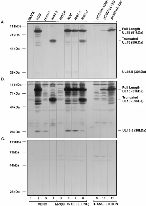
The abilities ofhr81-1 andhr81-2 to express UL15 proteins
were determined by Western blot analysis. Total cell extracts from infected cells were prepared and assessed for the
pres-ence of UL15 gene products by using an a-UL15 polyclonal
antibody raised against an MBP-UL15 exon 2 fusion protein as described in Materials and Methods. The following observa-tions were made. (i) In the KOS-infected Vero cell extract, there was a band of 81 kDa (Fig. 3A and B, lanes 2) which was
absent in bothhr81-1- andhr81-2-infected Vero cells (Fig. 3A
and B, lanes 3 and 4). The size of this band was consistent with the predicated molecular mass of 81 kDa for the full-length UL15 protein of 735 aa. When M-3 cells were infected with
eitherhr81-1 orhr81-2, the 81-kDa band was detected (Fig. 3A
and B, lanes 7 and 8), indicating that expression of the full-length UL15 protein in the cell line could be induced by viral infection. The 81-kDa band was also detected in the extract from Vero cells transfected with expression plasmid pCMV-UL15G or pCMV-UL15C (Fig. 3A and B, lanes 10 and 11), whereas this band was absent in the extract from Vero cells transfected with the control plasmid pCDNAI/Amp (Fig. 3A and B, lanes 9). Thus, these results confirm that the 81-kDa band is the authentic full-length product of the UL15 gene. (ii)
Sincehr81-2 contains the ICP6::lacZcassette inserted at amino
acid residue 509, it might be expected that a 59-kDa truncated derivative of the UL15 protein lacking its 226 C-terminal amino acid residues would still be expressed (Fig. 1). In fact, in hr81-2-infected cells, the predicted 59-kDa band was detected (Fig. 3A and B, lanes 4 and 8). The inability of the truncated UL15 protein to support viral growth indicates that the C terminus of the full-length UL15 protein is essential. Since the
ICP6::lacZcassette is inserted at aa 153 inhr81-1, it might be
[image:4.612.63.557.83.155.2]expected that the 153-aa C-terminally truncated UL15 protein with the predicted molecular mass of 20 kDa would be ex-pressed as well; however, this product would not be detected by the antibody which was raised against the second exon of
FIG. 2. The ICP6::lacZcassette is inserted at the expected positions in the viral genome. Vero and M-3 cells were infected with KOS,hr81-1, orhr81-2 at an MOI of 3 PFU per cell for 12 h at 378C. Total DNA was extracted, digested withXhoI, separated by conventional agarose electrophoresis, and blotted to a GeneScreen Plus membrane, which was hybridized with either the32P-labeled EcoRIlacZfragment (A) or the32P-labeledAflII/MluI UL15 cDNA fragment
(B). Lanes 1 to 4 represent DNA from infected Vero cells, and lanes 5 to 8 represent DNA from infected M-3 cells. Lanes 1 and 5, mock-infected cells; lanes 2 and 6, KOS-infected cells; lanes 3 and 7,hr81-1-infected cells; lanes 4 and 8,
hr81-2-infected cells. Fragment sizes are indicated.
TABLE 1. Plaquing efficiencies ofhr81-1 andhr81-2 on Vero, C-2, G-27, and M-3 cells
Cell Integrated UL15genea Copy no.
Titer (PFU/ml)
KOS hr81-1 hr81-2
Vero N/A NA 1.53108 ,53103 ,53103
C-2 cDNA 200 1.63108 2.23108 3.83108
G-27 Genomic DNAc 60 1.23108 1.23108 1.53108
M-3b Genomic DNAd 20 1.03108 1.93108 2.73108
aRefers to type of the UL15 gene integrated into the cell genome. bUsed to make virus stocks of the UL15 mutants.
[image:4.612.58.299.498.643.2]cApoI fragment containing UL15, UL16, and UL17 genes. dHindIII fragment containing UL13 to UL19 genes.
TABLE 2. Transient complementation ofhr81-1 andhr81-2 with UL15 expression plasmidsa
Plasmid CI
b
hr81-1 hr81-2
None 1 1
pCDNAI/Amp 0.5 0.7
pCMV-UL15C 670 125
pCMV-UL15G 230 45
aVero cells were transfected with the indicated plasmid and superinfected with eitherhr81-1 orhr81-2 as described in Materials and Methods. Progeny virus was titered on M-3 cells. This experiment was repeated three times, and similar results were obtained each time.
bComplementation index (CI) is calculated as PFU of progeny virus from cultures transfected with the indicated plasmid/PFU of progeny virus from mock-transfected cultures.
on November 9, 2019 by guest
http://jvi.asm.org/
[image:4.612.315.556.598.672.2]UL15. (iii) Thea-UL15 antibody specifically recognized a 30-kDa protein in KOS-infected cells (Fig. 3B, lanes 2 and 6), which was consistent with the observation by Baines et al. that UL15 encodes two proteins with molecular masses of 75 and 35
kDa (6). However, more interestingly, we found that hr81-1
was still able to express the 30-kDa protein whereas hr81-2
failed to express either the 81- or 30-kDa protein (Fig. 3B, lanes 3 and 4). Both mutants as well as KOS were able to express the 30-kDa UL15 protein on M-3 cells (Fig. 3B, lanes 6 to 8). Quantification of the 30-kDa protein bands indicated
that the levels of 30-kDa protein expressed inhr81-1-infected
Vero cells were comparable with those expressed by wild-type virus-infected Vero cells (data not shown). Furthermore, the
absence of the 30-kDa protein inhr81-2-infected cells was not
the result of lower overall expression from the UL15 gene
since the 59-kDa truncated UL15 protein was still expressed at significant levels (Fig. 3A and B, lane 4). This observation has two implications. First, the presence of the 30-kDa version and the absence of the 81-kDa version of the UL15 protein in hr81-1-infected cells indicates that the 30-kDa protein is not likely to be a proteolytic breakdown product of the 81-kDa protein; it may be translated from an internal in-frame methi-onine downstream of residue 153 of UL15 where the
ICP6::lacZcassette is inserted (see Discussion). Second, the
30-kDa version of UL15 is unable to functionally substitute for the 81-kDa full-length UL15 protein, based on the growth
phenotype of hr81-1. Figure 3C shows that the preimmune
serum failed to detect either the 81- or 30-kDa band in infected or transfected cell extracts, confirming that both bands
de-tected by thea-UL15 antibody are authentic UL15 gene
prod-ucts rather than nonspecific cross-reacting proteins. Although the origin of the 30-kDa protein is not clear, we tentatively designate it UL15.5 (see Discussion). In summary, we conclude that the full-length UL15 protein is essential for viral growth and that its functions cannot be substituted by the 59-kDa
C-terminally truncated UL15 protein expressed by hr81-2 or
the 30-kDa UL15.5 protein expressed byhr81-1.
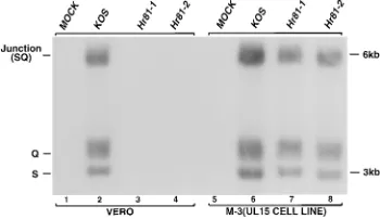
hr81-1 andhr81-2 are defective in DNA cleavage while re-taining the ability to synthesize viral DNA.The temperature-sensitive mutant of UL15 (ts66.4) is defective in DNA cleavage and packaging at the nonpermissive temperature (6, 44).
hr81-1- orhr81-2-infected Vero cells exhibit wild-type levels of
viral DNA synthesis, as assessed by dot blot hybridization (data not shown). To confirm that UL15 is required for the viral
DNA cleavage, cells infected withhr81-1 orhr81-2 were
exam-ined for the presence of free genomic termini. Total DNA
from Vero or M-3 cells infected with KOS, hr81-1, orhr81-2
was digested withBamHI and subjected to Southern blot
hy-bridization with aBamHI SQ-specific probe as described in the
legend to Fig. 4. This probe specifically hybridizes to both
terminalBamHI S and Q fragments as well as junctionBamHI
SQ fragments. The levels of SQ junction fragments present in
hr81-1- andhr81-2-infected cells are similar to those in
KOS-infected cells (Fig. 4, lanes 2 to 4), confirming that the UL15 mutants were capable of wild-type levels of DNA replication. However, S and Q terminal fragments were absent in both
hr81-1- and hr81-2-infected Vero cells, while these terminal
fragments were readily detectable in KOS-infected Vero cells
as well as inhr81-1- orhr81-2-infected M-3 cells (Fig. 4, lanes
[image:5.612.57.298.72.412.2]2 and 6 to 8), indicating that UL15 mutant viruses were unable
FIG. 3. Expression of UL15 gene products in infected and transfected cells. Vero and M-3 cells were infected with the indicated virus or transiently trans-fected with UL15 expression constructs. Lysates of 43105cells were prepared,
resolved by SDS-PAGE, and immunoblotted by using an ECL protocol as de-scribed in Materials and Methods. (A) One-minute exposure using thea-UL15 polyclonal antibody as the primary antibody; (B) 20-min exposure using the
a-UL15 antibody as the primary antibody (the densities of the 30-kDa bands in this panel were within the linear response range and thus could be quantified); (C) 20-min exposure using preimmune serum as the primary antibody. Lanes 1 to 4 represent infected Vero cells, lanes 5 to 8 represent infected M-3 cells, and lanes 9 to 11 represent transfected Vero cells. Lanes 1 and 5, mock-infected cells; lanes 2 and 6, KOS-infected cells; lanes 3 and 7,hr81-1-infected cells; lanes 4 and 8,hr81-2-infected cells. Lanes 9 to 11 are Vero cells transfected with control plasmid pCDNAI/Amp and UL15 expression plasmids pCMV-UL15G and pCMV-UL15C, respectively. The positions of the full-length UL15 protein, the truncated UL15 protein expressed inhr81-2-infected cells, and the UL15.5 pro-tein are indicated.
FIG. 4. UL15 mutants are defective in viral DNA cleavage. Vero or M-3 cells were infected with the indicated virus at an MOI of 3 PFU per cell at 378C for 12 h. Five micrograms of total DNA extracts was prepared from infected cells, digested withBamHI, and analyzed by Southern blot hybridization as described in the legend to Fig. 1, using the32P-labeledBamHI SQ junction fragment (Fig.
1) as a probe. Lanes 1 to 4 represent infected Vero cells, and lanes 5 to 8 represent infected M-3 cells. Lanes 1 and 5, mock-infected cells; lanes 2 and 6, KOS-infected cells; lanes 3 and 7,hr81-1-infected cells; lanes 4 and 8,hr 81-2-infected cells. The positions of junction (SQ) and terminal (S and Q) fragments are indicated.
on November 9, 2019 by guest
http://jvi.asm.org/
to cleave viral DNA concatemers into monomers to generate free viral genomic termini under nonpermissive conditions.
The DNA cleavage defects inhr81-1 andhr81-2 were
con-firmed by analyzing the structure of viral DNA which accumu-lates in KOS- and UL15 mutant-infected cells. Viral DNA replication results in the accumulation of concatemeric DNA intermediates, which are subsequently cleaved into mature mo-nomeric genomes. PFGE of DNA from HSV-infected cells results in the separation of viral DNA into two bands, one which does not enter the gel and represents replicating DNA intermediates (well DNA) and one which migrates as a 152-kb genome-length DNA (35, 48, 60). Total DNA from Vero or
M-3 cells infected with either KOS, hr81-1, or hr81-2 was
subjected to PFGE and subsequently analyzed by Southern blot using the UL15 cDNA fragment as a probe. DNA from infected Vero cells as well as UL15 mutant- or KOS-infected M-3 cells contains both replicating well DNA and monomeric DNA (Fig. 5, lanes 2 and 6 to 8). However, DNA
fromhr81-1 andhr81-2-infected Vero cells contains only well
DNA (Fig. 5, lanes 3 and 4). The absence of 152-kb
genome-length viral DNA in hr81-1- and hr81-2-infected Vero cells
indicates that inactivation of the UL15 gene results in the failure to cleave DNA concatemers into mature monomeric genomes. The comparable amounts of viral DNA detected in
KOS-,hr81-1-, andhr81-2-infected Vero cells (Fig. 5, lanes 2 to
4) is again in agreement with the conclusion that UL15 is not required for viral DNA synthesis.
hr81-1 andhr81-2 are defective in DNA packaging.To assess
the ability ofhr81-1 andhr81-2 to encapsidate DNA, DNase
I-resistant DNA from infected cells was measured. DNase I treatment would be expected to degrade all DNA in infected cells except for encapsidated viral DNA protected by capsids. DNase I-protected DNA from Vero and M-3 cells infected with KOS or UL15 mutants was prepared as described in the legend to Fig. 6 and analyzed by Southern blot using the
32P-labeledBamHI SQ fragment as a probe. Figure 6 shows
that DNase I-resistant S and Q terminal fragments were ob-served in KOS-infected Vero cells and in KOS- or UL15 mu-tant-infected M-3 cells, indicating that the viral monomeric DNA was encapsidated efficiently (Fig. 6, lanes 2 and 6 to 8).
In contrast, DNA fromhr81-1 andhr81-2-infected Vero cells
was completely degraded upon DNase I treatment (Fig. 6, lanes 3 and 4), suggesting that DNA packaging is defective in these two UL15 mutants. In summary, these results suggest
that the UL15 gene is essential for both cleavage and packag-ing of viral DNA.
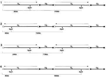
hr81-1 and hr81-2 are capable of DNA inversion.During
viral DNA replication, the two unique sequences (ULand US)
invert relative to one another, resulting in four types of viral monomeric isomers (28). It has been suggested that genomic inversion, a result of a recombination event between the
in-verted repeats flanking the UL and US sequences, may be
stimulated by the presence of free termini generated by DNA cleavage and packaging (53). Therefore, we decided to assess genomic inversion in UL15 mutants incapable of DNA cleav-age and packaging to see whether the terminal ends generated by this mechanism are essential for inversion. The inversion event can be assessed by examining the restriction pattern of replicating well DNA. For instance, as diagrammed in Fig. 7,
digestion with SpeI, which cleaves once per monomer unit,
would be expected to release bands of 118, 152, and 186 kb from the replicating intermediate had the inversion event oc-curred. Vero cells were infected with UL15 mutant viruses for
various times, and well DNA was isolated, digested withSpeI,
[image:6.612.103.253.68.175.2]and analyzed by Southern blotting as described in the legend to
Fig. 8. Using the32P-labeledBamHI SQ fragment as a probe,
the predicted 118-, 152-, and 186-kb bands were detected in
KOS-infected as well as in hr81-1- and hr81-2-infected Vero
cells as early as at 6 h postinfection (Fig. 8, lanes 4 to 6). At each of the times tested (from 3 to 15 h), the levels of three
isomer bands inhr81-1- andhr81-2-infected cells were
compa-rable to those in KOS-infected cells and increased with time. These results clearly indicate that DNA inversion occurs in UL15 mutants as efficiently as in the wild-type virus. Lamberti and Weller demonstrated that in a mutant virus defective in UL6, another gene required for DNA cleavage and packaging, DNA inversion occurs normally as well (32). Taken together, these results indicate that recombination and genomic inver-sion is likely linked to DNA replication rather than to DNA cleavage and packaging, which is in agreement with the model proposed by Sarisky and Weber (47).
In this experiment, well DNA was also analyzed for the presence of free termini. It has been reported that wild-type well DNA contains only one type of free genomic termini
corresponding to the UL portion of the HSV-1 genome (35,
[image:6.612.348.523.519.620.2]60). Furthermore, the ULtermini can be released as either an
FIG. 5. UL15 mutants fail to process concatemeric replicating DNA to mo-nomeric viral genomes. Vero or M-3 cells were infected with the indicated virus at an MOI of 5 PFU per cell at 378C. At 18 h postinfection, 7.53105infected
cells were suspended in low-melting-point agarose gel blocks, lysed in situ, subjected to PFGE as described previously (35), and analyzed by Southern blot hybridization using the32P-labeledAflII/MluI UL15 cDNA fragment as a probe.
Lanes 1 to 4 represent infected Vero cells, and lanes 5 to 8 represent infected M-3 cells. Lanes 1 and 5, mock-infected cells; lanes 2 and 6, KOS-infected cells; lanes 3 and 7,hr81-1-infected cells; lanes 4 and 8,hr81-2-infected cells. The positions of well (concatemeric) and 152-kb (monomeric) DNAs are indicated.
FIG. 6. UL15 mutants fail to encapsidate viral DNA. Vero and M-3 cells were infected with the indicated virus at an MOI of 3 PFU per cell at 378C for 12 h. Cells were lysed and treated with 50mg of DNase I per ml for 2 h. Samples were resuspended in 0.6% SDS–10 mM EDTA–10 mM Tris (pH 7.4)–100mg of proteinase K per ml for 4 h to inactivate DNase I and to remove protein. Samples were subsequently incubated with 100mg of RNase per ml for 30 min to remove RNA. All enzyme treatments were carried out at 378C. After phenol extraction, 5 mg of DNase I-resistant DNA was digested withBamHI and analyzed by Southern blot hybridization as described in the legend to Fig. 5. Lanes 1 to 4 represent infected Vero cells, and lanes 5 to 8 represent infected M-3 cells. Lanes 1 and 5, mock-infected cells; lanes 2 and 6, KOS-infected cells; lanes 3 and 7,
hr81-1-infected cells; lanes 4 and 8,hr81-2-infected cells. The positions ofBamHI junction (SQ) and terminal (S and Q) fragments are indicated.
on November 9, 2019 by guest
http://jvi.asm.org/
80-kb or a 46-kb band uponSpeI digestion, depending on the
orientation of the terminal ULarm as diagrammed in Fig. 7,
and these two bands were found to hybridize with a UL
termi-nus-specific probe but not with a US-specific probe in Southern
blot analysis (32, 60). In Fig. 8, two extra bands, corresponding
to the 80- and 46-kb SpeI UL-terminal fragments, were
de-tected in theSpeI-treated wild-type well DNA as early as at 9 h
postinfection as reported previously, while they were absent in
the SpeI-treated well DNA from either hr81-1- or
hr81-2-in-fected cells at all times tested. The presence of ULtermini in
wild-type replicating DNA led Zhang et al. to propose that these free termini may result either from the initiation and termination of DNA replication or from the
cleavage/packag-ing process (60). The absence of these two bands in the
SpeI-treated well DNA from hr81-1- or hr81-2-infected cells
re-ported in this paper, together with the observation by Lamberti
and Weller that ULtermini were also absent inSpeI-treated
well DNA from the UL6 mutant (32), suggests that these free
ULtermini present in the replicating concatemeric DNA
in-termediates are products of DNA cleavage and packaging rather than DNA replication.
Analysis of capsid formation inhr81-1 andhr81-2. Herpes-virus-infected cells contain three types of capsid structures (A, B, and C), as identified by sucrose gradient velocity centrifu-gation. It was reported that only B-type capsids were found in Vero cells infected with mutants defective in cleavage and packaging (1–3, 32, 42, 52, 55). The absence of A- and C-type capsids suggests that DNA packaging is not even attempted. In contrast, infection of Vero cells with a mutant defective for viral alkaline nuclease (UL12) results in the appearance of B capsids, an increased abundance of A capsids, and few C cap-sids. This result suggests that packaging was attempted but was either interrupted or aborted (49). In this study, lysates of Vero
cells infected with KOS,hr81-1, orhr81-2 were subjected to 15
to 50% sucrose gradient centrifugation as described in Mate-rials and Methods. In KOS-infected Vero cells, all three capsid bands, A, B, and C, were clearly observed (Fig. 9, lane 1).
However, in bothhr81-1- andhr81-2-infected cells, only B-type
capsids were seen (Fig. 9, lanes 2 and 3). The absence of A and
C capsids in hr81-1- and hr81-2-infected Vero cells suggests
[image:7.612.127.492.67.330.2]that, unlike the UL12 mutant, in which DNA encapsidation is attempted but aborted, both UL15 mutants were defective at
FIG. 7. Schematic diagram of replicating HSV-1 DNA showing expected fragments released bySpeI digestion. Four different relative orientations of neighboring ULarms due to genomic inversion (lines 1 to 4) give rise to three discrete isomeric forms of the linear HSV-1 genome with sizes of 118, 152, and 186 kb uponSpeI
digestion. The expected 46-kb (lanes 2 and 4) and 80-kb (lanes 1 and 3) ULterminal fragments released bySpeI digestion are also indicated, depending on the
orientation of the terminal ULarm. Hypothetical USterminal fragments of 72 or 106 kb released bySpeI digestion are not shown.
FIG. 8. UL15 mutants retain the ability to undergo DNA inversion. A total of 7.53105cells were infected with the indicated virus at an MOI of 5 PFU per
cell at 378C for 3 h (lanes 1 to 3), 6 h (lanes 4 to 6), 9 h (lanes 7 to 9), 12 h (lanes 10 to 12), and 15 h (lanes 13 to 15). Well DNA was twice purified away from monomeric DNA as described previously (35), digested withSpeI in situ, sub-jected to PFGE, and analyzed by Southern blot hybridization using the32
P-labeledBamHI SQ junction fragment as a probe. Lanes 1, 4, 7, 10, and 13, KOS-infected cells; lanes 2, 5, 8, 11, and 14,hr81-1-infected cells; lanes 3, 6, 9, 12, and 15,hr81-2-infected cells. Bands corresponding to positions of well, 46-kb, 80-kb, 118-kb, 152-kb, and 186-kb DNAs are indicated.
on November 9, 2019 by guest
http://jvi.asm.org/
an earlier stage, in which DNA encapsidation has not even been attempted.
DISCUSSION
In this study, permissive cells were constructed in order to
isolate and propagate HSV-1 mutantshr81-1 andhr81-2
con-taining the disrupted UL15 gene in which the ICP6::lacZ
mu-tagenic cassette was inserted in exon 1 and exon 2, respectively.
hr81-1 and hr81-2 can grow and form plaques on
UL15-per-missive cells but not on Vero cells, confirming that UL15 is an essential gene for viral growth. Both mutants are defective in DNA cleavage and packaging, and they accumulate only B-type capsids although they still retain the ability to synthesize wild-type levels of DNA. Only cells containing the UL15 gene either stably as in permissive cell lines or transiently as in the transient complementation assay are able to support the
growth ofhr81-1 andhr81-2, indicating that the phenotypes of
UL15 mutant viruses are due to loss of UL15 function. Our results are consistent with those obtained from studies of the
UL15 temperature-sensitive mutantts66.4 (6, 44).
The wild-type virus synthesizes two proteins with molecular masses of 81 and 30 kDa, detected by Western blot analysis using a polyclonal antibody raised against the UL15 exon 2 sequence. These results confirm those of Baines et al. for assays using an independently isolated UL15 antibody (6). Furthermore, a C-terminally truncated derivative of the UL15
protein expressed inhr81-2-infected cells was not able to
sup-port viral growth, indicating that the C-terminal 226 aa are
essential for UL15 function. Interestingly,hr81-1 was able to
synthesize the 30-kDa protein but not the 81-kDa UL15 pro-tein. This result indicates that the 30-kDa protein is a separate translation product rather than a degradation product of the
full-length 81-kDa UL15 protein. Analysis of the UL15 protein sequence reveals five internal methionines situated at amino acid residues 370, 418, 443, 460, and 500, which, if used for initiation, would encode proteins with predicted molecular masses of 40, 34, 32, 30, and 25 kDa, respectively; methionine residues 370 and 443 appear to be in a more optimal context for translation than the other three (31). Costa et al. have identified a 2.7-kb mRNA in the UL15 region which presum-ably encodes the 81-kDa protein; however, two smaller mRNAs which may have been generated either from indepen-dent promoters or by alternative splicing events were observed (15). It is possible that one of these smaller mRNAs encodes the 30-kDa protein; alternatively, the protein may be trans-lated from the 2.7-kb mRNA, using an internal methionine as a start codon. In this paper, we have tentatively designated this 30-kDa protein as UL15.5.
Although our results indicate that UL15 is essential for viral growth and for DNA cleavage and packaging, its precise role in these processes is still unknown. It has been speculated that the UL15 protein may have a terminase function, based on its homology with the large subunit of T4 terminase complex (gp17) (17). For most dsDNA bacteriophages, the terminase is composed of two subunits, a 10- to 25-kDa low-molecular-mass protein and a 60- to 75-kDa high-molecular-mass protein, for instance, gp16 and gp17 of T4, gp18 and gp19 of T3, and gpA and gpNu1 of lambda. Large subunits of the terminase usually possess prohead binding and DNA-dependent ATPase activi-ties while the small subunits exhibit sequence-specific DNA binding activities (10, 26). Furthermore, in these dsDNA phages, genes for two subunits are often situated adjacent to each other in the genome. In the HSV-1 UL15 gene region, all five possible start codons for translation of UL15.5 are situated in UL15 exon 2 and downstream of the potential ATP binding site (GKT [amino acid residues 263 to 265] and DE [amino acid residues 356 to 357]), suggesting that the UL15.5 protein may not have ATP binding activity. It is possible that the UL15.5 protein acts as the small subunit of the terminase. Alternatively, it is possible that another viral protein such as one of the other cleavage and packaging proteins performs this function or that the subunit structure of the HSV terminase is unlike that of the dsDNA bacteriophages.
Since DNA cleavage and packaging are biologically linked with capsid formation, it is expected that some DNA cleavage/ packaging proteins may interact with capsids in vivo. It has been reported that UL6 is present stably in all three types of capsids and mature virion particles and that UL25 is associated with purified HSV-1 virions as well (4, 33, 41). Preliminary immunoblot analysis reveals that the full-length 81-kDa UL15 protein expressed in KOS-infected cells is associated with B capsids (data not shown). Thus, at least three minor-capsid proteins, UL6, UL15, and UL25, have also been implicated in cleavage and packaging, although their roles are not clear. In dsDNA bacteriophage systems, terminase has been found to interact with proheads. The finding of UL15 in B capsids is consistent with its putative function as the HSV-1 terminase. In addition, by analogy to dsDNA bacteriophage systems, if there is a unique portal vertex, it is intriguing to speculate that some of the minor-capsid components may be part of this structure. Alternatively, herpesviruses may utilize a different DNA pack-aging mechanism which does not require the presence of a unique portal.
The reagents generated in this study will facilitate the fur-ther functional analysis of the UL15 gene. The availability of
UL15-permissive cell lines and ICP6::lacZinsertional mutants
[image:8.612.112.240.69.305.2]which are capable of expressing blue color in the presence of X-Gal will facilitate the isolation of viruses bearing subtle
FIG. 9. UL15 mutants fail to make A and C capsids. A total of 1.53107
Vero cells were infected with KOS,hr81-1, orhr81-2 at an MOI of 10 PFU per cell at 378C. At 20 h postinfection, the cells were lysed in 1% Nonidet P40-containing lysis buffer by sonication and subjected to 15 to 50% sucrose gradient centrifugation as described in Materials and Methods. Gradients 1, 2, and 3 represent the capsid profiles of lysates from KOS-,hr81-1-, andhr81-2-infected Vero cells, respectively. The positions of A, B, and C capsid bands of wild-type HSV-1 are indicated.
on November 9, 2019 by guest
http://jvi.asm.org/
site-specific mutations. The null background of UL15 inser-tional mutants will allow us to develop biochemical assays in vitro and to carry out in vivo complementation assays for the detailed assessment of the functions of individual domains within the UL15 gene.
ACKNOWLEDGMENTS
We are grateful to all members of our laboratories for critical dis-cussion and review of the manuscript. We thank Bernard Roizman for providing the UL15 temperature-sensitive mutantts66.4 and antisera against the UL15 exon 2 sequences, Min Gao (Bristol-Myers Squibb) for providing the HSV-1 KOSHindIII J-fragment clone, and, espe-cially, Todd Wilson for purifying MBP-UL15 fusion protein for gen-eration of antisera.
This work was supported by National Institute of Health grant AI 37549.
REFERENCES
1.Addison, C., F. J. Rixon, J. W. Palfreyman, M. O’Hara, and V. G. Preston. 1984. Characterisation of a herpes simplex virus type 1 mutant which has a temperature-sensitive defect in penetration of cells and assembly of capsids. Virology138:246–259.
2.Addison, C., F. J. Rixon, and V. G. Preston.1990. Herpes simplex virus type 1 UL28 gene product is important for the formation of mature capsids. J. Gen. Virol.71:2377–2384.
3.Al-Kobaisi, M. F., F. J. Rixon, I. McDougall, and V. G. Preston.1991. The herpes simplex virus UL33 gene product is required for the assembly of full capsids. Virology180:380–388.
4.Ali, M. A., B. Forghani, and E. M. Cantin.1996. Characterization of an essential HSV-1 protein encoded by the UL25 gene reported to be involved in virus penetration and capsid assembly. Virology216:278–283.
5.Backhaus, H.1985. DNA packaging initiation ofSalmonellabacteriophage P22: determination of cut sites within the DNA sequence coding for gene 3. J. Virol.55:458–465.
6.Baines, J. D., A. P. W. Poon, J. Rovnak, and B. Roizman.1994. The herpes simplex virus 1 UL15 gene encodes two proteins and is required for cleavage
of genomic viral DNA. J. Virol.68:8118–8124.
7.Bazinet, C., and J. King.1985. The DNA translocating vertex of DSDNA bacteriophage. Annu. Rev. Microbiol.39:109–129.
8.Becker, A., and H. Murialdo.1990. Bacteriophage lambda DNA: the begin-ning of the end. J. Bacteriol.172:2819–2824.
9.Ben-Porat, T., and F. J. Rixon.1979. Replication of herpesvirus DNA. IV. Analysis of concatemers. Virology94:61–70.
10. Black, L.1989. DNA packaging in dsDNA bacteriophages. Annu. Rev. Microbiol.43:267–292.
11. Black, L. W., A. L. Zachary, and V. Manne.1981. Studies of the mechanism of bacteriophage T4 DNA encapsidation, p. 111–126.InM. DuBow (ed.), Bacteriophage assembly. Alan R. Liss, Inc., New York, N.Y.
12. Casjens, S., W. M. Huang, M. Hay, and R. Parr.1987. Initiation of bacte-riophage P22 DNA packaging sequence analysis of a mutant which alters DNA target specificity of the packaging apparatus. J. Mol. Biol.194:411–420. 13. Casjens, S. R.1985. Nucleic acid packaging by viruses, p. 75–147.InS. R. Casjens (ed.), Virus structure and assembly. Jones and Bartlett, Portola Valley, Calif.
14. Chou, J., and B. Roizman.1985. Isomerization of herpes simplex virus 1 genome: identification of the cis-acting and recombination sites within the domain of the a sequence. Cell41:803–811.
15. Costa, R. H., K. G. Draper, T. J. Kelly, and E. K. Wagner.1985. An unusual spliced herpes simplex virus type 1 transcript with sequence homology to Epstein-Barr virus DNA. J. Virol.54:317–328.
16. Dargan, D. J.1986. The structure and assembly of herpesviruses, p. 359–437.
InJ. Harris and R. Horne (ed.), Electron microscopy of proteins, vol. 5. Academic Press Inc., London, England.
17. Davison, A. J.1992. Channel catfish virus: a new type of herpesvirus. Virol-ogy186:9–14.
18. Deiss, L. P., J. Chou, and N. Frenkel.1986. Functional domains within the
a sequence involved in the cleavage-packaging of herpes simplex virus DNA. J. Virol.59:605–618.
19. Dolan, A., M. Arbuckle, and D. J. McGeoch.1991. Sequence analysis of the splice junction in the transcript of herpes simplex virus type 1 gene UL15. Virus Res.20:97–104.
20. Gibson, W., and B. Roizman.1972. Proteins specified by herpes simplex virus. VIII. Characterization and composition of multiple capsid forms of subtypes 1 and 2. J. Virol.10:1044–1052.
21. Goldstein, D. J., and S. K. Weller.1988a. Factor(s) present in herpes simplex virus type 1-infected cells can compensate for the loss of the large subunit of the viral ribonucleotide reductase: characterization of an ICP6 deletion mutant. Virology166:41–51.
22. Goldstein, D. J., and S. K. Weller.1988b. Herpes simplex virus type 1-in-duced ribonucleotide reductase activity is dispensable for virus growth and DNA synthesis: isolation and characterization of an ICP6::lacZinsertion mutant. J. Virol.62:196–205.
23. Goldstein, D. J., and S. K. Weller.1988c. An ICP6::lacZinsertional mutagen is used to demonstrate that the UL52 gene of herpes simplex virus type 1 is required for virus growth and DNA synthesis. J. Virol.62:2970–2977. 24. Graham, F. L., and A. J. van der Eb.1973. A new technique for the assay of
infectivity of human adenovirus 5 DNA. Virology52:456–467.
25. Guo, P., S. Grimes, and D. Anderson.1986. A defined system for in vitro packaging of DNA-gp3 of the Bacillus subtilis bacteriophage phi29. Proc. Natl. Acad. Sci. USA83:3505–3509.
26. Guo, P., and M. Trottier.1994. Biological and biochemical properties of the small viral RNA (pRNA) essential for the packaging of the dsDNA of phage phi29. Semin. Virol.5:27–37.
27. Hamada, K., H. Fujisawa, and T. Minagawa.1986. A defined in vitro system for packaging of bacteriophage T3 DNA. Virology151:119–123.
28. Hayward, G. S., R. J. Jacob, S. C. Wadsworth, and B. Roizman.1975. Anatomy of herpes simplex virus DNA: evidence for four populations of molecules that differ in the relative orientations of their long and short components. Proc. Natl. Acad. Sci. USA72:4243–4247.
29. Hirsch, I., G. Cabral, M. Patterson, and N. Biswal.1977. Studies on intra-cellular replicating DNA of herpes simplex virus type 1. Virology81:48–61. 30. Jacob, R. J., L. S. Morse, and B. Roizman.1979. Anatomy of herpes simplex virus DNA. XII. Accumulation of head-to-tail concatemers in the nuclei of infected cells and their role in the generation of four isomeric arrangements of viral DNA. J. Virol.29:448–457.
31. Kozak, M.1987. At least six nucleotides preceding the AUG initiator codon enhance translation in mammalian cells. J. Mol. Biol.196:947–950. 32. Lamberti, C., and S. K. Weller.1996. The herpes simplex virus type 1 UL6
protein is essential for cleavage and packaging but not for genomic inversion. Virology226:403–407.
33. Lamberti, C., and S. K. Weller.1996. Unpublished results.
34. Maniatis, T., E. F. Fritsch, and J. Sambrook.1982. Molecular cloning: a laboratory manual. Cold Spring Harbor Laboratory, Cold Spring Harbor, N.Y.
35. Martinez, R., R. Sarisky, P. Weber, and S. K. Weller.1996. Herpes simplex virus type 1 alkaline nuclease is required for efficient processing of viral DNA replication intermediates. J. Virol.70:2075–2085.
36. Martinez, R., L. Shao, and S. K. Weller.1992. The conserved helicase motifs of the herpes simplex virus type 1 origin-binding protein UL9 are important for function. J. Virol.66:6735–6746.
37. McGeoch, D. J., M. A. Dalrymple, A. J. Davison, A. Dolan, M. C. Frame, D. McNab, L. J. Perry, J. E. Scott, and P. Taylor.1988. The complete DNA sequence of the long unique region in the genome of herpes simplex virus type 1. J. Gen. Virol.69:1531–1574.
38. Nasseri, M., and E. S. Mocarski.1988. The cleavage recognition signal is contained within sequences surrounding an a-a junction in herpes simplex virus DNA. Virology167:25–30.
39. Newcomb, W. W., F. L. Homa, D. R. Thomsen, F. P. Booy, B. L. Trus, A. C. Steven, J. V. Spencer, and J. C. Brown.1996. Assembly of the herpes simplex virus capsids: characterization of intermediates observed during cell-free capsid formation. J. Mol. Biol.263:431–446.
40. Newcomb, W. W., B. L. Trus, F. P. Booy, A. C. Steven, J. S. Wall, and J. C. Brown.1993. Structure of the herpes simplex virus capsid: molecular com-position of the pentons and the triplexes. J. Mol. Biol.232:499–511. 41. Patel, A. H., and J. B. Maclean.1995. The product of the UL6 gene of herpes
simplex virus type 1 is associated with virus capsids. Virology206:465–478. 42. Patel, A. H., F. J. Rixon, C. Cunningham, and A. J. Davison.1996. Isolation and characterization of herpes simplex virus type-1 mutant defective in the UL6 gene. Virology217:111–123.
43. Perdue, M. L., J. C. Cohen, C. C. Randall, and D. J. O’Callaghan.1976. Biochemical studies of the maturation of herpesvirus nucleocapsid species. Virology74:194–208.
44. Poon, A. P., and B. Roizman.1993. Characterization of a temperature-sensitive mutant of the UL15 open reading frame of herpes simplex virus 1. J. Virol.67:4497–4503.
45. Roizman, B.1979. The structure and isomerization of herpes simplex virus genomes. Cell16:481–494.
46. Roizman, B., and A. E. Sears.1990. Herpes simplex viruses and their repli-cation, p. 1796–1841.InB. N. Fields and D. M. Knipe (ed.), Virology, vol. 2. Raven Press, New York, N.Y.
47. Sarisky, R. T., and P. C. Weber.1994. Requirement for double-strand breaks but not for specific DNA sequences in herpes simplex virus type 1 genome isomerization events. J. Virol.68:34–47.
48. Severini, A., A. R. Morgan, D. R. Tovell, and L. J. Tyrrel.1994. Study of the structure of replicative intermediates of HSV-1 DNA by pulsed-field gel electrophoresis. Virology200:428–435.
49. Shao, L., L. M. Rapp, and S. K. Weller.1993. Herpes simplex virus 1 alkaline nuclease is required for efficient egress of capsids from the nucleus. Virology 196:146–162.
50. Sheldrick, P., and N. Berthelot.1975. Inverted repetitions in the
on November 9, 2019 by guest
http://jvi.asm.org/
some of herpes simplex virus. Cold Spring Harbor Symp. Quant. Biol.2:667– 678.
51. Sherman, G., and S. Bachenheimer.1987. DNA processing in temperature-sensitive morphogenic mutants of HSV-1. Virology158:427–430. 52. Sherman, G., and S. L. Bachenheimer.1988. Characterization of
intranu-clear capsids made by ts morphogenic mutants of HSV-1. Virology163:471– 480.
53. Smiley, J. R., J. Duncan, and M. Howes.1990. Sequence requirements for DNA rearrangements induced by the terminal repeat of herpes simplex virus type 1 KOS DNA. J. Virol.64:5036–5050.
54. Stow, N. D., E. C. McMonagle, and A. J. Davison.1983. Fragments from both termini of the herpes simplex virus type 1 genome contain signals required for the encapsidation of viral DNA. Nucleic Acids Res.11:8205–8220. 55. Tengelsen, L. A., N. E. Pederson, P. R. Shaver, M. W. Wathen, and F. L.
Homa.1993. Herpes simplex virus type 1 DNA cleavage and encapsidation require the product of the UL28 gene: isolation and characterization of two UL28 deletion mutants. J. Virol.67:3470–3480.
56. Vlazny, D. A., A. Kwong, and N. Frenkel.1982. Site-specific cleavage/pack-aging of herpes simplex virus DNA and the selective maturation of nucleo-capsids containing full-length viral DNA. Proc. Natl. Acad. Sci. USA79: 1423–1427.
57. Walker, J. E., M. Saraste, M. J. Runswick, and N. J. Gay.1982. Distantly
related sequences in the a andb-subunits of ATP synthase, myosin, kinases and other ATP-requiring enzymes and a common nucleotide binding fold. EMBO J.1:945–951.
58. Weller, S. K., E. P. Carmichael, D. P. Aschman, D. J. Goldstein, and P. A. Schaffer.1987. Genetic and phenotypic characterization of mutants in four essential genes that map to the left half of HSV-1 UL DNA. Virology 161:198–210.
59. Weller, S. K., K. J. Lee, D. J. Sabourin, and P. A. Schaffer.1983. Genetic analysis of temperature-sensitive mutants which define the gene for the major herpes simplex virus type 1 DNA-binding protein. J. Virol.45:354– 366.
60. Zhang, X., S. Efstathiou, and A. Simmons.1994. Identification of novel herpes simplex virus replicative intermediates by field inversion gel electro-phoresis: implications for viral DNA amplification strategies. Virology202: 530–539.
61. Zhu, L., and S. K. Weller.1992. The UL5 gene of the herpes simplex virus type 1: isolation of alacZinsertion mutant and association of the UL5 gene product with other members of the helicase-primase complex. J. Virol.66: 458–468.
62. Zhu, L., and S. K. Weller.1988. UL5, a protein required for HSV DNA synthesis: genetic analysis, overexpression in Escherichia coli, and generation of polyclonal antibodies. Virology166:366–378.