Copyright © 1999, American Society for Microbiology. All Rights Reserved.
V3 Recombinants Indicate a Central Role for CCR5 as a
Coreceptor in Tissue Infection by Human
Immunodeficiency Virus Type 1
STEPHEN Y. CHAN,
1,2ROBERTO F. SPECK,
1CHRISTOPHER POWER,
3SARAH L. GAFFEN,
1BRUCE CHESEBRO,
4ANDMARK A. GOLDSMITH
1,2*
Gladstone Institute of Virology and Immunology
1and Department of Medicine, School of Medicine, University of
California San Francisco,
2San Francisco, California; Department of Clinical Neurosciences, University of Calgary,
Calgary, Alberta, Canada
3; and Laboratory of Persistent Viral Diseases, Rocky Mountain Laboratories,
National Institute of Allergy and Infectious Diseases, Hamilton, Montana
4Received 6 July 1998/Accepted 4 November 1998
Binding of the human immunodeficiency virus type 1 (HIV-1) envelope glycoprotein gp120 to both CD4 and
one of several chemokine receptors (coreceptors) permits entry of virus into target cells. Infection of tissues
may establish latent viral reservoirs as well as cause direct pathologic effects that manifest as clinical disease
such as HIV-associated dementia. We sought to identify the critical coreceptors recognized by HIV-1
tissue-derived strains as well as to correlate these coreceptor preferences with site of infection and dementia
diagnosis. To reconstitute coreceptor use, we cloned HIV-1 envelope V3 sequences encoding the primary
determinants of coreceptor specificity from 13 brain-derived and 6 colon-derived viruses into an isogenic
(NL4-3) viral background. All V3 recombinants utilized the chemokine receptor CCR5 uniformly and efficiently
as a coreceptor but not CXCR4, BOB/GPR15, or Bonzo/STRL33. Other receptors such as CCR3, CCR8, and
US28 were inefficiently and variably used as coreceptors by various envelopes. CCR5 without CD4 present did
not allow for detectable infection by any of the tested recombinants. In contrast to the pathogenic switch in
coreceptor specificity frequently observed in comparisons of blood-derived viruses early after HIV-1
serocon-version and after onset of AIDS, the characteristics of these V3 recombinants suggest that CCR5 is a primary
coreceptor for brain- and colon-derived viruses regardless of tissue source or diagnosis of dementia. Therefore,
tissue infection may not depend significantly on viral envelope quasispeciation to broaden coreceptor range but
rather selects for CCR5 use throughout disease progression.
Entry into target cells by human immunodeficiency virus
type 1 (HIV-1) depends critically on binding of the viral
enve-lope glycoprotein (gp120) to both CD4 and a cellular
corecep-tor (31). Recently, both definitive and putative corecepcorecep-tors
have been identified as members of the G-protein-coupled
chemokine receptor family that confer onto cells susceptibility
to infection by various isolates of HIV-1.
HIV-1 coreceptor utilization is the principal determinant of
cellular tropism. While macrophage-tropic viruses
characteris-tically employ the
b
-chemokine receptor CCR5 (3, 15, 21, 29,
30), T-cell line-tropic viruses use the
a
-chemokine receptor
CXCR4 (38). Changes in tropism and coreceptor specificity
correlate with progression of AIDS. Early after infection,
pri-mary viral isolates from the blood are homogeneous in
enve-lope sequence and are largely or exclusively CCR5 using or
macrophage-tropic (18, 85, 110, 111). As AIDS develops,
ap-proximately 50% of individuals experience a switch in cellular
tropism to a more heterogeneous population in the blood that
carries CXCR4-using or T-cell line-tropic viruses (18, 98–100).
The importance of CCR5 in mediating HIV-1 infection was
established by the natural occurrence of the CCR5
D
32
loss-of-function mutation. Persons homozygous for CCR5
D
32 display
resistance to initial HIV-1 infection, while heterozygotes
dem-onstrate a slower progression to AIDS after seroconversion
(19, 45, 60, 77, 84). The contribution of CXCR4 to
pathogen-esis has also been highlighted by studies in various models of
HIV-1 immunodepletion (41, 71).
High levels of viral replication are associated with genetic
evolution in vivo. This allows for production of a range of
quasispecies with distinct envelopes that have been
hypothe-sized to use a broader range of coreceptors to infect a larger
number of host cell types (103). Accordingly, a number of
HIV-1 strains that can utilize alternate chemokine receptors in
addition to CCR5 and CXCR4 under various in vitro
condi-tions have been described. These receptors include CCR2b
(29), CCR3 (15, 43), CCR8 (50), BOB/GPR15 (22, 37), Bonzo/
STRL33 (22, 59), GPR1 (37), V28/CX
3CR1 (82), ChemR23
(83), leukotriene B4 receptor (69), Apj (14, 32), and human
cytomegalovirus (HCMV)-encoded US28 (72). However, the
significance of each alternate coreceptor in HIV-1 disease
re-mains undefined.
Previous work that explored coreceptor use and disease
pro-gression focused mainly on primary blood isolates (18). Viral
entry into tissues may also be a principal determinant of HIV-1
dissemination and pathogenesis (58), and studies have begun
to examine this issue (26, 88). Tissue infection may allow for
establishment of viral reservoirs that function as separate
rep-lication sites from blood. Viruses isolated from the central
nervous system (CNS) (1, 6, 10, 27, 35, 46, 55, 70, 76, 87, 107),
bowel (8), and other tissues (6, 27, 35, 48, 87, 112) possess
genetic and phenotypic differences compared to viruses
iso-lated from peripheral blood mononuclear cells. In addition,
various cell types that reside in tissues and express alternate
coreceptors may play critical roles in disease progression (5,
20, 36, 40, 53, 66, 81, 97, 102). It is unknown whether a separate
* Corresponding author. Mailing address: Gladstone Institute of
Virology and Immunology, P.O. Box 419100, San Francisco, CA
94110-9100. Phone: (415) 695-3775. Fax: (415) 695-1364. E-mail:
2350
on November 9, 2019 by guest
http://jvi.asm.org/
evolution of coreceptor use also occurs in viruses replicating in
tissues. Preliminary coreceptor specificity studies have also
im-plicated tissue-invasive strains as direct contributors to clinical
disease. Such a paradigm exists for the CNS and brain, where
neurotropic strains are hypothesized to infect specifically
mac-rophages, microglia (74, 96, 102), and other components of
neural tissue (5, 97). Neurovirulent strains evolving from these
may subsequently trigger the development of HIV-associated
dementia (HIVD) (51, 61, 62).
The V3 hypervariable region of the gp120 envelope protein
carries the main determinants of both cellular tropism (11, 12,
47, 86, 89, 105, 106) and coreceptor use (15, 16, 94, 108).
Transfer of a V3 loop from one virus into a distinct HIV-1
background typically retains the coreceptor profile of the V3
donor (15, 94). In addition, single amino acid changes in the
V3 loop can drastically alter coreceptor specificities (94). As a
result, V3 recombinants allow not only for mapping
determi-nants of tropism but also for a methodology of screening viral
coreceptor preferences. Although other regions of gp120 may
influence coreceptor recognition in some settings (13, 92),
rel-atively few unpassaged HIV-1 strains have been isolated from
tissues as molecular clones that carry full-length envelope
se-quences (26, 88).
Thus, the most feasible approach to screening in vivo
core-ceptor specificity is to use the V3 and flanking sequences
pre-viously identified by postmortem PCR analyses of brain and
colon tissue from AIDS patients (27, 76). Isolates from brain
tissue were chosen to identify selection pressures present in the
CNS viral reservoir (10, 54) that may affect development of
HIVD. Isolates from colon tissue were chosen for comparison
to explore the possible existence of an alternate viral reservoir
that imposes distinct selection pressures. Using an isogenic
proviral backbone, we constructed V3 recombinants that
car-ried the specific coreceptor usages of the original clinical
iso-lates. Subsequent functional analyses allowed us to identify the
biologically relevant coreceptors that mediate tissue infection
of the brain and colon and then correlate coreceptor specificity
with site of infection (brain versus colon) and the diagnosis of
HIVD.
MATERIALS AND METHODS
Cell lines. 293T (American Type Culture Collection, Rockville, Md.) and human osteosarcoma (GHOST) indicator (provided by D. Littman) cell lines were cultured in Dulbecco modified Eagle medium (Mediatech, Herndon, Va.) supplemented with 10% fetal bovine serum (FBS; Gemini Bio-Products, Cala-basas, Calif.) and 100mg of penicillin-streptomycin (Mediatech) per ml. The parental GHOST clone 34 stably maintaining human CD4 expression was under constant selection with G418 (500mg/ml; Life Technologies, Grand Island, N.Y.) and hygromycin B (100mg/ml; Boehringer Mannheim, Mannheim, Germany). GHOST transfectants stably expressing a specific coreceptor (CCR5, CXCR4, BOB/GPR15, or Bonzo/STRL33) were under additional selection with puromy-cin (1mg/ml; Sigma Chemical Co., St. Louis, Mo.). COS-7 cells (American Type Culture Collection) were routinely cultured in Iscove’s medium (Mediatech) supplemented with 10% FBS, 100mg of penicillin-streptomycin per ml, and 2 mML-glutamine (Mediatech). COS-7 transfections were carried out with Lipo-fectAMINE (Life Technologies) according to the manufacturer’s instructions. All infections were performed in Dulbecco modified Eagle medium–10% FBS– 100mg of penicillin-streptomycin per ml.
Plasmids and construction of recombinant proviruses. Molecular clones pYU-2, pNL4-3, and pNL-Luc-E2R2(17) (the NL4-3 provirus backbone with a
luciferase reporter gene insertion) were obtained from the AIDS Research and Reference Reagent Program, Division of AIDS, National Institute of Allergy and Infectious Diseases. Expression plasmids encoding CCR3 (pcDNA3-CCR3p), HCMV US28 (pRC/CMV-US28), ADA gp160 envelope (ADApEnv), CD4 (pCD4neo), and CCR5 (pCMVFCCR5) were previously described (4, 42, 72, 82, 105). The expression plasmid encoding CCR8 (pAW-CCR8F) was pro-vided by I. Charo.
Plasmids (p2-1, p10-1, p14-2, p15-2, p17-2, p19-1, p26-4, p26-5, p48-1, p54-1, p59-1, and p62-1) encoding chimeric NL4-3 provirus with a 428-bp insertion (StuI-NheI) that includes the V3 and portions of the C2 and C3 regions of brain-derived gp120 sequences were constructed as previously described (76).
Plasmid p4-14 (11) encoding NL4-3 provirus (with a 16-bp polylinkerMluI-NheI inserted in place of the 126-bp region coding portions of gp120 V3-C3 region) was used as the isogenic viral backbone for constructing additional V3 replace-ments based on published amino acid sequences (27, 74, 76). V3 nucleotide sequences were deduced by inserting degenerate codons most prevalent in the mammalian genome. An oligonucleotide pair representing the deduced V3 se-quence for brain-derived 5-1 (Fig. 1) along with deduced 59C3 sequence for NL4-3 was annealed as described by the manufacturer (Oligos, Etc., Wilsonville, Oreg.) and contained restriction site overhangs 59MluI-39NheI with unique sites
ApaI,SmaI, andXbaI inserted internally. The annealed oligonucleotides were cloned initially into a holding vector. After digestion of both p4-14 and the holding vector containing 5-1 withMluI andNheI and ligation of 5-1 insert with p4-14 backbone, the full-length chimeric provirus p5-1 was used as the parental vector for construction of other proviruses (p4-7, p4-8, p4-9, p6-2, p6-3, and p6-4) since it carried convenient restriction sites. Oligonucleotide pairs 4-7 and 4-8 were annealed (59SmaI-39XbaI), inserted into a holding vector, digested (SmaI/
XbaI), and ligated with the linearized p5-1 backbone (SmaI/XbaI) to make p4-7 and p4-8 proviruses. Oligonucleotide pairs 4-9, 6-2, 6-3, and 6-4 were annealed (59MluI-39XbaI), inserted into a holding vector, digested (MluI/XbaI), and ligated with the linearized p5-1 (MluI/XbaI) to make p4-9, p6-2, p6-3, and p6-4 proviruses. Inserted sequences were verified by ABI Prism Dye terminator cycle sequencing (Perkin-Elmer, Foster City, Calif.).
Preparation of virus stocks.To prepare experimental V3 recombinant viruses and full-length control viruses (YU-2 and NL4-3) for spreading infections, pro-viral plasmids were transfected into 293T cells by the CaPO4method (Promega,
Madison, Wis.) as previously described (42). Recombinant viral stocks were sterile filtered (0.45-mm-pore-size-filters) and harvested after both 36- and 60-h incubations. The p24Gagconcentration was assessed by enzyme-linked
immu-nosorbent assay (ELISA; New England Nuclear Life Sciences, Boston, Mass.), and stocks greater than 1,500 ng/ml were used for infections. Simian immuno-deficiency virus (SIV) strain Mac239 (550 ng of p27Gagper ml) (52) was provided
by R. Grant.
To prepare pseudotype virus carrying the luciferase gene (Luc1) with the
ADA envelope for single-round infections, pNL-Luc-E2R2(2 mg/well) was
cotransfected with ADApEnv (2mg/well) in 293T cells growing in six-well plates as previously described (22). Pseudotype Luc1viruses carrying the tissue-derived
V3 envelopes were similarly prepared by cotransfecting pNL-Luc-E2R2(3mg/
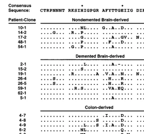
[image:2.612.313.550.71.292.2]well) along with full-length proviral plasmids described above (1 mg/well). FIG. 1. Three groups of V3 loop amino acid sequences, isolated by postmor-tem PCR from brain tissue and colon tissue of AIDS patients, show homology to a macrophage-tropic consensus sequence (11) deduced from blood-borne strains. A period denotes identity with the consensus, while a dash denotes deletion. Bullets denote V3 positions 13 and 25 in the consensus sequence. Sequences of colon-derived viruses and demented brain-derived virus (5-1) were isolated from three AIDS patients (27). Remaining brain-derived virus se-quences were isolated from 11 AIDS patients diagnosed with HIVD (76). Re-combinant virus 59-1 was previously termed 59-3. ReRe-combinant viruses 26-4 and 26-5 (previously termed 26-1 and 26-2, respectively) have identical V3 sequences as noted above but vary at gp120 position 335 in the C3 region, as described elsewhere (76).
V
OL. 73, 1999
HIV-1 CORECEPTOR USE IN TISSUE INFECTION
2351
on November 9, 2019 by guest
http://jvi.asm.org/
Pseudotype recombinant virus stocks greater than 110 ng/ml were used for infections.
Coreceptor utilization assay for spreading infection.GHOST cell lines ex-pressing CD4 alone or in combination with CCR5 or CXCR4 were plated in 96-well plates (7,000 cells/well) and infected as previously described (93). Cells were washed once with phosphate-buffered saline, and medium was replaced 12 h after infection. Day 0 and day 8 supernatant aliquots (10ml) were removed for secreted p24Gagmeasurements. Cell lines were infected with equivalent
inocula of recombinant virus normalized by utilization of CCR5 as a coreceptor (Fig. 2). The amount of recombinant virus added to different GHOST cell lines never varied for a given infection. Infection of GHOST cell lines stably express-ing CD4 with BOB or Bonzo was assessed as described above except that cells were plated (20,000 cells/well) in 12-well plates. The amount of added recombi-nant virus never varied between these cell lines for a given infection. To detect infection by SIV Mac239, secreted p27Gagconcentration was measured by
ELISA (Coulter, Miami, Fla.). For all infections, microscopic observations of syncytium formation were also recorded up to 14 days after infection.
Coreceptor utilization assay for single-round infection.COS-7 cells were plated in six-well plates and transfected with pCMVFCCR5 alone, pCD4neo alone, or pCD4neo in combination with pCMVFCCR5, pcDNA3-CCR3p, pAW-CCR8F, or pRC/CMV-US28 as previously described (4). After 24 h, cells were infected with pseudotype Luc1recombinant viruses. V3 recombinant viruses
were normalized by utilization of CCR5 as a coreceptor (Fig. 3A), and amounts of added virus never varied between cell lines in a specific transfection/infection experiment. After 48 h, luciferase expression was assessed by enzymatic activity (light units/milligram of protein) as previously described (65), as instructed by the manufacturer (Analytical Luminescence Laboratories, Sparks, Md.).
RESULTS
Envelope sequences from brain and colon tissue isolates of
HIV-1 inserted into the NL4-3 provirus.
Previous studies
dem-onstrated that V3 loop insertion into an alternate viral
back-bone such as NL4-3 can reconstitute the tropism (47, 106) and
coreceptor phenotype (15, 16, 94) of the V3 donor. For the
present study, we surveyed the literature for V3 loop amino
acid sequences derived from colon and brain samples of AIDS
patients for whom HIVD diagnosis was documented (27, 76).
To correlate coreceptor preferences with presence of HIVD,
we chose five sequences from five nondemented patients and
eight sequences from seven demented patients for analysis. To
correlate coreceptor preferences with site of infection, we
se-lected six colon sequences from two patients for comparison to
the brain-derived recombinants (Fig. 1).
All brain- and colon-derived V3 sequences showed
homol-ogy to a macrophage-tropic consensus sequence (76).
Further-more, all samples carried a moderately positive net V3 loop
charge (ranging from
1
2 to
1
6), in contrast to the highly
positive net charge commonly observed in CXCR4-using V3
loops (49). Of note, V3 loop position 13 appeared to favor
proline in nondemented subjects compared to a consensus
histidine in demented patients when a larger collection than
this listed set was considered (76). In addition, all
colon-de-rived viral sequences carried either an aspartate or a glutamine
residue at V3 loop position 25 rather than the consensus
glu-tamate. In the present study, DNA sequences representing the
selected V3 loops with adjacent conserved sequences (for
de-tails, see Materials and Methods) were inserted into the NL4-3
backbone. The resulting chimeric viruses were used to test
definitively the coreceptor utilization of tissue-specific HIV-1
isolates.
Efficient use of CCR5, but not of CXCR4, as coreceptors for
cellular entry.
Human osteosarcoma (GHOST) cell lines
sta-bly expressing human CD4/CCR5 or human CD4/CXCR4
were infected with each recombinant virus in vitro. Detection
of p24
Gagby ELISA was performed on the culture
superna-tants at days 0 and 8 after infection as a marker of spreading
infection. Since it demands multiple rounds of infection, such
an assay is most biologically pertinent and interpretable. All
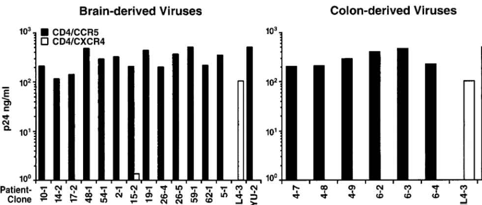
V3 recombinants representing brain-derived (nondemented
and demented) and colon-derived viruses displayed uniformly
high replication in GHOST CCR5/CD4 cells (Fig. 2). By day 8,
microscopic examination revealed syncytia in all cultures.
Con-versely, no brain- or colon-derived recombinant propagated
significant spreading infection in GHOST CXCR4/CD4 cells,
and no syncytia were observed at any time in the cultures (Fig.
2). The parental NL4-3 virus, known to employ CXCR4 as a
coreceptor, infected these cells robustly. Therefore, all of the
experimental recombinant viruses displayed efficient CCR5
co-receptor use, while none demonstrated detectable use of
CXCR4.
Nearly uniform failure to use either BOB/GPR15 or Bonzo/
STRL33 as an alternate coreceptor for cellular entry.
We
sought to determine if BOB/GPR15 or Bonzo/STRL33, two
FIG. 2. Efficient and uniform use of CCR5 but not CXCR4 as a coreceptor by all V3 recombinant viruses. HIV-1 YU-2 (CCR5 dependent) and NL4-3 (CXCR4 dependent) were used as controls. Displayed values are representative of day 8 supernatant p24 concentrations seen in two separate infections performed with these recombinants.
on November 9, 2019 by guest
http://jvi.asm.org/
[image:3.612.57.548.72.281.2]orphan chemokine receptor-like proteins identified as putative
HIV-1 coreceptors (22, 59), could be used for cellular entry.
We employed SIV Mac239 (52) as a positive control since it
uses both coreceptors more efficiently than some HIV-1 strains
(data not shown) and is able to establish more robust spreading
infections in culture (as detected in a p27 ELISA). The
super-natant p24 measurements of infection of GHOST cells stably
expressing CD4/BOB or CD4/Bonzo were compared to
Mac239 p27 levels after days 0 and 10. One brain-derived
recombinant (2-1) inefficiently employed BOB/GPR15 to
sus-tain spreading infection (4 ng of p24 per ml measured at day
10), but no syncytia were observed by day 14. In contrast, no
other V3 recombinant representing a brain-derived virus
(non-demented or (non-demented) or colon-derived virus detectably used
BOB/GPR15 or Bonzo/STRL33 for spreading infection (data
not shown).
Inefficient and variable use of CCR3, CCR8, and US28 as
alternate coreceptors for cellular entry.
In contrast to CCR5,
CXCR4, BOB/GPR15, and Bonzo/STRL33, the chemokine
receptors CCR3, CCR8, and HCMV US28 presented several
technical challenges to studying their role as coreceptors.
Im-portantly, our initial experiments revealed that HIV-1 strains
previously reported to recognize these alternate coreceptors
(15) may not utilize them efficiently for cellular entry. Thus, we
used an enzymatic assay in which COS-7 cells transiently
ex-pressing CD4 and a putative coreceptor were infected with
pseudotype viruses carrying the envelopes of interest and the
luciferase reporter gene (22). Viral infection was quantitated
by measuring entry of the pseudotype Luc
1recombinant
strains through readout of luciferase enzymatic activity. This
allowed for a sensitive assessment of coreceptor specificity that
was not achieved in a p24 spreading infection assay.
In corroboration of the results from the spreading infection
assay, all of the V3 recombinants representing the brain- and
colon-derived strains efficiently used CCR5 for cellular entry
(Fig. 3A), generating signals in the range of 10
6to 10
7light
units/mg of protein. Since several recent studies have identified
CD4-independent infection by a neurovirulent strain of SIV
(33) and select strains of HIV-2 (34, 79), viral infection in the
presence of CCR5 without CD4 was also assessed. None of the
brain- or colon-derived recombinants infected cells detectably
in the absence of CD4 expression (Fig. 3A).
Substantially lower and more variable luciferase signals were
observed in single-round infections of COS-7 cells expressing
CD4/CCR3 compared with CD4/CCR5 (Fig. 3B). Whereas all
recombinants used CCR5 as a coreceptor for entry, one (10-1)
did not detectably use CCR3, two (17-2, 2-1) slightly
recog-nized CCR3, while others (for example, 14-2, 54-1; 26-4, 62-1,
and 6-4) measurably used CCR3, albeit less efficiently than
CCR5. Of those that used CCR3, nearly all recombinants
generated luciferase counts less than 10
6light units/mg of
protein, which were typically 90 to 99% lower than comparable
readings seen in the CCR5-dependent infections. Importantly,
comparison of V3 recombinants representing brain-derived
vi-ruses from nondemented and demented patients revealed no
distinct pattern of CCR3 use, with both groups displaying a
spectrum of viral phenotypes with regard to CCR3. Similarly
variable phenotypes of CCR3 use were observed in the
colon-derived collection compared with the brain-specific set.
Inefficient and variable infections were also observed with
the putative coreceptor CCR8 after single-round infection
(Fig. 3C). While some brain-derived recombinants from the
nondemented (14-2 and 48-1) and demented (2-1, 26-4, 26-5,
62-1, and 5-1) groups exhibited modest infection, other
recom-binants from both brain collections (10-1, 17-2, 54-1, 15-2, 19-1,
and 59-1) showed little infection using CCR8 as a coreceptor.
Similarly, only one colon-derived recombinant (4-9) failed to
use CCR8, while the others recognized CCR8 to a slight
de-gree. Signals from the CCR8 infections were less than 10
5light
units/mg of protein and at least 90% lower than those for
CCR5 infections.
Similar results of low and variable infectivity were obtained
with HCMV US28 (Fig. 3D). Because it is encoded by HCMV
rather than the human host genome, US28 has been proposed
to enhance HIV-1 disease progression during HCMV
coinfec-tion commonly seen in AIDS patients (7). Inefficient infeccoinfec-tion
through US28 was observed with recombinants from certain
nondemented (14-2, 17-2, and 48-1) and demented (2-1, 26-4,
26-5, 62-1, and 5-1) patients, while other strains (10-1, 54-1,
15-2, 19-1, and 59-1) from both groups displayed no cellular
entry. Similar to the CCR8 profile, one colon-derived
recom-binant (4-9) did not use US28, while the rest of the set
recog-nized it to a small degree. Nearly all recombinant viral
infec-tions mediated by US28 exhibited luciferase readings less than
10
5light units/mg of protein and at least 90% lower than those
from CCR5.
Further comparison of the virus infection profiles revealed
no uniform correlation among particular coreceptor
specifici-ties in any recombinant collection. One recombinant (10-1)
exhibited virtually no use of any of the alternate coreceptors
tested, while others (14-2, 48-1, 2-1, 62-1, 5-1, 4-7, and 6-2)
used all three alternate coreceptors to some degree. Moreover,
recombinants such as 26-5 recognized CCR3 and CCR8 but
only slightly US28, while 17-2 recognized CCR3 and US28 but
not CCR8, and 4-9 recognized CCR3 but not CCR8 or US28.
Thus, these V3 recombinant viruses did not display a simple
and predictable pattern in their coreceptor specificity profiles.
Rather, they all demonstrated variable alternate coreceptor
patterns compared to their highly efficient and uniform
utili-zation of CCR5.
DISCUSSION
In this study, we sought to characterize coreceptor
prefer-ences among V3 recombinants representing brain- and
colon-derived HIV-1 isolates as well as to correlate HIVD diagnosis
and site of infection with these coreceptor recognition profiles.
We determined that all demented and nondemented
brain-specific recombinants and colon-brain-specific recombinants utilized
CCR5 efficiently but did not use CXCR4, BOB/GPR15, or
Bonzo/STRL33 detectably by spreading infection. In addition,
these recombinants recognized CCR3, CCR8, and US28
rela-tively inefficiently and variably as coreceptors as assayed by
sensitive single-round infection methods.
Incomplete eradication of HIV-1 after triple-drug therapy
has been attributed in part to the hypothesized separation and
relative protection of tissue reservoirs of latent virus from viral
populations residing in blood (54, 101, 107). Previous
compar-isons of V3 envelope sequences (9, 24, 27, 35, 48, 55, 56, 76, 78,
101) and, in some cases, cellular target specificities (26, 74, 88)
of unpassaged, tissue-derived viruses sought to define a
pat-tern(s) necessary for viral propagation in tissues different from
that observed in blood. In the present study, we examined V3
recombinant viruses carrying some sequence variability within
the tissue-specific V3 loops, particularly at positions 13 and 25
(see Results). Brain-derived HIV-1 strains such as those
rep-resented here are thought to replicate in vivo as an isolated
population, while colon-derived strains may not evolve
sepa-rately from blood-borne viruses, based on a recent
phyloge-netic analysis (101). Despite these differences, we discovered a
pattern of uniform CCR5 use that indicated a higher degree of
functional similarity among tissue-specific viruses than initially
V
OL. 73, 1999
HIV-1 CORECEPTOR USE IN TISSUE INFECTION
2353
on November 9, 2019 by guest
http://jvi.asm.org/
on November 9, 2019 by guest
http://jvi.asm.org/
suspected. Indeed, additional sequence comparisons of these
recombinants with the V3 loops of known CCR5-using strains
(64) revealed extensive similarities at other potentially critical
amino acid positions. Although we were unable to detect the
presence of a distinct viral reservoir present in colon tissue
based on a lack of differences in coreceptor specificity, the
uniformity of CCR5 utilization did indicate the probable
exis-tence of a common selective pressure in multiple tissues. These
results strengthen the claim that antigen-presenting cells, such
as microglia, macrophages, and follicular dendritic cells, which
commonly reside in tissues and express CD4 and CCR5, are
the principal cells that establish and maintain virus populations
in all tissues (23, 53, 74, 91, 95, 102).
The nearly absent usage of CXCR4, BOB/GPR15, and
Bonzo/STRL33 suggests the inability of these coreceptors to
mediate tissue infection despite their expression on certain
tissue-resident cell types (22, 57, 109). While no evidence has
directly demonstrated the in vivo importance of BOB/GPR15
and Bonzo/STRL33, the role of CXCR4 in pathogenesis has
been described for certain viruses residing in the blood (41,
71). From our results, it is reasonable to postulate that
uniden-tified pressures in the brain and colon select against viral use of
CXCR4, perhaps in part by the exclusion of infected
CXCR4-expressing T cells from trafficking into these tissues.
While most of the brain- and colon-derived recombinants
were found to recognize CCR3 as a coreceptor to a variable
degree, all could use CCR5 better than CCR3, with most
generating at least a 10-fold difference in infection levels.
Pre-viously, it was demonstrated that the level of HIV-1 infection
mediated by CCR3 varies with the level of coreceptor
expres-sion, and inefficient use of CCR3 (15, 82) was attributed to the
difficulty of maintaining high levels of expression in cell culture
(73). For our analyses, we used the best-reported expression
construct (pcDNA3-CCR3p) (82) and a sensitive enzymatic
readout to increase our ability to detect CCR3-dependent
en-try. Conceivably, the presence of at least some degree of CCR3
recognition in nearly all tested recombinants may point to a
positive selective pressure in the brain and colon in light of
recent evidence of high expression levels in vivo of CCR3 in
the CNS and lamina propria of the colon (109). Nonetheless,
the global efficiency of CCR5 utilization strongly highlights
CCR5 as a predominant coreceptor in mediating tissue-specific
infections.
Recently, it was reported that certain HIV-1 strains isolated
from brain-derived cells can use CCR8 as a coreceptor (50). In
addition, US28 has been implicated in HIV-1 pathogenesis
through HCMV opportunistic coinfections in various tissues
(39, 63, 67, 90). Our results indicated that neither CCR8 nor
HCMV US28 supports efficient infection by any of the
recom-binants tested. Furthermore, the variability in infection
effi-ciency among different recombinants suggested that no
consis-tent selection pattern is imposed on viruses based on CCR8 or
HCMV US28 use. Of note, all colon-derived recombinants
from patient 6 did display relatively uniform use of US28. It is
tempting to speculate that this coreceptor specificity may be
related to the presence of HCMV in the colon, perhaps
caus-ing the mucosal atrophy and inflammation observed in this
patient (28). However, in light of the rest of our results and the
fact that US28 is expressed only during the HCMV lytic growth
phase in vitro (104), it is unlikely that US28 has major in vivo
significance by itself as a coreceptor for infection of the brain
or colon.
We also sought to correlate coreceptor preferences with the
presence of HIVD (74, 76, 96). Evidence that both supports
and argues against the hypothesis that CCR3 is preferentially
recognized by neurotropic and/or neurovirulent viruses (43,
88) that may dictate dementia development has been
pre-sented. We observed uniform coreceptor recognition of CCR5
along with inefficient and variable use of alternate putative
coreceptors including CCR3 by all demented and
nonde-mented brain-derived V3 recombinants. Importantly,
compar-ison of coreceptor use by brain-derived recombinants with that
by presumably nonneurotropic colon-derived recombinants
yielded no significant difference in relative specificity profiles.
These results imply that a change or selection for CCR3 or
other coreceptor specificity is not sufficient to trigger HIVD
development. In addition, our results suggest that
CD4-inde-pendent infection may not contribute to progression of
demen-tia. Alternatively, it has been postulated that HIV-1 may use
CCR3 in combination with CCR5 or other coreceptors to
enhance infection of the brain (43). However, our initial
ex-periments showed no significant difference in levels of
infec-tion among cells transiently expressing CD4 with both CCR5
and CCR3 and cells expressing CD4 with CCR5 alone (data
not shown). Collectively, these observations argue against
al-ternate coreceptors or viral CD4 independence as key factors
in the pathogenesis of HIVD.
Our strategy employing an isogenic viral backbone allowed
us to normalize postentry events, such as viral replication and
transcription rates that may vary among different primary
iso-lates. However, it also limited our ability to test all possible
determinants of coreceptor specificity and cellular tropism of
these clinical strains. Although the V3 loop primarily controls
coreceptor specificity, other envelope regions such as V1/2 can
be important with certain viruses for specific coreceptor
utili-zation (44, 68, 80). To address this issue, we obtained
recom-binant NL4-3 proviruses carrying regions V1 to V3 of five of
the original brain-derived viruses, including two from the
de-mented group and three from the nondede-mented group (75).
Comparable coreceptor utilization profiles were achieved for
both the V1-V3 strains and the matched V3 strains upon
test-ing CCR5, CXCR4, BOB/GRRI5, Bonzo/STRL33, CCR3,
CCR8, and US28. Therefore, these results further substantiate
the claim that usage patterns derived from the V3
recombi-nants constructed for this study approximate the coreceptor
specificities of the original primary tissue-derived isolates.
It is also conceivable that coreceptor recognition and
infec-tion of primary cells in vivo are modulated by parameters
different from those that allow for in vitro infections (25).
However, of the 19 recombinant strains, 13 had previously
FIG. 3. Variable use of CCR3, CCR8, and US28 as coreceptors by all V3 recombinant viruses compared to use of CCR5. (A) Comparison of infections by pseudotype V3 recombinant viruses of COS-7 cells transiently expressing CCR5 with and without CD4. No tested recombinant displayed infection mediated by CCR5 in the absence of CD4. (B) Infection of COS-7 cells transiently expressing CD4 and CCR3. Recombinant virus 10-1 displayed no detectable infection mediated by CCR3. (C) Infection of COS-7 cells transiently expressing CD4 and CCR8. Recombinant viruses 17-2, 54-1, 15-2, 19-1, 59-1, and 4-9 displayed no detectable infection mediated by CCR8. (D) Infection of COS-7 cells transiently expressing CD4 and US28. Recombinant viruses 10-1, 54-1, 15-2, 19-1, 59-1, and 4-9 displayed no detectable infection mediated by US28. In all studies, Luc1pseudotype HIV-1 carrying the ADA envelope was used as a positive control. Background readings were obtained
from infection of COS-7 cells transiently expressing CD4 without coreceptor and ranged from 0 to 5,000 light units/mg of protein. Displayed values were normalized by subtraction of background signals, and they are representative of the relative ratios of coreceptor specificity seen in two to three separate infections performed with Luc1pseudotype recombinant viruses.
V
OL. 73, 1999
HIV-1 CORECEPTOR USE IN TISSUE INFECTION
2355
on November 9, 2019 by guest
http://jvi.asm.org/
been shown to infect macrophages in culture (74),
demonstrat-ing that the efficiency of CCR5 use by these viruses correlates
well with macrophage tropism. Furthermore, previous reports
have emphasized that different coreceptor assays display a
range of intrinsic sensitivities that can lead to conflicting results
in specificity (2, 13, 88). Therefore, differences in the various
assays must be noted in order to compare accurately
corecep-tor utilization data among different studies.
Despite these caveats, the consistent patterns and large
dif-ferences in coreceptor specificity among these representative
recombinants strengthen our conclusion of the predominant
role of CCR5 in tissue invasion. Therefore, the coreceptor
switch from CCR5 to CXCR4 characteristically seen in
blood-borne viruses does not seem typical of tissue-specific isolates.
Absence of a predictable coreceptor evolution pattern also
suggests that other pathogenic events in the brain are
respon-sible for causing HIVD. It remains an intriguing possibility that
other recently described putative coreceptors, such as V28/
CX
3CR1 (79, 82) and Apj (14, 32), that are highly expressed in
the brain play a role in development of HIVD. Alternatively,
differences observed in the V3 loop sequence between isolates
from demented and nondemented patients may have an
im-pact on HIVD progression by differentially altering production
of soluble neurotoxic factors by infected macrophages (75),
perhaps through alterations in signal transduction that are
independent of tropism determination. Nonetheless, the
re-sults of the present study should provide optimism for the
potential effectiveness of drug therapies aimed at preventing
CCR5-mediated cellular entry.
ACKNOWLEDGMENTS
We thank Marc Alizon, Israel Charo, Robert Grant, Beatrice Hahn,
Nathaniel Landau, Dan Littman, Malcolm Martin, and Stephen Peiper
for reagents, Yang He for technical assistance, Gary Howard for
edi-torial assistance, and John Carroll for preparation of the figures. We
also thank Ninan Abraham, Robert Atchison, Michael Penn, and
Jonathan Snow for critical review of the manuscript.
This work was supported by the J. David Gladstone Institutes and by
Pfizer, Inc. (M.A.G.). S.Y.C. was supported by the NIH Medical
Sci-entist Training Program and the UCSF Biomedical Sciences Graduate
Program, R.F.S. was supported by the Swiss National Science
Foun-dation, C.P. was supported by the Medical Research Council of
Can-ada, and S.L.G. was supported by the Bank of America-Giannini
Foundation.
REFERENCES
1.Ait-Khaled, M., J. McLaughlin, M. Johnson, and V. Emery.1995. Distinct HIV-1 long terminal repeat quasispecies present in nervous tissues com-pared to that in lung, blood and lymphoid tissues of an AIDS patient. AIDS 9:675–683.
2.Alkhatib, G., E. Berger, P. Murphy, and J. Pease.1997. Determinants of HIV-1 coreceptor function on CC chemokine receptor 3. Importance of both extracellular and transmembrane/cytoplasmic regions. J. Biol. Chem. 272:20420–20426.
3.Alkhatib, G., C. Combadiere, C. C. Broder, Y. Feng, P. E. Kennedy, P. M. Murphy, and E. A. Berger.1996. CC CKR5: a RANTES, MIP-1a, MIP-1b receptor as a fusion cofactor for macrophage-tropic HIV-1. Science272: 1955–1958.
4.Atchison, R. E., J. Gosling, F. S. Monteclaro, C. Franci, L. Digilio, I. F. Charo, and M. A. Goldsmith. 1996. Multiple extracellular elements of CCR5 and HIV-1 entry: dissociation from response to chemokines. Science 274:1924–1926.
5.Bagasra, O., E. Lavi, L. Bobroski, K. Khalili, J. Pestaner, R. Tawadros, and R. Pomerantz.1996. Cellular reservoirs of HIV-1 in the central nervous system of infected individuals: identification by the combination of in situ polymerase chain reaction and immunohistochemistry. AIDS10:573–585. 6.Ball, J., E. Holmes, H. Whitwell, and U. Desselberger. 1994. Genomic
variation of human immunodeficiency virus type 1 (HIV-1): molecular analyses of HIV-1 in sequential blood samples and various organs obtained at autopsy. J. Gen. Virol.75:67–79.
7.Balter, M.1997. Does a common virus give HIV a helping hand? Science 276:1794.
8.Barnett, S., A. Barboza, C. Wilcox, C. Forsmark, and J. Levy.1991. Char-acterization of human immunodeficiency virus type 1 strains recovered from the bowel of infected individuals. Virology182:802–809.
9.Chang, J., R. Jozwiak, B. Wang, T. Ng, Y. Ge, W. Bolton, D. Dwyer, C. Randle, R. Osborn, A. Cunningham, and N. Saksena.1998. Unique HIV type 1 V3 region sequences derived from six different regions of brain: region-specific evolution within host-determined quasispecies. AIDS Res. Hum. Retrovirol.14:25–30.
10.Cheng-Mayer, C., C. Weiss, D. Seto, and J. Levy.1989. Isolates of human immunodeficiency virus type 1 from the brain may constitute a special group of the AIDS virus. Proc. Natl. Acad. Sci. USA86:8575–8579. 11.Chesebro, B., K. Wehrly, J. Nishio, and S. Perryman.1992.
Macrophage-tropic human immunodeficiency virus isolates from different patients ex-hibit unusual V3 envelope sequence homogeneity in comparison with T-cell-tropic isolates: definition of critical amino acids involved in cell tropism. J. Virol.66:6547–6554.
12.Chesebro, B., K. Wehrly, J. Nishio, and S. Perryman.1996. Mapping of independent V3 envelope determinants of human immunodeficiency virus type 1 macrophage tropism and syncytium formation in lymphocytes. J. Vi-rol.70:9055–9059.
13.Cho, M., M. Lee, M. Carney, J. Berson, R. Doms, and M. Martin.1998. Identification of determinants on a dualtropic human immunodeficiency virus type 1 envelope glycoprotein that confer usage of CXCR4. J. Virol. 72:2509–2515.
14.Choe, H., M. Farzan, M. Konkel, K. Martin, Y. Sun, L. Marcon, M. Cayabyab, M. Berman, M. Dorf, N. Gerard, C. Gerard, and J. Sodroski. 1998. The orphan seven-transmembrane receptor Apj supports the entry of primary T-cell-line-tropic and dualtropic human immunodeficiency virus type 1. J. Virol.72:6113–6118.
15.Choe, H., M. Farzan, Y. Sun, N. Sullivan, B. Rollins, P. D. Ponath, L. Wu, C. R. Mackay, G. LaRosa, W. Newman, N. Gerard, C. Gerard, and J. Sodroski.1996. Theb-chemokine receptors CCR3 and CCR5 facilitate infection by primary HIV-1 isolates. Cell85:1135–1148.
16.Cocchi, F., A. DeVico, A. Garzino-Demo, A. Cara, R. C. Gallo, and P. Lusso. 1996. The V3 domain of the HIV-1 gp 120 envelope glycoprotein is critical for chemokine-medicated blockade of infection. Nat. Med.2:1244–1247. 17.Connor, R., B. Chen, S. Choe, and N. Landau.1995. Vpr is required for
efficient replication of human immunodeficiency virus type-1 in mononu-clear phagocytes. Virology206:935–944.
18.Connor, R. I., K. E. Sheridan, D. Ceradini, S. Choe, and N. R. Landau. 1997. Change in coreceptor use correlates with disease progression in HIV-1-infected individuals. J. Exp. Med.185:621–628.
19.Dean, M., M. Carrington, C. Winkler, G. A. Huttley, M. W. Smith, R. Allikemts, J. J. Goedert, S. P. Buchbinder, E. Vittinghoff, E. Gomperts, S. Donfield, D. Vlahov, R. Kaslow, A. Saah, C. Rinaldo, R. Detels, Hemophilia Growth and Development Study, Multicenter AIDS Cohort Study, Multi-center Hemophilia Cohort Study, San Francisco City Cohort, ALIVE Study, and S. J. O’Brien.1996. Genetic restriction of HIV-1 infection and progression to AIDS by a deletion allele of the CKR5 structural gene. Science273:1856–1862.
20.Delezay, O., N. Koch, N. Yahi, D. Hammache, C. Tourres, C. Tamalet, and J. Fantini.1997. Co-expression of CXCR4/fusin and galactosylceramide in the human intestinal epithelial cell line HT-29. AIDS11:1311–1318. 21.Deng, H., R. Liu, W. Ellmeier, S. Choe, D. Unutmaz, M. Burkhart, P. Di
Marzio, S. Marmon, R. E. Sutton, C. M. Hill, C. B. Davis, S. C. Peiper, T. J. Schall, D. R. Littman, and N. R. Landau.1996. Identification of a major co-receptor for primary isolates of HIV-1. Nature381:661–666. 22.Deng, H., D. Unutmaz, V. KewalRamani, and D. R. Littman.1997.
Expres-sion cloning of new receptors used by simian and human immunodeficiency viruses. Nature388:296–299.
23.Dickson, D., L. Mattiace, K. Kure, K. Hutchins, W. Lyman, and C. Bros-nan.1991. Microglia in human disease, with an emphasis on acquired immune deficiency syndrome. Lab. Investig.64:135–156.
24.Di Stefano, M., S. Wilt, F. Gray, M. Dubois-Dalcq, and F. Chiodi.1996. HIV type 1 V3 sequences and the development of dementia during AIDS. AIDS Res. Hum. Retrovirol.12:471–476.
25.Dittmar, M., A. McKnight, G. Simmons, P. Clapham, and R. Weiss.1997. HIV-1 tropism and co-receptor use. Nature385:495–496.
26.Dittmar, M., G. Simmons, Y. Donaldson, P. Simmonds, P. Clapham, T. Schulz, and R. Weiss.1997. Biological characterization of human immu-nodeficiency virus type 1 clones derived from different organs of an AIDS patient by long-range PCR. J. Virol.71:5140–5147.
27.Donaldson, Y., J. Bell, E. Holmes, E. Hughes, H. Brown, and P. Simmonds. 1994. In vivo distribution and cytopathology of variants of human immu-nodeficiency virus type 1 showing restricted sequence variability in the V3 loop. J. Virol.68:5991–6005.
28.Donaldson, Y., J. Bell, J. Ironside, R. Brettle, J. Robertson, A. Busuttil, and P. Simmonds.1994. Redistribution of HIV outside the lymphoid system with onset of AIDS. Lancet343:383–385.
29.Doranz, B. J., J. Rucker, Y. Yi, R. J. Smyth, M. Samson, S. C. Peiper, M. Parmentier, R. G. Collman, and R. W. Doms.1996. A dual-tropic primary HIV-1 isolate that uses fusin and the b-chemokine receptors CKR-5,
on November 9, 2019 by guest
http://jvi.asm.org/
CKR-3, and CKR-2bas fusion cofactors. Cell85:1149–1158.
30.Dragic, T., V. Litwin, G. P. Allaway, S. R. Martin, Y. Huang, K. A. Na-gashima, C. Cayanan, P. J. Maddon, R. A. Koup, J. P. Moore, and W. A. Paxton.1996. HIV-1 entry into CD41cells is mediated by the chemokine
receptor CC-CKR-5. Nature381:667–673.
31.D’Souza, M., and V. Harden.1996. Chemokines and HIV-1 second recep-tors. Nat. Med.2:1293–1300.
32.Edinger, A., T. Hoffman, M. Sharron, B. Lee, Y. Yi, W. Choe, D. Kolson, B. Mitrovic, Y. Zhou, D. Faulds, R. Collman, J. Hesselgesser, R. Horuk, and R. Doms. 1998. An orphan seven-transmembrane domain receptor ex-pressed widely in the brain functions as a coreceptor for human immuno-deficiency virus type 1 and simian immunoimmuno-deficiency virus. J. Virol.72: 7934–7940.
33.Edinger, A., J. Mankowski, B. Doranz, B. Margulies, B. Lee, J. Rucker, M. Sharron, T. Hoffman, J. Berson, M. Zink, V. Hirsch, J. Clements, and R. Doms.1997. CD4-independent, CCR5-dependent infection of brain capil-lary endothelial cells by a neurovirulent simian immunodeficiency virus strain. Proc. Natl. Acad. Sci. USA94:14742–14747.
34.Endres, M. J., P. R. Clapham, M. Marsh, M. Ahuja, J. Davis Turner, A. McKnight, J. F. Thomas, B. Stoebenau-Haggarty, S. Choe, P. J. Vance, T. N. C. Wells, C. A. Power, S. S. Sutterwala, R. W. Doms, N. R. Landau, and J. A. Hoxie.1996. CD4-independent infection by HIV-2 is mediated by fusin/CXCR4. Cell87:745–756.
35.Epstein, L., C. Kuiken, B. Blumberg, S. Hartman, L. Sharer, M. Clement, and J. Goudsmit.1991. HIV-1 V3 domain variation in brain and spleen of children with AIDS: tissue-specific evolution within host-determined qua-sispecies. Virology180:583–590.
36.Fantini, J., N. Yahi, and J. Chermann.1991. Human immunodeficiency virus can infect the apical and basolateral surfaces of human colonic epi-thelial cells. Proc. Natl. Acad. Sci. USA88:9297–9301.
37.Farzan, M., H. Choe, K. Martin, L. Marcon, W. Hoffmann, G. Karlsson, Y. Sun, P. Barrett, N. Marchand, N. Sullivan, N. Gerard, C. Gerard, and J. Sodroski. 1997. Two orphan seven-transmembrane segment receptors which are expressed in CD4-positive cells support simian immunodeficiency virus infection. J. Exp. Med.186:405–411.
38.Feng, Y., C. C. Broder, P. E. Kennedy, and E. A. Berger.1996. HIV-1 entry cofactor: functional cDNA cloning of a seven-transmembrane, G protein-coupled receptor. Science272:872–877.
39.Finkle, C., M. Tapper, K. Knox, and D. Carrigan.1991. Coinfection of cells with the human immunodeficiency virus and cytomegalovirus in lung tissues of patients with AIDS. J. Acquired Immune Defic. Syndr.4:735–737. 40.Ghorpade, A., M. Xia, B. Hyman, Y. Persidsky, A. Nukuna, P. Bock, M.
Che, J. Limoges, H. Gendelman, and C. Mackay.1998. Role of beta-chemokine receptors CCR3 and CCR5 in human immunodeficiency virus type 1 infection of monocytes and microglia. J. Virol.72:3351–3361. 41.Glushakova, S., J. Grivel, W. Fitzgerald, A. Sylwester, J. Zimmerberg, and
L. Margolis.1998. Evidence for the HIV-1 phenotype switch as a causal factor in acquired immunodeficiency. Nat. Med.4:346–349.
42.Goldsmith, M. A., M. T. Warmerdam, R. E. Atchison, M. D. Miller, and W. C. Greene.1995. Dissociation of the CD4 downregulation and viral infectivity enhancement functions of human immunodeficiency virus type 1 Nef. J. Virol.69:4112–4121.
43.He, J., Y. Chen, M. Farzan, H. Choe, A. Ohagen, S. Gartner, J. Busciglio, X. Yang, W. Hofmann, W. Newman, C. R. Mackay, J. Sodroski, and D. Gabuzda.1997. CCR3 and CCR5 are co-receptors for HIV-1 infection of microglia. Nature385:645–649.
44.Hoffman, T., E. Stephens, O. Narayan, and R. Doms.1998. HIV type I envelope determinants for use of the CCR2b, CCR3, STRL33, and APJ coreceptors. Proc. Natl. Acad. Sci. USA95:11360–11365.
45.Huang, Y., W. A. Paxton, S. M. Wolinsky, A. U. Neumann, L. Zhang, T. He, S. Kang, D. Ceradini, Z. Jin, K. Yazdanbakhsh, K. Kunstman, D. Erikson, E. Dragon, N. R. Landau, J. Phair, D. D. Ho, and R. A. Koup.1996. The role of a mutant CCR5 allele in HIV-1 transmission and disease progres-sion. Nat. Med.2:1240–1243.
46.Hughes, E., J. Bell, and P. Simmonds.1997. Investigation of the dynamics of the spread of human immunodeficiency virus to brain and other tissues by evolutionary analysis of sequences from the p17gagandenvgenes. J. Vi-rol.71:1272–1280.
47.Hwang, S. S., T. J. Boyle, H. K. Lyerly, and B. R. Cullen.1991. Identifica-tion of the envelope V3 loop as the primary determinant of cell tropism in HIV-1. Science71:71–74.
48.Itescu, S., P. Simonelli, R. Winchester, and H. Ginsberg.1994. Human immunodeficiency virus type 1 strains in the lungs of infected individuals evolve independently from those in peripheral blood and are highly con-served in the C-terminal region of the envelope V3 loop. Proc. Natl. Acad. Sci. USA91:11378–11382.
49.Jiang, S.1997. HIV-1-co-receptors binding. Nat. Med.3:367–368. 50.Jinno, A., N. Shimizu, Y. Soda, Y. Haraguchi, T. Kitamura, and H.
Hoshino.1998. Identification of the chemokine receptor TER1/CCR8 ex-pressed in brain-derived cells and T cells as a new coreceptor for HIV-1 infection. Biochem. Biophys. Res. Commun.243:479–502.
51.Johnson, R., J. Glass, J. McArthur, and B. Chesebro.1996. Quantitation of
human immunodeficiency virus in brains of demented and nondemented patients with acquired immunodeficiency syndrome. Ann. Neurol.39:392– 395.
52.Kestler, H. W., III, D. J. Ringler, K. Mori, D. L. Panicali, P. K. Sehgal, M. D. Daniel, and R. C. Desrosiers.1991. Importance of the nef gene for maintenance of high virus loads and for development of AIDS. Cell65: 651–662.
53.Koenig, S., H. Gendelman, J. Orenstein, M. Dal Canto, G. Pezeshkpour, M. Yungbluth, F. Janotta, A. Aksamit, M. Martin, and A. Fauci.1986. Detec-tion of AIDS virus in macrophages in brain tissue from AIDS patients with encephalopathy. Science233:1089–1093.
54.Kolson, D., E. Lavi, and F. Gonzalez-Scarano.1998. The effects of human immunodeficiency virus in the central nervous system. Adv. Virus Res. 50:1–47.
55.Korber, B., K. Kunstman, B. Patterson, M. Furtado, M. McEvilly, R. Levy, and S. Wolinsky. 1994. Genetic differences between blood- and brain-derived viral sequences from human immunodeficiency virus type 1-in-fected patients: evidence of conserved elements in the V3 region of the envelope protein of brain-derived sequences. J. Virol.68:7467–7481. 56.Kuiken, C., J. Goudsmit, G. Weiller, J. Armstrong, S. Hartman, P.
Porte-gies, J. Dekker, and M. Cornelissen.1995. Differences in human immuno-deficiency virus type 1 V3 sequences from patients with and without AIDS dementia complex. J. Gen. Virol.76:175–180.
57.Lavi, E., J. Strizki, A. Ulrich, W. Zhang, L. Fu, Q. Wang, M. O’Connor, J. Hoxie, and F. Gonzalez-Scarano.1997. CXCR-4 (fusin), a co-receptor for the type 1 human immunodeficiency virus (HIV-1), is expressed in the human brain in a variety of cell types, including microglia and neurons. Am. J. Pathol.151:1035–1042.
58.Levy, J.1993. Pathogenesis of human immunodeficiency virus infection. Microbiol. Rev.57:183–289.
59.Liao, F., G. Alkhatib, K. Peden, G. Sharma, E. A. Berger, and J. M. Farber. 1997. STRL33, a novel chemokine receptor-like protein, functions as a fusion cofactor for both macrophage-tropic and T cell line-tropic HIV-1. J. Exp. Med.185:2015–2023.
60.Liu, R., W. A. Paxton, S. Choe, D. Ceradini, S. R. Martin, R. Horuk, M. E. McDonald, H. Stuhlmann, R. A. Koup, and N. R. Landau.1996. Homozy-gous defect in HIV-1 coreceptor accounts for resistance of some multiply-exposed individuals to HIV-1 infection. Cell86:367–377.
61.Mankowski, J., M. Flaherty, J. Spelman, D. Hauer, P. Didier, A. Amedee, M. Murphey-Corb, L. Kirstein, A. Munoz, J. Clements, and M. Zink.1997. Pathogenesis of simian immunodeficiency virus encephalitis: viral determi-nants of neurovirulence. J. Virol.71:6055–6060.
62.Mankowski, J., J. Spelman, H. Ressetar, J. Strandberg, J. Laterra, D. Carter, J. Clements, and M. Zink.1994. Neurovirulent simian immunode-ficiency virus replicates productively in endothelial cells of the central nervous system in vivo and in vitro. J. Virol.68:8202–8208.
63.Mentec, H., C. Leport, C. Marche, M. Harzic, and J. Vilde.1994. Cyto-megalovirus colitis in HIV-1-infected patients: a prospective research in 55 patients. AIDS8:461–467.
64.Milich, L., B. Margolin, and R. Swanstrom.1997. Patterns of amino acid variability in NSI-like and SI-like V3 sequences and a linked change in the CD4-binding domain of the HIV-1 Env protein. Virology239:108–118. 65.Molden, J., Y. Chang, Y. You, P. Moore, and M. Goldsmith. 1997. A
Kaposi’s sarcoma-associated herpesvirus-encoded cytokine homolog (vIL-6) activates signaling through the shared gp130 receptor subunit. J. Biol. Chem.272:19625–19631.
66.Moyer, M., and H. Gendelman.1991. HIV replication and persistence in human gastrointestinal cells cultured in vitro. J. Leukoc. Biol.49:499–504. 67.Nelson, J., C. Reynolds-Kohler, M. Oldstone, and C. Wiley.1988. HIV and HCMV coinfect brain cells in patients with AIDS. Virology165:286–290. 68.Owen, S., D. Ellenberger, M. Rayfield, S. Wiktor, P. Michel, M. Grieco, F.
Gao, B. Hahn, and R. Lal.1998. Genetically divergent strains of human immunodeficiency virus type 2 use multiple coreceptors for viral entry. J. Virol.72:5425–5432.
69.Owman, C., A. Garzino-Demo, F. Cocchi, M. Popovic, A. Sabirsh, and R. Gallo. 1998. The leukotriene B4 receptor functions as a novel type of coreceptor mediating entry of primary HIV-1 isolates into CD4-positive cells. Proc. Natl. Acad. Sci. USA95:9530–9534.
70.Pang, S., H. Vinters, T. Akashi, W. O’Brien, and I. Chen.1991. HIV-1 env sequence variation in brain tissue of patients with AIDS-related neurologic disease. J. Acquired Immune Defic. Syndr.4:1082–1092.
71.Picchio, G., R. Gulizia, K. Wehrly, B. Chesebro, and D. Mosier.1998. The cell tropism of human immunodeficiency virus type 1 determines the kinet-ics of plasma viremia in SCID mice reconstituted with human peripheral blood leukocytes. J. Virol.72:2002–2009.
72.Pleskoff, O., C. Treboute, A. Brelot, N. Heveker, M. Seman, and M. Alizon. 1997. Identification of a chemokine receptor encoded by human cytomeg-alovirus as a cofactor for HIV-1 entry. Science276:1874–1878.
73.Ponath, P., S. Qin, T. Post, J. Wang, L. Wu, N. Gerard, W. Newman, C. Gerard, and C. Mackay.1996. Molecular cloning and characterization of a human eotaxin receptor expressed selectively on eosinophils. J. Exp. Med. 183:2437–2448.
V
OL. 73, 1999
HIV-1 CORECEPTOR USE IN TISSUE INFECTION
2357
on November 9, 2019 by guest
http://jvi.asm.org/
74.Power, C., J. McArthur, R. Johnson, D. Griffin, J. Glass, R. Dewey, and B. Chesebro.1995. Distinct HIV-1 env sequences are associated with neuro-tropism and neurovirulence. Curr. Top. Microbiol. Immunol.202:89–104. 75.Power, C., J. McArthur, A. Nath, K. Wehrly, M. Mayne, J. Nishio, T. Langelier, R. Johnson, and B. Chesebro.1998. Neuronal death induced by brain-derived human immunodeficiency virus type 1 envelope genes differs between demented and nondemented AIDS patients. J. Virol.72:9045– 9053.
76.Power, C., J. C. McArthur, R. T. Johnson, D. E. Griffin, J. D. Glass, S. Perryman, and B. Chesebro.1994. Demented and nondemented patients with AIDS differ in brain-derived human immunodeficiency virus type 1 envelope sequences. J. Virol.68:4643–4649.
77.Rana, S., G. Besson, D. Cook, J. Rucker, R. Smyth, Y. Yi, J. Turner, H. Guo, J. Du, S. Peiper, E. Lavi, M. Samson, F. Libert, C. Liesnard, G. Vassart, R. Doms, M. Parmentier, and R. Collman.1997. Role of CCR5 in infection of primary macrophages and lymphocytes by macrophage-tropic strains of human immunodeficiency virus: resistance to patient-derived and prototype isolates resulting from theDccr5mutation. J. Virol.71:3219–3227. 78.Reddy, R., C. Achim, D. Sirko, S. Tehranchi, F. Kraus, F. Wong-Staal, and
C. Wiley.1996. Sequence analysis of the V3 loop in brain and spleen of patients with HIV encephalitis. AIDS Res. Hum. Retrovirol.12:477–482. 79.Reeves, J., A. McKnight, S. Potempa, G. Simmons, P. Gray, C. Power, T.
Wells, R. Weiss, and S. Talbot.1997. CD4-independent infection by HIV-2 (ROD/B): use of the 7-transmembrane receptors CXCR-4, CCR-3, and V28 for entry. Virology231:130–134.
80.Ross, T., and B. Cullen.1998. The ability of HIV type 1 to use CCR-3 as a coreceptor is controlled by envelope V1/V2 sequences acting in conjunction with a CCR-5 tropic V3 loop. Proc. Natl. Acad. Sci. USA95:7682–7686. 81.Rubbert, A., C. Combadiere, M. Ostrowski, J. Arthos, M. Dybul, E.
Machado, M. Cohn, J. Hoxie, P. Murphy, A. Fauci, and D. Weissman.1998. Dendritic cells express multiple chemokine receptors used as coreceptors for HIV entry. J. Immunol.160:3933–3941.
82.Rucker, J., A. L. Edinger, M. Sharron, M. Samson, B. Lee, J. F. Berson, Y. Yi, B. Margulies, R. G. Collman, B. J. Doranz, M. Parmentier, and R. W. Doms.1997. Utilization of chemokine receptors, orphan receptors, and herpesvirus-encoded receptors by diverse human and simian immunodefi-ciency viruses. J. Virol.71:8999–9007.
83.Samson, M., A. Edinger, P. Stordeur, J. Rucker, V. Verhasselt, M. Sharron, C. Govaerts, C. Mollereau, G. Vassart, R. Doms, and M. Parmentier.1998. ChemR23, a putative chemoattractant receptor, is expressed in monocyte-derived dendritic cells and macrophages and is a coreceptor for SIV and some primary HIV-1 strains. Eur. J. Immunol.28:1689–1700.
84.Samson, M., F. Libert, B. J. Doranz, J. Rucker, C. Liesnard, C.-M. Farber, S. Saragosti, C. Lapoumeroulie, J. Cognaux, C. Forceille, G. Muyldermans, C. Verhofstede, G. Burtonboy, M. Georges, T. Imai, S. Rana, Y. Yi, R. J. Smyth, R. G. Collman, R. W. Doms, G. Vassart, and M. Parmentier.1996. Resistance to HIV-1 infection in Caucasian individuals bearing mutant alleles of the CCR-5 chemokine receptor gene. Nature382:722–725. 85.Schuitemaker, H., M. Koot, N. A. Kootstra, M. W. Dercksen, R. E. Y. De
Goede, R. P. van Steenwijk, J. M. A. Lange, J. Schattenkerk, F. Miedema, and M. Tersmette.1992. Biological phenotype of human immunodeficiency virus type 1 clones at different stages of infection: progression of disease is associated with a shift from monocytotropic to T-cell-tropic virus popula-tions. J. Virol.66:1354–1360.
86.Sharpless, N., W. O’Brien, E. Verdin, C. Kufta, I. Chen, and M. Dubois-Dalcq.1992. Human immunodeficiency virus type 1 tropism for brain mi-croglial cells is determined by a region of the Env glycoprotein that also controls macrophage tropism. J. Virol.66:2588–2593.
87.Sheehy, N., U. Desselberger, H. Whitwell, and J. Ball.1996. Concurrent evolution of regions of the envelope and polymerase genes of human immunodeficiency virus type 1 during zidovudine (AZT) therapy. J. Gen. Virol.77:1071–1801.
88.Shieh, J., A. Albright, M. Sharron, S. Gartner, J. Strizki, R. Doms, and F. Gonzalez-Scarano.1998. Chemokine receptor utilization by human immu-nodeficiency virus type 1 isolates that replicate in microglia. J. Virol.72: 4243–4249.
89.Shioda, T., J. Levy, and C. Cheng-Mayer.1992. Small amino acid changes in the V3 hypervariable region of gp120 can affect the T-cell-line and macrophage tropism of human immunodeficiency virus type 1. Proc. Natl. Acad. Sci. USA89:9434–9438.
90.Skolnik, P., R. Pomerantz, S. de la Monte, S. Lee, G. Hsiung, R. Foos, G. Cowan, B. Kosloff, M. Hirsch, and J. Pepose.1989. Dual infection of retina with human immunodeficiency virus type 1 and cytomegalovirus. Am. J. Ophthalmol.107:361–372.
91.Smith, P., G. Meng, G. Shaw, and L. Li.1997. Infection of gastrointestinal tract macrophages by HIV-1. J. Leukoc. Biol.62:72–77.
92.Smyth, R., Y. Yi, A. Singh, and R. Collman.1998. Determinants of entry cofactor utilization and tropism in a dualtropic human immunodeficiency
virus type 1 primary isolate. J. Virol.72:4478–4484.
93.Speck, R., M. Penn, J. Wimmer, U. Esser, B. Hague, T. Kindt, R. Atchison, and M. Goldsmith.1998. Rabbit cells expressing human CD4 and human CCR5 are highly permissive for human immunodeficiency virus type 1 infection. J. Virol.72:5728–5734.
94.Speck, R. F., B. Chesebro, R. E. Atchison, K. Wehrly, I. F. Charo, and M. A. Goldsmith.1997. Selective employment of chemokine receptors as human immunodeficiency virus type 1 coreceptors determined by individual amino acids within the envelope V3 loop. J. Virol.71:7136–7139.
95.Spiegel, H., H. Herbst, G. Niedobitek, H.-D. Foss, and H. Stein.1992. Follicular dendritic cells are a major reservoir for human immunodeficiency virus type 1 in lymphoid tissues facilitating infection of CD41T-helper
cells. Am. J. Pathol.140:15–22.
96.Strizki, J., A. Albright, H. Sheng, M. O’Connor, L. Perrin, and F. Gonzalez-Scarano.1996. Infection of primary human microglia and monocyte-de-rived macrophages with human immunodeficiency virus type 1 isolates: evidence of differential tropism. J. Virol.70:7654–7662.
97.Takahashi, K., S. Wesselingh, D. Griffin, J. McArthur, R. Johnson, and J. Glass.1996. Localization of HIV-1 in human brain using polymerase chain reaction/in situ hybridization and immunocytochemistry. Ann. Neurol.39: 705–711.
98.Tersmette, M., R. de Goede, B. Al, I. Winkel, R. Gruters, H. Cuypers, H. Huisman, and F. Miedema.1988. Differential syncytium-inducing capacity of human immunodeficiency virus isolates: frequent detection of syncytium-inducing isolates in patients with acquired immunodeficiency syndrome (AIDS) and AIDS-related complex. J. Virol.62:2026–2032.
99.Tersmette, M., R. A. Gruters, F. de Wolf, R. E. Y. de Goede, J. M. A. Lange, P. T. A. Schellekens, J. Goudsmit, H. G. Huisman, and F. Miedema.1989. Evidence for a role of virulent human immunodeficiency virus (HIV) vari-ants in the pathogenesis of AIDS: studies on sequential HIV isolates. J. Virol.63:2118–2125.
100. Tersmette, M., J. Lange, R. de Goede, F. de Wolf, J. Eeftink-Schattenkerk, P. Schellekens, R. Coutinho, J. Huisman, J. Goudsmit, and F. Miedema. 1989. Association between biological properties of human immunodefi-ciency virus variants and risk for AIDS and AIDS mortality. Lanceti:983– 985.
101. van’t Wout, A., L. Ran, C. Kuiken, N. Kootstra, S. Pals, and H. Schuite-maker.1998. Analysis of the temporal relationship between human immu-nodeficiency virus type 1 quasispecies in sequential blood samples and various organs obtained at autopsy. J. Virol.72:488–496.
102. Watkins, B., H. Dorn, W. Kelly, R. Armstrong, B. Potts, F. Michaels, C. Kufta, and M. Dubois-Dalcq.1990. Specific tropism of HIV-1 for microglial cells in primary human brain cultures. Science249:549–553.
103. Weiss, R.1996. HIV receptors and the pathogenesis of AIDS. Science 272:1885–1886.
104. Welch, A., L. McGregor, and W. Gibson.1991. Cytomegalovirus homologs of cellular G protein-coupled receptor genes are transcribed. J. Virol. 65:3915–3918.
105. Westervelt, P., H. Gendelman, and L. Ratner.1991. Identification of a determinant within the human immunodeficiency virus 1 surface envelope glycoprotein critical for productive infection of primary monocytes. Proc. Natl. Acad. Sci. USA88:3097–3101.
106. Westervelt, P., D. Trowbridge, L. Epstein, B. Blumberg, Y. Li, B. Hahn, G. Shaw, R. Price, and L. Ratner.1992. Macrophage tropism determinants of human immunodeficiency virus type 1 in vivo. J. Virol.66:2577–2582. 107. Wong, J., C. Ignacio, F. Torriani, D. Havlir, N. Fitch, and D. Richman.
1997. In vivo compartmentalization of human immunodeficiency virus: ev-idence from the examination ofpolsequences from autopsy tissues. J. Virol. 71:2059–2071.
108. Wu, L., N. P. Gerard, R. Wyatt, H. Choe, C. Parolin, N. Ruffing, A. Borsett, A. A. Cardoso, E. Desjardin, W. Newman, C. Gerard, and J. Sodroski.1996. CD4-induced interaction of primary HIV-1 gp120 glycoproteins with the chemokine receptor CCR-5. Nature384:179–183.
109. Zhang, L., T. He, A. Talal, G. Wang, S. Frankel, and D. Ho.1998. In vivo distribution of the human immunodeficiency virus/simian immunodefi-ciency virus coreceptors: CXCR4, CCR3, and CCR5. J. Virol.72:5035– 5045.
110. Zhang, L., P. MacKenzie, A. Cleland, E. Holmes, A. Brown, and P. Sim-monds.1993. Selection for specific sequences in the external envelope protein of human immunodeficiency virus type 1 upon primary infection. J. Virol.67:3345–3356.
111. Zhu, T., H. Mo, N. Wang, D. S. Nam, Y. Cao, R. A. Koup, and D. D. Ho. 1993. Genotypic and phenotypic characterization of HIV-1 in patients with primary infection. Science261:1179–1181.
112. Zhu, T., N. Wang, A. Carr, D. S. Nam, R. Moor-Jankowski, D. A. Cooper, and D. D. Ho.1996. Genetic characterization of human immunodeficiency virus type 1 in blood and genital secretions: evidence for viral compartmen-talization and selection during sexual transmission. J. Virol.70:3098–3107.
on November 9, 2019 by guest
http://jvi.asm.org/