This is a repository copy of The use of a radiolucent template to improve bone age X-ray
quality (BASIC study).
White Rose Research Online URL for this paper:
http://eprints.whiterose.ac.uk/125221/
Version: Accepted Version
Article:
Cockill, T.G., Hewitt, A., Heafey, C. et al. (2 more authors) (2017) The use of a radiolucent
template to improve bone age X-ray quality (BASIC study). Journal of Pediatric
Endocrinology and Metabolism , 30 (12). pp. 1277-1280. ISSN 0334-018X
https://doi.org/10.1515/jpem-2017-0233
Reuse
Items deposited in White Rose Research Online are protected by copyright, with all rights reserved unless
indicated otherwise. They may be downloaded and/or printed for private study, or other acts as permitted by
national copyright laws. The publisher or other rights holders may allow further reproduction and re-use of
the full text version. This is indicated by the licence information on the White Rose Research Online record
for the item.
Takedown
If you consider content in White Rose Research Online to be in breach of UK law, please notify us by
Title:
The use of a radiolucent template to improve bone age X-ray quality (BASIC study)
Running or short title:
Bone Age Study In Children (BASIC Study)
T G Cockill1, A Hewitt2, C Heafey2, N P Wright2, C J Elder1,2
1University of Sheffield, Sheffield, UK
2S C NHS F T , Sheffield, UK
Corresponding Author:
Dr Charlotte Elder
Academic Clinical Lecturer in Paediatrics,
Academic Unit of Child Health,
S C H
Western Bank
Sheffield
S10 2TH
Word Count: Abstract: 200. Manuscript: 1254128.
Additional Material: Figures: 23. Tables: 1.
Abstract
Background: Left hand and wristX-rays are conventionally used to assess skeletal maturity
using methods such as Tanner-Whitehouse3 (TW3). We noted a number were poor quality,
caused by difficulty with hand placement. We introduced a simple radiolucent hand template
to assist in hand positioning and assessed changes in X-ray quality and repeat X-ray rates.
Method: The position of fingers, thumb and overall clarity of bone age X-rays were
prospectively scored. In the absence of a validated tool to assess quality a 1-3 scale (poor,
borderline, good) was devised. A radiolucent hand template was introduced for use in the
intervention group. Need for repeat X-ray was determined by set criteria.
Results: The intervention improved scores. More patients scored 3 (good) for positioning of
fingers (89.29% and 85.33%, p=0.38), thumb (98.21% and 89.96%, p=0.06) and overall clarity
(76.79% and 70.27%, p=0.41) for the intervention (N=56) and control groups (N=259)
respectively. No patient required repeat X-ray from the intervention group, compared with
28 in the control group (p=0.007).
Discussion: Achieving good quality bone age X-rays is more difficult than previously assumed.
The use of a radiolucent hand template has been shown to improve hand position and
Introduction
Bone age studies traditionally require X-ray of the left hand and wrist to assess skeletal
maturity. The Tanner-Whitehouse 3 (TW3) scoring method provides an objective framework
for calculating bone age and specifies exact placement of the hand.1
In our service we have noted a number of poor quality films caused by difficulty with hand
placement, e.g. scrunching of the fingers. This compromises the ability to score the X-rays
accurately and can necessitate repeat X-ray with the associated inconvenience for the patient,
increased radiation exposure and financial implications.
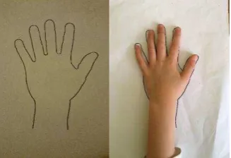
We introduced a simple radiolucent hand template to which the patient could match their
hand in order to assist positioning (figure 1). Three separate hand template sizes were
devised to accommodate for the variety of patient ages and hand sizes.
The aim of the study was to assess changes in X-ray quality through objectively analysing the
positioning of fingers and thumb and clarity of X-ray. The need for repeat X-ray was similarly
assessed to determine if repeat X-ray rates could be reduced.
Patients and Methods
Study Design
This was a prospective intervention study. A service evaluation of the quality of bone age
X-rays was conducted from June 2013 to February 2014. This information was then used as
control data against which our hand template intervention was measured. The study period
commenced in March 2014 and ran to March 2015, during which all consented individuals
used the template.
All bone age X-rays during the study period were evaluated by a single Auxology Nurse blinded
as to whether or not the template had been used. To evaluate the effectiveness of the
intervention, all bone age X-rays were scored against set criteria. Due to the absence of a
validated tool, the position of fingers, thumb and overall clarity of bone age X-rays were
scored by a simple 1-3 scale (poor, borderline, good), devised in conjunction with an academic
radiologist.
Primary outcome measure
Following introduction of the hand template the degree of improvement in quality of bone
age X-rays was objectively assessed using the simple scale outlined above. A reduction of 5%
of X-rays scoring borderline or poor in each category was felt to indicate clinically significant
improvement.
Secondary outcome measure
In addition, an evaluation of the effect the intervention had on repeat X-ray rates was made.
result of suboptimal hand placement. When X-rays were judged by the auxology nurse to be
of insufficient quality to enable accurate scoring, the family was contacted and a repeat X-ray
request made.
In the intervention cohort, the need for repeat X-ray was determined by criteria set by the
auxology nurse as follows:
1. X-ray whose score for either finger positioning or clarity was equal to 1 (poor quality).
2. X-ray where the thumb positioning scored 2 (borderline quality) with an overall
aggregate score of 7 or less (i.e. where any of the other categories scored <3). Thumb
positioning was deemed the most important criteria when scoring bone age X-rays
using the TW3 method and was most commonly linked with a need to repeat X-ray in
practice.
Patients
Patients were recruited if they were undergoing a bone age X-ray as part of their routine
management in the growth and endocrine clinic at a single centre, S C
Hospital, UK. Children and young people were aged between 0 and 20 years of age. The
control arm data were derived from patients who had formed part of the service evaluation.
Exclusion criteria:
Study materials were not translated into multiple languages therefore patients were
only recruited if their English was deemed of a standard to ensure informed consent. Any child with a left hand abnormality causing difficulty in placing their hand flat e.g.
Children with an inadequate level of understanding to comply with the instructions
required to match their hand to the outline placed on the X-ray plate.
The study was approved by Yorkshire and Humber Research Ethics Committee. Written
informed consent was given by all participants. This study complies with the World Medical
Association Declaration of Helsinki regarding ethical conduct of research involving human
subjects.
Results
Study population
There were 259 children in the initial service evaluation (123 female) aged between 1.92 to
18.48 years (mean 10.21 years). The intervention arm comprised 56 participants (28 female)
ranging from 0.9 to 15.77 years (mean 8.95 years). The groups were equally matched for
gender however the intervention group were younger (p=0.03) (table 1). All P values in this
F
Table 1, Study population age profile
Quality of bone age X-rays
The intervention improved X-ray quality scores. Fewer patients scored less than 3 for the
position of fingers (10.7% and 14.7%, p=0.38), thumb (1.8% and 10.0%, p=0.06) and overall
2graph 1). This equates to an improvement of 3.7% for positioning of fingers, 8.3% for positioning of thumb and 6.5% for overall clarity.
Repeat X-ray rates
The template significantly reduced the numbers requiring repeat X-ray. No patient required
a repeat X-ray from the intervention group, compared to 28 (10.81%) in the control group
(p=0.007).
Limitations
Due to the nature of bone age X-rays and their appropriate indication, we were only able to
obtain a small number of patients aged 15.00-19.99 years old. This is unlikely to adversely
affect the significance of this study in the context of such a simple and cheap intervention
that can be seen to have a greater impact on those younger users. This is illustrated in the
figure below (figure 3) which shows the percentage of patients in each age group with a net X-ray score that qualified as I
from 72.2% to 100%;
whilst in the 5-9.99 age range, this increased from 84.5% to 96.7%. Figure 2, X-ray quality scores.
Figure 3, Percentage net X-ray score by age group. .
Discussion
Achieving good quality films for bone age studies is important for the optimal assessment of
skeletal maturity. The data in this study suggest that achieving this may be more difficult than
previously assumed.
The use of a simple radiolucent hand template to which children and young people can match
their hand significantly improved the positioning of the hand and the clarity of the X-rays
produced. This improvement may be due to the increased likelihood of the patient positioning
their hand in such a way as to more closely reflect the ideal positioning described by Tanner1,
enhanced awareness by both the radiographer and child about accurate hand placement and
a greater chance that the child will keep their hand still as the X-ray is taken.
The need for repeat X-ray was eliminated in this study. Given that the radiation dose of a
bone age X-ray is quantified as 0.0001mSv (0.1 microSieverts)2, this represents an important
reduction in radiation exposure.
The results of this study support the introduction of this simple and cost-effective
intervention. The implementation of such a simple and safe intervention has been shown to
significantly reduce the need for repeat X-ray in those patients who require bone age X-rays.
Acknowledgements
We would like to thank the patients and their parents who agreed to take part in this study
Disclosure Statement
The authors have nothing to disclose.
References
1. Tanner J M, Healy M, Goldstein H, et al. Assessment of skeletal maturity and prediction of
adult height (TW3 Method), 3rd edn. London: WB Saunders, Harcourt Publishers Ltd, 2001.
2. Huda W, Gkanatsios NA, Radiation dosimetry for extremity radiographs. Health Phys
1998;75;492-999.
[image:10.595.55.219.403.515.2]Figures
Figure 1, Radiolucent hand template (with and without hand).
Table 1, Study population age profile
Age range
(years) 0-5 5.01-10 10.01-15 15.01-20 Total Mean SD
Control 1.92-18.48 18 109 106 26 259 10.21 3.87
Figure 2, X-ray quality scores.
Figure 3,Percentage net X-ray score by age group
0% 10% 20% 30% 40% 50% 60% 70% 80% 90% 100%
I C I C I C
Fingers Thumb Overall Clarity
1 - Poor 2 - Borderline 3 - Good
I - Intervention
C Control
0% 10% 20% 30% 40% 50% 60% 70% 80% 90% 100%
I C I C I C I C
0-4.99 5-9.99 10-14.99 15-19.99