Open Access Journal of Sports Medicine
Dove
press
O r i g i n A l r e S e A r c h
open access to scientific and medical research
Open Access Full Text Article
Magnetic resonance imaging arthrography following
type ii superior labrum from anterior to posterior
repair: interobserver and intraobserver reliability
hafeez M Kurji1
Yohei Ono2,3
Atiba A nelson2
Kristie D More2
Ben Wong4
corinne Dyke4
richard S Boorman2
gail M Thornton2,5
ian KY lo2
1college of Medicine, University of
Saskatchewan, Saskatoon, SK, canada;
2Department of Surgery, Section of
Orthopaedic Surgery, Mccaig institute for Bone and Joint health, University of calgary, calgary, AB, canada;
3Department of Orthopedic Surgery,
nagoya University graduate School of Medicine, nagoya, Japan; 4Department
of radiology, University of calgary, calgary, AB, canada; 5Department of
Orthopaedics, University of British columbia, Vancouver, Bc, canada
correspondence: ian KY lo
Department of Surgery, The University of calgary, 3280 hospital Drive, nW, calgary, AB, canada Tel +1 40 3220 2745 Fax +1 40 3210 8197 email [email protected]
Background: Arthroscopic repair of type II superior labrum from anterior to posterior (SLAP) lesions is a common surgical procedure. However, anatomic healing following repair has rarely been investigated. The intraobserver and interobserver reliability of magnetic resonance imaging arthrography (MRA) following type II SLAP repair has not previously been investigated. This is of particular interest due to recent reports of poor clinical results following type II SLAP lesion repair.
Purpose: To evaluate the MRA findings following arthroscopic type II SLAP lesion repair and determine its intraobserver and interobserver reliability.
Study design: Cohort study (diagnosis), Level of Evidence, 2.
Methods: Twenty-five patients with an isolated type II SLAP lesion (confirmed via diagnostic arthroscopy) underwent standard suture anchor-based repair. At a mean of 25.2 months post-operatively, patients underwent a standardized MRA protocol to investigate the integrity of the repair. MRAs were independently reviewed by two radiologists and a fellowship trained shoulder surgeon. The outcomes were classified as healed SLAP repair or re-torn SLAP repair.
Results: On average, 54% of MRAs were interpreted as healed SLAP repairs while 46% of MRAs were interpreted as having a re-torn SLAP repair. Overall, only 43% of the studies had 100% agreement across all interpretations. The intraobserver reliability ranged from 0.71 to 0.81 while the interobserver reliability between readers ranged from 0.13 to 0.44 (Table 1).
Conclusion: The intraobserver agreement of MRA in the evaluation of type II SLAP repair was substantial to excellent. However, the interobserver agreement of MRA was poor to fair. As a result, the routine use of MRA in the evaluation of type II SLAP lesion repair should be utilized with caution. A global evaluation of the patient, including detailed history and physi-cal examination, is paramount in determining the cause of failure and one should not rely on MRA alone.
Keywords: SLAP, MRA, labrum, postoperative
Introduction
Tears of the superior labrum were first described by Andrews et al1 in 1985. Subsequent
to this, Snyder et al2 coined the term superior labrum from anterior to posterior (SLAP)
lesion and classified them into four types. SLAP lesions have reported incidences rang-ing from 4% to 26%2–7 with the type II SLAP lesion being the most common surgical
lesion. However, controversy still exists in the treatment of this condition in part due to the difficulty in the clinical diagnosis of a symptomatic SLAP lesion.
In North America, magnetic resonance imaging (MRI) has become the imaging modality of choice when evaluating soft tissue lesions of the shoulder. With improved assessment of intra-articular pathology, magnetic resonance imaging arthrography
Open Access Journal of Sports Medicine downloaded from https://www.dovepress.com/ by 118.70.13.36 on 26-Aug-2020
For personal use only.
Number of times this article has been viewed
This article was published in the following Dove Press journal: Open Access Journal of Sports Medicine
Dovepress Kurji et al
Figure 1 Arthroscopic view of a right shoulder through a posterior glenohumeral
portal demonstrating the immediate postoperative appearance of a type ii SlAP lesions repair using a standard suture anchor based technique.
Abbreviation: SlAP, superior labrum from anterior to posterior.
(MRA) is widely used when evaluating lesions of the labrum (eg, SLAP lesions, Bankart lesions).
While MRI or MRA have been routinely utilized preop-eratively, their utility in the postoperative scenario has been less frequently reported. In particular, MRI has been more commonly applied in the assessment of rotator cuff integrity following repair.8 However, the use of MRA has only rarely
been reported.9,10 In particular, its utility in the postoperative
assessment of superior labrum tears is unclear.
Recently, the intraobserver and interobserver reliability of various diagnostic tests to detect type II SLAP lesions has been investigated. In particular, MRA has been extensively investigated and has shown to be a sensitive, minimally inva-sive procedure for detection and grading of SLAP lesions, with substantial to excellent intraobserver and interobserver reliability.11–13
However, the intraobserver and interobserver reliability of MRA in the postoperative scenario following type II SLAP repair has not previously been investigated. In addition, the anatomic healing of a type II SLAP lesion following repair has rarely been investigated. This is particularly relevant in light of reports of poor results following type II SLAP lesion repairs.14–16
Therefore, the purpose of this study is to evaluate the MRA findings following arthroscopic type II SLAP lesion repairs and determine its intraobserver and interobserver reliability. We hypothesized that the intraobserver and interobserver reliability would be highly variable in this patient population.
Methods
The study was a retrospective cohort study and patients were identified from a billing review of the senior author’s practice. Patients were contacted by the research coordinator to deter-mine their eligibility and interest in participating in the study. The University of Calgary Conjoint Health Research Ethics Board approved this research.
Between March 2003 and June 2006, the senior author performed isolated repairs of 25 type II SLAP lesions in 25 patients (18 male, 7 female) with a mean age of 40 ±
12 years. Exclusion criteria included: previous surgery, sig-nificant concomitant pathology requiring treatment (eg, subacromial decompression, distal clavicle excision, rotator cuff repair, biceps tenodesis, biceps tenotomy, Bankart repair, posterior labral repair, osteoarthritis), significant cervical spine pathology (and/or radiculopathy), significant medical issues precluding surgery, secondary gain issues (Workers’ Compensation Board claim, litigation), unwillingness to
complete study outcomes, and unable to provide informed consent.
All patients underwent a diagnostic arthroscopy with confirmation of an isolated type II SLAP lesion. Standard suture anchor based repair was then performed. One anchor (3.0 mm Bio-FASTak or Bio-SutureTak double loaded with #2 Fiberwire; Arthrex, Inc., Naples, FL, USA) was placed underneath the biceps root and sutures were passed anterior and posterior to the biceps tendon. For larger tears extending posteriorly, subsequent anchors were placed through separate percutaneous transtendon portals (eg, Port of Wilmington) into the posterior superior aspect of the glenoid neck. Nine patients had one anchor inserted under the biceps, 13 patients had two anchors inserted, and three patients had three anchors inserted (Figure 1).
MrA imaging
At a mean of 25.2 months postoperatively (range: 13–44 months), 21 of 25 patients underwent a standardized MRA protocol to investigate the integrity of the type II SLAP repair. All sequences were obtained immediately post intra-articular injection of 10–15 mL dilute gadolinium into the glenohumeral joint, performed under fluoroscopic guidance prior to MRI.
A 1.5-T MRI scanner (Symphony; Siemens Healthcare Global) was utilized with the patient’s arm positioned in the neutral position. Standardized coronal oblique T1FS, T2FS, sagittal oblique T1, T2FS, Axial T1FS, and Axial GRE images were obtained. Parameters for all sequences were a section thickness of 3 mm with an intersecting gap of
Open Access Journal of Sports Medicine downloaded from https://www.dovepress.com/ by 118.70.13.36 on 26-Aug-2020
Dovepress MRA findings in arthroscopic type II SLAP lesion repairs
(κ) were used to determine intraobserver agreement (,0 poor agreement; 0.00–0.20 slight; 0.21–0.40 fair; 0.41–0.60 moderate; 0.61–0.80 substantial; 0.81–1.00 excellent). Fleiss’ kappa coefficients used to determine interobserver agreement among the three readers (,0.4 poor; 0.4–0.75 fair to good; .0.75 excellent).
Results
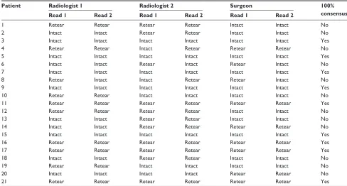
Twenty-one patients were included in the analysis. The results of each reader’s interpretation are summarized in Table 1. On average, 11.4 (54.3%) (range: 9–14) of MRAs were inter-preted as healed SLAP repairs (Figure 2) while 9.6 (45.7%) (range: 7–12) MRAs were interpreted as having re-torn SLAP repairs (Figure 3). Overall, only nine of 21 MRIs (42.9%) had 100% agreement across all interpretations.
The intraobserver reliability was 0.81 for radiologist one (excellent agreement), 0.71 for radiologist two (substantial agreement), and 0.71 for the surgeon (substantial agreement). The interobserver reliability between readers was ranged considerably but was considered poor to fair (range: 0.13–0.44).
Confirmatory arthroscopy was not performed routinely. However, in one patient with a poor clinical outcome, a second operative procedure was performed. This case was a 27-year-old male who developed acute onset, dominant-sided 0.3 mm, a field of view of 16 cm and an in-plane resolution
of 0.4×0.3–0.5 mm.
image analysis
All magnetic resonance images were reviewed on a com-mercial picture archiving and communication system (PACS) workstation (EasyVision; Philips, Best, the Netherlands). MRAs were independently reviewed by two radiologists with .10 years of experience in musculoskeletal imaging. In addition, the MRAs were evaluated by a fellowship-trained shoulder surgeon with .10 years experience in shoulder surgery. All readers were blinded to previous imaging reports, patient demographics, and all clinical findings and outcomes. MRAs were read twice in random order separated by a mini-mum of 6 months.
Outcomes were classified according to:
1. Healed SLAP repair: minimal to no dye leakage (and/ or improvement when compared to the preoperative imaging) under the labrum (Figure 2).
2. Re-torn SLAP repair: detached superior labrum and dye present between the labrum and superior glenoid at the 12 o’clock position or posteriorly (Figure 3).
Statistical analysis
Statistical analysis was performed using SPSS (version 18; SPSS Inc., Chicago IL, USA). Cohen’s kappa coefficients
Figure 2 T1-weighted fat-suppressed para-coronal MrA image demonstrating a
healed type ii SlAP repair with no gadolinium insinuating between the superior labrum and glenoid. This MrA was interpreted as healed by all three readers.
Abbreviations: MrA, magnetic resonance imaging arthrography; SlAP, superior
labrum from anterior to posterior.
Figure 3 T1-weighted fat-suppressed para-coronal MrA imaging demonstrating a
re-torn type ii SlAP repair with gadolinium insinuating between the superior labrum and glenoid adjacent to the anchor site. This MrA was interpreted as re-torn by all three readers.
Abbreviations: MrA, magnetic resonance imaging arthrography; SlAP, superior
labrum from anterior to posterior.
Open Access Journal of Sports Medicine downloaded from https://www.dovepress.com/ by 118.70.13.36 on 26-Aug-2020
Dovepress Kurji et al
Table 1 results of each reader’s interpretation of postoperative MrA following type ii SlAP lesion repair
Patient Radiologist 1 Radiologist 2 Surgeon 100%
consensus
Read 1 Read 2 Read 1 Read 2 Read 1 Read 2
1 retear retear retear retear intact intact no
2 intact intact retear retear intact intact no
3 intact intact intact intact intact intact Yes
4 retear retear intact retear retear retear no
5 intact intact intact intact intact intact Yes
6 intact intact retear intact retear intact no
7 intact intact intact intact intact intact Yes
8 retear intact intact retear retear intact no
9 intact intact intact intact intact intact Yes
10 retear retear intact intact intact intact no
11 retear retear retear retear retear retear Yes
12 retear retear retear retear intact intact no
13 intact intact retear retear intact intact no
14 intact intact retear retear retear retear no
15 intact intact intact intact intact intact Yes
16 retear retear retear retear retear retear Yes
17 retear retear retear retear retear retear Yes
18 intact intact retear retear intact intact no
19 retear retear intact intact intact intact no
20 intact intact intact intact retear retear no
21 retear retear retear retear retear retear Yes
Abbreviations: MrA, magnetic resonance imaging arthrography; SlAP, superior labrum from anterior to posterior.
shoulder pain following an overhead pressing exercise in the gym. Arthroscopy demonstrated a type II SLAP lesion and a repair was performed with the use of two anchors. The patient initially did reasonably well, but continued to have difficulties with overhead activities and reaching activities, and became fatigued easily. A postoperative MRA performed 42 months after surgery was read as intact on five of six interpretations (Figure 4A). Fifty-eight months following surgery the patient underwent a second-look arthroscopy. Diagnostic arthroscopy demonstrated an intact superior labrum (Figure 4B) but there was evidence suggesting nonhealing of the transtendon pos-terolateral portal (Port of Wilmington), which was felt to be responsible for the patient’s symptoms (Figure 4C).
Discussion
Despite its initial description almost 30 years ago and the subsequent multitude of articles pertaining to SLAP lesions, the clinical diagnosis of a type II SLAP lesion still remains controversial.17 For this reason, various imaging modalities
used to evaluate superior labral tears have been investigated. In 2001, Jee et al18 retrospectively reviewed the results of
80 patients who underwent MRA and arthroscopy during a 54-month period. They concluded that the interobserver agreement was substantial to moderate with a reported accuracy between 74% and 85%. In 2010, Holzapfel et al13
evaluated the intraobserver and interobserver reliability for the detection and classification of SLAP lesions; they
dem-onstrated excellent intraobserver and excellent interobserver agreement for the detection of SLAP lesions and an overall accuracy .82%.
While these results appear promising, not all authors have reported substantial observer agreement or accuracy. In 2011, Halma et al19 evaluated the interobserver and intraobserver
agreement and accuracy of MRI imaging in the shoulder between both radiologists and orthopedic surgeons, which were confirmed at arthroscopy. While intraobserver agree-ment was considered moderate to substantial, there was only fair interobserver agreement between radiologists and also between radiologist and surgeons.
The intraobserver or interobserver reliability of MRA in the postoperative evaluation of type II SLAP lesions has not previously been investigated. This is particularly relevant since a number of studies have been recently published evaluating clinical failure following type II SLAP repairs.14–16
In 2009, Katz et al14 published the results of 39 patients with
poor clinical outcomes who presented to their institution fol-lowing a type II SLAP repair. They noted a large percentage of their patients having a history of Worker’s Compensation claims, trauma leading to symptoms, and involvement of the dominant shoulder. Similarly in 2011, Weber16 presented his
results of 24 shoulders with clinical failure following type II SLAP repair. In both these studies, the authors noted a number of different failure mechanisms (eg, failure of SLAP healing, partial biceps tearing, stiffness, chondral injuries,
Open Access Journal of Sports Medicine downloaded from https://www.dovepress.com/ by 118.70.13.36 on 26-Aug-2020
Dovepress MRA findings in arthroscopic type II SLAP lesion repairs
Figure 4 Patient with a failed SlAP repair following type ii SlAP repair.
Notes: (A) Post-operative T1-weighted fat-suppressed para-coronal MrA imaging
demonstrating an intact superior labrum with no gadolinium contrast intervening between the superior labrum and superior glenoid; (B) Arthroscopic view of a right
shoulder from the posterior portal demonstrating an intact superior labrum. The superior labrum was stable under direct palpation with a probe and with range of motion; (C) Arthroscopic view of a right shoulder from the posterior portal
demonstrating a persistent partial thickness rotator cuff tear corresponding to the previous posterior lateral portal site (Port of Wilmington).
Abbreviations: SlAP, superior labrum from anterior to posterior; MrA, magnetic
resonance imaging arthrography.
treatment options. This is even more relevant since only 29% of their patients were satisfied with conservative management and eventually required surgery.14
In the current study, while substantial to excellent intrao-bserver agreement was demonstrated, there was only poor to fair interobserver agreement. There are multiple reasons for this outcome. First, while the overall interobserver agreement may be good in the primary setting, it may in fact be poor in the postoperative setting due to interference by postoperative artifact. This may include anchors or implants, suture mate-rial, and debris (metallic or other). This artifact may affect postoperative imaging, obscure detail, and consequently cloud interpretation. Second, the actual normal healing mech-anism of a type II SLAP repair is unknown; that is, how a type II SLAP lesion heals over time is unknown. Even though all MRAs were obtained at a minimum of 1 year postopera-tively, it is possible that further remodeling of granulation material may occur and affect interpretation. Third, while we attempted to utilize strict criteria when interpreting MRAs, a formal training program with confirmatory arthroscopy was not employed, nor did the observers meet to discuss classifica-tions prior to the study. This may have improved the results but could have affected reader interpretation by subjecting them to previous biases, education, thus limiting the study’s generalizability. A formal training period with interpretation consensus could have improved interobserver reliability. Fourth, interpreter experience may be a factor.20 In our study,
all interpreters were musculoskeletal trained radiologists or shoulder surgeons with .10 years of experience in muscu-loskeletal medicine. However, while all had vast experience in evaluating the primary cases of type II SLAP lesions, the assessment of type II SLAP lesions postoperatively is not routinely performed. Therefore, because of the infre-quency of postoperative imaging of type II SLAP lesions, the actual experience in interpreting this scenario would be significantly less. Furthermore, continued self-assessment with arthroscopic confirmation would be even rarer.
In fact, it is important to consider that there was no routine arthroscopic correlation to confirm the radiologist’s or surgeon’s MRA interpretation. Therefore, the accuracy of each reader’s interpretation of MRAs is unknown. Nevertheless, one patient with ongoing symptoms did have revision surgery. In this patient, the majority of read-ers interpreted the postoperative MRA as demonstrating a healed superior labrum with subsequent arthroscopic confirmation.
While routine arthroscopic correlation would have signifi-cantly strengthened our study, even arthroscopic evaluation of the superior labrum can be difficult.12,21,22 In 2008, Gobezie
hardware problems, arthritis, rotator cuff tears) with various subsequent revision procedures (eg, revision SLAP repair, biceps tenodesis, removal of loose hardware, rotator cuff repair, total shoulder arthroplasty). These reports highlight the importance of careful diagnostic evaluation of failed type II SLAP repairs with multiple mechanisms of failure and
Open Access Journal of Sports Medicine downloaded from https://www.dovepress.com/ by 118.70.13.36 on 26-Aug-2020
Dovepress Kurji et al
et al12 used recorded video “vignettes” of diagnostic
arthros-copies to evaluate the reliability of arthroscopy in the diagnosis and treatment of type II SLAP lesions. In what was considered a “normal” shoulder, only 68% of surgeons made the same “normal” diagnosis and 10% of surgeons felt the labrum was torn and repair was indicated. Furthermore, in “type II SLAP” lesion shoulders, only 54% of surgeons made the same diag-nosis of a type II SLAP lesion and 22% felt the labrum was normal. These results and others21,22 have lead some to question
the use of diagnostic arthroscopy as the gold standard for the diagnosis of superior labrum tears.13 We would expect that
with the further influence of surgical artifact, the postoperative healing interpretation of diagnostic arthroscopy following type II SLAP repairs would be even more variable.
Overall, following a type II SLAP repair, approximately 46% were interpreted to be re-torn and 54% of patients were interpreted as demonstrating healed superior labrums by MRA. However, the MRA results did not correlate with patient outcomes. Patients with intact or re-torn type II SLAP repairs by MRA demonstrated similar outcomes.22
This suggests that patients with good outcomes may have persistent dye insinuation between the glenoid and labrum similar to persistent defects following rotator cuff repair.23–25 Furthermore, in the evaluation of the failed type
II SLAP repair, the presence of a recurrent tear should not routinely be interpreted as the cause of failure and careful clinical evaluation should ensue to rule out the multiple different causes of a failed SLAP repair.14,16
Conclusion
In conclusion, the intraobserver agreement of MRA in the evaluation of type II SLAP repair was substantial. However, the interobserver agreement of MRA was poor to fair. As a result, the routine use of MRA in the evaluation of type II SLAP lesion repair should be utilized with caution.
Disclosure
The authors report no conflicts of interest in this work.
References
1. Andrews JR, Carson WG, McLeod WD. Glenoid labrum tears related to the long head of the biceps. Am J Sports Med. 1985;13(5):337–341. 2. Snyder SJ, Karzel RP, Del Pizzo W, Ferkel RD, Friedman MJ. SLAP
lesions of the shoulder. Arthroscopy. 1990;6(4):274–279.
3. Handelberg F, Willems S, Shahabpour M, Huskin JP, Kuta J. SLAP lesions: a retrospective multicentre study. Arthroscopy. 1998;14(8): 856–862.
4. Kim TK, Queale WS, Cosgarea AJ, McFarland EG. Clinical features of the different types of SLAP lesions. An analysis of one hundred and thrity-nine cases. J Bone Joint Surg. 2003;85(A):66–71.
5. Kampa RJ, Clasper J. Incidence of SLAP lesions in a military population.
J R Army Med Corps. 2005;151(3):171–175.
6. Maffet MW, Gartsman GM, Moseley B. Superior labrum-biceps tendon complex lesions of the shoulder. Am J Sports Med. 1995;23:93–98. 7. Snyder SJ, Banas MP, Karzel RP. An analysis of 140 injuries to the
superior glenoid labrum. J Shoulder Elbow Surg. 1995;4:243–248. 8. Saccomanno MF, Cazzato G, Fodale M, Sircana G, Milano G. Magnetic
resonance imaging criteria for the assessment of the rotator cuff after repair: a systematic review. Knee Surg Sports Traumatol Arthrosc. 2015;23(2):423–442.
9. Rand T, Freilinger W, Breitenseher M, et al. Magnetic resonance arthrog-raphy (MRA) in the postoperative shoulder. Magn Reson Imaging. 1999;17(6):843–850.
10. Thakkar RS, Thakkar SC, Srikumaran U, McFarland EG, Favad LM. Comlications of rotator cuff surgery – the role of postoperative imaging in patient care. Br J Radiol. 2014;87(1039):20130630. [Review]. 11. Amin MF, Youssef AO. The diagnostic value of magnetic resonance
arthrography of the shoulder in detection and grading of SLAP lesions: comparison with arthroscopic findings. Eur J Radiol. 2012;81(9): 2343–2347.
12. Gobezie R, Zurakowski D, Lavery K, Millett PJ, Cole BJ, Warner JJ. Analysis of interobserver and intraobserver variability in the diagnosis and treatment of SLAP tears using the Snyder classification. Am J Sports
Med. 2008;36(7):1373–1379.
13. Holzapfel K, Waldt S, Bruegel M, et al. Inter- and intraobserver vari-ability of MR arthrography in the detection and classification of superior labral anterior posterior (SLAP) lesions: evaluation in 78 cases with arthroscopic correlation. Eur Radiol. 2010;20:666–673.
14. Katz LM, Hsu S, Miller S, et al. Poor outcomes after SLAP repair: descriptive analysis and prognosis. Arthroscopy. 2009;25(8): 849–855.
15. Park SD, Glousman RE. Outcomes of revision arthroscopic type II superior labral anterior posterior repairs. Am J Sports Med. 2011;39(6): 1290–1294.
16. Weber SC. Surgical management of the failed SLAP repair. Sports Med
Arthrosc Rev. 2010;18:162–166.
17. Higgins LD, Warner JJ. Superior labral lesions. Clin Orthop Relat Res. 2013;201:73–82.
18. Jee WH, McCauley TR, Katz LD, Matheny JM, Ruwe PA, Daigneault JP. Superior labral anterior posterior (SLAP) lesions of the glenoid labrum: reliability and accuracy of MR arthrography for diagnosis. Radiology. 2001;218:127–132.
19. Halma JJ, Eshuis R, Krebbers YM, Weits T, de Gast A. Interdisciplinary inter-observer agreement and accuracy of MR imaging of the shoulder with arthroscopic correlation. Arch Orthop Trauma Surg. 2012;132: 311–320.
20. Dinter DJ, Martetschlager F, Busing KA, Schonberg SO, Scharf HP, Lehmann LJ. Shoulder injuries in overhead athletes: utility of MR arthography. Sportverletz Sportschaden. 2008;22(3):146–152. 21. Jia X, Yokota A, McCarty EC, et al. Reproducibility and reliability of
the Snyder classification of superior labrum anterior posterior lesions among shoulder surgeons. Am J Sports Med. 2011;39(5):986–991. 22. Trantalis JN, Lo IK, Boorman RS, Sohmer S, Pletsch KD, Woods T.
Arthroscopic repair of type II SLAP lesions: clinical and magnetic resonance imaging arthrogram follow-up. Arthroscopy. 2008;24 (6 Suppl):e7.
23. Dodson CC, Kitay A, Verma NN, et al. The long-term outcome of recurrent defects after rotator cuff repair. Am J Sports Med. 2010;38(1): 35–39.
24. Spielmann AL, Forster BB, Kokan P, Hawkins RH, Janzen DL. Shoulder after rotator cuff repair: MR imaging findings in asymptomatic individuals – initial experience. Radiology. 1999;213(3):705–708. 25. Zanetti M, Jost B, Hodler J, Gerber C. MR imaging after rotator cuff
repair: full-thickness defects and bursitis-like subacromial abnormalities in asymptomatic subjects. Skeletal Radiol. 2000;29(6):314–319.
Open Access Journal of Sports Medicine downloaded from https://www.dovepress.com/ by 118.70.13.36 on 26-Aug-2020
Open Access Journal of Sports Medicine
Publish your work in this journal
Submit your manuscript here: http://www.dovepress.com/open-access-journal-of-sports-medicine-journal Open Access Journal of Sports Medicine is an international,
peer-reviewed, open access journal publishing original research, reports, reviews and commentaries on all areas of sports medicine. The manuscript management system is completely online and includes a very quick and fair peer-review system.
Visit http://www.dovepress.com/testimonials.php to read real quotes from published authors.
Dovepress
Dove
press
MRA findings in arthroscopic type II SLAP lesion repairs
Open Access Journal of Sports Medicine downloaded from https://www.dovepress.com/ by 118.70.13.36 on 26-Aug-2020