GANTT, JOHN ARTHUR. Comparative Analysis of RNase P. (Under the direction of Dr. James W. Brown.)
This Master’s Thesis contains a description of the results of two research projects that were worked on over the course of two years. The first was an attempt to heterologously
reconstitute the Bacillus subtilis RNase P RNA with the RNase P protein subunits of
Methanothermobacter thermoautotrophicus, to identify a functional homology between one or more of the archaeal proteins with the Bacillus subtilis RNase P protein.
Unfortunately, the reconstitution experiment could not be completed due to the poor quality of the pre-tRNAasp substrate synthesized. The second project dealt with
COMPARATIVE ANALYSIS OF RNASE P
by
JOHN ARTHUR GANTT
A thesis submitted to the Graduate Faculty of The North Carolina State University
in partial fulfillment of the requirements for the Degree of
Master of Science
MICROBIOLOGY
Raleigh
2003
APPROVED BY:
_______________________
_______________________
Committee Member
Committee Member Dr. Stephen J. Libby Dr. Amy M. Grunden_______________________
BIOGRAPHY
I, John Gantt, was born in Houston, Texas on December 16, 1978. After moving a
number of times my family settled in North Carolina in the small town of Morganton,
where my parents still reside. In 1997 I was accepted into High Point University where I
majored in Biology and minored in Chemistry and Mathematics. There I learned that
diversity and understanding is a treasure and that GOD has a place in his heart for all of us.
After graduating from HPU, I entered into the Graduate department of Microbiology at
North Carolina State University. Upon completion of the Master of Science requirements I
iii
ACKNOWLEDGEMENTS
Through the support of my family and friends I have completed this arduous task. I
would like to thank Dr. James Brown and the Brown lab for their continual support in my
scientific growth. Drs. Steve Libby and Amy Grunden, my committee members, have also
been invaluable in my progression. I would like to dedicate this thesis and my work to my
TABLE OF CONTENTS
List of Table v
List of Figures vi
RNase P Literature Review 1
The RNase P Reaction 1
Bacterial RNase P 3
Eukaryotic RNase P 6
Archaeal RNase P 8
References 17
Chapter 1: RNase P proteins: The homology between that of archaeal and bacterial
subunits. 21
Abstract 21
Introduction 21
Materials and Methods 22
Results and Discussion 25
References 33
Chapter 2: Comparative analysis of the bacterial RNase P RNA
structure. 34
Abstract 34
Introduction 34
Materials and Methods 35
Results and Discussion 37
v
LIST OF TABLES
RNase P Literature Review
Table 1: Comparison of subunits in RNase P and RNase MRP from yeast, human,
and Archaea. 16
Chapter 2: Comparative analysis of the bacterial RNase P RNA structure.
LIST OF FIGURES
RNase P Literature Review
Figure 1: The RNase P reaction. 10
Figure 2: RNase P RNA:pre-tRNA interactions. 11 Figure 3: Bacterial RNase P protein. 12
Figure 4: Bacterial type A RNase P RNA secondary structure,
represented by E.coli, the minimum consensus bacterial RNase P RNA, and bacterial type B RNase P RNA secondary structure,
represented by Bacillus subtilis. 13
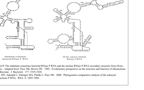
Figure 5: The minimum consensus bacterial RNase P RNA and
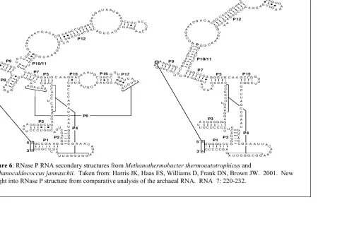
the nuclear RNase P RNA secondary structure from Homo sapiens. 14 Figure 6: RNase P RNA secondary structures from Methanothermobacter thermoautotrophicus and Methanocaldococcus jannaschii. 15
Chapter 1: RNase P Proteins: The homology between that of archaeal and bacterial subunits.
Figure 1:Expression of M. thermoautotrophicus RNase P recombinant proteins (A)11p, (B)1618p, (C)688p, and (C)687p. 28 Figure 2:RNase P ribozyme assay. 29 Figure 3: A titration gel-shift assay (shown diagrammatically). 30 Figure 4: Gel-shift binding assay (shown diagrammatically). 31 Figure 5: B. subtilis RNase P protein reconstituting activity
in archaeal RNase P RNAs. 32
Chapter 2: Comparative analysis of the bacterial RNase P RNA structure.
Figure 1: Bacterial type A RNase P RNA secondary structure, represented by
E.coli, the minimum consensus bacterial RNase P RNA, and bacterial type B RNase P RNA secondary structure, represented by Bacillus subtilis. 42 Figure 2: Comparative analysis of eleven new RNase P RNAs and their
derivatives. 43
1
RNASE P LITERATURE REVIEW
In 1970 Sidney Altman and his colleagues, through the use of mutant tRNA
precursors, discovered an enigmatic enzyme ribonuclease P (RNase P), involved in the
biosynthesis of tRNA (1). All transfer-RNAs are initially transcribed as precursors
containing extra nucleotides 3′ and 5′ to the mature ends. These extra sequences are removed by a complex set of enzymes and the resulting mature transfer RNA is
transported to the ribosome where it aids in protein synthesis (2, 23, 24). RNase P is the
ubiquitous endoribonuclease responsible for removal of the 5′ leader sequence of precursor tRNA (pre-tRNA) (3, 4, 25). RNase P has now been characterized from representatives of
all three Domains of life, Bacteria, Archaea, and Eucarya, and those from mitochondria
and chloroplasts. RNase P is a ribozyme; the RNA is the catalytic subunit of the enzyme
and the protein is a structural enhancer (4). In 1989 Sidney Altman and Thomas Cech
shared the Nobel Prize in Chemistry for their discovery of the catalytic properties of RNA;
for their discoveries that group I introns are self-splicing (Cech) and that the RNA subunit
of RNase P is a true catalytic ribozyme (Altman) (1).
The RNase P Reaction
The RNase P reaction has been studied in Bacteria, Archaea, and Eucarya, but is
best characterized and described in the bacterial ribozyme (RNA only) reaction. This
enzymatic reaction consists of three basic steps; enzyme-substrate binding, scissile bond
cleavage, and product release (6).
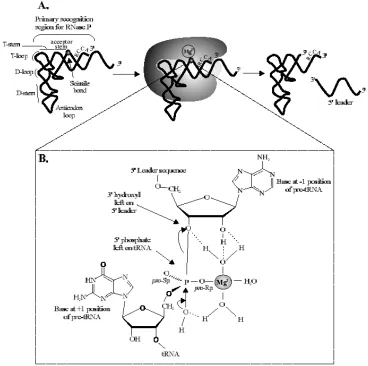
Although perhaps most critical in cleaving many types of pre-tRNAs, RNase P is
also required to recognize and cleave precursors from a wide variety of natural RNA
of substrates poses a problem in that even pre-tRNAs show considerable sequence
variation. Because it cannot rely solely on sequences for substrate recognition, RNase P
instead recognizes the overall tertiary structure of pre-tRNA with a minimal determinant of
the T-stem, T-loop and acceptor stems. (Figure 1a) (12). Pre-tRNA contains several
conserved features proximal to the cleavage site that are crucial to the ribozyme reaction
(3, 6, 11, 4, 27). The 3′ RCCA “tail” is known to be responsible for positioning and anchoring of the substrate phosphodiester bond into the active site of the RNase P by
base-pairing to the GGU in J16/15; this enzyme: substrate interaction is known as the 73/294
interaction (10, 5, 3). In addition to the 73/294 interaction, it has recently been determined
that the 2′ hydroxyl of U(-1) in pre-tRNA and A(248) in RNase P RNA interact. Zahler et al. shows that mutagenesis of A(248), in J5/15, resulted in considerable miscleavage of the
pre-tRNA. Lastly the G(1)-C(72) base-pair is known to contribute to affinity and catalysis,
although the details of the contribution remain undefined (3) (Figure 3).
Even though RNA-RNA associations play an important role in catalysis, the
protein subunit of RNase P also plays a role in substrate binding. The protein subunit
assists by directly contacting the 5′ leader in a sequence non-specific manner thus ameliorating dependence on the CCA tail of the pre-tRNA (11, 8, 29, 30). The RNase P
holoenzyme (RNA plus protein) has the ability to discriminate between substrate and
product because of the protein interaction to the 5′ leader and, as a result product inhibition by mature tRNA is minimized (28).
3 requires monovalent cations, preferably K+ and NH4+, whose role is apparently in
counteracting electrostatic repulsion between RNA phosphates (12, 32). Divalent cations,
preferably Mg2+, contribute directly to catalysis. Mg2+ is known to be coordinated by a
phosphate at the cleavage site and oxygens on the RNA subunit, thus allowing a hydroxyl
attack by water on the same phosphate that coordinated Mg2+ (Figure 1b) (12, 33, 34).
This is an Sn2-type nucleophilic reaction.
After the 3′O of the precursor chain is displaced the products dissociate from the reaction, the 5′ leader is degraded, and the resulting mature-length tRNA is further modified and finally utilized by the translation apparatus (2).
Bacterial RNase P
A majority of the information about RNase P has been derived from Bacteria,
primarily E. coli and B. subtilis, because of the opportunity to correlate catalytic activity of
the RNA with structure (12). Bacterial RNase P is a heterodimer consisting of a catalytic
RNA, 350-400nts, and a single protein cofactor, 13-14kDa, which are known under normal
conditions to be both essential in vivo. However, at high ionic strength in vitro, the protein
subunit is unnecessary for catalysis (13, 4).
The bacterial RNase P protein is encoded by the single-copy rnpA gene, that is part
of a well-conserved operon with rmpH, encoding ribosomal protein L34. The RNase P
protein functions on at least two levels. It was first thought that the proteins only role was
to shield unfavorable electrostatic repulsion between the ribozyme and the substrate,
because it was catalytically replaceable by high monovalent salt concentrations. Recently
it has been found that the protein alleviates product inhibition, enhances the relative rate of
to those of two ribosomal proteins, S5 and elongation factor G. This suggests that the
protein subunit either arose early in protein evolution and thus mediated the conversion
from all-RNA to an RNP world, or that one of these proteins was later recruited to function
in RNase P (14).
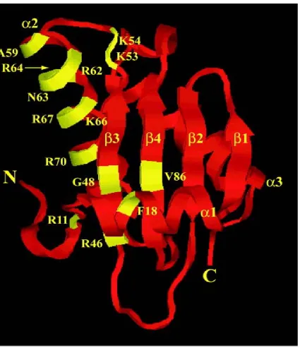
The RNase P protein structure is three α-helices surrounding a central β-sheet (Figure 4) (4). Two conserved sets of residues in the tertiary structure, including the RNR
motif and a large central cleft, are thought to assist in the interaction of the pre-tRNA
substrate and the RNase P RNA subunit. The RNR (Arg-Asn-Arg) motif, located in α2, is thought to specifically bind the RNase P RNA, although the details of the interaction are
not known (13). Most Arg-rich sequences of RNA-binding proteins specifically recognize
RNA, thus setting a precedence for the binding motif of RNR (35, 36). Other evidence
includes mutagenesis of conserved residues in the RNR motif of the RNase P protein
hindering enzymatic activity in in vitro experiments (37). The large central cleft, formed
by packing of α1 against the β-sheet, is thought to form weak interactions with the single stranded 5’ leaders of pre-tRNA. Photocrosslinking experiments showed that nucleotides
–4 to –7 in the pre-tRNA are proximal to Phe16(α1), Phe20(α1), Val32(β2), Tyr34(β2), Ser49(β3), and Ile86(β4) in the cleft (38).
Neither the RNA nor the holoenzyme of RNase P has been crystallized for
structural analysis successfully. A different approach has been taken to examine the RNA
structure, in which similar RNA sequences from a variety of organisms are compared and
5 structure has been thoroughly studied by comparative analysis, which requires a substantial
number of sequences for high resolution (11). RNAs from Bacteria have fallen into two
main structural categories; type A (ancestral type), represented by various groups of
Gram-negative and high G+C Gram-positive Bacteria and bear considerable sequence and
structural similarity to most archaeal RNase P RNAs; and type B, which are restricted to
low G+C Gram-positives including Bacillus and relatives. Figure 4 illustrates the RNA
secondary structures of E. coli, representing type A, and B. subtilis, representing type B,
and shows that although these RNAs are the most disparate of the bacterial RNase P
RNAs, a minimal consensus sequence and structure can be identified that contains all of
the essential determinants of the RNA. These two types of RNAs are able to interchange
proteins, in vivo and in vitro, indicating an overall similarity in tertiary structure (11).
A standard nomenclature based primarily on the type-A RNA, represented by that
of E. coli, has been applied to RNase P RNAs. Base-paired helices are labeled “P” one
through twenty, unpaired joining regions “J” between helices, and terminal loops “L” (11).
Comparative analysis indicates that although type A RNAs lack P5.1, P10.1, and P15.1,
type B lack P13, P14, P16, and P17, indicating that these helices may play interchangeable
functional and structural roles (11, 39). It is known that the evolutionary change leading
from type A to type B RNA occurred abruptly and no intermediates have been identified
Eukaryotic RNase P
Nuclear
RNase P from the nuclear region has been purified from a variety of eukaryotes,
but is best understood from humans and the yeast Saccharomyces cerevisiae. The single
eukaryotic nuclear RNase P RNA contains a conserved catalytic core similar to the
bacterial RNA (Figure 6), strongly suggesting that the nuclear RNA function is
homologous to that of the bacterial RNA, and further that the nuclear RNase P RNA is also
a ribozyme (6, 40, 41). However, with a protein complement of 50%, compared to 10% in
Bacteria, the nuclear RNA has become absolutely dependent on protein for function. The
purpose of this protein addition includes structural stabilization, spatial organization of
pre-tRNA biosynthesis, RNase P holoenzyme maturation, and perhaps participation in
undiscovered processing pathways (6, 16, 17).
The human and S. cerevisiae RNase P holoenzymes exhibit many similarities in
sequence (Table 1), but also share a significant sequence similarity with the ribosomal
RNA processing enzyme RNase MRP. Of the nine protein subunits of the S. cerevisiae
RNase P, eight are shared with RNase MRP (5, 6).
Organellar
The endosymbiotic theory states that organelles are the direct descendents of
Bacteria (42). Eukaryotic cells containing such organelles, including mitochondria and
chloroplasts, contain distinct RNase P complexes in these organelles, although the data is
7 the mitochondrial genome and a single protein subunit (Rpm2p) of 105kDa encoded by a
nuclear gene (5, 6, 43). During the purification of the enzyme, any fragmentation of the
RNA that occurs does not interfere with its processing activity, suggesting that the protein
is primarily responsible for structural integrity of the enzyme. Rpm2p was also found to
be involved in cell growth, interdependent of mitochondrial translation (44). Human
mitochondrial RNase P has been suggested to be purely protein based and any RNA
associations still are considered a contaminant. Alternatively, the mitochondrial enzyme
may be identical to that of the nuclear RNase P RNA (17).
The chloroplast RNase P from spinach has been isolated and may be purely protein.
The buoyant density in CsCl of 1.28g/mL, the micrococcal nuclease insensitivity, and the
inability to detect an RNA component using sensitive assays confirms this finding (5). A
lack of inhibition by phosphothioate at the scissile bond may indicate that the bacterial
cleavage mechanism is not being used. Another unknown aspect of this complex is that it
has not yet been proven essential for pre-tRNA maturation in vivo (6).
Unlike higher plants, primitive algae, such as Cyanophora paradoxa, contain
cyanelles with traditional RNase P activity. These photosynthetic genomes encode a
type A bacterial RNase P RNA, although this RNA is not catalytic in itself (18). This
resembles some archaeal RNase P RNAs in that the RNA conforms to the bacterial
consensus but are not true ribozymes. The proteins have not been characterized, but they
constitute 80% of the holoenzyme mass. This is very similar to eukaryotes in that it is
Archaeal RNase P
Archaeal RNase P has been an enigma compared to its eukaryal and bacterial
homologs. The first characterized archaeal holoenzymes, those of Haloferax volcanii and
Sulfolobus acidocaldarius, were found to be very different biochemically (22). Haloferax
volcanii, a halophilic euryarchaeon, was found to be sensitive to micrococcal nuclease
treatment and to have a buoyant density in Cs2SO4 of 1.61, and so seemed to be composed
primarily of RNA and thus resembling the bacterial enzyme. However, it was later shown
that Cs2SO4 inactivated the enzyme. The RNase P of Sulfolobus acidocaldarius, a
thermophilic sulfur-oxidizing acid crenarchaeon, was found to be resistant to the
micrococcal nuclease treatment and had a lower buoyant density in Cs2SO4 of 1.27, thus
indicating that it was predominantly protein. Protein content indicated that this might
resemble the eukaryal RNase P. Recently, two RNase P enzymes from distantly related
methanogenic Archaea, Methanothermobacter thermoautotrophicus and
Methanocaldococcus jannaschii, have been characterized and it was determined that the
buoyant densities, and other properties, were so alike that they argued against the idea that
archaeal RNase P enzymes are so dissimilar (1.39g/mL for M. jannaschii and 1.42g/mL for
M. thermoautotrophicus) (20, 21, 22).
A compilation of a variety of archaeal RNase P RNAs have been sequenced, and it
has been determined through comparative analysis that two structural types exist. The first
is the common structural class, type A (ancestral type), represented by M.
9 P12. Various bacterial RNase P RNAs lack structural elements P18 or P13/14, but these
are rare (20). The second type of archaeal RNase P RNA, type M, found only in
Archaeoglobus and Methanococcus, lack P8, L15/P16/P17/P6 and contain alterations in P7
and P10/11 (Figure 7). Evolutionarily, the loss of these essential structures, ie. P8 which is
involved in substrate T-loop recognition and L15 which is involved in 3′-RCCA
recognition, may have required the protein subunits to take an additional role in substrate
binding. Type M RNAs are not capable of catalysis in the absence of protein, unlike
type-A RNtype-As under high ionic strength (20).
Four proteins have been recently identified in M. thermoautotrophicus with
homologues in M. jannaschii that share sequence conservation to four of the nine RNase P
proteins in S. cerevisiae (Table 1). Mth11p, Mth1618p, Mth687p, and Mth688p were
identified as bona fide RNase P proteins by copurification with the RNase P holoenzyme
and the ability of antibodies against each of the recombinant proteins to immunoprecipitate
RNase P activity (19). The conservation of archaeal protein subunits in nuclear RNase P is
consistent with the view that eukaryotic RNase P has an archaeal-like origin (17).
11
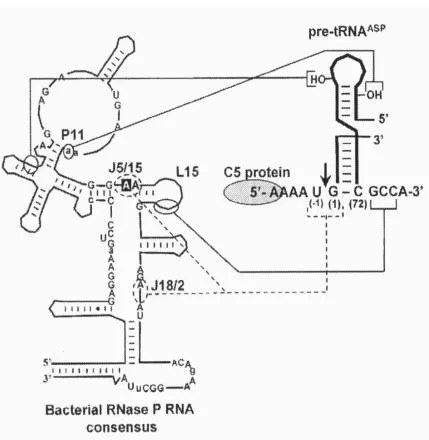
Figure 2: RNase P RNA:pre-tRNA interactions. The RCCA/GGU (73/294 interaction),
13
Type A Bacterial RNase P RNA Bacterial Minimum Consensus Type B Bacterial RNase P RNA
Eschericia coli Bacillus subtilis
15
Figure 6: RNase P RNA secondary structures from Methanothermobacter thermoautotrophicus and
Table 1: Comparison of subunits in RNase P and RNase MRP from yeast, human, and Archaea. Adapted from: Jarrous N. 2002. Human ribonuclease P: Subunits, function, and intranuclear localization. RNA 8: 1-7.
Xiao S, Scott F, Fierke CA, Engelke DR. 2002. Eukaryotic ribonuclease P: A plurality of ribonucleoprotein enzymes. Annu. Rev. Biochem. 71: 165-189.
Yeast Gene Human Gene
Subunit Type RNase P RNase MRP RNase P RNase MRP Archaeal RNase P Function/ Interaction
RNA RPR1 H1 Catalysis
RNA NME1 (7-2) Catalysis
Protein POP1 POP1 hPOP1 hPOP1 Localization
Protein POP3 POP3
Protein POP4 POP4 RPP29 RPP29 MTH11
Localization/ Substrate binding Protein POP5 POP5 hPOP5 hPOP5 MTH687
Protein POP6 POP6
Protein POP7 POP7 RPP20 ATPase/ helicase;
Protein POP8 POP8
Protein RPP1 RPP1 RPP30 RPP30 MTH688 H1 RNA binding
Protein RPR2 RPP21 MTH1618
Substrate
binding/(ATPase/ helicase)
Protein SNM1
Protein RPP38 RPP38 Localization/H1 RNA binding
Protein RPP40
Protein RPP25 H1 RNA binding
17
REFERENCES
1. Altman S. 2000. The road to RNase P. Nat. Struct. Biol. 7: 827-828.
2. Wolin SL, Matera AG. 1999. The trials and travels of tRNA. Genes Dev. 13: 1-10.
3. Zahler NH, Christian EL, Harris ME. 2003. Recognition of the 5′ leader of pre-tRNA substrates by the active site of ribonuclease P. RNA 9:734-745.
4. Frank DN, Pace NR. 1998. Ribonuclease P: Unity and diversity in a tRNA processing ribozyme. Annu. Rev. Biochem. 67: 153-180.
5. Schon A. 1999. Ribonuclease P: The diversity of a ubiquitous RNA processing enzyme. FEMS Microbio. Rev. 23: 391-406.
6. Xiao S, Scott F, Fierke CA, Engelke, DR. 2002. Eukaryotic ribonuclease P: A plurality of ribonucleoprotein enzymes. Annu. Rev. Biochem. 71: 165-189.
7. Kirsebom LA. 2002. RNase P RNA-mediated catalysis. Biochem. Soc. Trans. 30: 1153-1158.
8. Christian EL, Zahler NH, Kaye NM, Harris ME. 2002. Analysis of substrate recognition by the ribonucleoprotein endonuclease RNase P. Meth. Enz. 28: 307-322.
9. Kaye NM, Zayler NH, Christian EL, Harris ME. 2002. Conservation of helical structure contributes to functional metal ion interactions in the catalytic domain of ribonuclease P RNA. J. Mol. Biol. 324: 429-442.
10. Brannvall M, Pettersson BMF, Kirsebom LA. 2003. Importance of the 73/249 Interaction in E. coli RNase P RNA substrate complexes for cleavage and metal ion coordination. J. Mol. Biol. 325: 697-709.
11. Hall TA, Brown JW. 2001. The ribonuclease P family. Meth. Enz. 341: 56-77.
12. Pace NR, Brown JW. 1995. Evolutionary perspective on the structure and function of ribonuclease P, a ribozyme. J. Bacteriol. 177: 1919-1928.
13. Jovanovic M, Sanchez R, Altman S, Gopalan V. 2002. Elucidation of structure-function relationships in the protein subunit of bacterial RNase P using a genetic complementation approach. Nucl. Acids Res. 30: 5065-5073.
15. Haas ES, Brown JW. 1998. Evolutionary variation in bacterial RNase P RNAs. Nucl. Acids Res. 26: 4093-4099.
16. Houser-Scott F, Xiao S, Millikin CE, Zengel JM, Lindahl L, Engelke DR. 2002. Interactions among the protein and RNA subunits of Saccharomyces cerevisiae nuclear RNase P. Proc. Natl. Acad. Sci. USA. 99: 2684-2689.
17. Jarrous N. 2002. Human ribonuclease P: Subunits, function, and intranuclear localization. RNA 8: 1-7.
18. Cordier A, Schon A. 1999. Cyanelle RNase P: RNA structure analysis and
holoenzyme properties of an organellar ribonucleoprotein enzyme. J. Mol. Biol. 289: 9-20.
19. Hall TA, Brown JW. 2002. Archaeal RNase P has multiple protein subunits homologous to eukaryotic nuclear RNase P proteins. RNA 8: 296-306.
20. Harris JK, Haas ES, Williams D, Frank DN, Brown JW. 2001. New insight into RNase P RNA structure from comparative analysis of the archaeal RNA. RNA 7: 220-232.
21. Andrews AJ, Hall TA, Brown JW. 2001. Characterization of RNase P holoenzymes from Methanococcus jannaschii and Methanothermobacter thermoautotrophicus. Biol. Chem. 382: 1171-1177.
22. Haas ES, Armbruster DW, Vucson BM, Daniels CJ, Brown JW. 1996. Comparative analysis of ribonuclease P RNA structure in Archaea. Nucl. Acids Res. 24: 1252-1259.
23. Li Z, Deutscher MP. 1996. Maturation pathways for E.coli tRNA precursors: A random multienzyme pathway in vivo. Cell 86: 503-512.
24. Westaway SK, Abelson J. 1995. Splicing of tRNA precursors. In tRNA: Structure, biosynthesis, and function (ed. D. Soll and U.L. RajBhandary), pp. 79-92. American Society for Microbiology Press, Washington, D.C.
25. Stark BC, Kole R, Bowman EJ, Altman S. 1978. Ribonuclease P: An enzyme with an essential RNA component. Proc. Natl. Acad. Sci. USA. 75: 3717-3721.
19 27. Ziehler WA, Day JJ, Fierke CA, Engelke DR. 2000. Effects of 5′ leader and 3′ trailer structures on pre-tRNA processing by nuclear RNase P. Biochemistry 39: 9909-10016.
28. Kurz JC, Niranjanakumari S, Fierke CA. 1998. Protein component of Bacillus subtilis RNase P specifically enhances the affinity for precursor-tRNAAsp. Biochemistry 37: 2393-2400.
29. Niranjanakumari S, Kurz JC, Fierke CA. 1998. Expression, purification and characterization of the recombinant ribonuclease P protein component from Bacillus subtilis. Proc. Natl. Acad. Sci. USA. 95: 15212-15217.
30. Crary SM, Niranjanakumari S, Fierke CA. 1998. The protein component of Bacillus subtilis ribonuclease P increases catalytic efficiency by enhancing interactions with the 5′ leader sequence of pre-tRNAAsp. Biochemistry 37: 9409-9416.
31. Krasilnikov AS, Yang X, Pan T, Mondragon A. 2003. Crystal structure of the specificity domain of ribonuclease P. Nature 421: 760-764.
32. Gardiner KJ, Marsh TL, Pace NR. 1985. Ion dependence of the Bacillus subtilis RNase P reaction. J. Biol. Chem. 260: 5415-5419.
33. Oh BK, Frank DN, Pace NR. 1998. Participation of the 3′-CCA of tRNA in the binding of catalytic Mg2+ ions by ribonuclease P. Biochemistry 37: 7277-7283.
34. Smith D, Pace NR. 1993. Multiple magnesium ions in the ribonuclease P reaction mechanism. Biochemistry 32. 5273-5281.
35. Nagai K. 1996. RNA-protein complexes. Curr. Opin. Struct. Biol. 6: 53-61.
36. Draper DE. 1999. Themes in RNA-protein recognition. J. Mol. Biol. 293: 255-275.
37. Gopalan V, Baxevanis A, Landsman D, Altman S. 1997. Functional analysis of conserved amino acid residues in the protein subunit of ribonuclease P from Escherichia coli. J. Mol. Biol. 267: 818-829.
38. Niranjanakumari S, Stams T, Crary S, Christianson DW, Fierke CA. 1998. Protein component of the ribozyme ribonuclease P alters substrate recognition by directly contacting precursor tRNA. Proc. Natl. Acad. Sci. USA. 95: 15212-15217.
40. Frank DN, Adamidi C, Ehringer MA, Pitulle C, Pace NR. 2000. Phylogenetic-comparative analysis of the eukaryal ribonuclease P RNA. RNA 6: 1895-1904.
41. Pitulle C, Garcia-Paris M, Zamudio KR, Pace NR. 1998. Comparative structure analysis of vertebrate ribonuclease P RNA. Nucl. Acids Res. 26: 3333-3339.
42. Gray MW. 1989. The evolutionary origins of organelles. Trends Genet. 5: 294-299.
43. Dang YL, Martin NC. 1993. Yeast mitochondrial RNase P. Sequence of the RPM2 gene and demonstration that its product is a protein subunit of the enzyme. J. Biol. Chem. 268: 19791-19796.
21
Chapter 1. RNase P proteins: The homology between that of archaeal
and bacterial subunits.
ABSTRACT
No sequence similarity has been found between archaeal and bacterial protein
subunits, and yet heterologous reconstitution between the Bacillus subtilis RNase P protein
and some archaeal RNase P RNAs has been demonstrated. One of those archaeal RNAs is
that of Methanothermobacter thermoautotrophicus, that has recently been shown to be
associated with at least four protein subunits: Mth11p, Mth687p, Mth688p, and Mth1618p.
By determining a functional interchangeability between one or more of these proteins with
the Bacillus subtilis RNase P protein, an evolutionary relationship (homology) may be
identified between the protein subunits of Archaea and Bacteria. Unfortunately, in this
study, a relationship could not be identified due to poor quality pre-tRNAasp substrate
synthesized.
INTRODUCTION
RNase P is essential throughout the Domains of life, including subcellular
organelles that are involved in tRNA biogenesis. RNase P is responsible for the removal
of the 5′ leader sequence from all known precursor tRNAs (pre-tRNAs) (2, 4). The RNA subunit of RNase P by itself in Bacteria and some Archaea is capable of catalyzing the
hydrolysis of pre-tRNA in the absence of its protein cofactor (at artificially high ionic
RNA is the catalytic subunit of the enzyme (3). Most bacterial and archaeal RNase P
RNAs have retained their ancestral type (type A) secondary structure. It has also been
shown that the single bacterial RNase P protein can reconstitute enzymatic activity in the
archaeal RNase P RNA of M.thermoautotrophicus (6). Four of the nine proteins subunits
from the nuclear RNase P of Saccharomyces cerevisiae (Pop4p, Pop5p, Rpp1p, and Rpr2p)
are homologous to four methanogenic archaeal RNase P proteins; in M.
thermoautotrophicus these are Mth11p, Mth687p, Mth688p, and Mth1618p. (2, 6). These
relationships between RNase P subunits in Bacteria, Archaea, and Eukarya suggest that
heterologous reconstitution of enzymatic activity might be possible using the RNA and
protein subunits from different Domains. Our goal in this study was to determine the
functional homology between RNase P protein subunits found in Bacteria and Archaea.
Unfortunately, due to complications in the RNase P assay the homology could not be
assessed.
MATERIALS AND METHODS
Cloning and expression of recombinant Mth11p, Mth687p, Mth688p, and Mth1618p
archaeal proteins
The Mth11, Mth687, Mth688, and Mth1618 ORFs were previously amplified from
Methanothermobacter thermoautotrophicus genomic DNA and cloned as in-frame fusions
into the pET16b (Novagen) plasmid creating amino-terminal 6X His-tagged constructs
23 (100µg/ml/34µg/ml) LB-plates to select for plasmid carriers. Mth11 was amplified using primers Mth11 5′Bg1II (GGAAGATCTGATAACCCCCAGGAATATTTTCAGGC) and Mth11 3′BamHI (CGCGGATCCTATGGTTTTCTAAATTTCTTC), Mth687 was
amplified using primers Mth687 5′XhoI
(CCGCTCGAGATGAAGATACTGCCACCAACACTG) and cMth687 3′BamHI (CGGGATCCTCAGTATTTATCTTTCTTGGAAGG), Mth688 was amplified using
primers cMth688 5′XhoI (CCGCTCGAGTTGATTCCCCAAAGGATTCTG) and Mth688 3′BamHI (CGGGATCCTTAACTCTCAGGGAGGAGCC), and Mth1618 was amplified using primers Mth1618 5′Bg1II (GGAAGATCTGAGGAGAGGAAAGAGACCACG) and Mth1618 3′BamHI (CGCGGATCCTATCTGTCACTCTGGCTTTCCCC) at 100ng each, along with 10X reaction buffer (500mM KCl, 300mM Tris-Cl pH 8.3, and 15mM MgCl2),
2.5µM each dNTPs, template, 3 units of Pfu Polymerase, and nuclease-free water up to volume in 50µl reactions (Perkin Elmer DNA Thermal Cycler).
The recombinant proteins were expressed via induction of the T7 promoter with
1mM IPTG at a culture O.D. of 0.5-0.7 at 600nm approximately 4-6 hours into growth.
Four hours after induction with IPTG, cells from the 1L culture were harvested by
centrifuging at 8,000 rpm for 30 min. in the Sorvall SS-34 rotor. Four milliliters of lysis
buffer pH 8.0 (50mM NaH2PO4, 300mM NaCl, 10mM imidazole) per gram of cell
material was then added to the cell pellet. One mg/ml of lysozyme was added and then
incubated on ice for 30 min. They were then sonicated on ice and again centrifuged at
8,000 rpm for 30 min and the supernatant was collected. One ml of this cleared lysate was
Each lysate-Ni-NTA mixture was loaded into a separate column and washed twice with
25mL of wash buffer pH 8.0 (50mM NaH2PO4, 300mM NaCl, 20mM imidazole) then
eluted with elution buffer pH 8.0 (50mM NaH2PO4, 300mM NaCl, 250mM imidazole).
Protein in fractions from each wash were electrophoresed in 12% SDS-PAGE gels. The
gels were then stained, while shaking, with Coomassie-R250 and then destained, while
shaking, to visualize the recombinant protein.
In vitro transcription
α32P labeled RNA transcripts of Bacillus subtilis (pre-tRNAasp) were generated by run-off transcription from the T7 promoter on plasmid pDW128. Cleavage of pDW128 for
run-off termination was achieved via restriction digest of pDW128 (David Waugh) using
BstNI. Reaction contents include transcription optimized 5X buffer(200mM Tris-Cl pH
7.9 at 25°C, 50mM NaCl, 30mM MgCl2, and 10mM spermidine), 100mM DTT, 1.25µl RNasin, 10mM (ATP, CTP, UTP) and 2mM GTP, approximately 0.5µg of template, 50 units of recombinant T7 polymerase, and 100 µg α32P labeled GTP. Reactions were
incubated for 2 hours at 37°C. Products were separated on an 8% urea-polyacrylamide gel, visualized by autoradiography, excised and passively eluted into 10mM Tris pH 8, 1mM
EDTA, and 0.1% SDS by shaking overnight at room temperature. The elutates were then
phenol extracted, RNA was ethanol precipitated, and quantitated in the Beckman LS 6500
scintillation counter.
Plasmids containing the genes encoding RNase P RNAs (E. coli and
25 RNase P RNA containing plasmid pDW98 was digested with SnaBI while M.
thermoautotrophicus RNase P RNA containing plasmid ∆H trimmed #18 was digested with BsaI.
RNase P Ribozyme Assay
RNase P assay reactions of E. coli and M. thermoautotrophicus RNase P RNAs,
were performed using a 90 µl premix of dried substrate (1 kcpm/reaction), 10X P Buffer (25mM MgCl2, 0.05% nonidet P-40, and 50mM Tris) (9µl), 5M NH4OAc (18µl for E.coli RNase P RNA and 54µl for M. thermoautotrophicus RNase P RNAs), and nuclease-free water (54µl for E.coli and 18µl for M. thermoautotrophicus RNase P RNAs) added to one µl of RNase P RNA prepared in serial 1:5 dilutions. Each 10µl reaction mixture was incubated for 10 min. at 37°C. The products were separated on an 8% urea-polyacryamide gel and visualized by phosphoimagery.
RESULTS AND DISCUSSION
Expression of recombinant Mth11, Mth687, Mth688, and Mth1618 Archaeal Proteins
After transformation of the recombinant protein plasmids into E.coli there was
some difficulty in confirming the presence of the open reading frames (ORFs) by
amplification. The initial method used after transformation to prepare the cells for
expression of the recombinant proteins was: (1) plating onto LB (AMP-CAM), (2)
checking for archaeal inserts in the plasmid via PCR amplification, (3) transferring to
LB (AMP-CAM) broth, then again (4) checking for archaeal inserts in the plasmid via
M. jannashii ORF 1625, may cause the cells to either die or lose the plasmid (personal
communication C. Ellis). This protocol was optimized by excluding the testing for the
presence of the insert via amplification, eliminating the need for repeated subcultivation.
Induction of protein over-expression was achieved by addition of IPTG (Figure 1).
Mth11p, Mth1618p, Mth687p, and Mth688p have been previously shown to run in 12%
SDS-PAGE gels at 15.5, 23.5, 18 and 31.5 kDa, respectively. These proteins were to be
used in gel-shift binding assay and the enzymatic cleavage assays, although time
constraints made this impossible.
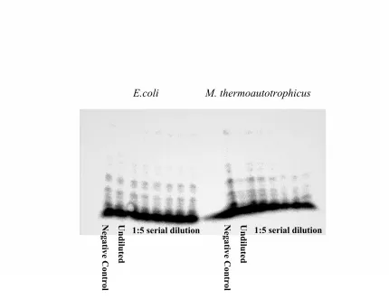
RNase P Ribozyme Assay
In a typical ribozyme reaction the RNase P RNA would remove the 5′ leader sequence from the pre-tRNAAsp substrate (under high ionic strength), although E. coli and
M. thermoautotrophicus RNase P RNAs ability to catalyze the cleavage of α32P labeled Bacillus subtilis pre-tRNAAsp substrate was impaired. More degradation bands were found
than were to be expected in the 8% urea-polyacrylamide gel, attributed to poor substrate
quality (Figure 2).
Titration, Gel-shift Binding, and Enzymatic Cleavage Assays
Three more assays were planned to be performed to determine the functional
homology between the bacterial and archaeal protein subunits: (1) a gel-shift binding assay
would be used to determine the organization of the archaeal protein subunits that bind to
the bacterial RNA (Figure 4), (2) A titration assay would be used to determine the Kd of
27 would be the inverse of the B. subtilis protein: M. thermoautotrophicus RNA experiment
already published (Figure 5). It remains to be shown which, if any, of the archaeal/nuclear
RNase P proteins are functionally homologous to the bacterial protein.
A. B.
W E W E
LMW L F I II III I II III IV LMW F I II I
C. D.
W E W E
LMW L F I II III IV I II III IV F I II I LMW
Figure 1: Expression of M. thermoautotrophicus RNase P recombinant proteins (A) 11p,
(B)1618p, (C)688p, and (C)687p. (A) 12% SDS-PAGE gel of Mth11p, contents from left to right: low molecular weight marker (LMW), lysate (L), flow-through (F), wash (W) I-III, and elution (E) I-IV. (B) 12% SDS-PAGE gel of Mth1618p, contents from left to right: low molecular weight (LMW), flow-through (F), wash (W) I and II, and elution (E) I. (C) 12% SDS-PAGE gel of Mth688p, contents from left to right: low molecular weight marker (LMW), lysate (L), flow-through (F), wash (W) I-IV, and elution (E) I-IV. (D) 12% SDS-PAGE gel of Mth687p, contents from left to right: flow-through (F), wash (W) I and II, elution (E) I, and low molecular weight marker (LMW). The Low molecular weight marker is Phosphorylase at 94kDa, BSA at 67kDa, Ovalbumin at 43kDa, Carbonic Andydmi at 30kDa, Soybean Trypsin at 20.1kDa, and α-Lactolbumin at 14.4kDa.
29
Figure 2: RNase P ribozyme assay. E. coli RNase P RNA ribozyme reaction on left and
M. thermoautotrophicus RNase P RNA ribozyme reaction on right. Lane contents from left: (1) Negative control with no RNase P RNA, (2) Undiluted RNase P RNA, and (3-8) 1:5 serial dilutions of RNase P RNA.
Undiluted
Negative Control
1:5 serial dilution
E.coli M. thermoautotrophicus
Negative Control Undiluted
31
Figure4: Gel-shift binding assay (shown diagrammatically). This is an example of how
33
REFERENCES
1. Andrews AJ, Hall TA, Brown JW. 2001. Characterization of RNase P holoenzymes from Methanococcus jannaschii and Methanothermobacter thermoautotrophicus. Biol. Chem. 382: 1171-1177.
2. Xiao S, Scott F, Fierke CA, Engelke DR. 2002. Eukaryotic ribonuclease P: A plurality of ribonucleoprotein enzymes. Annu. Rev. Biochem. 71: 165-189.
3. Pannucci JA, Haas ES, Hall TA, Harris JK, Brown JW. 1999. RNase P RNAs from some Archaea are catalytically active. Proc. Natl. Acad. Sci. USA. 96: 7803-7808.
4. Kirsebom LA. 2002. RNase P RNA-mediated catalysis. Biochem. Soc. Trans. 30: 1153-1158.
5. Pace NR, Brown JW. 1995. Evolutionary perspective on the structure and function of ribonuclease P, a ribozyme. J. Bacteriol. 177: 1919-1928.
Chapter 2. Comparative analysis of the bacterial RNase P RNA
structure
ABSTRACT
Eleven new RNase P RNA sequences from a range of negative and
Gram-positive Bacteria have been amplified, sequenced, and their secondary structures derived
through comparative analysis. These sequences and structures will contribute to the
ongoing refinement of the model of RNase P RNA structure and evolution.
INTRODUCTION
Ribonuclease P (RNase P) is the endoribonuclease that catalyzes removal of the
leader sequence in all known precursor-tRNAs (pre-tRNAs), generating the mature 5′ end of the tRNA. (1-3). In all organisms, RNase P is composed of conserved RNA and
variable protein subunit(s). The RNA subunit (not the protein) is the catalyst; RNase P is a
ribozyme. In Bacteria and most Archaea, the catalytic RNA is functional in vitro in the
absence of protein. In Eukarya and other Archaea, the RNA depends absolutely on protein
for function.
Construction of the RNase P RNA secondary structure is needed to understand the
mechanisms of substrate recognition, catalytic activity, and evolution of the ribozyme (6).
The first RNase P RNA sequences determined were those of Escherichia coli and Bacillus
subtilis. Comparative analysis of the RNase P RNA sequences of these organisms and
35 RNAs that were first characterized, those of Escherichia coli and Bacillus subtilis (8).
Type A, the ancestral type, is the most common type of RNase P RNA secondary structure
and is represented by that of E. coli. This type can be found in among various groups of
Gram-negative and Gram-positive Bacteria, whereas type B RNAs, represented by that of
B. subtilis, are restricted to a subset of the low G+C Gram-positive Bacteria. The
secondary structure of type B RNase P RNAs are quite different than their type A
counterparts, but they contain a core of conserved sequence and structure (Figure 1). In
addition, the ability to interchange protein subunits between Escherichia coli and Bacillus
subtilis RNase P enzymes indicates an overall similarity in the tertiary structure of these
RNAs (7).
In this study eleven new RNase P RNAs have been amplified and sequenced as part
of the ongoing refinement of the RNase P RNA structure through comparative analysis.
The organisms from which the RNase P RNAs originated were chosen from Carolina
Biological Supply, because the genera were not represented in the RNase P Database.
MATERIALS AND METHODS
Growth of Organisms
Fifteen bacterial organisms were ordered from Carolina Biological Supply with the
intent of characterizing their RNase P RNAs. The catalog number, organism, phylogenetic
group, medium, and growth temperature can be found in Table 1. All of these organisms
Polymerase Chain Reaction
Polymerase chain reaction (PCR) was carried out in a Perkin Elmer GeneAmp PCR
System 2400. The ReadyMix Taq PCR Reaction Mix (SIGMA), used to amplify RNase P,
contained Taq DNA Polymerase in a 2X concentrate containing the nucleotides and
reagents necessary for standard PCR: 25µl of 2X ReadyMix (1.5 units of Taq DNA Polymerase, 10mM Tris-HCl (pH 8,0), 50mM KCl, 1.5mM MgCl2, 0.001% gelatin,
0.2mM dNTP), one µl each of appropriate forward and reverse primers (2ng/mL final concentration) (59FBam (CGGGATCCGIIGAGGAAAGTCCIIGC), 347REco
(CGGAATTCRTAAGCCGGRTTCTGT), or 347RXba
(GCTCTAGATAAGCCRYGTTYTGT) (I=Inosine, Y=C+T, R=A+G)), circa 1/10thµl of cell material, and water up to a volume of 50µl. The amplifications were performed with an initial holding temperature of 94°C at 3 min. followed by 30 cycle sets of 94°C for 1.5 min., 50°C for 1.5 min., and 75°C for 2 min, and two final holding temperature of 75°C for 15 min. and 10°C until the tubes were collected for storage at 4ºC. PCR products were purified from reactions using the QIAquick PCR purification kit.
Sequencing
Sequences of the RNase P RNA genes were determined from double-stranded PCR
products using an ABI Prism 377 sequencer (Iowa State University DNA Sequencing and
37
Sequence/Structure Construction and Analysis
The forward and reverse sequences of the RNase P RNA PCR products were
assembled and edited using ABI Prism EditView. A BLAST Search of Genbank was
performed using the forward sequence to identify the nearest related available sequence,
and this guide secondary structure was obtained from the RNase P Database (8).
Secondary structures were then constructed manually. Conserved elements were included
by superimposing homologous regions within their related structure, while the structure of
variable elements were predicted using MFOLD (Michael Zukar). The sequences were
then added to the existing RNase P RNA alignment on the basis of their structures, using
SeqApp and SeqPup (Don Gilbert, Indiana University). Phylogenetic trees were garnered
using Phylip by the Neighbor-Joining Method (10).
RESULTS AND DISCUSSION
Of the fifteen organisms from Carolina Biological Supply, eleven RNase P RNAs
were successfully amplified and sequenced. These organisms were chosen, because the
genera were not represented in the RNase P Database. The RNase P RNA genes from
these organisms were amplified with primers targeted to the conserved P4 region. Of
these, ten were of the bacterial secondary structure type A: Citrobacter freundii,
Enterobacter aerogenes, Shigella flexneri, Acinetobacter calcoaceticus, Branhamella
catarrhalis, Aquaspirillum serpens, Chromobacterium violaiceum, Spirillum volutans,
Acetobacter aceti and Gluconobacter oxydans. One was type B, that of Sporosarcina
ureae. The type A RNAs were all from the Kingdom Proteobacteria, whereas
The Citrobacter freundii, Enterobacter aerogenes, Shigella flexneri, Acinetobacter
calcoaceticus, Branhamella catarrhalis sequence are consistent with their placement in the
and categorized as χ-purple Bacteria (proteobacteria). The Acinetobacter calcoaceticus RNase P RNA structure was derived largely by comparison to Volunteer ESH7-9 RNase P
sequence, although it contained a shorter P12 and a few nucleotide changes (Figure 2A).
P12 in bacterial type A RNAs is an extremely variable region in sequence, length, and
structure after the conserved base of the first five base-pairs that are thought to interact
with P13 (9).
The Branhamella catarrhalis RNase P RNA was derived by comparison to the
Haemophilus influenza RNase P RNA (Figure 2B). The helices are similar in length, but
vary in sequence. This is typical for most RNase P RNAs in that there is significant
sequence variability in the helices, indicating that the end-loops are more significant in
substrate, protein, self, or cationic binding or that it is the base pairs rather than nucleotide
identities that contribute to RNA structure and function. The last three χ-purple Bacteria are from a specific branch of the Kingdom known as Enterobacteriaceae, or enterics.
These Bacteria are typically found in the intestinal tract of animals.
The Enterobacter aerogenes RNase P RNA was derived by comparison to that of
Klebsiella pneumoniae, also an enteric (Figure 2C). Two nucleotide substitutions at the
end-loop of P9 (L9) are the only differences between the RNAs of E. aerogenes and K
pneumoniae. In bacterial RNase P RNAs, the GNRA L9 forms a tertiary interaction with
39 The Citrobacter freundii RNase P RNA was most similar in sequence to that of
another enteric, Salmonella typhimurium (Figure 2D). The only difference found was a
nucleotide substitution in the four-nucleotide bulge between P16 and P17. This bulge is
extremely variable in size and sequence, and probably serves only as a point of flexibility
between P16 and P17.
The final enteric RNase P RNA was from Shigella flexneri, which was compared to
that of Escherichia coli K-12 (Figure 2E). There is only sequence variation in P12, which
is typical for very closely related RNase P RNAs. The conserved nature of the enteric
RNase P RNAs as well as those of Acinetobacter calcoaceticus and Branhamella
catarrhalis can be seen in the multiple-alignment of type A bacterial RNase P RNAs
(Figure 3) and the phylogenetic tree based on this alignment (Figure 4).
The Aquaspirillum serpens, Chromobacterium violaceum, and Spirillum volutans
RNase P RNA sequences are consistent with their placement in the β-purple bacteria (proteobacteria). The Aquaspirillum serpens RNase P RNA was modeled after the RNase
P RNA of Neisseria meningitidis (Figure 2F). Although this RNA is its closest available
relative, it contains multiple differences, including a number of nucleotide substitutions in
the helices, a conserved GNRA tetra-loop in P12, and the presence of P19. Nucleotide
substitutions in helices are common among different RNase P RNAs, and the GNRA
tetraloop of P12, according to the sequence logo of type A RNase P RNAs (Figure 7), is
not highly conserved. The most significant difference is the addition of P19, which where
present is thought to serve to stabilize adjacent helices via stacking (probably P2) (1).
The Chromobacterium violaceum RNase P RNA was compared to the Volunteer
while C. violaceum has an additional cruciform located in P12. The Volunteer ESH7-4
RNase P RNA sequence was previously considered to be of uncertain affiliation. The
similarity of this RNA to that of C. violaceum indicates that this unknown RNase P RNA
may belong to a β-purple bacterium.
Finally, the RNase P RNA of Spirillum volutans, a β-proteobacteria; was derived by comparison to the χ-purple bacteria E. coli (Figure 2H). Comparative analysis of a β -purple to that of a χ-purple bacteria may seem inappropriate, but this sequence is only distantly-related to any other known RNase P RNA. The RNase P RNAs from the β -purple Bacteria vary more in sequence and structure than the χ-purple bacteria. In the phylogenetic tree (Figure 4), the S. volutans RNA does not group with the other β-purple Bacteria, but seems to reside somewhere near Chlamydia or Spirochaetes, as does the α
-purple bacterium B. aphidicola.
The Acetobacter aceti and Gluconobacter oxydans RNase P RNAs sequences are
consistent with those of other α-purple Bacteria. The partial RNase P RNA from Acetobacter aceti is similar to that of the unknown RNase P RNA CP-A62 (Figure 2I).
CP-A62 RNase P RNA has been considered to be of uncertain affiliation, although its
similarity to A. aceti implies that it may be an α-purple Bacteria. Overall the structures are the same, with typical length variations in P17 and P19 (9). P10/11 helices in Acetobacter
aceti and CP-A62 appear different, although the single-stranded RNA fork after P10/11 in
41 The RNase P RNA from Gluconobacter oxydans was derived by comparison to
that of Rhodospirillum rubrum A348 RNase P RNA (Figure 2J). These RNAs share
significant structural similarities, although there is sequence and length variation in P12
and P17/18/19. These two α-purple bacteria were found to coincide with typical α-purple RNase P RNAs (Figures 3 and 4).
The RNase P RNA of Sporosarcina ureae was the only type B RNase P RNA
obtained. It is phylogenetically characterized as a low G+C Gram positive bacterium. The
structure of the RNA was derived by comparison to that of Bacillus subtilis 168 (Figure
2K). Type B RNase P RNAs contain helices that differ from type A RNAs, ie. P5.1,
P10.1, and P15.1. There are significant structural similarities between S. ureae and B.
subtilis, with typical sequence variations (Figure 2K, 5). Known tertiary interactions are
conserved.
The addition of eleven new partial RNase P RNAs to the already available 427
bacterial RNase P RNAs in the RNase P Database is crucial to the refinement of those and
Type-A Bacterial RNase P RNA Bacterial Minimum Consensus Type-B Bacterial RNase P RNA
Escherichia coli Bacillus subtilis
43
A.
Figure 2: Comparative analysis of eleven new RNase P RNAs and their derivatives.
45
C.
47
E.
49
G.
51
I.
53
K.
Taken From:
A.serpens 1 --- 1
C.violacium 1 --- 1
S.flexneri 1 --- 1
A.calcoa 1 --- 1
A.aceti 1 --- 1
G.oxydan 1 --- 1
B.catarr 1 --- 1
C.freund 1 --- 1
S.volutan 1 --- 1
E.aerogen 1 --- 1
E.coli 1 GAAGCUGACCAGACAGUCGCCGCUUCGUCGUCGUCCUCUUCGGGGGAGACGGGCGGAGGG 60 A.serpens 1 GAGGAAAGUCCGGGCUUCAUAGAG-CAGAAUGCUGGCUAACGGCCAGGCGUCGU--AAGG 57 C.violacium 1 GAGGAAAGUCCGGGCUCCGCAGAG-CAGGAUGCCGGCUAACGGCCGGGCGCCGCG--AGG 57 S.flexneri 1 GAGGAAAGUCCGGGCUCCAUAGGG-CAGGGUGCCAGGUAACGCCUGGGGGGGA----AAC 55 A.calcoa 1 GAGGAAAGUCCGGGCUUCAUAGGG-CAGGGUGCCAGGUAACGCCUGGGCGGUG--AAAGC 57 A.aceti 1 GAGGAAAGUCCGGGCUCCACGGGAGAACGGUGCCGGCUAACGGCCGGCGAGGGUG--ACC 58 G.oxydan 1 GAGGAAAGUCCGGGCUC-AUGGGAAGACGGUACCGGCUAAUUGCCGGCGGGGGCGU-ACC 58 B.catarr 1 GAGGAAAGUCCGGGCUACAUAGGG-CAAUGUGCUAGCUAACAGCUAGGGGGCG--AAAGC 57 C.freund 1 GAGGAAAGUCCGGGCUCCAUAGG--CAGGGUGCCAGGUAACGCCUGGGGGGGA----AAC 54 S.volutan 1 GAGGAAAGUCCGGGCUGCAUAGAG-CAAAAUGCCAGUUAAUGGCUGGAAGUUGU---GAA 56 E.aerogen 1 GAGGAAAGUCCGGGCUCCAUAGGG-CAGG-UGCCAGGUAACGCCUGGGGGGUGUCACAGC 58 E.coli 61 GAGGAAAGUCCGGGCUCCAUAGGG-CAGGGUGCCAGGUAACGCCUGGGGGGGA----AAC115 A.serpens 58 CGACGGAA-AGUGGAACAGAGCAGCUGAACCGCCGA---UGGCUUC--- 99
C.violacium 58 CGACGGAA-AGUGGAACAGAGAG-CUGAACCGCCGAUGGCCGGACUUGUCGACCGCGAGG115 S.flexneri 56 CCACGACC-AGUGCAACAGAGAG--CAAACCGCCGA---UGGCCCG--- 95
A.calcoa 58 CGACGGAA-AGUGCAGCAGAGAG--UAGACCGCC---UCAUUCG--- 95
A.aceti 59 UCAGGGAA-AGUGCCACAGAAAA--CAGACCGCCUU---CCGGGCC--- 98
G.oxydan 59 CCAGGGNACAGUGCCACAGAAAA--CAGACCGCCCG---CCGGACUGUGCC104 B.catarr 58 CUACGACC-AGUGCAGCAGAGAA--UAGACCGCCU---AUUA--- 93
C.freund 55 CCACGACC-AGUGCAACAGAGAG--CAAACCGCCGA---UGGCCCA--- 94
S.volutan 57 ACUUGGAA-AGUGCCACAGAAAA--UAAACCGCCUC---UUUGUUA--- 96
E.aerogen 59 CCACGACC-AGUGCAACAGAGAG--CAAACCGCCGA---UGGCCCG--- 98
E.coli 116 CCACGACC-AGUGCAACAGAGAG--CAAACCGCCGA---UGGCCCG---155
A.serpens 99 ---GCAAGAAGA---UCAGGUAAG 117
C.violacium 116 UCGACAAAGUCUUUUCCCGGGGCUUGCCCCGGAAAAACCCGCAAGGGCGGAUCAGGCAAG 175
S.flexneri 95 ---U---GCAAGCGGG---A---UCAGGUAAG 115
A.calcoa 95 ---U---G---AGGUAAG 104
A.aceti 98 ---U---GUUCGCAGG---CUGGAGGUAAG 119
G.oxydan 105 UCAUGCGUUC---AUUCGCGGGGGAACAUGUUGCGGGUAAG 142
B.catarr 93 ---A---AU---AGGUAAG 103
C.freund 94 ---C---GUAAGUGGG---A---UCAGGUAAG 114
S.volutan 96 ---U---AAA-G---AGGUAAG 108
E.aerogen 98 ---C---GCAAGCGGG---A---UCAGGUAAG 118
E.coli 155 ---C---GCAAGCGGG---A---UCAGGUAAG 175
Figure 3: Type A bacterial RNase P RNA multiple sequence alignment. These sequences
were aligned manually on the basis of secondary structure using Bioedit. Shaded regions indicate the most conserved nucleotide. A. serpens is Aquaspirillum serpens , C.
55
A.serpens 118 GGUGAAAAGGUG---UG 131
C.violacium 176 GGUGAAAAGGUGUACCGGGAGGCGCAAGCCGGCCCGGACGGGCGGGGCAACCCGCUCGCG 235
S.flexneri 116 GGUGAAAGGGUG---CG 129
A.calcoa 105 GGUGAAAGGGUG---CG 118
A.aceti 120 GGUGAAACGGUG---CG 133
G.oxydan 143 GGCGAAACGGUG---CG 156
B.catarr 104 GGUGAAACGGUG---GG 117
C.freund 115 GGUGAAAGGGUG---CG 128
S.volutan 109 GGUGAAACGGCG---UG 122
E.aerogen 119 GGUGAAAGGGUG---CG 132
E.coli 176 GGUGAAAGGGUG---CG 189
A.serpens 132 GUAAGAGCACACCGCGUAACUGGUAACAGGCUACGGCAGGCUAAACCCCAUUCGAAGCAA 191
C.violacium 236 GUAAGAGCGCACCGCGGACGUGGCGACA-CGGACGGCAGGCUAAACCCCAUCCGGAGCAA 294
S.flexneri 130 GUAAGAGCGCACCGCGCGGCUGGUAACAGUCCGUGGCACGGUAAACUCCACCCGGAGCAA 189
A.calcoa 119 GUAAGAGCGCACCGCGUGUCUGGCAACAGUUCACGGCAUGGUAAACCCCACCAGAAGCAA 178
A.aceti 134 GUAAGAGCGCACCGCGCUUCCGGUAACGGCGGCGG-CAGGGCAAACCCCAUCGGGAGCAA 192
G.oxydan 157 GCAAGAGCGCACCGCAUCUCCGGCAACGGCGAUGGUCAUGGUAAACCCUACCGGGAGCAA 216
B.catarr 118 GUAAGAGCCCACCGCGUGACUGGCGACAGCUCACGGCAAGGUAAACUCCACAUGUAGCAA 177
C.freund 129 GUAAGAGCGCACCGCGCGGCUGGUAACAGUCCGUGGCACGGUAAACUCCACCCGGAGCAA 188
S.volutan 123 GUAAGAGCACACCGCUGGCUUGGUAACAAGACA-GGCAAGGUAAACCCCAUUUGCAGCAA 181
E.aerogen 133 GUAAGAGCGCACCGCGCGGCUGGUAACAGUCCGCGGCACGGUAAACUCCACCCGGAGCAA 192
E.coli 190 GUAAGAGCGCACCGCGCGGCUGGUAACAGUCCGUGGCACGGUAAACUCCACCCGGAGCAA 249
A.serpens 192 GACCAAGCAGGGCG---CG---CUAACGUGGCUCGCGUUGUGCCCG 231
C.violacium 295 GACCAAAUAGGGGGA---CG---CCAACGUGGCUCGCGUUGUCCCCG 335
S.flexneri 190 GGCCAAAUAGGGGUU---CA---UAAGGUACGGCCCGUACUGAACCCG 231
A.calcoa 179 GACCAAAUAGGAAUC---C---UGAGGUACGGCCCGUACUGGAUUCG 219
A.aceti 193 GACCGAAUAGGGACGGUAGG---CAGCCUGACUGCCAAAGGUUCUCCCGCCUUCCGUCCG 249
G.oxydan 217 GACCGAAUAGGAACGGCAGGUUUCCGCAAGGAAACCGGGGGUUUCUCGCCCCGUCGUUCG 276
B.catarr 178 GACCAAAUAGGAACU---C---AAUGGGUUGCCCGCUCAGAGUUCG 217
C.freund 189 GGCCAAAUAGGGGUU---CA---CAAGGUACGGCCCGUACUGAACCCG 230
S.volutan 182 GACCAAAUAAGGAU---CA---AAUCAUGUGGCUCGCAUGUGAUUCG 222
E.aerogen 193 GGCCAAAUAGGGGUU---CA---UAAGGUACGGCCCGUACUGAACCCG 234
E.coli 250 GGCCAAAUAGGGGUU---CA---UAAGGUACGGCCCGUACUGAACCCG 291
A.serpens 232 GGUAGGUUGCUGGAGCCUGUCAGUAAUGGCAGGCCUAGAGGAAUGACUGUCUGGGGCGUU 291
C.violacium 336 GGUAGGUUGCUUGAGCCUGUCGGCAACGGCAGGCCUAGAGGAAUGAUUGUCCAA----C- 390
S.flexneri 232 GGUAGGCUGCUUGAGCCAGUGAGCGAUUGCUGGCCUAGAUGAAUGACUGUCCAC--- 285
A.calcoa 220 GGUAGGUCGCUUGAGCGUAUGAGUGAUUGUACGCCUAGAGGAAUGACCAUCCUC--- 273
A.aceti 250 GGUUGGUCGCGUGAGGCGUGUUGCAAGACACGUCCCAGAGGAAUGAUCGUCACGGUCUCU 309 G.oxydan 277 GGUUGGUUGCGUGAGGCCUGCAGCAAUGCAGGUCCCAGAGGAAUGAUCGUCCCG----CU 332 B.catarr 218 GGUAGGUUGCUUGAGCAUAUCAGCAAUGAUAUGCCUAGAUGAAUGAUUGUCCAC--- 271
C.freund 231 GGUAGGCUGCUUGAGCCAGUGAGCGAUUGCUGGCCUAGAUGAAUGACUGUCCAC--- 284
S.volutan 223 GGUAGGUCGCUUGAGCAAUAGUGUGAACUAUUGCCUAGAUGAAUGACUGUUCAC--- 276
E.aerogen 235 GGUAGGCUGCUUGAGCCAGUGAGCGAUUGCUGGCCUAGAUGAAUGACUGUCCAC--- 288
E.coli 292 GGUAGGCUGCUUGAGCCAGUGAGCGAUUGCUGGCCUAGAUGAAUGACUGUCCAC--- 345
A.serpens 292 UAUCGCCUAACACAGAANCCGGCUUA--- 317
C.violacium 390 ---G---ACAAAACACGGCUUA--- 406
S.flexneri 285 ---G---ACAAAACACGGCUUA--- 301
A.calcoa 273 ---G---ACAAAACACGGCUUA--- 289
A.aceti 310 UCGGAGAACGGACAAAACACGGCUU--- 334
G.oxydan 333 UUC--GGGCGGACAAAACACGGCUUA--- 356
B.catarr 271 ---G---ACANAACNCGGCUUA--- 287
C.freund 284 ---G---ACAAAACACGGCUUA--- 300
S.volutan 276 ---G---ACAAAACACGGCUUU--- 292
E.aerogen 288 ---G---ACAAAACACGGCUUA--- 304
E.coli 345 ---G---ACAGAACCCGGCUUAUCGGUCAGUU 371
57
Figure 4: Type A bacterial RNase P RNA neighbor-joining tree. New RNase P RNAs are
59
B.subtilis 1 GUUCUUAACGUUCGGGUAAUCGCUGCAGAUCUUGAAUCUGUAGAGGAAAGUCCAUGCUCG 60
S.ureae 1 ---GAGGAAAGUCCGGGCUCA 18
B.subtilis 61 CACGGUGCUGAGAUGCCCGUAGUGUUCGUGCCUAGCGAAGUCAUAAGCUAGGGCAGUCUU 120
S.ureae 19 CACGGAUCUGAGAUGACCGUAGUGUUCGUGCUCGGUGAAAAAAUAAGCCGGGGCAAUGCC 78
B.subtilis 121 UAGAGGCUGACGGCAGGAAAAAAGCCUACGUCUUCGGAUAUGGCUGAGUAUCCUUGAAAG 180
S.ureae 79 UCUGCAUUGAUGGCGGGAAUAACUCCUAAGUCUUCGGAUAUGGACUAGAUUCUCUGAAAG 138
B.subtilis 181 UGCCACAGUGACGAAGUCUCACUAGAAAUGGUGAGAGUGGAACGCGGUAAACCCCUCGAG 240
S.ureae 139 UGCCACAGUGACGUAGUCGGCAUGGAAACAUGUCGAGUGGAACG-GGUAAACCCCACGAG 197
B.subtilis 241 CGAGAAACCCAAAUUUUGGUAGGGGAACCUUCUU--AACGGAAUUCAACGGAGAGAAGGA 298
S.ureae 198 UGAGAAACCCAAAUUAUGGUAGGGG--CGUUCUUCCGAAAGAAUUCAAUGGAGGAAGGAA 255
B.subtilis 299 CAGAAUGCUUUCUGUAGAUAGAUGAUUGCCGCCU-GAGUACGAGGUGAUGAGCCGUUUGC 357
S.ureae 256 CAGGCAAUUGUCUGUAGAUAGAUGAUUACCACUUCAAGUACGAGGCG-CGAGCCGUUUG- 313
B.subtilis 358 AGUACGAUGGAACAAAACAUGGCUUACAGAACGUUAGACCACU 400
S.ureae 314 AGUACCGAAGAACAAAACACGGCUUA--- 339
Figure 5: Type B bacterial RNase P RNA multiple sequence alignment. These sequences
61
Figure 7: Sequence logo of type A RNase P RNAs. Numbering is based on the E. coli
Table 1: List of Organisms providing new RNase P RNAs. Carolina Supply Catalog # Organism Phylogenetic Group Medium Growth Temp.
WW-15-4810 Acetobacter aceti α
-proteobacteria
Mannitol
Special Agar 30°C WW-15-4822A Acinetobacter
calcoaceticus χproteobacteria -
Nutrient
Agar 37°C WW-15-4828 Aeromonas
sobria χproteobacteria -
Nutrient
Agar 25°C WW-15-4835A Alcaligenes
faecalis βproteobacteria
-Nutrient
Agar 37°C WW-15-4838 Aquaspirillum
serpens βproteobacteria
-Nutrient
Agar 30°C WW-15-4840 Arthrobacter
globiformis
High G+C Gram Positive
Nutrient
Agar 30°C WW-15-4929 Branhamella
catarrhalis χproteobacteria -
Brain Heart Infusion Agar
37°C
WW-15-4931A Chromobacterium
violaceum βproteobacteria
-Nutrient
Agar 30°C WW-15-4941 Citrobacter
freundii χproteobacteria -
Nutrient
Agar 37°C WW-15-5030 Enterobacter
aerogenes χproteobacteria -
Nutrient
Agar 30°C WW-15-5087 Gluconobacter
oxydans αproteobacteria
-Gluconobact
er Agar 25°C WW-15-5270 Rhizobium
leguminosarum αproteobacteria
-Rhizobium
X Agar 25°C WW-15-5470A Shigella flexneri χ-
proteobacteria
Nutrient
Agar 37°C WW-15-5512 Spirillum
volutans βproteobacteria
- Peptone-Succinate Medium
30°C
WW-15-5518 Sporosarcina ureae
Low G+C Gram Positive
Nutrient
63
REFERENCES
1. Haas ES, Brown JW. 1998. Evolutionary variation in bacterial RNase P RNAs. Nucl. Acids Res. 26: 4093-4099.
2. Frank DN, Pace NR. 1998. Ribonuclease P: Unity and diversity in a tRNA processing ribozyme. Annu. Rev. Biochem. 67: 153-180.
3. Christian EL, Zahler NH, Kaye NM, Harris ME. 2002. Analysis of substrate recognition by the ribonucleoprotein endonuclease RNase P. Meth. Enz. 28: 307-322.
4. Jovanovic M, Sanchez R, Altman S, Gopalan V. 2002. Elucidation of structure-function relationships in the protein subunit of bacterial RNase P using a genetic complementation approach. Nucl. Acids Res. 30: 5065-5073.
5. Haas ES, Banta AB, Harris JK, Pace NR, Brown JW. 1996. Structure and evolution of ribonuclease P RNA in Gram-positive Bacteria. Nucl. Acids Res. 24: 4775-4782.
6. Brown JW, Haas ES, James BD, Hunt DA, Pace NR. 1991. Phylogenetic analysis and evolution of RNase P RNA in proteobacteria. J. Bacteriol. 173: 3855-3863.
7. Hall TA, Brown JW. 2001. The ribonuclease P family. Meth. Enz. 341: 56-77.
8. Brown JW. 1999. The ribonuclease P database. Nucl. Acids Res. 27: 314.
9. Harris JK, Haas ES, Williams D, Frank DN, Brown JW. 2001. New insight into RNase P RNA structure from comparative analysis of the archaeal RNA. RNA 7: 220-232.
10. Felsenstein J. 1989. Phylip- phylogeny interference package (Version 3.2). Cladistics. 5: 164-166.