Drug Design, Development and Therapy
Dovepress
O r i g i n a l r e s e a r c h
open access to scientific and medical research
Open access Full Text article
indole and synthetic derivative activate chaperone
expression to reduce polyQ aggregation in sca17
neuronal cell and slice culture models
Pin-Jui Kung1,*
Yu-chen Tao1,*
ho-chiang hsu1
Wan-ling chen1
Te-hsien lin1
Donala Janreddy2
ching-Fa Yao2
Kuo-hsuan chang3
Jung-Yaw lin1
Ming-Tsan su1
chung-hsin Wu1
guey-Jen lee-chen1
hsiu-Mei hsieh-li1
1Department of life science, 2Department of chemistry, national
Taiwan normal University, Taipei, Taiwan; 3Department of neurology,
chang gung Memorial hospital, chang gung University college of Medicine, Taipei, Taiwan
*These authors contributed equally to this work
Abstract: In spinocerebellar ataxia type 17 (SCA17), the expansion of a translated CAG repeat
in the TATA box binding protein (TBP) gene results in a long polyglutamine (polyQ) tract in the TBP protein, leading to intracellular accumulation of aggregated TBP and cell death. The molecular chaperones act in preventing protein aggregation to ameliorate downstream harmful events. In this study, we used Tet-On SH-SY5Y cells with inducible SCA17 TBP/ Q79-green fluorescent protein (GFP) expression to test indole and synthetic derivative NC001-8 for neuroprotection. We found that indole and NC001-8 up-regulated chaperone expression to reduce polyQ aggregation in neuronal differentiated TBP/Q79 cells. The effects on promoting neurite outgrowth and on reduction of aggregation on Purkinje cells were also confirmed with cerebellar primary and slice cultures of SCA17 transgenic mice. Our results demonstrate how indole and derivative NC001-8 reduce polyQ aggregation to support their therapeutic potentials in SCA17 treatment.
Keywords: spinocerebellar ataxia type 17, TATA box binding protein, polyQ aggregation,
indole and derivative, therapeutics
Introduction
A large number of human disorders are caused by protein misfolding interfering
with diverse cellular processes.1 This is well illustrated by polyglutamine
(polyQ)-mediated diseases such as Huntington’s disease and some forms of spinocerebellar
ataxias (SCA).2−9 As misfolded polyQ proteins result in aberrant protein aggregate
accumulation, the inhibition of aggregation is expected to also inhibit the associated
downstream pathogenic events.10
Among the polyQ-mediated SCA, SCA type 17 (SCA17) is caused by an expanded
polyQ in the TATA-box binding protein (TBP).9 In humans, the polyQ tract of TBP
normally contains 25–42 glutamine residues.11 TBP repeat expansions of greater than
43 repeats were found not only in the patients with cerebellar degeneration9,12−14 but
also in those with Parkinson’s disease, Alzheimer’s disease, multiple system
atrophy-cerebellar type, and psychiatric disorders.15−18
Heat shock proteins (HSPs) are a group of evolutionarily conserved chaperones that possess housekeeping function under physiological conditions to assist in protein
folding, transport, and refolding.19 Under conditions of stress, inducible HSPs are
highly up-regulated by heat shock factors (HSFs) to maintain cellular homeostasis and
develop cell survival functions.20,21 HSP70, the most conserved of the HSP families,
includes the cytosolic and nuclear constitutive HSC70 (HSPA8, heat shock 70 kDa protein 8) and the stress-inducible HSP70 (HSPA1A, heat shock 70 kDa protein 1A) correspondence: guey-Jen lee-chen
Department of life science, national Taiwan normal University, 88 Ting-chou rd, sec 4, Taipei 11677, Taiwan Tel +886 2 7734 6359 Fax +886 2 2931 2904 email [email protected]
hsiu-Mei hsieh-li
Department of life science, national Taiwan normal University, 88 Ting-chou rd, sec 4, Taipei 11677, Taiwan Tel +886 2 7734 6354 Fax +886 2 2931 2904 email [email protected]
Journal name: Drug Design, Development and Therapy Article Designation: Original Research
Year: 2014 Volume: 8
Running head verso: Kung et al
Running head recto: Chaperone expression, polyQ aggregation in SCA17 neuronal cell and slice culture models DOI: http://dx.doi.org/10.2147/DDDT.S67376
Drug Design, Development and Therapy downloaded from https://www.dovepress.com/ by 118.70.13.36 on 22-Aug-2020
For personal use only.
This article was published in the following Dove Press journal: Drug Design, Development and Therapy
16 October 2014
Dovepress
Kung et al
proteins.22 Overexpression of the HSP70 chaperone suppresses
neuropathology and improves motor functions in mouse and
Drosophila models of polyglutamine diseases.23−25
Indole is composed of a benzene ring fused to a nitrogen-containing pyrrole ring. Its derivatives are potent anticancer
agents widely studied in treating cancers.26,27 In contrast, their
potential in neurodegenerative diseases has not been well known. In this study, we found the antiaggregation effect of indole and synthetic derivative NC001-8 in SCA17 TBP/
Q79 cell model by enhancing chaperone expression. This
antiaggregation effect was further confirmed by cerebellar primary and slice cultures from SCA17 transgenic mice. Our findings provide evidence that indole and its synthetic derivative NC001-8 may have therapeutic potential in treat-ing polyQ-mediated SCA.
Materials and methods
cell culture and cell proliferation assay
Human neuroblastoma SH-SY5Y cells (American Type Cul-ture Collection no CRL-2266) were cultivated in Dulbecco’s Modified Eagle’s Medium F12 medium containing 10%
fetal bovine serum at 37°C in an incubator with 5% CO2
atmosphere. Cell proliferation was measured by using
Hoechst-propidium iodide (PI) staining. Briefly, cells (5×10⁴)
were plated into 48 well dishes, grown for 20 hours, and
treated with 100 nM–100 µM geranylgeranylacetone (GGA,
a potent HSP inducer; Sigma), indole (Sigma), and synthetic
derivative NC001-8.28 After 24 hours, cells were stained
with Hoechst 33342 (0.1 µg/mL, Sigma) and PI (5 µg/mL,
Sigma), and images of the cells were automatically obtained using MetaXpress Image Acquisition and Analysis Software (Molecular Devices), with excitation/emission wavelengths at 346/460 (Hoechst) and 535/617 (PI).
Triple fluorescent reporter cells
and fluorescent assay
A pcDNA5/FRT/TO-derived fluorescent reporter plasmid with mCherry, ZsYellow1, and AmCyan1 reporters driven by HSF1 (to enhance chaperone expression), heat shock cognate protein (HSPA8, to drive constitutively expressed HSP70), and heat-inducible HSP70 chaperone (HSPA1A, to drive heat-inducible HSP70) promoter fragments, respectively,
was cloned as described.29 The resulting plasmid was used
to generate triple fluorescent reporter cells and maintained in accordance with the supplier’s instructions (Invitrogen).
GGA, indole, or NC001-8 (100 nM~100 µM) was added to
the medium for 24 hours. The red (HSF1, mCherry), yel-low (HSPA8, ZsYelyel-low1), and cyan (HSPA1A, AmCyan1)
fluorescence colors were analyzed simultaneously at 453/486 (mCherry), 531/540 (ZsYellow1), and 587/610 nm (AmCyan1), using automated microscopy combined with an automated image analysis system (ImageXpressMICRO; Molecular Devices).
TBP cDna constructs and
sh-sY5Y TBP cell lines
The cloning of full-length TBP/Q36~79 cDNAs in pGEM-T
Easy was as described.30 The EcoRI (in MCS of pGEM-T
Easy)-RsaI fragment containing the N terminus of TBP
(160 amino acids for TBP/Q36) was removed and cloned into
pEGFP-N1. Next, a HindIII-NotI DNA fragment containing
in-frame TBP/Q36~79-GFP was cloned into the pcDNA5/FRT/
TO, and the resulting plasmids were used to establish the
Flp-In TBP/Q36~79 cells using the SH-SY5Y-derived Flp-In
host cell line described previously.30 The CAG repeats
in these TBP cell lines were confirmed by polymerase chain reaction and sequencing.
sh-sY5Y TBP/Q
36~79aggregation
and outgrowth assays
SH-SY5Y TBP/Q36~79-GFP cells were seeded in six-well
(2×10⁵/well) plates in medium containing all-trans
retinoic acid (10 µM; Sigma). The next day, doxycycline
(5 µg/mL) was added to induce TBP/Q36~79-GFP expression
for 7~21 days. After that, cells were stained with Hoechst
33342 (0.1 µg/mL; Sigma), and aggregation percentage
was assessed by a high content analysis system at 482 (excitation)/536 (emission) wavelengths. The morphologic
differentiation of TBP/Q36~79 cells, including processes
and branches, was assessed by using Metamorph micros-copy automation and image analysis software (Molecular Devices). For testing the aggregation reduction potential of GGA, indole, and NC001-8, cells were pretreated with GGA,
indole, or NC001-8 (0.1 µM) for 8 hours before inducing
TBP/Q36~79-GFP expression for 7 days.
real-time quantitative Pcr
Total RNA from SH-SY5Y TBP lines was extracted using Trizol reagent (Invitrogen). The DNase-treated RNA was
quantified and reverse-transcribed to cDNA (SuperScriptTM
III reverse transcriptase; Invitrogen). The ABI PRISM® 7000
Sequence Detection System (Applied Biosystems) was used to perform real-time quantitative polymerase chain reaction (PCR) experiments. Gene-specific TaqMan fluorogenic probes Hs00920494_ml for TBP and 4326321E for HPRT1 (endogenous control) (Applied Biosystems) and 20 ng cDNA
Drug Design, Development and Therapy downloaded from https://www.dovepress.com/ by 118.70.13.36 on 22-Aug-2020
Dovepress chaperone expression, polyQ aggregation in sca17 neuronal cell and slice culture models
were added to the amplification reaction. Fold change was
calculated using the formula 2ΔCt, ΔC
T= CT (control) – CT
(target), in which CT indicates cycle threshold.
Western blot analysis
Total proteins were prepared using buffer containing 50 mM Tris-HCl at pH 8.0, 150 mM NaCl, 1 mM ethyl-enediaminetetraacetic acid at pH 8.0, 1 mM ethylene glycol tetraacetic acid at pH 8.0, 0.1% sodium dodecyl sulfate, 0.5% sodium deoxycholate, 1% TritonX-100, and protease
inhibitor cocktail (Sigma). Proteins (20 µg) were separated
by sodium dodecyl sulfate-polyacrylamide gel electrophore-sis (10%) and blotted onto nitrocellulose membranes. After blocking, the membrane was probed with HSF1 (1:1,000 dilution; Abnova), HSPA8 (1:500 dilution; Santa Cruz), HSPA1A (1:500 dilution; Santa Cruz), GFP (1:500 dilution;
Santa Cruz), or β-actin (1:5,000 dilution; Millipore) at 4°C
overnight. Then horseradish peroxidase-conjugated goat antimouse or goat anti-rabbit IgG antibody (1:5,000 dilution; GeneTex) and chemiluminescent substrate (Millipore) were used to detect the immune complexes.
cerebellar primary culture
and immunostaining
The culture protocol was modified from several previous
reports.31−33 Culture medium was based on Neurobasal®
medium (Invitrogen) with supplements of 2% B-27 (v/v; Invitrogen), 1 mM adenine (Sigma), 2 mM GlutaMax-I
(Invitrogen), 3 mM KCl (Sigma), 5 µg/mL gentamicin
(Invit-rogen), 100 U/mL penicillin (Invit(Invit-rogen), and 100 µg/mL
streptomycin (Invitrogen). Cerebella were isolated from
postnatal day 0~1 (P0–P1) SCA17 mice,34 cut into small
pieces, and incubated with culture medium containing 0.05% trypsin/ethylenediaminetetraacetic acid (Invitrogen) and
20 U/mL DNase I (Sigma) for 15 minutes at 37°C. To stop
the proteolytic reaction, medium was replaced with 10% fetal bovine serum and 20 U/mL DNase I. After centrifugation, cells were resuspended in medium with 1% fetal bovine serum. Finally, cells were seeded into 96 well culture plates
coated with 100 µg/mL poly-L-lysine (Sigma). All treatments
were applied to cells with medium containing 4 µM cytosine
arabinoside at day 5 (DIV5). After culture for 18 days, cells were fixed with 4% paraformaldehyde and immunostained with primary antibodies IP3R-1 (for Purkinje cells; 1:1,000 dilution; Santa Cruz) and 1TBP18 (for aggregation; 1:30,000 dilution; QED Bioscience), fluorescence-conjugated
second-ary antibodies (1:500, Invitrogen), and 4′
,6-diamidino-2-phenylindole (DAPI, 1:10,000; Sigma). The staining results
were observed by live-cell image microscope (Leica DMI 4000) and high content analysis (HCA) system.
Organotypic cerebellar slice culture
and immunostaining
The cerebellar slice culture protocol was modified from a
previous report.35 Whole brains were isolated from postnatal
day 7 SCA17 mice and transferred to ice-cold culture medium containing 50% basal Medium Eagle (Invitrogen), 25% Hank’s buffered salt solution (Invitrogen), 25% horse serum (Invitrogen), 0.5% D-glucose (Sigma), 1 mM GlutaMax-I,
100 U/mL penicillin, and 100 µg/mL streptomycin. The
cerebellum was separated from the other brain regions in ice-cold medium, and the hemisphere was embedded with low-melting-point agarose (Bio Basic) in D-PBS
(Invitro-gen). The cerebellum was then cut into 350 µm parasagittal
sections with a Vibratome (VT1200S; Leica). To improve the survival rate of cerebellar slices, we continuously bubbled
the buffer with 95% O2 and 5% CO2 during the sectioning.
The slices were then cultured on 0.4 µm pore size culture
plate inserts (Millipore) in six-well plates. All treatments were applied to the slices at day 2. After culture for 7 days, cells were immunostained with primary antibodies IP3R-1 and 1TBP18, fluorescence-conjugated secondary antibodies,
and 4′,6-diamidino-2-phenylindole, as described earlier. The
staining results were observed by confocal microscope.
statistical analysis
Three independent experiments, each done in triplicate, were
performed and values were expressed as the means ± standard
deviation. Differences between groups were evaluated by Student’s t-test or one-way analysis of variance with post hoc Fisher’s Least Significant Difference test where
appro-priate. All P-values were two-tailed, with values of P0.05
considered significant.
Results
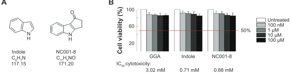
indole compounds and cytotoxicity
We have previously shown that novel synthetic indole com-pound 1,1,3-tri(3-indolyl)cyclohexane inhibits cancer cell
growth in lung cancer cells and xenograft models.36 As an
indole compound, indomethacin was reported to induce the expression of HSPs to suppress polyQ aggregation in a
cel-lular model of spinal and bulbar muscular atrophy.37 Indole
and synthetic derivative NC001-8 (Figure 1A) were selected
to test their potentials to reduce the polyQ aggregation. GGA,
a potent HSP inducer,38 was included for comparison. Cell
viability assays were performed with human neuroblastoma
Drug Design, Development and Therapy downloaded from https://www.dovepress.com/ by 118.70.13.36 on 22-Aug-2020
Dovepress
Kung et al
SH-SY5Y cells after treatment with GGA, indole, or
NC001-8 (0.1~100 µM) for 24 hours. The half maximal
inhibitory concentrations of the GGA and indoles were calcu-lated using the interpolation method. As shown in Figure 1B, GGA, indole, and NC001-8 had half maximal inhibitory concentrations of 3.02, 0.71, and 0.88 mM, respectively, in SH-SY5Y cells. GGA, indole, and derivative NC001-8 had
at least 85% cell viability up to the tested 100 µM, suggesting
their low cytotoxicity.
indole/nc001-8 enhanced hsF1
and hsP70 chaperone expression
on heK-293 cells
To examine the potential of indole/NC001-8 to enhance HSF1 and HSP70 chaperone expression, triple fluorescent reporter cells with mCherry, ZsYellow1, and AmCyan1 reporters driven by HSF1, HSPA8, and HSPA1A promoters were used. As shown in Figure 2A, 1 day GGA treatment
(100 nM~100 µM) significantly increases HSF1, HSPA8,
and HSPA1A promoter activity (HSF1, 110%~112%
[P=0.010~0.002]; HSPA8, 106%~116% [P=0.024~0.000];
HSPA1A, 108%~118% [P=0.034~0.001]). This is also true for
100 nM~100 µM indole treatment, with 117%~125% HSF1
(P=0.045~0.030), 118%~125% HSPA8 (P=0.046~0.016),
and 116%~123% HSPA1A (P=0.043~0.011) promoter
activi-ties compared with no treatment. For NC001-8 treatment (100
nM~100 µM), HSF1 (111%~123%; P=0.042~0.007), HSPA8
(109%~118%; P=0.048~0.004), and HSPA1A (106%~121%;
P=0.042~0.003) promoter activities were also significantly
increased. The enhancement of indole and NC001-8
(100 nM) on HSF1 (113%~114%; P=0.021~0.007), HSPA8
(108%~109%; P=0.046~0.028), and HSPA1A (119%~120%;
P=0.035~0.001) expression was verified by the Western blot
in HEK-293 cells after 2 days of treatment (Figure 2B).
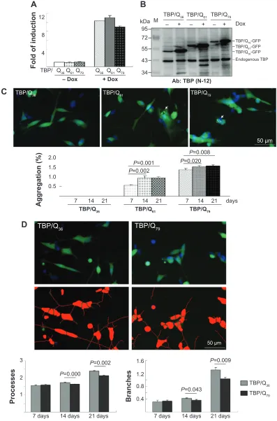
construction of sh-sY5Y
TBP/Q
36~79lines
To test the aggregation reduction potential of indole and NC001-8 in neuronal cells, we constructed Flp-In SH-SY5Y
SCA17 cells with N-terminal TBP/Q36~79-GFP expression
in an inducible fashion. As shown in Figure 3A, real-time polymerase quantification of these TBP lines shows
9~11 times TBP expression after induction with doxycycline
for 2 days. In immunoblot, TBP antibody detected 52~62 kDa
TBP/Q36~79-GFP protein in addition to the endogenous 43 kDa
TBP protein (Figure 3B). When TBP/Q36~79 SH-SY5Y cells
were differentiated for 7 to 21 days, using retinoic acid,39,40
a Q length-dependent and expression time-dependent
aggre-gate formation was seen in TBP/Q61~79-GFP cells, whereas
no aggregate was seen in TBP/Q36-GFP cells (Figure 3C).
When neuronal phenotype was examined after 7~21 days
of differentiation, significantly less process and branch in
TBP/Q79-GFP cells was observed at 2~3 weeks of
differen-tiation compared with TBP/Q36-GFP cells (1.63~2.12 versus
1.72~2.38 for process [P=0.000~0.002]; 0.37~1.03 versus
0.43~1.31 for branch [P=0.043~0.009]) (Figure 3D).
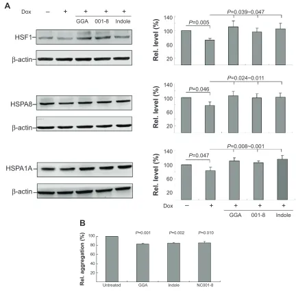
indole/nc001-8 enhanced hsF1
and hsP70 chaperone expression
and reduced TBP/Q
79aggregation
on sh-sY5Y cell model
The Flp-In SH-SY5Y TBP/Q79-GFP cells discussed
ear-lier were used to examine whether indole and NC001-8 up-regulate HSF1, HSPA8, and HSPA1A expression to reduce aggregation. As shown in Figure 4A, induced
expression of TBP/Q79 for 6 days attenuated the
expres-sion of HSF1 (72%; P=0.005), HSPA8 (78%; P=0.046),
and HSPA1A (84%; P=0.047) compared with uninduced
A
N H
Indole C8H7N 117.15
NC001-8 C11H9NO 171.20
N O
H
Cell viability (%
)
IC50 cytotoxicity:
3.02 mM 0.71 mM 0.88 mM
GGA 20
60 100
Indole NC001-8
50%
B
Untreated 100 nM 1 µM 10 µM 100 µM
Figure 1 indole and derivative nc001-8 and cytotoxicity.
Notes: (A) structure, formula, and molecular weight of indole and synthetic derivative nc001-8. (B) cytotoxicity of gga, indole, and nc001-8 against sh-sY5Y cells, using
hoechst-propidium iodide staining. cells were treated with 100 nM~100 µM tested compounds, and cell proliferation was measured the next day (n=3). The half maximal inhibitory concentration of each compound was shown under the columns. To normalize, the relative viability in untreated cells is set as 100%.
Abbreviations: gga, geranylgeranylacetone; ic50, inhibitory concentration at 50% level.
Drug Design, Development and Therapy downloaded from https://www.dovepress.com/ by 118.70.13.36 on 22-Aug-2020
Dovepress chaperone expression, polyQ aggregation in sca17 neuronal cell and slice culture models
cells (100%). This reduction can be rescued by the addition of GGA, indole, or NC001-8 (100 nM), with significantly
increased HSF1 (96%~111%; P=0.039~0.004), HSPA8
(99%~105%; P=0.024~0.011), and HSPA1A (106%~117%;
P=0.008~0.001) expression compared with untreated cells
(72%~84%). The treatment of GGA, indole, and NC001-8
led to 17% (P=0.001), 15% (P=0.002), and 14% (P=0.010),
respectively, aggregation reduction in TBP/Q79 expressed
differentiated neuronal cells (Figure 4B). These findings indicate that indole and NC001-8 up-regulated HSF1 and
A
Fluorescence (%
)
HSF1-mCherry
GGA 60
20 100
140 P=0.010∼0.002
P=0.045∼0.030
P=0.042∼0.007
PHSF1 mCherry PHSPA8 ZsYellow1 PHSPA1A AmCyan1
P=0.024∼0.000
P=0.046∼0.016
P=0.048∼0.004
P=0.034∼0.001
P=0.043∼0.011
P=0.042∼0.003
Indole NC001-8 GGA Indole NC001-8 GGA Indole NC001-8
100 nM 1 µM 10 µM 100 µM
HSPA8-ZsYellow1 HSPA1A-AmCyan1
B
- GGA
-HSF1
β-actin
β-actin HSPA8
HSPA1A
β-actin
001-8 Indole
120 P=0.034 0.021 0.007
P=0.046 0.028
0.035
P=0.026 0.001
001-8
GGA Indole
001-8
GGA Indole
001-8
GGA Indole
80
40
120
80
40
20 60 100 140
Rel. level (%
)
Rel. level (%
)
Rel. level (%
)
Figure 2 enhancement of chaperone expression by indole and nc001-8 in heK-293 cells.
Notes: (A) Fluorescent reporters mcherry, ZsYellow1, and amcyan1 driven by hsF1, hsPa8, and hsPa1a promoter fragments, respectively (top), and effects of gga,
indole, and nc001-8 (100 nM~100 µM) on HSF1, HSPA8, and HSPA1A promoter activities (bottom). To normalize, the fluorescence level in untreated cells is set as 100%. Three independent experiments were performed, with P0.05 considered significant. (B) representative Western blot images of heK-293 cells treated with gga, indole
and nc001-8 (100 nM) for two days, using hsF1, hsPa8, hsPa1a, and β-actin antibodies. levels of hsF1, hsPa8, and hsPa1a were normalized with a loading control (β-actin). Data are expressed as the mean ± standard deviation values from three independent experiments.
Abbreviations: gga, geranylgeranylacetone; 001-8, nc001-8; hsF1, heat shock transcription factor 1; rel., relative; hsPa8, heat shock 70 kDa protein 8; hsPa1a, heat
shock 70 kDa protein 1a.
Drug Design, Development and Therapy downloaded from https://www.dovepress.com/ by 118.70.13.36 on 22-Aug-2020
Dovepress
Kung et al
D
TBP/Q36Processes
3
P=0.000
P=0.043
7 days 14 days 21 days 7 days 14 days 21 days
P=0.002 P=0.009
TBP/Q36
TBP/Q79
2
1
0.4 0.8 1.2 1.6
Branches
TBP/Q79
50 µm
A
Fold of induction
Aggregation (%
)
12
kDa M – + – + – +
Ab: TBP (N-12)
TBP/Q36
TBP/Q36
TBP/Q36
TBP/Q36-GFP Endogenous TBP TBP/Q61-GFP TBP/Q79-GFP
TBP/Q61
TBP/Q61
TBP/Q61
TBP/Q79
TBP/Q79
TBP/Q79
50 µm
95 72 55 43 34 8
4
0.5
7 14 21 7
P=0.001
P=0.002
P=0.008
P=0.020
14 21 7 14 21 days
1.0 1.5 2.0
TBP/ Q36Q61
– Dox + Dox
Q79 Q36 Q61Q79
C
B
Dox
Figure 3 sh-sY5Y cells with induced TBP/Q36~79-gFP expression and neuronal phenotype.
Notes: (A) Real-time polymerase chain reaction quantification (n=3) of TBP/Q36~79-gFP mrna level relative to hPrT1 mrna after 2 days of induction with doxycycline
(+ Dox) or not (− Dox). (B) Western blot analysis of TBP/Q36~79-gFP protein level using TBP (n-12) antibody after 2 days of induction with doxycycline (+ Dox) or not
(− Dox). (C) representative microscopic images (top) of TBP/Q36~79-gFP cells after induced differentiation with retinoic acid (+ RA) for 7 days and aggregate quantification
(bottom; n=3) of cells with induced differentiation for 7~21 days. P-values were evaluated by one-way analysis of variance with post hoc lsD test. (D) representative
microscopic images (top) of neuronal differentiated TBP/Q36 and TBP/Q79 cells (for 14 days) and quantification (n=3) of neuronal processes and branches (bottom) of cells
with induced differentiation for 7~21 days (blue, nuclei; green, expressed TBP/Q36~79-gFP protein; red, cell body and outgrowth segmentation).
Abbreviations: Dox, doxycycline; TBP, TATA box binding protein; GFP, green fluorescent protein; mRNA, messenger RNA; Ab, antibody; LSD, Fisher’s least significant difference.
Drug Design, Development and Therapy downloaded from https://www.dovepress.com/ by 118.70.13.36 on 22-Aug-2020
Dovepress chaperone expression, polyQ aggregation in sca17 neuronal cell and slice culture models
A
Dox
HSF1
HSPA8
HSPA1A
β-actin
β-actin
β-actin
GGA 001-8 Indole 140 P=0.005
P=0.046
P=0.039~0.047
P=0.024~0.011
P=0.047
P=0.008~0.001
100
60
20
140
100
60
20
140
100
60
20
Rel. level (%
)
Rel. level (%
)
Rel. level (%
)
– + + + +
Dox
GGA 001-8 Indole
– + + + +
P=0.047
Rel. aggregation (%
)
B
Untreated
P=0.001 P=0.002 P=0.010
100 80
60 40 20
GGA Indole NC001-8
Figure 4 enhancement of hsF1 and chaperone expression and reduction of aggregation by indole and nc001-8 in neuronal sh-sY5Y TBP/Q79 cells.
Notes: (A) cells were pretreated with gga, indole, or nc001-8 (100 nM) for 8 hours and TBP/Q79-gFP expression induced for 6 days. relative hsF1, hsPa8, and hsPa1a
expressions were analyzed by immunoblot analysis, using β-actin as a loading control (n=3). P-values were evaluated by one-way analysis of variance with post hoc lsD test. (B) cells were treated with gga, indole, or nc001-8 (100 nM) for 7 days, and relative aggregation assessed by hca system (n=3). To normalize, the relative aggregation level in untreated cells is set as 100%.
Abbreviations: Dox, doxycycline; gga, geranylgeranylacetone; hsF1, heat shock transcription factor 1; hsPa8, heat shock 70 kDa protein 8; hsPa1a, heat shock 70 kDa
protein 1A; Rel., relative; GFP, green fluorescent protein; HCA: high content analysis; 001-8, NC001-8; LSD, Fisher’s least significant difference.
HSP70 chaperone expression to reduce TBP/Q79 aggregation
in differentiated neuronal cell models.
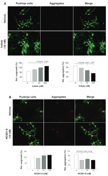
indole/nc001-8 promoted Purkinje
cell neurite outgrowth and reduced
aggregation on sca17 mouse primary
and slice cultures
To further confirm the neuroprotective potential of the indole compounds, we applied indole and NC001-8 to the cerebellar primary and slice cultures established from
SCA17 mice.34 As shown in Figure 5A-B, with 10~100 nM
compound concentration, significantly (indole: 134%~149%;
P=0.016~0.002) or notably (NC001-8: 114%~119%;
P0.05) increased Purkinje cell neurite outgrowth was
observed. Both compounds at concentrations 1~100 nM
significantly reduced the Purkinje cell aggregation on
the primary culture (indole: P=0.039~0.001; NC001-8:
P=0.042~0.016). The percentage of cells with aggregates
under indole treatment is 86% (1 nM), 71% (10 nM), and 53% (100 nM) in Figure 5A, and the percentage of cells with aggregates under NC001-8 treatment is 60% (1 nM), 68% (10 nM), and 60% (100 nM) in Figure 5B. On SCA17 mouse cerebellar slice culture, although indole at 10 nM could significantly reduce the Purkinje cell aggregation (28%;
Drug Design, Development and Therapy downloaded from https://www.dovepress.com/ by 118.70.13.36 on 22-Aug-2020
Dovepress
Kung et al
A
Purkinje cellsVe
hicle
Indole (10 nM
)
Rel. outgrowth (%
)
Indole (nM)
180
135
90
45
0 1 10
P=0.016 0.002 P=0.039 0.001 0.001
100 0 1 10 100
120 90
60
30
Indole (nM)
Rel. aggregation (%
)
Aggregates Merge
B
Purkinje cellsVe
hicle
NC001-8
(10 nM
)
Aggregates Merge
Rel. outgrowth (%
)
NC001-8 (nM) NC001-8 (nM)
P=0.016 0.042 0.018
100 10 1
0 0 1 10 100
30 60 90 120 150
100
50
Rel. aggregation (%
)
Figure 5 (Continued)
Drug Design, Development and Therapy downloaded from https://www.dovepress.com/ by 118.70.13.36 on 22-Aug-2020
Dovepress chaperone expression, polyQ aggregation in sca17 neuronal cell and slice culture models
Figure 5 indole and nc001-8 promoted neurite outgrowth and reduced aggregation of Purkinje cells in spinocerebellar ataxia type 17 mouse cerebellar primary and slice
cultures.
Notes: The primary culture was treated with 0~100 nM indole (A) or nc001-8 (B) for 13 days. The representative microscopic images of treatment with 100 nM indole or
nc001-8 are shown, and the relative Purkinje cell neurite outgrowth (green) and aggregation (shown in white in the middle column and in red in the right merged column) were quantified (n=3). (C) The slice culture was treated with 10 nM indole or 10 µM nc001-8 for 6 days. The representative microscopic images of treatment are shown, and the relative Purkinje cell aggregation (red) was quantified (n=3). To normalize, the relative neurite outgrowth length and aggregation level in vehicle-treated cells or slices is set as 100%. iP3r-1 and 1TBP18 antibodies were used to detect the Purkinje cells and TBP aggregation, respectively.
Abbreviations: iP3r-1, inositol 1,4,5 trisphosphate receptor type 1; 1TBP18, mouse monoclonal antibody to TaTa binding protein (TBP); rel., relative; DaPi, 4 ′,6-diamidino-2-phenylindole.
C
Purkinje cellsVe
hicle
Indole (10 nm
)
NC001-8
(10 µm)
Rel. aggregation (%
)
Vehicle 20 40 60 80 100
P=0.001 0.000
Indole (10 nM) NC001-8(10 µM)
Aggregates DAPI Merge
P=0.001), 1,000 folds of NC001-8 (10 µM) were required
to obtain a significant reduction of the aggregation (16%;
P0.001) (Figure 5C). Thus, indole worked more efficiently
than NC001-8 in reducing the Purkinje cell aggregation on SCA17 mouse cerebellar slice culture.
Discussion
An amount of evidence has indicated indole compounds as efficacious low-molecular drugs for the treatment of cancers. For example, indole-3-carbinol has been shown to suppress the proliferation and induce apoptosis and cell cycle arrest of
a wide variety of cancer cells.41,42 Nevertheless, attempts to
apply these chemicals in treating polyQ diseases are still too
few. Previously, indole compound indomethacin induced the expression of HSPs at physiological temperatures to suppress the protein aggregation and apoptosis caused by an expansion
of the polyQ tract in the androgen receptor.37 In this study,
we revealed that indole and its synthetic derivative NC001-8 reduced TBP aggregates in cell line and mouse cerebellar primary and slice cultures. The intact cellular interaction within the slice culture makes inadequate penetration of the medium into the inner part of the slice; therefore, 1,000 folds of NC001-8 were required to reduce polyQ-induced aggre-gation in slice culture compared with cell line and primary culture. In addition, our data show that indole and NC001-8 reduced TBP aggregates through the activation of HSF1 to
Drug Design, Development and Therapy downloaded from https://www.dovepress.com/ by 118.70.13.36 on 22-Aug-2020
Dovepress
Kung et al
increase cellular levels of HSP70 chaperone. Together, these data suggest the therapeutic potential of indole and derivative for polyQ-expanded SCA.
In polyQ-mediated SCA, the inclusion of the
disease-causing proteins affects molecular chaperone pathways.43
Previously, we showed the decreased heat shock cog-nate HSPA8 protein (a constitutive HSP70) expression
underlying pathogenesis of SCA17.44,45 HSP70 has been
reported to suppress polyQ-mediated neurodegeneration in
Drosophila.23,24 Overexpression of heat-inducible HSP70
chaperone (HSPA1A) rescues the severity of polyQ-mediated
degeneration and improves motor function in SCA1 mice.25
Expression of molecular chaperones is regulated by HSF1, and HSF1-activating compounds have been indicated as
therapeutic candidates for polyQ disorders.46,47 Our results
of neuroprotection via enhancement of chaperone system provide a novel mechanism of indole and derivative NC001-8 to decelerate the neurodegenerative process.
Although we have shown that both indole and NC001-8 performed neuroprotection via enhancement of chaperone system, pleiotropic effects of these compounds may exist to lead to the neuroprotective effect. For example, indole-3-carbinol from Brassica family vegetables was shown
to have anti-inflammatory and antioxidative activities,48,49
which could also benefit polyQ-mediated diseases.50,51 Future
genome-wide expression studies could explore whether there is any other underlying mechanism of these compounds.
To summarize, we provided strong evidence that indole and NC001-8 could be novel therapeutics for SCA17. As indole is the precursor to many pharmaceuticals, and indole
and derivatives can be synthesized by all kinds of methods,52,53
the development of indole-based compounds offers a promis-ing strategy for the treatment of polyQ diseases. Future inves-tigations of indole, NC001-8, and derivatives in more SCA or other polyQ animal models would strengthen their potential in aggregation reduction and disease amelioration.
Acknowledgments
We thank the Molecular Imaging Core Facility of National Taiwan Normal University for the technical assistance. This work was supported by grants MOST103-2325-B-003-001 and MOST103-2325-B-003-003 from the Ministry of Science and Technology and grants 103T3040B05 and 103T3040B07 from National Taiwan Normal University, Taipei, Taiwan.
Disclosure
The authors report no conflicts of interest in this work.
References
1. Gomes CM. Protein misfolding in disease and small molecule thera-pies. Curr Top Med Chem. 2012;12(22):2460–2469.
2. MacDonald M; The Huntington’s Disease Collaborative Research Group. A novel gene containing a trinucleotide repeat that is expanded and unstable on Huntington’s disease chromosomes. Cell. 1993;72(6): 971–983.
3. Orr HT, Chung MY, Banfi S, et al. Expansion of an unstable trinucle-otide CAG repeat in spinocerebellar ataxia type 1. Nat Genet. 1993; 4(3):221–226.
4. Pulst SM, Nechiporuk A, Nechiporuk T, et al. Moderate expansion of a normally biallelic trinucleotide repeat in spinocerebellar ataxia type 2.
Nat Genet. 1996;14(3):269–276.
5. Kawaguchi Y, Okamoto T, Taniwaki M, et al. CAG expansions in a novel gene for Machado-Joseph disease at chromosome 14q32.1. Nat
Genet. 1994;8(3):221–228.
6. Zhuchenko O, Bailey J, Bonnen P, et al. Autosomal dominant cerebel-lar ataxia (SCA6) associated with small polyglutamine expansions in the α 1A-voltage-dependent calcium channel. Nat Genet. 1997;
15(1):62–69.
7. David G, Abbas N, Stevanin G, et al. Cloning of the SCA7 gene reveals a highly unstable CAG repeat expansion. Nat Genet. 1997;17(1):65–70. 8. Moseley ML, Zu T, Ikeda Y, et al. Bidirectional expression of CUG and
CAG expansion transcripts and intranuclear polyglutamine inclusions in spinocerebellar ataxia type 8. Nat Genet. 2006;38(7):758–769. 9. Koide R, Kobayashi S, Shimohata T, et al. A neurological disease
caused by an expanded CAG trinucleotide repeat in the TATA-binding protein gene: a new polyglutamine disease? Hum Mol Genet. 1999; 8(11):2047–2053.
10. Denny RA, Gavrin LK, Saiah E. Recent developments in targeting protein misfolding diseases. Bioorg Med Chem Lett. 2013;23(7):1935–1944. 11. Gostout B, Liu Q, Sommer SS. “Cryptic” repeating triplets of purines
and pyrimidines (cRRY(i)) are frequent and polymorphic: analysis of coding cRRY(i) in the proopiomelanocortin (POMC) and TATA-binding protein (TBP) genes. Am J Hum Genet. 1993;52(6):1182–1190. 12. Nakamura K, Jeong SY, Uchihara T, et al. SCA17, a novel autosomal
dominant cerebellar ataxia caused by an expanded polyglutamine in TATA-binding protein. Hum Mol Genet. 2001;10(14):1441–1448. 13. Fujigasaki H, Martin JJ, De Deyn PP, et al. CAG repeat expansion in the
TATA box-binding protein gene causes autosomal dominant cerebellar ataxia. Brain. 2001;124(Pt 10):1939–1947.
14. Silveira I, Miranda C, Guimarães L, et al. Trinucleotide repeats in 202 families with ataxia: a small expanded (CAG)n allele at the SCA17 locus. Arch Neurol. 2002;59(4):623–629.
15. Wu YR, Lin HY, Chen CM, et al. Genetic testing in spinocerebellar ataxia in Taiwan: expansions of trinucleotide repeats in SCA8 and SCA17 are associated with typical Parkinson’s disease. Clin Genet. 2004; 65(3):209–214.
16. Wu YR, Fung HC, Lee-Chen GJ, et al. Analysis of polyglutamine-coding repeats in the TATA-binding protein in different neurodegen-erative diseases. J Neural Transm. 2005;112(4):539–546.
17. Chen CM, Lane HY, Wu YR, et al. Expanded trinucleotide repeats in the TBP/SCA17 gene mapped to chromosome 6q27 are associated with schizophrenia. Schizophr Res. 2005;78(2–3):131–136.
18. Lin IS, Wu RM, Lee-Chen GJ, Shan DE, Gwinn-Hardy K. The SCA17 phenotype can include features of MSA-C, PSP and cognitive impair-ment. Parkinsonism Relat Disord. 2007;13(4):246–249.
19. Hartl FU, Hayer-Hartl M. Molecular chaperones in the cytosol: from nascent chain to folded protein. Science. 2002;295(5561):1852–1858. 20. Welch WJ, Gething M-J, Clarke AR, et al. Heat shock proteins func-tioning as molecular chaperones: their roles in normal and stressed cells. Philos Trans R Soc Lond B Biol Sci. 1993;339(1289):327–333. 21. Jäättelä M. Heat shock proteins as cellular lifeguards. Ann Med. 1999;
31(4):261–271.
22. Daugaard M, Rohde M, Jäättelä M. The heat shock protein 70 family: Highly homologous proteins with overlapping and distinct functions.
FEBS Lett. 2007;581(19):3702–3710.
Drug Design, Development and Therapy downloaded from https://www.dovepress.com/ by 118.70.13.36 on 22-Aug-2020
Drug Design, Development and Therapy
Publish your work in this journal
Submit your manuscript here: http://www.dovepress.com/drug-design-development-and-therapy-journal Drug Design, Development and Therapy is an international,
peer-reviewed open-access journal that spans the spectrum of drug design and development through to clinical applications. Clinical outcomes, patient safety, and programs for the development and effective, safe, and sustained use of medicines are a feature of the journal, which
has also been accepted for indexing on PubMed Central. The manu-script management system is completely online and includes a very quick and fair peer-review system, which is all easy to use. Visit http://www.dovepress.com/testimonials.php to read real quotes from published authors.
Dovepress
Dovepress
chaperone expression, polyQ aggregation in sca17 neuronal cell and slice culture models23. Warrick JM, Chan HY, Gray-Board GL, Chai Y, Paulson HL, Bonini NM. Suppression of polyglutamine-mediated neurodegeneration in
Droso-phila by the molecular chaperone HSP70. Nat Genet. 1999;23(4):
425–428.
24. Iijima-Ando K, Wu P, Drier EA, Iijima K, Yin JC. cAMP-response element-binding protein and heat-shock protein 70 additively suppress polyglutamine-mediated toxicity in Drosophila. Proc Natl Acad Sci
U S A. 2005;102(29):10261–10266.
25. Cummings CJ, Sun Y, Opal P, et al. Over-expression of inducible HSP70 chaperone suppresses neuropathology and improves motor function in SCA1 mice. Hum Mol Genet. 2001;10(14):1511–1518.
26. Ahmad A, Biersack B, Li Y, et al. Targeted regulation of PI3K/Akt/ mTOR/NF-κB signaling by indole compounds and their derivatives: mechanistic details and biological implications for cancer therapy.
Anticancer Agents Med Chem. 2013;13(7):1002–1013.
27. Biersack B, Schobert R. Indole compounds against breast cancer: recent developments. Curr Drug Targets. 2012;13(14):1705–1719. 28. Janreddy D, Kavala V, Bosco JWJ, Kuo CW, Yao CF. An easy access
to carbazolones and 2,3-disubstituted indoles. Eur J Org Chem. 2011; 12(12):2360–2365.
29. Chang KH, Chen WL, Lee LC, et al. Aqueous Extract of Paeonia lacti-flora and Paeoniflorin as Aggregation Reducers Targeting Chaperones in Cell Models of Spinocerebellar Ataxia 3. Evid Based Complement
Alternat Med. 2013;2013:471659.
30. Lee LC, Chen CM, Wang HC, et al. Role of the CCAAT-binding protein NFY in SCA17 pathogenesis. PLoS ONE. 2012;7(4):e35302. 31. Furuya S, Makino A, Hirabayashi Y. An improved method for culturing
cerebellar Purkinje cells with differentiated dendrites under a mixed monolayer setting. Brain Res Brain Res Protoc. 1998;3(2):192–198. 32. Gimenez-Cassina A, Lim F, Diaz-Nido J. Gene transfer into Purkinje
cells using herpesviral amplicon vectors in cerebellar cultures.
Neurochem Int. 2007;50(1):181–188.
33. Tanaka M, Yanagawa Y, Hirashima N. Transfer of small interfering RNA by single-cell electroporation in cerebellar cell cultures. J Neurosci
Methods. 2009;178(1):80–86.
34. Chang YC, Lin CY, Hsu CM, et al. Neuroprotective effects of granu-locyte-colony stimulating factor in a novel transgenic mouse model of SCA17. J Neurochem. 2011;118(2):288–303.
35. Birgbauer E, Rao TS, Webb M. Lysolecithin induces demyelination in vitro in a cerebellar slice culture system. J Neurosci Res. 2004;78(2): 157–166.
36. Lee CH, Yao CF, Huang SM, et al. Novel 2-step synthetic indole com-pound 1,1,3-tri(3-indolyl)cyclohexane inhibits cancer cell growth in lung cancer cells and xenograft models. Cancer. 2008;113(4):815–825. 37. Ishihara K, Yamagishi N, Hatayama T. Suppression of heat- and
polyglutamine-induced cytotoxicity by nonsteroidal anti-inflammatory drugs. Eur J Biochem. 2004;271(22):4552–4558.
38. Yamanaka K, Takahashi N, Ooie T, Kaneda K, Yoshimatsu H, Saikawa T. Role of protein kinase C in geranylgeranylacetone-induced expression of heat-shock protein 72 and cardioprotection in the rat heart.
J Mol Cell Cardiol. 2003;35(7):785–794.
39. Sidell N. Retinoic acid-induced growth inhibition and morphologic differentiation of human neuroblastoma cells in vitro. J Natl Cancer
Inst. 1982;68(4):589–596.
40. Cheung YT, Lau WK, Yu MS, et al. Effects of all-trans-retinoic acid on human SH-SY5Y neuroblastoma as in vitro model in neurotoxicity research. Neurotoxicology. 2009;30(1):127–135.
41. Aggarwal BB, Ichikawa H. Molecular targets and anticancer poten-tial of indole-3-carbinol and its derivatives. Cell Cycle. 2005;4(9): 1201–1215.
42. Rogan EG. The natural chemopreventive compound indole-3-carbinol: state of the science. In Vivo. 2006;20(2):221–228.
43. Chai Y, Koppenhafer SL, Bonini NM, Paulson HL. Analysis of the role of heat shock protein (Hsp) molecular chaperones in polyglutamine disease. J Neurosci. 1999;19(23):10338–10347.
44. Lee LC, Chen CM, Chen FL, et al. Altered expression of HSPA5, HSPA8 and PARK7 in spinocerebellar ataxia type 17 identified by 2-dimensional fluorescence difference in gel electrophoresis. Clin Chim
Acta. 2009;400(1–2):56–62.
45. Chen CM, Lee LC, Soong BW, et al. SCA17 repeat expansion: mildly expanded CAG/CAA repeat alleles in neurological disorders and the functional implications. Clin Chim Acta. 2010;411(5–6):375–380. 46. Fujimoto M, Takaki E, Hayashi T, et al. Active HSF1 significantly
suppresses polyglutamine aggregate formation in cellular and mouse models. J Biol Chem. 2005;280(41):34908–34916.
47. Fujikake N, Nagai Y, Popiel HA, Okamoto Y, Yamaguchi M, Toda T. Heat shock transcription factor 1-activating compounds suppress polyglutamine-induced neurodegeneration through induction of multi-ple molecular chaperones. J Biol Chem. 2008;283(38):26188–26197. 48. Choi Y, Kim Y, Park S, Lee KW, Park T. Indole-3-carbinol prevents
diet-induced obesity through modulation of multiple genes related to adipogenesis, thermogenesis or inflammation in the visceral adipose tissue of mice. J Nutr Biochem. 2012;23(12):1732–1739.
49. Wu TY, Saw CL, Khor TO, Pung D, Boyanapalli SS, Kong AN. In vivo pharmacodynamics of indole-3-carbinol in the inhibition of prostate cancer in transgenic adenocarcinoma of mouse prostate (TRAMP) mice: involvement of Nrf2 and cell cycle/apoptosis signaling pathways. Mol
Carcinog. 2012;51(10):761–770.
50. Crotti A, Benner C, Kerman BE, et al. Mutant Huntingtin promotes autonomous microglia activation via myeloid lineage-determining factors. Nat Neurosci. 2014;17(4):513–521.
51. Johri A, Beal MF. Antioxidants in Huntington’s disease. Biochim
Biophys Acta. 2012;1822(5):664–674.
52. Humphrey GR, Kuethe JT. Practical methodologies for the synthesis of indoles. Chem Rev. 2006;106(7):2875–2911.
53. Cacchi S, Fabrizi G. Update 1 of: Synthesis and functionalization of indoles through palladium-catalyzed reactions. Chem Rev. 2011;111(5): PR215–PR283.
Drug Design, Development and Therapy downloaded from https://www.dovepress.com/ by 118.70.13.36 on 22-Aug-2020