Copyright1998 by the Genetics Society of America
Genetic Analysis of the Caenorhabditis elegans MAP Kinase Gene mpk-1
Mark R. Lackner
1and Stuart K. Kim
Department of Developmental Biology, Stanford University School of Medicine, Stanford, California 94305-5329
Manuscript received March 17, 1998 Accepted for publication May 26, 1998
ABSTRACT
The Caenorhabditis elegans mpk-1 gene encodes a MAP kinase protein that plays an important role in Ras-mediated induction of vulval cell fates. We show that mutations that eliminate mpk-1 activity result in a highly penetrant, vulvaless phenotype. A double mutant containing a gain-of-function mpk-1 mutation and a gain-of-function mek mutation (MEK phosphorylates and activates MPK-1) exhibits a multivulva phenotype. These results suggest that mpk-1 may transduce most or all of the anchor cell signal. Epistasis analysis suggests that mpk-1 acts downstream of mek-2 (encodes a MEK homolog) and upstream of lin-1 (encodes an Ets transcription factor) in the anchor cell signaling pathway. Finally, mpk-1 may act together with let-60 ras in multiple developmental processes, as mpk-1 mutants exhibit nearly the same range of developmental phenotypes as let-60 ras mutants.
M
AP kinases are key mediators of cellular differenti- In Drosophila, there is currently a single known MAP ation and proliferation in all animals, and they kinase, which is encoded by the rolled gene (Biggset al. function in receptor tyrosine kinase/Ras signaling path- 1994). A mutation (termed Sevenmaker) that activates ways (reviewed inMarshall1994). MAP kinase plays rolled MAP kinase results in the formation of extra R7 an important role in the Ras signaling pathway because cells, similar to the phenotype caused by constitutively it can activate downstream substrates that directly medi- activated Ras1 (Brunneret al. 1994).ate the cellular response to growth factors, suggesting In Caenorhabditis elegans, there is a single known MAP that MAP kinase acts near or at the end of this signaling kinase that is encoded by the mpk-1/sur-1 gene ( Lack-pathway (reviewed inTreisman 1996). ner et al. 1994; Wu and Han 1994). Previous studies MAP kinases are activated when they become phos- have shown that mpk-1 plays a role in vulval induction phorylated by the protein kinase MEK (MAP or ERK in the C. elegans hermaphrodite. The vulva is induced kinase;AdamsandParker 1992;Crewset al. 1992b). from a set of six ectodermal blast cells, referred to as The major known substrate for MEK is currently MAP Pn.p cells (P3.p to P8.p), that lie in a row along the kinase, suggesting that the predominant function of ventral midline of the developing larva (reviewed in MEK may be to activate MAP kinase (Segeret al. 1992). Eisenmann
andKim1994). All six Pn.p cells are capable Once activated, a significant fraction of MAP kinase of adopting any one of three potential cell fates (18, 28, molecules translocate to the nucleus, and many impor- or 38), and these fates are predominantly determined tant MAP kinase substrates are localized in the nucleus by the sequential action of two signaling pathways. First, (e.g., the mammalian transcription factors Elk-1 and a signal from the anchor cell induces the nearest Pn.p Ets-1 (reviewed in Treisman 1996). In addition to
nu-cell (P6.p) to adopt the 18 cell fate, which is to divide clear targets, some MAP kinase substrates are located three times and generate eight descendant cells that in the cytoplasm, e.g., the guanine nucleotide exchange form the inner portion of the vulva. Then, a lateral factor SOS and the protein kinase pp90Rsk (reviewed signal from P6.p induces P5.p and P7.p to adopt the 28 inFerrell 1996).
cell fate (Sternberg1988;KogaandOhshima1995b; Vertebrates have two MAP kinases (Erk1 and Erk2) Simskeand Kim1995), which is to divide three times that have overlapping substrates (reviewed inCrewset
to give seven descendant cells that form the outer por-al. 1992a and Marshall1994). Saccharomyces cerevisiae
tions of the vulva. In addition to the lateral signal, some has two MAP kinase genes, fus3 and kss1, that act in experiments suggest that a graded signal from the an-the pheromone and invasive growth signaling pathways, chor cell can sometimes induce Pn.p cells to adopt the respectively (Herskowitz1995;Madhaniet al. 1997).
28cell fate (SternbergandHorvitz1986;Katzet al. 1995). The Pn.p cells furthest from the anchor cell (P3.p, P4.p, and P8.p) adopt uninduced (38) cell fates,
Corresponding author: Stuart K. Kim, Department of Developmental which is to divide once and then fuse with the
hypoder-Biology, Stanford University School of Medicine, Stanford, CA
94305-mal syncytium. 5329. E-mail: [email protected]
Genetic studies have shown that the anchor cell signal
1Present address: Department of Molecular and Cell Biology,
Univer-sity of California, Berkeley, CA 94720. activates a receptor tyrosine kinase/Ras pathway
viewed in Kornfeld 1997). The lin-3 gene encodes a development. First, we present evidence suggesting that mpk-1 transduces most if not all of the signal from let-protein similar to epidermal growth factor (EGF) that
is the inductive signal secreted by the anchor cell (Hill 23 receptor and let-60 ras. We used three genetic screens to recover new mpk-1 alleles (including three putative andSternberg1992). The let-23 gene encodes a
recep-tor tyrosine kinase similar to EGF receprecep-tor that is null alleles) and found that mpk-1–null mutants exhibit a strong vulvaless phenotype. We also showed that activa-thought to be the receptor for LIN-3 signal (Aroianet
al. 1990). Downstream of LIN-3 and LET-23 are a Grb2- tion of both MEK and MAP kinase activates the vulval signaling pathway, resulting in a multivulva phenotype. related adapter protein (encoded by sem-5; Clark et
al. 1992), a Ras protein (encoded by let-60 ; Han and Thus, mpk-1 is necessary for vulval induction, and mek and mpk-1 together are sufficient to activate vulval induc-Sternberg1990), a Raf homolog (encoded by lin-45;
Hanet al. 1993), and a protein similar to the serine/ tion. Second, genetic and molecular analyses suggest that mpk-1 acts downstream of all the signal transduction threonine protein kinase MEK (encoded by mek-2;
Korn-feldet al. 1995;Wuet al. 1995). Loss-of-function muta- genes in the let-23 RTK vulval signaling pathway, but upstream of lin-1 Ets (a vulval repressor gene encoding tions in any of these genes prevent P6.p from expressing
the 18cell fate and the lateral signal, so that all of the a putative transcription factor). Finally, our results sug-gest that mpk-1 may act together with let-60 in many, but Pn.p cells express 38 cell fates, resulting in a vulvaless
phenotype. Conversely, gain-of-function mutations in perhaps not all, of the tissues whose fates are deter-mined by let-60 ras signaling.
let-23 or let-60 cause the vulval precursor cells to adopt the 18 cell fate and to presumably express the lateral signal, resulting in a multivulva phenotype.
MATERIALS AND METHODS Previous studies have implicated mpk-1 in vulval
induc-tion. These studies showed that two mpk-1 alleles (n2521
General methods:Strain maintenance, genetic
manipula-and ku8) suppress the multivulva phenotype of the acti- tion, and ethyl methanesulfonate (EMS) mutagenesis were vated ras allele let-60(n1046) (Lackneret al. 1994; Wu performed as described by Brenner (1974). Animals were cultivated at 208 unless otherwise noted. The animals de-andHan 1994). This result indicates that mpk-1 most
scribed as wild type were C. elegans, variety Bristol, strain N2. likely acts downstream of let-60 ras in the vulval signaling
The following genes and alleles were used in this work [unless pathway. However, mpk-1(n2521) mutants did not
ex-otherwise noted, alleles used are described byWood(1988)]: hibit any defect in vulval induction, and mpk-1(ku8)
ex-LGI: mek-2(n2678) (Kornfeldet al. 1995), sup-11(n403), dpy-hibited only a weak defect in vulval induction. Thus,
5(e61), sur-2(ku9) (SinghandHan1996), smg-1(r861) (Pulak these mpk-1 alleles do not result in a strong reduction
and Anderson1993), unc-54(r293) (Pulakand Anderson of the anchor cell signaling pathway in P6.p. It is not 1993).
clear whether these mpk-1 alleles eliminate or might LGIII: unc-79(e1068), mpk-1(ku8) (WuandHan1994), mpk-1 only partially reduce mpk-1 activity, since both alleles (oz140) (Churchet al. 1995), dpy-17(e164), lin-12(n137sd), sDp3 (Rosenbluthet al. 1985), unc-119(ed3op) (Maduroand Pil-are missense mutations and gene dosage studies have
grim1995). not been possible because of the lack of a deficiency
LGIV: lin-1(sy254) (Beitelet al. 1995), lin-45(sy96) (Hanet that deletes the mpk-1 locus. al. 1993), let-60(n1046gf) (Beitelet al. 1990), dpy-20(e1282ts),
Because the mpk-1–null phenotype has not been es- unc-30(e191).
tablished, an important but unanswered question is LGV: him-5(e1490), lin-25(e1446) (Tuck and Greenwald 1995).
whether the vulval induction pathway is linear between
LGX: lin-15(n309), lin-3(syIs1) (gift fromR. Hill,Cal Tech), let-23 RTK and mpk-1, such that mpk-1 transduces all of
unc-7(e5).
the signal from activated let-23. If the pathway is linear, Transgenic arrays (transgenes; cotransformation markers): then null mutations in mpk-1 should prevent vulval in- gaEx36 {hs-mpk-1(gf ); hs-D-mek(gf ); rol-6(d)}, gaEx72 {lin-31-mpk-duction and result in a Vul phenotype. Alternatively, 1(gf ); lin-31-D-mek(gf ); unc-119(1)}, gaIs17{hs-mpk-1(gf); hs-D-mek(gf ); unc-30(1)}; gaIs36 {hs-mpk-1(1); EF1alpha-D-mek(gf );
the signaling pathway might be branched in a way that
unc-30(1)}.
other signaling molecules might act in parallel with
mpk-1. For instance, mammalian EGF receptor and Ras Genetic mapping:The oz140 allele was initially mapped to the left arm of chromosome III (T. Schedl,personal commu-are thought to transduce signals by activating targets in
nication). To determine if it mapped to the same interval as completely separate signaling branches (such as
phos-the mpk-1 locus, we mapped it relative to unc-79 and dpy-17. pholipase C gamma or PI3 kinase;Rocheet al. 1996)
Briefly, we picked Dpy non-Unc and Unc non-Dpy recombi-or by activating two MAP kinases (Erk1 and Erk2). If the nants from a strain of the genotype oz140/unc-79(e1068) dpy-C. elegans vulval signaling pathway is similarly branched, 17(e164). The progeny of these recombinant animals were screened for sterile (Ste) animals (the oz140 mutant pheno-then mpk-1 null mutations should result in only a partial
type). Eighteen out of 19 Dpy non-Unc animals segregated reduction in vulval induction because the mpk-1
muta-Ste animals, whereas 1 out of 17 Unc non-Dpy animals segre-tions should eliminate one but not all of the signaling
gated Ste animals. These data suggest that oz140 maps to the
branches. left of dpy-17 and very close or to the right of unc-79. This is
In this article, we have further characterized the role the approximate map position of the mpk-1 locus. The SD344 strain [1 mpk-1(oz140) dpy-17(e164)/unc-79(e1068) 1 dpy-17
TABLE 1
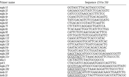
Oligonucleotide primers used in this study
Primer name Sequence (59to 39)a
mpk-1f GGTAGCTTACAGTAGCCGTAC
mpk-2f GAGAAGCGGTTATCTCGACGGTC
mpk-3r CATCCCGTAAGCACCTTCTCC
mpk-4r CGAAGTGTCCGTTGACAGAATG
mpk-5r TATCGACAGTCTCCGAACGGATG
mpk-6f CTGACACCCGTAGTTCAATTC
mpk-7f CTCTATCCAGGAGCTGATCCA
mpk-8r TCACAAACTGGCTCATCTCCTGG
mpk-9f CATTCTGTCAACGGACACTTCG
mpk-10f GTCTGGTCTGTCGGATGTATTC
mpk-11f CAAGCATTGGCTCACCCATAC
mpk-12r GGAGGAGGTGCATTCACATCAC
mpk-13f GTTCAATGCCTGATCGAGACTG
mpk14r GAATACATCCGACAGACCAGAC
mpk-15r TGGATCAGCTCCTGGATAGAG
mpk-20f GAGCTAGCATGGCCGACGGAGAAGCGGTT
mpk-21r GCGGTACCTCTAAACAGGATTCTGCCCTC
xba-1 CACTAGTTCTAGTGCGGCCG
gof-1 CTACGATCCAGGAAATGAGCCAGTTTG
mpk-27f GCGTCGACATGGCCGACGGAGAAGCGGTTATC
mpk-28r GAGCGGCCGCCTAAACAGGATTCTGCCCTCC
mek-1f GCGTCGACATGTCGAAGAACAAGCTGAATCTG
mek-2r GAGCGGCCGCCTAGTTGGGCGACGTATTACGC
aUnderlined sequences denote 8 to 12 nucleotide sequences used for subsequent cloning steps.
(e164)] was constructed by picking Dpy non-Unc recombinant assumed to be of genotype lin-45; gaEx36. lin-25; gaEx36 and
sur-2; gaEx36 strains were constructed similarly.
progeny of hermaphrodites of genotype
mpk-1(oz140)/unc-79(e1068) dpy-17(e164). All other newly isolated mpk-1 alleles Gonad ablations: Somatic gonad precursors Z1.aaa and Z4.ppp were ablated with a laser microbeam (Laser Science, were shown to be linked to unc-79 and dpy-17 by allowing
hermaphrodites of genotype mpk-1(rf)/unc-79 dpy-17 to self, Newton, MA) as described in Avery and Horvitz (1989). Animals were then subjected to a 30-min heat shock of 338as picking 20 Unc Dpy progeny and confirming that these
her-maphrodites did not segregate sterile mpk-1(rf) animals (this early L3 larvae and then scored as Muv or non-Muv.
Mutant sequence determination:We used oligonucleotide
experiment was performed at 258for ga111).
Three mutations isolated in a clonal screen for sterile and primers (mpk-1f and mpk-12r in Table 1) in polymerase chain reactions to amplify a 3.2-kb genomic DNA fragment con-vulvaless animals were mapped using STS polymorphisms
(data not shown;Williams et al. 1992). STS mapping and taining the entire mpk-1-coding region for the SLX2 form from wild-type strains and each mutant strain. The DNA sequence of two-factor mapping with dpy-5 and sup-11 indicated that ga180
and ga181 are linked to the left arm of chromosome I, close the mpk-1 gene (exons 1–6 and the exon/intron borders) from each mutant was then determined by direct sequencing to mek-2. We next showed that both ga180 and ga181 fail to
complement mek-2(n2678) for the Ste phenotype. STS map- of this fragment in low-melting-temperature agarose (Kretz
et al. 1989). DNA from two separate single-worm PCR reactions
ping suggested that ga119 is linked to chromosome III. Next,
we showed that ga119 fails to complement mpk-1(oz140) for was sequenced using the following primers: mpk-1f, mpk-2f, mpk-3r, mpk-4r, mpk-5r, mpk-6f, mpk-7f, mpk-8r, mpk-9f, the sterile phenotype.
Strain construction: Double mutants containing mpk-1 mpk-10f, mpk-11f, mpk-12r, mpk-13f, mpk-14r, and mpk-15r
(see Figure 1 and Table 1).
(ga117) and a Muv mutation (see Table 6) were all constructed
using a common strategy, which is illustrated here for lin- Germ line transformation experiments:Germ line transfor-mation experiments were done according to the method of
15(n309). First, 17(e164); lin-15(n309) was built. These
dpy-17(e164); lin-15(n309) hermaphrodites were then mated with Fireand Waterston (1989), as modified by Mello et al. (1991). DNAs were injected at concentrations of 100mg/ml. males of genotype mpk-1(ga117) dpy-17(e164); sDp3. sDp3 is a
chromosomal duplication that complements mutations in Cotransformation markers were rol-6(su1006), an unc-30(1)
plasmid and an unc-119(1) plasmid injected into N2, unc-mpk-1 and dpy-17. Non-Dpy cross-progeny of genotype ga117
e164/1e164; sDp3; n309/1were then allowed to self-fertilize. 30(e191), and unc-119(ed3op) hermaphrodites, respectively. For
each type of array, at least two transgenic lines were obtained, Muv non-Dpy progeny of this cross were then placed on
indi-vidual plates and allowed to self-fertilize. Muv non-Dpy animals and data from a representative line are shown. To integrate extrachromosomal arrays, L4 larvae containing the array of in which all the Dpy progeny were Ste were taken to be of
genotype mpk-1(ga117) dpy-17(e164); lin-15(n309); sDp3. interest were subjected to gamma irradiation from a Cesium 137 source (4000 rad). Strains with integrated arrays were The lin-45; gaEx36 strain was constructed by mating
lin-45/1 males with gaEx36 (Rol) hermaphrodites. Cross- backcrossed at least once to dpy-20; him-5.
Manipulation of DNA and RNA:Molecular biological
tech-progeny hermaphrodites were cloned, and their self-tech-progeny
Figure1.—Sequence alterations in mpk-1 mutants. The boxes represent exons 2–7 of the mpk-1 gene, slanted lines indicate introns, and arrows indicate locations in the coding sequence of the mpk-1 mutations isolated in this study. mpk-1 expresses two mRNAs, which differ in that one mRNA (132) includes exon 1 while the other mRNA (SL32) contains the SL1 trans-spliced leader joined to exon 2 (Lackneret al. 1994). The DNA sequence alteration and predicted protein coding change of each mutation is shown. Relative locations and orientations of all primers used to amplify and sequence the mpk-1-coding region from mutants are indicated below the boxes (Table 1). Arrows pointing to the right indicate that a given primer anneals to the antisense strand, while arrows pointing to the left indicate that a given primer anneals to the sense strand. Primers mpk-1f and mpk-12r were used in PCR experiments to amplify a 3.2-kb genomic fragment from single worms containing 292 bp of sequence 59to the coding region, the complete coding region, and 259 bp of 39untranslated region sequence. The 3.2-kb fragments from the wild type and each mutant were then sequenced using the internal primers shown, which span the entire coding region and all the intron-exon boundaries.
(1989), with minor modifications. Heat shock mpk-1(1) was Pn.p cell lineages shown in Tables 2 and 3 were determined by direct observation of cells or cell divisions using Nomarski constructed by PCR amplifying the mpk-1-coding region
con-tained in cDNA pML1 using primers mpk-20f and mpk-21r. optics, as described bySulstonandHorvitz(1977). Male mating assays were conducting by mating 20 individual These primers were used to amplify a 1.2-kb fragment starting
at the mpk-1 start codon and ending at the stop codon. Ampli- mpk-1(ga117) dpy-17 males with two unc-119 hermaphrodites
each. None produced non-Unc cross-progeny. Nine out of 10 fied fragments were cloned into the heat shock
promoter-containing vector JK465 (RoehlandKimble1993) to make control matings of dpy-17 males with unc-119 hermaphrodites gave cross-progeny.
plasmid pML54. To construct hs-mpk-1(gf), we used the unique
site elimination method to engineer the D324N substitution Western blotting:Western blots were prepared essentially as described in Hoskinset al. (1996), with minor modifica-in pML1 usmodifica-ing primers xba-1 and gamodifica-in-of-function-1 (Deng
andNickoloff1992). We then PCR amplified the insert of tions. For each lane, 50 adult worms were picked into Laemmli buffer and boiled for 5 min before loading. Filters were immu-pML14 using primers mpk-20 and mpk-21 and ligated this
fragment in the heat shock promoter vector JK465 to create noblotted overnight at 48in anti-Erk2 peptide antibody K-23 (Santa Cruz Biotechnology, Santa Cruz, CA) diluted 1:500. construct pML40. hs-D-mek constructs were kindly provided by
M. Koga(Koga and Ohshima1995a). lin-31-mpk-1(gf ) was constructed by amplifying the mpk-1-coding region from
pML14 with primers mpk-27 and mpk-28 (Table 1) and then RESULTS ligating the resulting fragment into the lin-31
promoter-con-taining vector pB255. lin-31-D-mek(gf ) was constructed by am- Isolation of new mpk-1 alleles:We wanted to deter-plifying the D-mek-coding region from construct Dsor1(su1) mine whether all of the anchor cell signal is transduced (M. Koga,personal communication) with primers mek-1 and
by mpk-1 (in which case elimination of mpk-1 activity mek-2 (Table 1) and then ligating the resulting fragment into
should prevent all vulval induction caused by the anchor pB255.
Phenotypic characterization: When examined with a dis- cell) or whether the anchor cell signaling pathway is
secting microscope, worms homozygous for strong mpk-1 al- branched (in which case elimination of mpk-1 activity leles have a characteristic appearance in which the gonad
should cause defects only in the mpk-1 branch of the appears mostly clear and devoid of embryos, with several
signaling pathway). To accomplish this, we isolated new clumps of dark granular material present in the gonad. They
charac-dites were mutagenized and allowed to self-fertilize. F1
male progeny (potentially heterozygous for a new mpk-1 mutation, new/1) were mated individually to two her-maphrodites of genotype mpk-1(oz140) dpy-17/unc-79 dpy-17. Non-Dpy cross-progeny on each plate were then screened for the presence of approximately one-quarter sterile animals, and the new mpk-1 alleles were recovered from these plates by picking the wild-type siblings (geno-type mpk-1(new)/1) of the sterile mutants (genotype mpk-1(new)/mpk-1(oz140)). From a screen involving 6008 haploid genomes, two mutations (ga117 and ga118) were isolated that mapped near to mpk-1 in the central Figure 2.—Western blot analysis of MPK-1. Lysates were
region of chromosome III and failed to complement prepared from 50 adult worms from wild-type (N2), mpk- the sterile phenotype of mpk-1(oz140). As described
be-1(ga117), and mek-2(n2678) strains. The extracts were analyzed
low, our molecular analyses indicate that both ga117 by Western blotting using 10% SDS-PAGE and anti–MAP
ki-and ga118 may severely reduce ki-and possibly eliminate nase antibodies (Santa Cruz Biochemical). The positions and
sizes (in Mr31023) of prestained molecular weight standards mpk-1 activity.
(Bio-Rad, Richmond, CA) are indicated on the left. The posi- The third approach used to isolate new alleles was tions of the two MPK-1 species predicted from analysis of to perform a general screen to identify heterozygous cDNAs and Northern blots (of Mr45 and 55 kD, respectively)
animals that segregated approximately one-quarter ster-are indicated by arrows.
ile, vulvaless self-progeny, and then to determine which were new mpk-1 alleles. This screen could identify muta-tions that would be missed in the first screen (because terization to determine if any of them were likely to be
null alleles, and then determined whether elimination it could identify mutations in any gene that cause a sterile, vulvaless phenotype), and it is simpler to perform of mpk-1 activity completely prevented the expression
of vulval cell fates. than the second screen (because it does not require
individual matings). Individual F1progeny of
mutagen-We used three approaches to isolate additional alleles
of mpk-1. The first approach was to screen for mutations ized hermaphrodites were placed on plates and allowed to self-fertilize, and then these plates were screened that failed to complement the egg-laying defective (Egl)
phenotype of mpk-1(ku8). This screen should identify for the presence of one-quarter sterile F2progeny. We
recovered three mutations from a screen involving 4020 mpk-1 alleles that predominantly affect vulval
develop-ment, but would miss null alleles if these alleles cause mutagenized genomes. Two of these mutations are al-leles of mek-2 because they map near to mek-2 on the sterile or lethal phenotypes when heterozygous with ku8.
Mutagenized N2 (wild-type) males were mated with mpk- left region of chromosome I and fail to complement the sterile phenotype caused by mek-2(n2678). One mu-1(ku8) dpy-17; unc-7 hermaphrodites, and the
cross-prog-eny hermaphrodites were screened for an Egl pheno- tation (ga119) is a new allele of mpk-1 because it maps to the central region of chromosome III, fails to comple-type. From a screen involving 7500 mutagenized
hap-loid genomes, we recovered two mutations (ga110 and ment the sterile phenotype of mpk-1(oz140), and con-tains a DNA sequence change in the mpk-1 gene (see ga111) that mapped near to mpk-1 in the central region
of chromosome III and failed to complement the mpk- below).
Molecular characterization of mpk-1 alleles:We used
1(ku8) Egl phenotype (data not shown). An additional
allele, mpk-1(oz140), was provided to us byT. Shedland molecular analysis of the new mpk-1 alleles to determine if any were likely to be null mutations. (Figure 1). DNA we subsequently showed that this allele results in the
same DNA sequence change as mpk-1(ga110) (see Figure from mpk-1(ga117) mutants had an AG to AA nucleotide substitution in the splice acceptor site upstream of exon 1). Based on the molecular analyses described below,
neither ga110, ga111, nor oz140 are likely to be null 3 (Figure 1), and this G nucleotide is present in all known splice acceptor sites in C. elegans (Blumenthal alleles.
Our second approach was to screen for mutations and Steward 1997). This result suggests that ga117 results in aberrant mRNA splicing and would be pre-that failed to complement the sterile phenotype of
mpk-1(oz140) (Churchet al. 1995). This screen could identify dicted to eliminate expression of MPK-1 sequences after amino acid 24 of the 376-amino-acid protein. We used mpk-1 null alleles if they resulted in sterility, but would
miss null alleles if they cause a lethal phenotype when anti-MAP kinase antibodies in Western blotting experi-ments to examine MPK-1 expression levels in extracts heterozygous with oz140. We screened individual males
for those that might be heterozygous for a new mpk-1 from adult wild-type worms and mpk-1(ga117) mutants (Figure 2). The antibodies detect one pair of bands allele and, hence, sire sterile cross-progeny when mated
hermaphro-TABLE 2
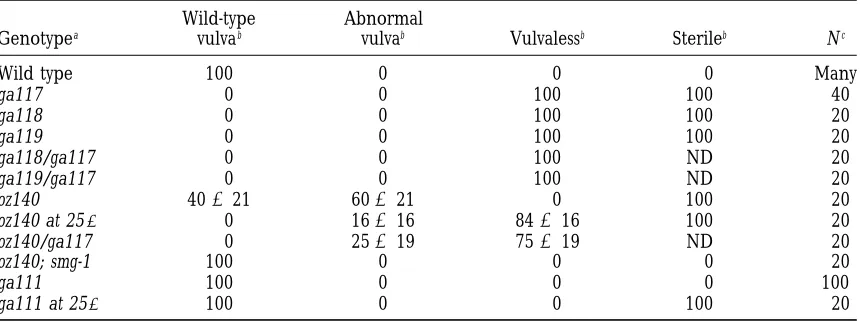
Phenotypes caused by mpk-1 alleles
Wild-type Abnormal
Genotypea vulvab vulvab Vulvalessb Sterileb Nc
Wild type 100 0 0 0 Many
ga117 0 0 100 100 40
ga118 0 0 100 100 20
ga119 0 0 100 100 20
ga118/ga117 0 0 100 ND 20
ga119/ga117 0 0 100 ND 20
oz140 40621 60621 0 100 20
oz140 at 258 0 16616 84616 100 20
oz140/ga117 0 25619 75619 ND 20
oz140; smg-1 100 0 0 0 20
ga111 100 0 0 0 100
ga111 at 258 100 0 0 100 20
aga111 was scored from homozygous ga111 hermaphrodites. In the case of all other alleles shown, animals
are homozygous mpk-1(rf ) progeny segregated from hermaphrodites of genotype mpk-1(rf ) dpy-17; sDp3. In the case of transheterozygous strains, the genotypes are mpk-1(ga117)/mpk-1(rf ) dpy-17(e164). sDp3 is a chromosomal duplication that complements mpk-1 and dpy-17. The complete genotype of the oz140; smg-1 strain is smg-1(r861)
unc-54; mpk-1(oz140) dpy-17. All strains were examined at 208unless otherwise indicated.
bWild type indicates that vulval development appeared normal at the L4 larval stage. Abnormal vulva indicates
that some vulval induction was observed, but less than in wild type. Vulvaless indicates that no vulval induction was observed. Sterile indicates that the hermaphrodite failed to produce progeny after 3 days. Numbers indicate the percentage6the 95% confidence interval (95% confidence interval51.96√(x(12x)/N ), where x is the
percentage of animals of a given phenotype and N is the total number of animals).
cNumber of animals scored.
pair of bands corresponds to phosphorylated and un- protein kinase resulted in a mutant protein with,0.9% of the activity of the wild-type protein (Gibbsand Zol-phosphorylated forms of one MPK-1 isoform. The sizes
of the lower band in each pair are the same as those ler1991). Similarly, the analogous mutation (E238K) in the C. elegans mek-2 gene results in a severe loss-of-predicted to result from translation of the two types of
mpk-1 RNA (SLX2 and 1X2, respectively;Lackneret al. function or null phenotype (Kornfeld et al. 1995). Thus, the mpk-1(ga119) mutation may nearly eliminate 1994). Neither pair of bands is present in extracts from
mpk-1(ga117) mutants, suggesting that mpk-1(ga117) re- MPK-1 kinase activity.
DNA from mpk-1(ga110) and mpk-1(oz140) mutants duces expression of wild-type MPK-1 to undetectable
levels as a result of improper RNA splicing. had the identical DNA sequence alteration, a G to A transition predicted to change tryptophan at amino acid DNA from mpk-1(ga118) mutants had a T to C
transi-tion that would change an arginine residue to a cysteine 351 to an amber stop codon (Figure 1). This substitution would result in truncation of the C-terminal 25 amino residue (R197C; Figure 1). This residue is conserved in
all known MAP kinases and is in a region referred to acids of the MPK-1 protein. These amino acids are not conserved in other MAP kinase proteins, suggesting that as the phosphorylation loop, which contains the MEK
phosphorylation site (TEY) and the kinase catalytic site these mutations may reduce but not eliminate gene activity. Premature termination of translation often re-(APE;Zhang et al. 1995). An arginine to alanine
sub-stitution at the analogous residue in human ERK1 sults in mRNA instability, so the oz140 phenotype might result from lower mRNA levels, decreased protein ki-(R208A) showed increased tyrosine phosphorylation
but decreased threonine phosphorylation by MEK, and nase activity, or both. smg-1 mutations increase the stabil-ity of nonsense messages (PulakandAnderson1993), it completely abolished kinase activity of Erk1 toward
its substrate, myelin basic protein (Butch and Guan and we found that the phenotypes caused by mpk-1(oz140) are suppressible by a smg-1 mutation (Table 1996). Thus, the R197C substitution is likely to eliminate
MPK-1 kinase activity. 2). Thus, C-terminal-truncated MPK-1 has MAP kinase
activity when the levels of mpk-1(oz140) mRNA are in-DNA from mpk-1(ga119) mutants had a G to A
transi-tion that would change the glutamic acid in the kinase creased by the smg-1 mutation. This result indicates that ga110 and oz140 partially reduce mpk-1 activity by de-catalytic site (APE) to a lysine (APK) (Figure 1). A
glu-tamic acid at this site is conserved in all known protein creasing mRNA levels rather than by affecting protein activity.
kinases (Hankset al. 1988). An alanine substitution at
transi-Figure 3.—Phenotypes caused by mpk-1 alleles. (A) Wild-type hermaphrodite in the middle of the L4 larval stage. The 22 descendants of P5.p, P6.p, and P7.p have been generated and are beginning vulval morphogenesis (arrow). (B) mpk-1(ga110) hermaphrodite in the middle of the L4 larval stage. The posterior daughter of P6.p has failed to divide, and the undivided cell is seen adhering to the cuticle (arrowhead). The 28cell lineages occurred normally in this animal. (C) mpk-1(ga117) hermaphrodite in the middle of the L4 larval stage. All the Pn.p cells have expressed the 38cell fate (indicated by lines), and no vulval tissue has been formed. (D) Tail of a wild-type adult male. The arrow indicates a spicule, the male copulatory organ. (E) Tail of an
mpk-1(ga117) adult male. The arrow indicates a crumpled spicule. mpk-1(ga117) males are unable to mate (seematerials and methods). All animals were viewed with Nomarski optics. Scale bar is 10mm.
tion that would be predicted to alter amino acid 148 20 homozygous 1(ga117), 1(ga118), and mpk-1(ga119) animals and found that all the vulval precursor from valine to glycine (Figure 1). This valine is
con-served in other MAP kinase proteins but not in other cells had expressed uninduced 38cell fates in each case (Table 2). These results show that mpk-1 null mutations types of protein kinases (Hankset al. 1988). This site
is near the predicted MEK-binding site (Brunet and cause a highly penetrant vulvaless phenotype.
mpk-1(ga110) and mpk-1(oz140) have the same DNA Pouyssegur 1996;Zhang et al. 1995), suggesting that
MPK-1(ga111) may be defective in activation by MEK. sequence alteration that results in a weak vulvaless phe-notype that is partially temperature sensitive. At 258,
Vulval phenotypes of mpk-1 alleles:Our first finding
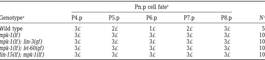
is that mpk-1 is required for vulval induction (Table 2). 84% of mpk-1(oz140) animals exhibited a Vul phenotype in which all six vulval precursor cells expressed 38cell Using the putative null mutation ga117, we determined
the cell lineages of the Pn.p cells in 20 mutant hermaph- fates, and the remaining 16% showed a partial Vul phe-notype (Table 2). At 208, none of mpk-1(oz140) mutants rodites and found that P5.p, P6.p, and P7.p expressed
the uninduced 38cell fate rather than induced 18or 28 showed a complete Vul phenotype, 60% showed partial vulval defects, and the remaining 40% had a wild-type cell fates in every animal (Figure 3, Tables 2 and 3).
TABLE 3
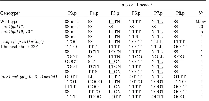
Pn.p cell lineages in mpk-1 mutants
Pn.p cell lineageb
Genotypea P3.p P4.p P5.p P6.p P7.p P8.p Nc
Wild type SS or U SS LLTN TTTT NTLL SS Many
mpk-1(ga117) SS or U SS SS SS SS SS 20
mpk-1(ga110) 208 SS or U SS LLTN TTTT NTLL SS 5
SS or U SS LLTN TTN NTLL SS 4
hs-mpk-(gf ); hs-D-mek(gf ) TTOO SS LLTN TOTT TLLL LTTT 1
1-hr heat shock 338 TTTO TTTT LTTT TOTT TTLL OOTT 1
SS TOTT LOTN TTTT NTLL SS 1
TOOT SS LTTN TTOO NOLL S OO 1
OOOT S TT LLON TOTT NTLL SS 1
TOOT TOTT LTON TTTT NTLL SS 1
SS TT S LLON TOTT NTLL SS 1
lin-31-mpk-(gf ); lin-31-D-mek(gf ) OOTT LL LLTT OTTT NTLL OTTT 1
TTOT OOOO LLTN OTTO TOOT TTTO 1
LLTT OOOT LLON TTTT TOOT OOTT 1
SS TTTO LLON TTTT TOOT OOTT 1
TTTT TOOO TOTT TTTT OOTT OOOL 1
aStrains containing mpk-1(ga117) and mpk-1(ga110) also contain the cis-linked mutation dpy-17(e164). hs-mpk-1(gf ); hs-D-mek(gf ) refers to the strain gaIs17. lin-31-mpk-hs-mpk-1(gf ); lin-31-D-mek(gf ) refers to the strain gaEx72 (see
materials and methods).
bCell fates are described in detail in the text. S, L, T, O, and N refer to the Pn.p cell that fused with the
hypodermal syncytium, a longitudinal cell division, a transverse cell division, an oblique cell division, or no cell division, respectively. Underlined letters indicate cells that adhered to the ventral hypodermis. Cell fates were determined by direct observation using Nomarski optics, using the criteria ofSulstonand Horvitz (1977). The 18cell fate is TTTT, the 28cell fate is LLTN or NTLL, and the 38cell fate is SS. Cell fates in wild-type animals are fromSulston and Horvitz(1977). P3.p often fails to divide (U) and, hence, does not undergo a 38cell fate (SS;SulstonandHorvitz1977).
cNumber of animals observed with this pattern of cell fates.
mpk-1(ga110) animals at 208. Four animals showed de- been identified. These results indicate that mpk-1(ku8) can have a more severe phenotype than putative mpk-fects in the expression of the 18 cell fate, and the
re-maining five animals showed wild-type cell lineage pat- 1(null) alleles in trans to mpk-1(ga111).
Activation of the vulval pathway with MEK and MAP
terns (Table 3). Gene dosage experiments suggest that
oz140 is a partial reduction-of-function mutation be- kinase is sufficient to cause vulval differentiation: Be-cause MAP kinase is the primary substrate for MEK in cause oz140 homozygotes have a weaker phenotype than
heterozygotes carrying oz140 in trans to a null allele, vertebrate cells (Segeret al. 1992), and because mek-2 and mpk-1 act at adjacent points in the vulval genetic ga117 (Table 2).
mpk-1(ga111) results in vulval phenotypes similar to pathway (see below), we have made the assumption that activating MEK results specifically in an increase in the phenotypes previously observed for mpk-1(n2521)
(Table 2). Homozygous mpk-1(ga111) animals exhibit MPK-1 activity. If the vulval induction pathway is lin-ear, then activating MEK and MAP kinase should give wild-type vulval development at all temperatures (Table
2). However, mpk-1(ga111) is a recessive suppressor of full vulval induction. Alternatively, if the pathway is branched, activating only MEK and MAP kinase should the multivulva phenotype caused by the gain-of-function
ras allele let-60(n1046). Specifically, let-60(n1046) single give rise to only partial induction because other branches should not be activated. We have addressed mutants have a Muv phenotype, but mpk-1(ga111);
let-60(n1046) double mutants have a wild-type vulval pheno- this issue by observing the effects of activated D-mek and mpk-1 on vulval development (Table 4).
type (data not shown).
Interestingly, mpk-1(ku8) might have a neomorphic All experiments with MEK were performed using a Drosophila MEK cDNA containing the gain-of-function activity in addition to partially reducing mpk-1 activity.
We found that mpk-1(ga111)/mpk-1(ga117) heterozy- Dsor1 mutation (referred to as D-mek(gf )) (Tsudaet al. 1993). Drosophila mek and C. elegans mek-2 are 55% gotes are wild-type, but that mpk-1(ga111)/mpk-1(ku8)
heterozygotes are Egl although they have a normal pat- identical (Wu et al. 1995), suggesting that D-mek may be functionally homologous to mek-2. We expressed tern of vulval cell fates (data not shown). The cause of
TABLE 4 found that 90% of these animals exhibited a Muv pheno-type (Table 4). We then followed the Pn.p cell lineages
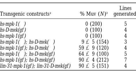
mpk-1(g f) and D-mek(g f) expression causes a
of five such animals (Table 3). In four animals, all six
multivulva phenotype
vulval precursor cells expressed induced (18and 28) cell fates. In one animal, five out of six Pn.p cells expressed Lines
Transgenic constructsa % Muv (N)b generatedc
induced cell fates. We observed 12 cases in which adja-cent Pn.p cells both expressed 18cell fates (out of 25
hs-mpk-1(1) 0 (200) 5
possible cases). These results suggest that expression
hs-D-mek(gf ) 0 (100) 4
of mek(gf ) and mpk-1(gf) is sufficient to induce vulval
hs-mpk-1(gf ) 0 (100) 4
hs-mpk-1(1); hs-D-mek(1) 965 (154) 3 precursor cells to express the 18cell fate in most cases.
hs-mpk-1(gf ); hs-D-mek(1) 5969 (120) 4 mpk-1 acts downstream of signal transduction genes
hs-mpk-1(1); hs-D-mek(gf ) 6469 (100) 5 but upstream of transcription factor genes in the vulval
hs-mpk-1(gf ); hs-D-mek(gf ) 9064 (212) 7 signaling pathway:We have used several complementary
lin-31-mpk-1(gf ); lin-31-D-mek(gf ) 9065 (151) 4
approaches to determine the position of mpk-1 in the
ampk-1(1) refers to a wild-type mpk-1 cDNA (derived from genetic pathway specifying vulval development. Our
re-the SLX2 mRNA form). D-mek(1) refers to a wild-type Dro- sults suggest that MPK-1 acts downstream of other
con-sophila mek cDNA. mpk-1(gf ) contains a single base pair change
served signal transduction molecules and upstream of predicted to cause a D324N substitution, which is analogous
transcription factors. The experiments are presented in to the Drosophila Sevenmaker gain-of-function MAP kinase
the order in which the tested gene (or signaling event) mutation. D-mek(gf ) (also known as Dsor1) is a gain-of-function
mutation in the D-mek cDNA. is thought to act in the anchor cell signaling pathway.
bTransgenic worms from a single representative transgenic
To determine whether MEK and MAP kinase act line were heat shocked at 338 for 30 min as L2 larvae, and downstream of the anchor cell signal, we used laser the Muv phenotype (percentage695% confidence interval)
microsurgery to ablate the entire somatic gonad, includ-was scored in adult animals. Numbers in parentheses indicate
ing the anchor cell, and then induced expression of hs-the number of transgenic worms scored for hs-the Muv
pheno-type. D-mek(gf ) and hs-mpk-1(gf ). We observed that P3.p, P4.p, cNumber of independent transgenic lines obtained.
or P8.p expressed vulval cell fates leading to ectopic vulval tissue in L4 stage animals and a Muv phenotype in adults (Figure 4, Table 5). The vulval phenotype promoter (hs-D-mek(gf )) or under the control of the lin- observed in these gonad-ablated animals was similar to 31 promoter, which predominantly drives expression that of mock-operated control animals. This result indi-in the vulval precursor cells (lindi-in-31-D-mek(gf )) (P. Tan, cates that the Muv phenotype caused by activation of unpublished results). Expression of hs-D-mek(gf ) in MEK and MAP kinase is independent of the anchor cell transgenic worms does not result in a multivulva pheno- signal.
type (Table 4). lin-3 encodes a protein similar to EGF that is likely
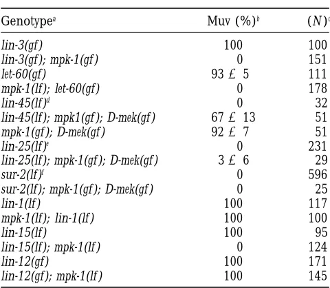
The amino acid substitution D334N is a gain-of-func- to be the inductive signal produced by the anchor cell, tion mutation of Drosophila MAP kinase (Brunneret and overexpression of lin-3 results in a Muv phenotype al. 1994). We used site-directed mutagenesis to engineer (HillandSternberg1992). The vulvaless phenotype the analogous substitution (D324N) into an mpk-1 cDNA of mpk-1(ga117) was found to be epistatic to the Muv clone. This cDNA construct was then placed under con- phenotype caused by overexpression of lin-3 because trol of a C. elegans heat shock promoter (hs-mpk-1(gf )) mpk-1(ga117); lin-3(d) double mutants have a vulvaless or the lin-31 promoter (lin-31-mpk-1(gf )). We found that phenotype (Tables 6 and 7). Furthermore, lin-3 is ex-expression of hs-mpk-1(gf) in transgenic worms did not pressed in the anchor cell, and a genetic mosaic analysis cause a multivulva phenotype (Table 4). shows that mpk-1 acts in the vulval precursor cells, show-We next constructed transgenic lines containing both ing that mpk-1(1) does not regulate lin-3 transcription hs-mpk-1(gf ) and hs-D-mek(gf) and found that heat shock (HillandSternberg1992;Lackneret al. 1994). Thus, expression of mpk-1(gf ) and D-mek(gf) resulted in 90% the Vul phenotype of mpk-1(ga117); lin-3(d) mutants of animals exhibiting a multivulva phenotype (Table 4). does not result from loss of lin-3 expression, but it indi-We followed the Pn.p cell lineages of seven animals cates that mpk-1 activity is necessary for the action of transgenic for hs-mpk-1(gf) and hs-D-mek(gf ) to determine the lin-3 gene and suggests that mpk-1 functions down-the pattern of cell fates expressed by down-the Pn.p cells stream of lin-3.
TABLE 6
Genetic interactions between mpk-1 and other vulval signaling genes
Genotypea Muv (%)b (N )c
lin-3(gf ) 100 100
lin-3(gf ); mpk-1(gf ) 0 151
let-60(gf) 9365 111
mpk-1(lf); let-60(gf) 0 178
lin-45(lf)d 0 32
lin-45(lf); mpk1(gf ); D-mek(gf ) 67613 51
mpk-1(gf ); D-mek(gf ) 9267 51
lin-25(lf)e 0 231
lin-25(lf); mpk-1(gf ); D-mek(gf ) 366 29
sur-2(lf)f 0 596
sur-2(lf); mpk-1(gf ); D-mek(gf ) 0 25
lin-1(lf ) 100 117
mpk-1(lf); lin-1(lf ) 100 100
lin-15(lf) 100 95
lin-15(lf); mpk-1(lf ) 0 124
lin-12(gf) 100 171
lin-12(gf); mpk-1(lf ) 100 145
aAlleles used: mpk-1(ga117) (strains with this allele also
con-tain the cis-linked mutation dpy-17(e164)), lin-3(syIs17) (overex-presses lin-3(1)), let-60(n1046), lin45(sy96) (a partial
reduc-tion-of-function allele), lin-1(sy254), lin-25(e1446), sur-2(ku9),
lin-15(n309), lin-12(n137sd). mpk-1(gf ); D-mek(gf ) strains
con-Figure 4.—The mpk-1(gf )-D-mek(gf) Muv phenotype is
go-tain hs-mpk-1(gf); hs-D-mek(gf ) and the cotransformation nad independent. (A) Nomarski photomicrograph of the
mid-marker rol-6(su1006sd). body region of an hs-D-mek(gf) hs-mpk-1(gf) (expressed from
bPercentage695% confidence interval.
the integrated array gaIs17) adult hermaphrodite that was
cNumber of animals analyzed.
subjected to a heat shock (338 for 30 min) as an L2 larva.
dData fromHanet al. (1993). Syncytial gonad nuclei are apparent, and the animal has a
eData fromTuckandGreenwald(1995). multivulva phenotype. (B) Nomarski photomicrograph of the
fData fromSinghandHan(1995). midbody region of a gaIs17 adult hermaphrodite in which
the somatic gonad precursor cells (Z1.aaa and Z4.ppp) were ablated at the L1 larval stage with a laser microbeam and that
was subjected to a heat shock (338for 30 min) as an L2 larvae. confirms and extends previous work indicating that No traces of the gonad are visible, and the animal has a
mpk-1 functions downstream of let-60. multivulva phenotype.
lin-45 is a homolog of the proto-oncogene raf, and loss-of-function lin-45 mutations cause a Vul phenotype (Hanet al. 1993). We found that the Muv phenotype whether activation of let-60(1) in P6.p required mpk-1
caused by D-mek(gf ) and mpk-1(gf ) is epistatic to the Vul activity. We found that mpk-1(ga117); let-60(gf ) double
phenotype caused by lin-45(lf ), suggesting that mek and mutants are Vul, indicating that mpk-1 activity is
neces-mpk-1 function downstream of lin-45 (Table 6). sary for let-60–mediated expression of vulval cell fates
To order the activity of mpk-1 relative to mek-2, we by any of the Pn.p cells (Tables 6 and 7). This result
determined whether a mek-2 null mutation prevents phosphorylation of MPK-1. mpk-1 expresses two RNAs
TABLE 5 (1X2 and SLX2) predicted to encode proteins of 45
and 55 kD, respectively (Lackner et al. 1994). Based
The mpk-1(gf); D-mek(gf ) Muv phenotype is
on studies of vertebrate MAP kinase, phosphorylation
gonad independent
of each of the MPK-1 protein isoforms by MEK would be expected to result in the appearance of a slightly Genotype Gonada % Muvb(N)c
slower migrating band on Western blots. As expected,
mpk-1(gf); D-mek(gf) 1 88615 (18)
Western blots of protein extracts from wild-type worms
mpk-1(gf); D-mek(gf) 2 100 (17)
immunoblotted with anti-MAP kinase antibodies detect
a2, animals in which a laser was used to ablate the somatic
two pairs of bands, with one pair migrating at z45 kD gonad precursor cells in L1 larvae;1, mock-operated animals. and the other at 55 kD (Figure 2). The upper bands of
bThe Muv phenotype was scored in adult gaIs17 animals
both the 45- and 55-kD doublets are missing in extracts that had been heat shocked at 338 for 30 min as L2 larvae,
from a mek-2 null mutant, suggesting that MPK-1 is not indicated as percentage695% confidence interval.
is required for MAP kinase activation, this result suggests of mpk-1(ga117) was found to be epistatic to the Muv phenotype caused by a lin-15 null mutation (Tables 6 that MPK-1 is not activated in a mek-2 mutant and, hence,
that mpk-1 is likely to function downstream of mek-2. and 7). This result suggests that mpk-1 functions down-stream of lin-15 or in a parallel pathway.
Genetic epistasis experiments suggest that lin-25 and
sur-2 may function at a step between let-60 ras and tran- The second pathway is a lateral signaling pathway in which a signal from the 18cell induces adjacent Pn.p scription factors such as lin-1 Ets (TuckandGreewald
1995; SinghandHan1996). Both lin-25 and sur-2 en- cells to adopt the 28cell fate. lin-12 encodes a protein with significant homology to the Notch family of trans-code novel gene products, and animals homozygous for
a null mutation in either gene exhibit decreased levels membrane receptors (YochemandGreenwald1989), and is likely to be the receptor for the lateral signal. of vulval differentiation and protruding vulva (Pvl)
phe-notypes. We found that the Pvl phenotype caused by lin-12(n137) is a gain-of-function mutation that causes a Muv phenotype in which all six Pn.p cells express the either a lin-25 or a sur-2 mutation was epistatic to the
Muv phenotype caused by a D-mek(gf ); mpk-1(gf) array. 28cell fate (YochemandGreenwald1989). We found that an mpk-1(ga117) lin-12(n137) double-mutant strain Specifically, lin-25; mek(gf); mpk-1(gf ) and sur-2;
hs-mek(gf ); hs-mpk-1(gf ) animals have an Egl and Pvl pheno- exhibited a Muv phenotype, suggesting that most or all of the vulval precursor cells expressed the 28cell fate type (indicating reduced expression of the 18cell fate by
P6.p), but not a Muv phenotype (which would indicate (Table 6). This result indicates that mpk-1 does not act downstream of lin-12 in the specification of the 28cell increased expression of vulval cell fates by P3.p, P4.p,
or P8.p; Table 6 and data not shown). Thus, the activity fate.
Constitutive activation of the anchor cell signaling
of lin-25 and sur-2 are necessary for the expression of
vulval cell fates caused by mek-2(gf ) and mpk-1(gf ), indi- pathway does not cause precocious vulval induction:
The vulval cell divisions beginz29 hr after egg laying cating that lin-25 and sur-2 may act downstream of mek-2
and mpk-1 or in a parallel pathway. when wild-type animals are raised at 258. The timing of vulval induction might be controlled by the time at Next, we ordered the activity of mpk-1 relative to lin-1,
which encodes a protein with a region of similarity to which the vulval signaling pathway is activated. For ex-ample, some component of the signaling pathway, such the Ets DNA-binding domain (Beitel et al. 1995). lin-1
loss-of-function mutations cause a Muv phenotype. mpk-1 as the anchor cell signal, might not be expressed until 29 hr after egg laying. Alternatively, the vulval precursor (ga117); lin-1(sy254) double-mutant worms also
exhib-ited a Muv phenotype (Table 6), indicating that lin-1 cells might not be competent to respond to activation of the let-23/let-60/mpk-1 signaling pathway until 29 hr likely acts downstream of mpk-1.
In addition to the let-23 RTK/let-60 ras/mpk-1 signal- after egg laying. We have constitutively activated the anchor cell signaling pathway by using animals that ing pathway, at least two other signaling pathways are
involved in specifying the wild-type pattern of vulval ubiquitously express D-mek(gf ) from the EF1a promoter and constitutively express hs-mpk-1(1) when grown at cell fates. The first pathway is an inhibitory pathway
postulated to arise from the surrounding hypodermis. 258. As shown above, constitutive activation of mek and mpk-1 should be sufficient to induce vulval cell fates lin-15 acts in this pathway and encodes two novel
pro-teins that function to produce an inhibitory signal in even if upstream components of the signaling pathway are not expressed before 29 hr after egg laying. In these the hypodermis (Hedgecock 1990;Clark et al. 1994;
Herman andHuang et al. 1994). The Vul phenotype transgenic worms, the vulval cell divisions beganz29–30
TABLE 7
Vulval cell lineages in double mutants
Pn.p cell fateb
Genotypea P4.p P5.p P6.p P7.p P8.p Nc
Wild type 38 28 18 28 38 5
mpk-1(lf ) 38 38 38 38 38 10
mpk-1(lf ); lin-3(gf ) 38 38 38 38 38 10
mpk-1(lf ); let-60(gf) 38 38 38 38 38 10
lin-15(lf); mpk-1(lf ) 38 38 38 38 38 10
aGenotypes for mpk-1(ga117) double mutants were mpk-1(ga117) 17(e164); lin-3(syIs1), mpk-1(ga117) dpy-17(e164); lin-15(n309), and mpk-1(ga117) dpy-dpy-17(e164); let-60(n1046gf ).
bCell fates were inferred by direct observation of L4 larvae with Nomarski optics. 38cell fate is S S, 28cell
fate is LLTN or NTLL, and 18cell fate is TTTT (abbreviations are defined in Table 3). P3.p was 38or U in every animal.
TABLE 9 TABLE 8
Timing of vulval precursor cell divisions in mpk-1 null mutations result in maternal
effect larval lethality activated MAP kinase strains
Number of animals in which Genotype of F1lethality
mother F1lethalityb (m1/m2 and
vulval precursor cell
divisions have begunb (m1/m2)a (m1/m1) m2/m2)b
hs-mpk-1(1); ga117/1 0 (120) 0 (344)
Hours after egg layinga Wild type EF1a-D-mek(gf )c
ga117/ga111 0 (458) NA
ga117/ga111 97 (195) 4 (400)
27 0/5 0/5
ga119/ga111 92 (140) 3 (372)
28 0/5 0/5
29 0/5 1/5 aFull genotypes of animals: ga117/unc-79 dpy-17, unc-79
30 2/5 1/5 ga111, ga117 dpy17/unc-79 ga111, and ga117 dpy-17/unc-79
31 4/5 3/5 ga111.
32 5/5 5/5 bPercentage lethality is shown. Parentheses indicate the
33 5/5 5/5 number of animals scored. Fertile non-Vul mothers were picked to individual plates, and entire F1broods were analyzed. aTwenty-five hermaphrodites were allowed to lay eggs for
Lethality was scored by counting dead Dpy larvae (m1/m1) 1 hr on seeded plates, after which they were removed and or dead non-Dpy larvae (m1/m2 and m2/m2).
progeny were placed in a 258incubator.
bAt each time point, five animals of each genotype were
examined under Nomarski optics to determine if any of the
eny segregated from mpk-1(ga117)/1 hermaphrodites Pn.p cells had divided.
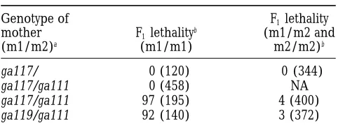
do not exhibit lethality (Table 9). We reasoned that
cThirty-three out of 35 of these animals expressed a Muv
phenotype as adults. maternal mpk-1(1) might rescue larval lethality in these animals, so we asked whether larval lethality was ob-served in a strain with less maternal mpk-1(1) activity. hr after the eggs were laid, as they do in wild-type worms We examined homozygous mpk-1(ga117) animals segre-(Table 8). The transgenes resulted in activation of the gated from mothers heterozygous for a partial loss-anchor cell signaling pathway because 90% of these of-function mpk-1 allele and a null allele, i.e., of geno-animals exhibited a Muv phenotype. This result indi- type ga111/ga117. The heterozygous mpk-1(ga111)/mpk-cates that the vulval precursor cells are not competent 1(ga117) mothers are not sterile, though they do show to divide in response to MEK and MAP kinase signaling reduced fecundity (data not shown). We observed that until at least 29 hr after egg laying. 97% of mpk-1(ga117)/mpk-1(ga117) progeny segregated mpk-1 acts in the let-60 ras signaling pathway in multi- from mpk-1(ga111)/mpk-1(ga117) parents arrested in the
ple cell types: let-60 ras functions in at least six cell fate L1 stage as rod-like larvae (Table 9). The remaining 3%
decisions because let-60 loss-of-function mutations affect of mpk-1(ga117)/mpk-1(ga117) animals were non-Let and at least six different cell types. An important question grew into sterile, vulvaless adults. Similar results were is whether let-60 signals through mpk-1 in all six cell obtained with another allele, mpk-1(ga119), which may types. If so, then mpk-1 mutants should show defects in strongly reduce mpk-1 function (Table 9). These results all the cell types that are affected in let-60 mutants. suggest that let-60 and mpk-1 may act in the same
signal-First, let-60 acts in the germ line, as loss-of-function ing pathway in early larval development.
mutations cause a sterile phenotype by preventing oo- Third, mutations in let-60 have been shown to affect cyte nuclei from exiting the pachytene stage of meiosis cell fate decisions in the male tail (Chamberlin and (Churchet al. 1995). The mpk-1(oz140) allele has pre- Sternberg1993, 1994) that result in defective spicule viously been shown to cause similar sterility in homozy- structure and defective male mating. mpk-1(ga117) ho-gous animals (Church et al. 1995). We report here mozygous males also have crumpled spicules and are that the mpk-1(ga117) null allele also causes this sterile unable to mate (Figure 3, materials and methods), phenotype and that the mpk-1(ga111) allele causes suggesting that mpk-1 and let-60 may act in the same a temperature-sensitive sterile phenotype (Table 2). signaling pathway during spicule development. These results suggest that let-60 and mpk-1 may act in Fourth, let-60 mutants have defects in the migration of the same signaling pathway in meiotic prophase pro- the sex myoblasts; the partial loss-of-function mutation
gression. mpk-1(ku1) causes weak defects in the migration of the
prog-morphic, such that the migration defect might reflect One possibility is that these two genes act in one linear pathway and that gain-of-function mutations at two se-interference from the mutant MPK-1(ku1) protein rather
than a requirement for wild-type MPK-1(1). Thus, it is quential steps are required to fully activate the pathway. It could be that full activation of MPK-1 requires both not clear whether mpk-1(1) acts in sex myoblast
migra-tion. the D324N missense mutation (analogous to Drosophila
Sevenmaker) and expression of D-MEK(gf). One finding Fifth, let-60 mutants have defects in the expression of
the P12.p cell fate (Sundaramet al. 1996). In contrast, that supports this idea is that a strong gain-of-function allele of mek-2 results in a Muv phenotype in single mpk-1(ga117) mutants segregated from mpk-1(ga117)/
mpk-1(ga111) mothers do not appear to have defects in mutants (Wuet al. 1995), showing that mek-2 is sufficient to activate vulval cell fates, even though the mek(gf) allele the expression of the P12.p cell fate. One possibility is
that maternal mpk-1 might be active in P12.p, so mpk-1 used in these studies does not cause full activation of mek. Another possibility is that these two genes act in mutants would not exhibit cell fate defects in these
cells because of maternal rescue of mpk-1(1) from their separate signaling branches and both branches need to be activated to cause a Muv phenotype.
heterozygous mothers. Another possibility is that let-60
ras transduces signals independently of mpk-1 in this mpk-1 acts downstream of signal transduction genes
but upstream of transcription factor genes: We have
cell, possibly involving another C. elegans MAP kinase
homolog. found that the MAP kinase homolog mpk-1 acts
down-stream of the Ras homolog let-60, the Raf homolog lin-Sixth, we have not observed mpk-1 phenotypes other
than the ones mentioned above. This result suggests 45, and the MEK homolog mek-2, consistent with the biochemical position of MAP kinase in vertebrate signal-that mpk-1 may be required only in let-60 ras signaling
pathways because all the mpk-1 phenotypes are also ob- ing pathways and the genetic position of the Drosophila rolled MAP kinase in the R7 photoreceptor signaling served in let-60 ras mutants.
pathway (Biggset al. 1994;Brunneret al. 1994; Treis-man1996).
DISCUSSION
Furthermore, we show that mpk-1 acts upstream of lin-1, which encodes a putative DNA binding protein
The anchor cell signaling pathway may be linear:
Pre-viously isolated mpk-1 alleles have weak vulval pheno- similar to mammalian Ets-1 and Drosophila Yan (Beitel et al. 1995). mpk-1 also acts upstream of lin-31, which types, whereas loss-of-function alleles in other genes in
the let-23 RTK/let-60 ras signaling pathway have a strong encodes a winged helix transcription factor similar to mammalian HNF-3 and Drosophila forkhead (Miller vulvaless phenotype. These results raised the possibility
that the let-23 signaling pathway may branch in a way et al. 1993;Tanet al. 1998). Both LIN-1 and LIN-31 are phosphorylated by MAP kinase in vitro and in vivo (Tan that another branch of the signaling pathway could
transduce the anchor cell signal in mpk-1 mutants. We et al. 1998). Unphosphorylated LIN-1 and LIN-31 bind to each other and form a protein complex that inhibits have tested this possibility and have shown that mpk-1
null mutations cause a highly penetrant, vulvaless phe- vulval induction. Phosphorylation of these proteins dis-rupts the LIN-1/LIN-31 protein complex and, thus, pre-notype. Thus, there is no evidence for a signaling branch
acting in parallel to mpk-1, and our data are consistent vents inhibition of vulval cell fates. Phosphorylation of LIN-31 may also create a transactivation domain and with the idea that the anchor cell signaling pathway is
linear through the mpk-1 step. However, our results do permit LIN-31 to act as a transcriptional activator, there-by promoting expression of vulval cell fates.
not rule out the possibility that the pathway may be
branched in a way that mpk-1 transduces the majority An interesting point is that the anchor cell signaling pathway may be predominantly linear between lin-3 and of the anchor cell signal and another branch has a
minor role. In the absence of mpk-1 activity, the minor mpk-1, but may branch downstream of mpk-1. mpk-1 null mutations have a highly penetrant vulvaless phenotype, branch may not be capable of mediating observable
amounts of vulval induction. but null mutations in genes that act downstream of mpk-1 (lin-1, lin-25, lin-31, and sur-2) neither completely The case for a linear signaling pathway is
strength-ened by our finding that coexpression of D-mek(gf) and activate nor completely prevent vulval induction. Spe-cifically, null mutations in lin-25 and sur-2 result in a mpk-1(gf ) results in a strong multivulva phenotype. Both
genes act at the end of the anchor cell signal transduc- partial vulvaless phenotype (TuckandGreewald1995; SinghandHan1996), lin-1 null mutations cause expres-tion pathway, and the primary known funcexpres-tion for MEK
is to activate MAP kinase. These results indicate that sion of partial or hybrid 18 or 28 cell fates (Beitel et al. 1995), and lin-31 null mutations result in partially the pathway is not branched upstream of mek and mpk-1,
as mek-2 and mpk-1 are both required for vulval induction penetrant Muv and Vul phenotypes (Milleret al. 1993; Tanet al. 1998). These results suggest that these genes and coexpression of mek(gf ) and mpk-1(gf ) is sufficient
for vulval induction. may function on separate branches downstream of MAP
kinase. Null mutations in any one gene may not com-However, expression of either gene alone [either