Mouse Protection of Anti-Pneumococcal Surface Protein A
Monoclonal Antibodies
Naeem Khan,a,b*Raies Ahmad Qadri,bDevinder Sehgala
Molecular Immunology Laboratory, National Institute of Immunology, New Delhi, Indiaa
; Department of Biotechnology, University of Kashmir, Srinagar, Indiab
The shortcomings of the licensed polysaccharide-based pneumococcal vaccine are driving efforts toward development of a
pro-tein-based vaccine that is serotype independent and effective in all age groups. An opsonophagocytic killing assay (OPKA) is
used to evaluate the antibody response against polysaccharide-based pneumococcal vaccines. However, the OPKA is not reliable
for noncapsular antigens. Thus, there is a need to develop an
in vitro
surrogate for protection for protein vaccine candidates like
pneumococcal surface antigen A (PspA). PspA is a serologically variable cell surface virulence factor. Based on its sequence, PspA
has been classified into families 1 (clade 1 and 2), 2 (clades 3, 4 and 5), and 3 (clade 6). Here, we report the characterization of 18
IgG anti-PspA monoclonal antibodies (anti-PspA
hkR36AMAbs) generated from mice immunized with heat-killed strain R36A
(clade 2). An enzyme-linked immunosorbent assay (ELISA)-based analysis of the reactivity of the MAbs with recombinant PspAs
from the 6 clades indicated that they were family 1 specific. This was confirmed by flow cytometry using a hyperimmune serum
generated against PspA from R36A. Eight MAbs that bind at least one clade 1- and clade 2-expressing strain were evaluated for
complement deposition, bactericidal activity, and passive protection. The anti-PspA
hkR36AMAb-dependent deposition of
com-plement on pneumococci showed a positive correlation with passive protection against strain WU2 (
r
ⴝ
0.8783,
P
ⴝ
0.0041). All
of our protective MAbs showed bactericidal activity; however, not all MAbs that exhibited bactericidal activity conferred
protec-tion
in vivo
. The protective MAbs described here can be used to identify conserved protection eliciting B cell epitopes for
engi-neering a superior PspA-based vaccine.
T
he bacterial pathogen
Streptococcus pneumoniae
(pneumococ-cus) is responsible for causing pneumonia, septicemia,
men-ingitis, and otitis media in humans (
1
). According to the estimate
made by the World Health Organization in 2005, 1.6 million
in-dividuals die of diseases caused by
S. pneumoniae
every year, and
most of these deaths occur in developing countries (
2
). In the year
2000, it was estimated that pneumococcal disease was responsible
for about 800,000 deaths of children
⬍
5 years of age (
3
). The
currently available pneumococcal polysaccharide vaccine is not
effective in children
⬍
2 years of age. Pneumococcal conjugate
vaccines, however, overcome this limitation and are effective in
children but have limited serotype coverage (
4
). The development
of antibiotic resistance and the emergence of nonvaccine
sero-types pose difficulties in the management of pneumococcal
infec-tions. Efforts are being made globally to develop a protein-based
pneumococcal vaccine that confers serotype-independent
protec-tion in all age groups (
5–7
).
A polysaccharide capsule envelops
S. pneumoniae
, and it serves
as the major virulence factor by shielding pneumococci from
im-mune attack. The unencapsulated strains of
S. pneumoniae
are
known to be avirulent or highly attenuated. In addition to the
capsule, several surface-associated proteins have been
demon-strated to be involved in pneumococcal virulence, and one such
protein is pneumococcal surface protein A (PspA) (
8
). A
well-established opsonophagocytic killing assay (OPKA) is available
for evaluating the pneumococcal polysaccharide-based vaccines.
The recent interest in the protein-based pneumococcal vaccines
has led to efforts toward development of an
in vitro
assay for
non-capsular antigens that can help in predicting and quantitating the
protective activity of antibodies against protein vaccine
candi-dates. Various investigators have tried to correlate anti-protein
antibody titers, surface binding (by a whole-cell enzyme-linked
immunosorbent assay [ELISA]), and a surface killing assay with
in
vivo
protection (
9–12
). The notion of
in vitro
antibody-mediated
complement deposition as a possible surrogate for predicting
in
vivo
protection has been proposed by Goulart et al. and Ochs et al.
(
13
,
14
). However, these investigators did not validate it with
in
vivo
protection experiments. Availability of a robust
in vitro
assay
would help in minimizing the use of animal models for testing
protein vaccine candidates.
PspA is a polymorphic, surface-associated choline-binding
protein (
15
). PspA has a predominantly
␣
-helical coiled coil
struc-ture (
16
,
17
). It is present in essentially all clinical isolates studied
to date and is being pursued as a promising vaccine candidate
(
18
). Based on the amino acid sequence, PspA has been classified
Received28 December 2013Returned for modification3 February 2014
Accepted11 November 2014
Accepted manuscript posted online19 November 2014
CitationKhan N, Qadri RA, Sehgal D. 2015. Correlation betweenin vitro
complement deposition and passive mouse protection of anti-pneumococcal surface protein A monoclonal antibodies. Clin Vaccine Immunol 22:99 –107.
doi:10.1128/CVI.00001-14.
Editor:T. S. Alexander
Address correspondence to Devinder Sehgal, [email protected]. * Present address: Naeem Khan, Department for Biomolecular Systems, Max Planck Institute of Colloids and Interfaces, Potsdam, Germany.
Supplemental material for this article may be found athttp://dx.doi.org/10.1128 /CVI.00001-14.
Copyright © 2015, American Society for Microbiology. All Rights Reserved.
doi:10.1128/CVI.00001-14
on August 17, 2020 by guest
http://cvi.asm.org/
into three families and six clades. Family 1 includes clades 1 and 2,
family 2 includes clades 3, 4, and 5, and family 3 includes clade 6
(
19
). Studies have shown that 94 to 99% of the pneumococcal
isolates analyzed belong to PspA families 1 and 2 (
20
,
21
).
The complement-mediated clearance of pneumococci is an
important component of the host defense mechanism (
22
). A
PspA-deficient strain is cleared faster than wild-type
pneumo-cocci, and an anti-PspA antibody facilitates
complement-depen-dent phagocytosis of
S. pneumoniae
(
23
). Ren and coworkers have
demonstrated that anti-PspA antibodies enhance complement
ac-tivation and deposition on pneumococcal surface and thus help in
clearance (
24
).
Active immunization with PspA in animal models has proven
to be protective against invasive disease and nasopharyngeal
car-riage (
25
). Mice immunized with DNA vaccine expressing the
extracellular domain of PspA were protected against an
intraper-itoneal challenge with a pneumococcal strain bearing PspA from
the same clade (
26
). The B cell epitopes recognized by protective
monoclonal antibodies (MAbs) have been mapped to the 192- to
260-amino acid region (
27
). Daniels et al. recently demonstrated
that the proline-rich region of PspA contains surface-accessible
epitopes that are protective in both active and passive mouse
pro-tection experiments (
28
). PspA has been shown to elicit high
an-tibody titers in humans, and human anti-PspA sera can protect
mice against pneumococcal challenge when transferred passively
(
18
,
29
).
There is evidence to suggest that not all anti-PspA antibodies
are protective. The goals of the present study were to identify
anti-PspA
hkR36AMAbs that recognize conserved cross-protective
B cell epitopes, and since the
in vitro
surrogate of protection is not
well established for noncapsular antigens, we evaluated the surface
binding, complement deposition, and bactericidal activity of
anti-PspA
hkR36AMAbs as potential
in vitro
correlates of protection. We
found that all of the 18 anti-PspA
hkR36AMAbs recognized family 1
PspAs and did not bind PspAs representing families 2 and 3. We
identified 4 anti-PspA
hkR36AMAbs (P1E11, M4F4, P2A4, and
P2B5) that augmented complement deposition on pneumococci,
exhibited bactericidal activity, and conferred protection against
PspA clade 1- and 2-bearing
S. pneumoniae
strains in passive
mouse protection experiments. Further, our data with strain WU2
suggested that anti-PspA
hkR36AMAb-dependent complement
de-position on pneumococci strongly correlated with
in vivo
protec-tion. We observed that all of the protective anti-PspA
hkR36AMAbs
exhibited bactericidal activity; however, not all of the
anti-PspA
hkR36AMAbs that showed bactericidal activity conferred
in
vivo
protection.
MATERIALS AND METHODS
Mice.Six- to 8-week-old BALB/c (female) and CBA/N (male/female) in-bred strains of mice were obtained from the Small Animal Facility of the National Institute of Immunology. Animals were rested and handled in accordance with the institutional animal ethics committee guidelines. Blood samples from healthy donors were taken with the approval of and following the guidelines of the institutional human ethics committee. Ex-periments involving recombinant DNA and handling ofS. pneumoniae
were carried out in accordance with the institutional biosafety committee guidelines.
Pneumococcal strains, plasmids, and culture conditions.The pneu-mococcal strains and plasmids used in this study are listed in Table S1 in the supplemental material. Pneumococcal strains were maintained in Todd-Hewitt broth supplemented with 0.5% yeast extract (THY) or on a
plate with tryptic soy agar (TSA) supplemented with 5% (vol/vol) sheep blood at 37°C in the presence of 5% CO2. The mid-logarithmic-phase pneumococcal cultures were stored with 15% (vol/vol) glycerol or 17% fetal calf serum, aliquoted, and stored at⫺70°C as described previously (30).
Escherichia colicells were maintained in Luria-Bertani broth or on a
Luria-Bertani agar plate with antibiotic(s) wherever required.
Molecular cloning, overexpression, and purification of recombi-nant PspA.The subfragments encoding the N-terminal (surface exposed) region of PspA for clade 3 (PspATIGR4) and clade 5 (PspAATCC 6303) were amplified by PCR using genomic DNA from the pneumococcal strains TIGR4 and ATCC 6303, respectively, following the cloning strategy and PCR conditions described by Rohatgi et al. (30). The corresponding plas-mid constructs for PspA clade 1 (pUAB069, strain L82016 [PspAL82016]), clade 4 (pUAB100, strain JCP#56 [PspAJCP#56]), and clade 6 (pUAB104, strain BG9300 [PspABG9300]) were kindly provided by Susan Hollings-head, University of Alabama, USA. The plasmid construct for PspA clade 2 (R36A [PspAR36A]) was published previously from our laboratory (30). For expression purposes, pQE-30 Xa- and pET-20b-based constructs were transformed into E. coli expression strains SG13009 and BL-21(DE3), respectively. Recombinant PspA was purified using nickle-ni-triloacetic acid (Ni-NTA) affinity chromatography (Sigma-Aldrich, USA). The purity of the protein preparation was found to be⬎95% by SDS-PAGE and was of the expected molecular size.
Enzyme-linked immunosorbent assay.The reactivities of the 18 IgG anti-PspAhkR36A MAbs with recombinant PspAs representing the six clades of PspA were analyzed by an ELISA. Briefly, 96-well polystyrene microtiter plates (Greiner Bio-One, Germany) were coated overnight at 4°C with recombinant PspA (50l of 2g/ml per well) in 100 mM car-bonate-bicarbonate buffer (pH 9.5). The plates were washed with phos-phate-buffered saline (PBS) containing 0.05% Tween 20 (PBST) and blocked with PBS containing 2% bovine serum albumin (BSA) at 37°C for 1 h. After washing with PBST, the plates were incubated with the culture supernatant from the 18 anti-PspAhkR36Ahybridomas (in duplicate) at 37°C for 1 h. The plates were washed with PBST and incubated with horseradish peroxidase-conjugated goat anti-mouse Ig antibody (diluted 1 in 2,500; Becton-Dickinson Bioscience, USA) followed by incubation at 37°C for 1 h. The color was developed using 3,3=,5,5= -tetramethylbenzi-dine-H2O2as the substrate, and absorbance was recorded at 450 nm.
Generation of polyclonal sera against recombinant PspAR36A.
Six-to 8-week-old female BALB/c mice were immunized subcutaneously with 25g recombinant PspAR36Aemulsified with Imject alum (1:1 [wt/wt]) (Pierce, USA). On days 14 and 28, mice were given a booster injection with the same amount of antigen emulsified as described above. The control mice received only Imject alum in PBS. One week after the second booster, hyperimmune serum was isolated after mice were bled retro-orbitally, and an ELISA was performed to determine the PspA-specific antibody titer.
Surface staining with anti-PspAhkR36AMAbs and anti-PspAR36A
hy-perimmune serum.The surface binding of anti-PspAhkR36AMAbs withS.
pneumoniaestrains that express clade 1 PspA (BG8838) and clade 2 PspA
(WU2 and D39) was analyzed using flow cytometry as described previ-ously (31). Briefly, mid-logarithmic-phase (optical density at 600 nm [OD600] of 0.4) pneumococci (107CFU) were washed with PBS and in-cubated with 200l of culture supernatant from the hybridomas at room temperature for 1 h. After washing with PBS, pneumococci were incu-bated with fluorescein isothiocyanate (FITC)-conjugated F(ab=)2 frag-ment goat anti-mouse IgG plus IgM(H⫹L) antibody (diluted 1 in 200) (Jackson ImmunoResearch Laboratory, USA) followed by incubation at room temperature for 1 h. After washing, pneumococci were fixed with 2% paraformaldehyde (PFA) for 10 min at 4°C, and surface staining was analyzed by flow cytometry (FACSCalibur; Becton-Dickinson Biosci-ence).
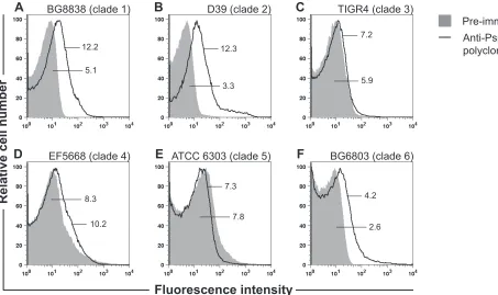
The surface binding of the anti-PspAR36Apolyclonal sera (diluted 1 in 200) was analyzed with strains BG8838 (clade 1), D39 (clade 2), TIGR4 (clade 3), EF5668 (clade 4), ATCC 6303 (clade 5), and BG6803 (clade 6)
on August 17, 2020 by guest
http://cvi.asm.org/
using flow cytometry as described above. Geometric mean fluorescence intensity (GMFI) values equal to or greater than twice the value obtained with the preimmune control were considered significant.
Purification of anti-PspAhkR36AMAbs.Hybridomas secreting
anti-PspAhkR36AMAbs were generated and cultured as previously described (30). Ascitic fluid was generated for the anti-PspAhkR36AMAb-secreting hybridomas. Briefly, BALB/c mice (3 mice per hybridoma) were injected intraperitoneally with 0.5 ml incomplete Freund’s adjuvant (Sigma-Al-drich). Three days later, 5⫻106hybridoma cells were injected intraperi-toneally. After 7 to 10 days, ascitic fluid from the peritoneal cavity was centrifuged at 500⫻gfor 10 min at 4°C, and the supernatant was collected and stored at⫺70°C. The MAbs were purified from ascitic fluid using protein G-Sepharose beads (GE Healthcare, USA) as described earlier (32). The protein concentration of the purified MAb was estimated using a micro BCA protein assay kit (Thermo-Scientific, USA).
Passive mouse protection assay.Groups of eight 6- to 8-week-old CBA/N mice were injected intraperitoneally with purified anti-PspAhkR36AMAb or a matched isotype control. The amounts of MAb administered for the high- and low-dose experiments were 5 and 1.25 mg/kg body weight, respectively. An hour later, mice were challenged intravenously with 107CFU (100 times the 50% lethal dose [LD
50]) of strain BG8838 and 103CFU (100 times the LD
50) of WU2 (27). The survival of mice was monitored every 12 h for the first 10 days and every 24 h for the next 11 days.
Blood bactericidal assay.The anti-PspAhkR36AMAbs were analyzed by a blood bactericidal assay as described previously (33). Briefly, human peripheral blood was collected using recombinant hirudin from yeast (100 U/ml blood) as an anticoagulant. Pneumococci (500 CFU in 10l) were incubated with 235l of blood in the presence of either purified anti-PspAhkR36AMAb or the corresponding isotype control (5l of 1 mg/ml). Samples were incubated at 37°C with rotation for 2 h for D39 and 3 h for BG8838. The surviving bacteria were enumerated by plating serial dilutions (in duplicate) on TSA plates, and the mean values obtained were used to calculate bactericidal activity as described below. The data are
presented as percent killing, which was arrived at using the formula ([col-ony count with appropriate isotype control MAb⫺colony count with anti-PspAhkR36AMAb] divided by colony count with the appropriate iso-type control MAb)⫻100.
Complement deposition assay.The complement deposition assay was performed as described by Ochs et al. with some modifications (14). Briefly, mid-logarithmic-phase pneumococci (107CFU) were washed with PBS and incubated with either an anti-PspAhkR36A MAb or a matched isotype control (100l of 20g/ml) for 1 h at room temperature. Treated pneumococci were incubated in 200l of normal human serum pooled from three healthy donors (diluted to 10% in Hanks’ balanced salt solution with Ca2⫹and Mg2⫹ions) (Biological Industries, Israel) for 30
min at 37°C. The mixture was incubated on ice for 10 min and washed with PBS, and the incubation was continued with PBS-1% BSA contain-ing mouse anti-human C3 MAbs (diluted 1 in 50) (Abcam, USA) for 45 min on ice. The bound anti-C3 antibody was detected using an FITC-conjugate F(ab=)2fragment goat anti-mouse IgG plus IgM(H⫹L) anti-body (diluted 1 in 200). Pneumococci were fixed with 2% PFA for 10 min on ice and analyzed by flow cytometry.
Statistical analysis. Statistical analysis was done using GraphPad Prism version 6 software (GraphPad Software Inc., USA). The passive mouse protection data were compared using the log rank test. We used a TABLE 1ELISA-based analysis for reactivity of anti-PspAhkR36AMAbs
with recombinant PspAs representing the 6 PspA cladesa
Hybridoma
Fold change for pneumococcal strain (PspA family/clade): L82016
(1/1)b R36A (1/2)b
TIGR4 (2/3)
JCP#56 (2/4)
ATCC 6303 (2/5)
BG9300 (3/6)
B3D12 16.5 17.2 0.8 0.8 0.8 1.2
B3H8 1.8 2.1 1.0 0.9 1.0 0.8
L5C8 17.1 16.7 0.8 0.8 0.8 0.8
L5F10 19.5 26.9 0.8 0.7 0.9 1.2
M4F4 30.0 32.5 1.1 1.2 1.6 1.2
P1E11 29.2 31.5 1.4 1.4 1.7 1.6
D1A5 10.6 11.9 0.9 0.8 0.7 0.9
K1B12 15.9 16.5 0.7 0.7 0.6 0.9
M6B2 13.5 14.5 1.0 1.4 1.3 1.9
P2F9 9.8 13.4 0.9 1.0 0.9 1.0
P2A4 21.6 19.9 1.0 1.5 0.9 1.0
P2B5 22.0 24.0 1.0 1.2 1.2 1.2
J4C1 28.2 29.3 1.0 0.8 0.7 0.8
P2C2 16.2 17.0 0.6 0.6 0.6 0.6
A1D9 2.0 2.7 0.6 0.7 0.7 0.6
C4B4 18.1 18.1 0.8 0.6 0.6 0.7
F4B6 24.7 26.3 1.0 0.9 0.8 1.1
D3H6 4.0 5.7 0.7 0.7 0.7 1.1
aThe culture supernatants from the 18 anti-PspAhkR36AMAb-secreting hybridomas
were tested for reactivity with recombinant PspAs representing the 6 clades of PspA (extracellular domain with or without the proline-rich region) by an ELISA. The numerical values represent fold change in absorbance relative to the control.
bThese columns show fold change values that were greater than twice the value
obtained with the control and were considered significant.
TABLE 2Surface binding of anti-PspAhkR36AMAbs with strains expressing family 1 PspAa
MAb
GMFI (fold change) for:
BG8838 clade 1 WU2 clade 2 D39 clade 2
IgG1 IC 4.77 (1.0) 3.75 (1.0) 3.67 (1.0)
B3D12 13.10 (2.8) 5.18 (1.4) 64.80 (17.7)
B3H8 14.70 (3.1) 5.49 (1.5) 8.75 (2.4)
L5C8 18.70 (3.9) 3.82 (1.0) 137.00 (37.3)
L5F10 18.90 (4.0) 7.22 (1.9) 66.20 (18.1)
M4F4 59.50 (12.5) 11.80 (3.2) 140.00 (38.2)
P1E11 81.80 (17.2) 14.20 (3.8) 146.00 (39.8)
D1A5 4.04 (0.8) 107.00 (28.5) 52.90 (14.4)
K1B12 3.86 (0.8) 9.39 (2.5) 69.80 (19.0)
M6B2 5.64 (1.2) 19.20 (5.1) 174.00 (47.4)
P2F9 4.80 (1.0) 4.12 (1.1) 14.30 (3.9)
IgG2a IC 4.82 (1.0) 2.82 (1.0) 4.68 (1.0)
P2A4 23.60 (4.9) 11.10 (3.9) 59.70 (12.8)
P2B5 17.60 (3.7) 16.10 (5.7) 33.90 (7.3)
J4C1 3.88 (0.8) 3.27 (1.2) 60.80 (13.0)
P2C2 3.52 (0.7) 8.93 (3.2) 34.80 (7.4)
IgG2b IC 4.16 (1.0) 2.28 (1.0) 3.21 (1.0)
A1D9 4.35 (1.1) 16.3 (7.2) 47.40 (14.8)
C4B4 10.80 (2.6) 22.10 (9.7) 45.00 (14.1)
F4B6 3.70 (0.89) 56.7 (24.9) 41.00 (12.8)
IgG3 IC 4.26 (1.0) 2.82 (1.0) 3.21 (1.0)
D3H6 4.41 (1.0) 4.32 (1.5) 25.80 (8.1)
a
Surface binding of 18 anti-PspAhkR36A
MAbs with strains BG8838 (clade 1) and WU2 and D39 (clade 2) was analyzed by flow cytometry. Strain D39 was included in the analysis as the anti-PspA MAbs were raised against R36A, an unencapsulated derivative of D39. Pneumococci were incubated with 200l of culture supernatant from the 18 anti-PspAhkR36A
MAb-secreting hybridomas followed by staining with the appropriate FITC-conjugated secondary antibody. Matched isotype control MAbs were included, and samples were analyzed using a flow cytometer. The surface binding is expressed as geometric mean fluorescence intensity (GMFI), and the fold change relative to the corresponding isotype control MAb (assigned 1.0) is given in parentheses. Aⱖ2-fold increase in the surface staining relative to that of the corresponding isotype control was considered significant. IgG1 IC, IgG2a IC, IgG2b IC, and IgG3 IC represent isotype control MAbs for the isotypes IgG1, IgG2a, IgG2b, and IgG3, respectively.
on August 17, 2020 by guest
http://cvi.asm.org/
parametric Pearson’s linear regression correlation analysis to evaluate a possible correlation between the anti-PspAhkR36AMAb-dependent depo-sition of complement on pneumococci and passive mouse protection. AP
value of⬍0.05 was considered statistically significant. RESULTS
Cross-reactivity and surface staining of anti-PspA
hkR36AMAbs
is restricted to PspA family 1.
We previously reported that the
relative avidity of the primary IgG anti-PspA polyclonal antibody
was higher than that of recombinant PspA
R36Ain the sera of mice
immunized with heat-killed R36A (
30
). In this study, we analyzed
18 IgG anti-PspA
hkR36AMAbs for the extent of cross-reactivity
with recombinant PspA belonging to the 6 PspA clades (i.e.,
PspA
L82016, PspA
R36A, PspA
TIGR4, PspA
JCP#56, PspA
ATCC 6303, and
PspA
BG9300) by an ELISA (
Table 1
). All of the 18 MAbs reacted
with PspAs representing clades 1 and 2 (family 1). None of the
MAbs bound PspAs representing families 2 (clades 3, 4, and 5) and
3 (clade 6), suggesting that the anti-PspA
hkR36AMAbs were family
1 specific.
Next, we assessed whether these 18 anti-PspA
hkR36AMAbs bind
to the surface of pneumococci. For this purpose, we tested strains
BG8838 (clade 1) and WU2 and D39 (clade 2). Of the 18 MAbs,
BG8838 and WU2 were recognized by 9 and 10 MAbs, respectively
(
Table 2
). As expected, all 18 anti-PspA MAbs stained D39. Nine
MAbs bound one clade 1 and two clade 2 PspA-expressing strains.
Eight (i.e., B3D12, B3H8, L5C8, L5F10, M4F4, P1E11, P2A4, and
P2B5) of these were selected at random for further analysis.
To confirm our observation that the anti-PspA antibody
re-sponse elicited against heat-killed R36A was family 1 specific, the
anti-PspA
R36Ahyperimmune serum was used to test surface
bind-ing with strains expressbind-ing PspAs belongbind-ing to families 1, 2, and 3
by flow cytometry (
Fig. 1
). The data demonstrate that
anti-PspA
R36Ahyperimmune serum binds to family 1 PspA-expressing
strains BG8838 (clade 1) and D39 (clade 2). No binding was
ob-served with PspA family 2-bearing strains TIGR4 (clade 3),
EF5668 (clade 4), and ATCC 6303 (clade 5) (
Fig. 1
). Similarly,
there was no surface staining with the PspA family 3-expressing
strain BG6803 (clade 6). Thus, our flow cytometry data
demon-strate that the binding of anti-PspA
R36Ahyperimmune serum to
pneumococcal strains was family 1 specific, and this corroborates
the data obtained with anti-PspA
hkR36AMAbs (
Table 2
).
In vivo
protective efficacy of anti-PspA
hkR36AMAbs.
The
rel-ative protective activities of anti-PspA
hkR36AMAbs B3D12, B3H8,
L5C8, L5F10, M4F4, P1E11, P2A4, and P2B5 were assessed by a
passive mouse protection assay using BG8838 (clade 1) and WU2
(clade 2) as the challenge strains. CBA/N mice were given either
purified anti-PspA
hkR36Aor the corresponding isotype control
MAb (high dose; 5 mg/kg body weight) intraperitoneally. One
hour later, mice were challenged intravenously with either
BG8838 or WU2. The mouse survival data with BG8838 as the
challenge strain (
Fig. 2A
,
C
, and
E
) suggested that anti-PspA
hkR36AMAbs M4F4, P1E11, and L5C8 provided 87.5, 75, and 50%
pro-tection, respectively, compared to that for the corresponding
iso-type control (12.5%) (
Fig. 2A
). The MAbs L5F10, B3H8, and
B3D12 provided 75, 62.5 and 37.5% protection, respectively,
when challenged with BG8838 (
Fig. 2C
). All mice in the set that
received the IgG1 isotype control MAb died within 7 days. The
MAbs P2A4 and P2B5 protected 100 and 62.5% of the mice,
re-spectively, when challenged with BG8838, compared to 12.5% in
A
B
C
D
E
F
BG8838 (clade 1)
D39 (clade 2)
TIGR4 (clade 3)
EF5668 (clade 4)
ATCC 6303 (clade 5)
BG6803 (clade 6)
Pre-immune sera
Anti-PspA (R36A)
polyclonal sera
12.2
5.1
12.3
3.3
7.2
5.9
8.3
10.2
7.3
7.8
4.2
2.6
FIG 1Surface binding of anti-PspAR36Apolyclonal sera with pneumococcal strains representing the 6 clades of PspA. Surface binding with strains BG8838 (A), D39 (B), TIGR4 (C), EF5668 (D), ATCC 6303 (E), and BG6803 (F) was analyzed by flow cytometry using anti-PspAR36Apolyclonal and preimmune sera. Preimmune serum (solid gray histogram) was used as the negative control. The GMFI values for the anti-PspAR36Apolyclonal and preimmune sera are shown. GMFI values ofⱖ2 times the value obtained with the preimmune sera were considered significant.
on August 17, 2020 by guest
http://cvi.asm.org/
the case of the isotype control (
Fig. 2E
). In experiments where
mice were challenged with WU2 (
Fig. 2B
,
D
, and
F
), M4F4 and
P1E11 provided 100% protection. L5C8 failed to confer any
pro-tection, like the IgG1 isotype control MAb (
Fig. 2B
). L5F10
con-ferred 62.5% protection and B3H8 and B3D12 provided
ⱕ
25%
protection when mice were challenged with WU2 (
Fig. 2D
). All of
the mice in the corresponding control group died within 3.5 days.
P2A4 provided 100% and P2B5 provided 87.5% protection when
mice were challenged with WU2, whereas the corresponding
IgG2a isotype control MAb protected only 12.5% of the mice
(
Fig. 2F
).
On the basis of results of the passive mouse protection
exper-iments described above, MAbs M4F4, P1E11, L5F10, P2A4, and
P2B5 were selected as they conferred
⬎
50% protection against the
2 challenge strains tested. We repeated the experiments with a
lower dose (1.25 mg/kg body weight) of the purified
anti-PspA
hkR36AMAbs and the corresponding isotype control. The
mouse survival data following challenge with BG8838 suggested
that P1E11, M4F4, and L5F10 provided 75, 62.5, and 37.5%
pro-tection, respectively, whereas all mice in the control group died
within 8 days (
Fig. 3A
), while P2A4 and P2B5 provided 62.5 and
50% protection, respectively, compared to the IgG2a isotype
con-trol for which 12.5% survival was observed (
Fig. 3C
). The mouse
survival data with WU2 as the challenge strain suggested that
P1E11, M4F4, and L5F10 provided 87.5, 75, and 37.5%
protec-tion, respectively (
Fig. 3B
), while P2A4 and P2B5 provided 100
and 75% protection, respectively (
Fig. 3D
). In both of the
exper-iments, all control mice died within 3.5 days. The consolidated
data from the passive protection experiments demonstrate that 4
anti-PspA
hkR36AMAbs (M4F4, P1E11, P2A4, and P2B5) confer
ⱖ
50% protection against intravenous challenge with PspA clade
1- and 2-expressing strains.
Correlation between
in vivo
protection and
in vitro
comple-ment deposition and bactericidal activity.
We were interested in
determining the possible correlation between
in vivo
protection
and
in vitro
bactericidal activity and the complement deposition
capability of anti-PspA
hkR36AMAbs. We employed a blood
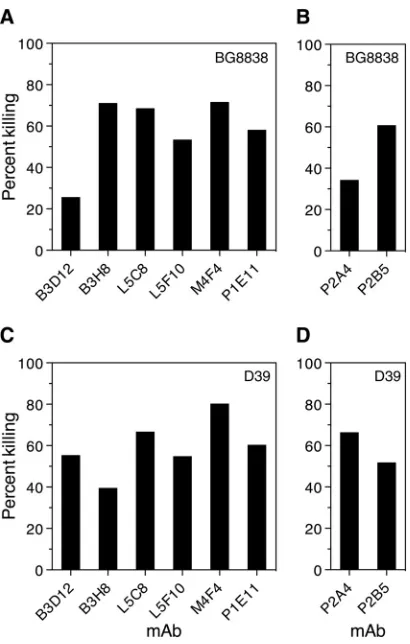
bacte-ricidal assay to evaluate the ability of the 8 anti-PspA
hkR36AMAbs
(i.e., B3D12, B3H8, L5C8, L5F10, M4F4, P1E11, P2A4, and P2B5)
to kill the PspA family 1-expressing strains BG8838 (clade 1) and
D39 (clade 2). Pneumococci were incubated with blood from
healthy donors in the presence of either purified anti-PspA
hkR36Aor a matched isotype control MAb, and the surviving
pneumo-cocci were enumerated by plating. The results suggest that all 8
anti-PspA
hkR36AMAbs exhibited significant bactericidal activity
(ranging from 25.7 to 80.3% compared with that of the matched
isotype control) against BG8838 and D39, although the extent of
bactericidal activity varied from one MAb to the other (
Fig. 4
).
We next tested whether these MAbs can enhance C3 deposition
on the surface of clade 1 (BG8838)-expressing and clade 2 (WU2
and D39)-expressing strains (
Table 3
). Monoclonal antibodies
A B
C D
E F
** ** *
*** ** **
*** **
** ns ns
*** ** *** ***
ns
FIG 2Relative efficacy of anti-PspAhkR36AMAbs to protect mice against in-travenous challenge. CBA/N mice were injected with purified anti-PspAhkR36A MAb M4F4, P1E11, or L5C8 (A and B), B3D12, B3H8, or L5F10 (C and D) or P2A4 or P2B5 (E and F) intraperitoneally at 5 mg/kg body weight (high dose). The corresponding isotype control MAb (IgG1 IC or IgG2a IC) was included in each set as the negative control. One hour later, mice were challenged with 107CFU of BG8838 (A, C, and E) or 103CFU of WU2 (B, D, and F), and mouse survival was recorded. The data for the group given anti-PspAhkR36AMAb were compared with those for the respective isotype control MAb using the log rank test. *,P⬍0.05; **,P⬍0.01; ***,P⬍0.001; ns, statistically not significant.
A B
C D
** ** ns
*** *** **
* *
*** **
FIG 3Anti-PspAhkR36AMAbs protect mice against pneumococcal infection even when given at a lower dose. CBA/N mice were injected intraperitoneally with 1.25 mg/kg body weight (low dose) of either anti-PspAhkR36AMAb M4F4, P1E11, or L5F10 (A and B) or P2A4 or P2B5 (C and D). The control group was given the respective isotype control MAb (IgG1 IC or IgG2a IC). Mice were challenged with BG8838 (A and C) or WU2 (B and D) 1 h later, and mouse survival was recorded. For other details, refer to the legend toFig. 2.
on August 17, 2020 by guest
http://cvi.asm.org/
M4F4, P1E11, P2A4, and P2B5 showed significant enhancement
in complement deposition on pneumococci compared to that of
the corresponding isotype control for the 3 strains analyzed. Seven
of the 8 MAbs augmented complement deposition on D39 with
B3H8 being the exception. B3H8, however, enhanced deposition
of complement on WU2. It was observed that the extent of the
enhancement in C3 deposition depended on the anti-PspA
hkR36AMAb and the target strain.
We wanted to find out which aspect or feature of the antibody
response correlated with
in vivo
protection. The surface staining,
complement deposition, bactericidal activity, and mouse
protec-tion data for MAbs B3D12, B3H8, L5C8, L5F10, M4F4, P1E11,
P2A4, and P2B5 are summarized in
Table 4
. The trend from the
data appears to be that the higher the extent of complement
de-position, the higher the bactericidal activity and
in vivo
protection.
This is well illustrated by MAbs M4F4, P1E11, P2A4, and P2B5.
While all the protective MAbs showed bactericidal activity, not all
MAbs that exhibited bactericidal activity showed passive
protec-tion. For example, MAbs B3D12, B3H8, and L5C8 exhibited
sig-nificant bactericidal activity but showed no to poor ability to
con-fer protection when given passively to mice.
To determine whether
in vitro
MAb-dependent deposition of
complement correlated with passive mouse protection, a
para-metric Pearson’s linear regression correlation analysis was
per-formed. The analysis indicated that there was a positive
correla-tion between
in vitro
complement deposition and the passive
protection for strains WU2 (
Fig. 5
) and BG8838 (data not shown).
The correlation was highly statistically significant for strain WU2
(
r
⫽
0.8783,
P
⫽
0.0041). Our data suggest that the
antibody-dependent deposition of complement on the pneumococcal
sur-face can be a potential
in vitro
correlate of protection for strains
like WU2.
DISCUSSION
PspA is a likely candidate for a protein-based vaccine against
pneumococcal infections; however, its serological variability
might restrict the coverage of a PspA-based vaccine. For this
rea-son, gaining insight into the nature of the variability of PspAs has
been the subject of several studies that are directed at development
of a protein-based pneumococcal vaccine. Studies aimed to
inves-tigate the level of cross-reactivity among PspAs in mice indicate
that antibodies generated against PspA show higher
cross-reactiv-ity with the strains expressing PspA of the same family than the
FIG 4Anti-PspAhkR36AMAbs promote pneumococcal killing by humanbloodin vitro.S. pneumoniaestrains BG8838 (A and B) and D39 (C and D) were incubated with blood from healthy donors and purified anti-PspAhkR36A or a matched isotype control MAb. Matched IgG1 and IgG2a isotype control MAbs were used as the comparator in panels A and C and B and D, respectively (not shown). The contents were rotated for (3 h for BG8838 and 2 h for D39) at 37°C, and the surviving pneumococci were enumerated by plating. The identity of the anti-PspAhkR36AMAb and the percent killing are plotted on the
xandyaxes, respectively. The percent bacterial killing was calculated as de-scribed in Materials and Methods. The assay was performed at least four times, and data from a single representative experiment are shown.
TABLE 3Flow cytometry-based analysis of complement C3 deposition on the surface of pneumococci in the presence of anti-PspAhkR36AMAbsa
Strains (clade)
GMFI (fold change) for:
Negative controlb
Positive controlc
IgG1 isotype
control B3D12d B3H8d L5C8d L5F10d M4F4d P1E11d
IgG2a isotype
control P2A4e P2B5e
BG8838 (1) 4.5 16.6 15.3 29.6 30.3 24.0 14.4 71.7f 233.0f 12.3 39.4f 106.0f
WU2 (2) 3.0 3.4 3.2 3.8 20.5f 3.4 5.2 482.0f 442.0f 3.6 320.0f 391.0f
D39 (2) 5.5 7.1 7.0 418.0f 12.9 516.0f 511.0f 568.0f 523.0f 6.1 429.0f 384.0f
a
The PspA family 1-expressing strains BG8838 (clade 1) and WU2 and D39 (clade 2) were incubated with either anti-PspAhkR36A
or a matched isotype control MAb. The bound complement C3 was detected by flow cytometry using anti-human C3 antibody followed by the appropriate FITC-conjugated secondary antibody.
b
Pneumococci were incubated with anti-human complement C3 antibody followed by the FITC-conjugated secondary antibody.
cPneumococci were incubated with 10% pooled normal human serum in Hanks’ balanced salt solution followed by anti-human C3 antibody and the FITC-conjugated secondary
antibody.
dThese anti-PspAhkR36AMAbs are of the IgG1 isotype.
e
These anti-PspAhkR36A
MAbs are of the IgG2a isotype.
fGMFI values greater than or equal to twice the value obtained with the corresponding isotype control were considered significant.
on August 17, 2020 by guest
http://cvi.asm.org/
strains that bear PspA of a different PspA family. We screened our
panel to identify the anti-PspA
hkR36AMAbs that exhibited the
maximum reactivity across PspA clades. Our ELISA and flow
cy-tometry-based surface staining data revealed that all of the 18
anti-PspA MAbs (raised against the clade 2-expressing strain
R36A) recognized family 1 PspA and not PspAs representing
fam-ilies 2 and 3. This is consistent with the observation that mice
immunized with DNA vaccine expressing the extracellular
do-main of PspA were protected from strains bearing PspA of the
same clade when tested in an intraperitoneal challenge mouse
model. Briles and coworkers, however, reported that human
anti-PspA antibodies, when administered to mice, conferred protection
against strains expressing PspA belonging to families 1 and 2 (
29
).
Antibody-dependent complement-mediated phagocytosis is a
well-established mechanism of pneumococcal clearance (
22
).
An-tibodies directed at pneumococci help in clearance by augmenting
opsonization. Antibodies to PspA enhance complement
deposi-tion on the pneumococcal surface, thereby contributing to their
protective effect (
24
). A previous report suggested that polyclonal
sera against PspA from families 1 and 2 help in enhancing
com-plement deposition (
34
). Our anti-PspA
hkR36AMAbs M4F4,
P1E11, P2A4, and P2B5 augmented complement deposition on
the 3 PspA family 1-expressing strains analyzed (
Table 3
). The
observed variation in the degree to which various anti-PspA
hkR36AMAbs augmented complement deposition across strains may have
to do with the chemical nature of the capsule on the target strain,
the thickness of the capsule, and the relative accessibility of the
pneumococcal surface.
There is evidence to suggest that not all antibodies to PspA
are protective indirectly, implying that not all PspA epitopes
elicit protective antibody responses. In our previous work, we
had observed that P1E11 and P2A4 compete for binding with a
PspA
R36Asubfragment corresponding to a 193- to
286-amino-acid stretch (PspA
R36A 193–286), indicating that these two MAbs
recognize the same or an overlapping epitope (
30
). Thus, M4F4,
P1E11, P2A4, and P2B5 recognize at least 3 topologically distinct
epitopes. The epitopes recognized by P1E11, P2A4, and P2B5 were
localized to the PspA
R36A 193–286subfragment. The epitope
recog-nized by M4F4 was mapped to the subfragment PspA
R36A 98 –192(
30
). Our data are consistent with those reported by Roche et al.,
who localized the cross protection-eliciting region of PspA to the
N-terminal 115 amino acid residues and
⬃
104 C-terminal
resi-dues of the extracellular domain from strain EF3296 (
12
).
Knowl-edge of the epitopes recognized by anti-PspA
hkR36AMAbs that do
TABLE 4In vivoprotective efficacies of anti-PspAhkR36AMAbs correlate with the extent of complement depositionaExperiment Strain
Result for anti-PspAhkR36AMAb:
B3D12 B3H8 L5C8 L5F10 M4F4b P1E11b P2A4b P2B5b
Surface staining (x)c BG8838 2.8 3.1 3.9 4.0 12.5 17.2 4.9 3.7
WU2 1.4 1.5 1.0 1.9 3.2 3.8 3.9 5.7
D39 17.7 2.4 37.3 18.1 38.2 39.8 12.8 7.3
Complement deposition (x)c BG8838 1.9 2.0 1.6 0.9 4.7 15.2 3.2 8.6
WU2 1.2 6.4 1.1 1.6 150.6 138.1 88.9 108.6
D39 59.7 1.8 73.7 73.0 81.1 74.7 70.3 62.9
Bactericidal activity (%)d BG8838 25.7 71.2 68.6 53.5 71.7 58.2 34.3 60.8
D39 55.4 39.6 66.8 54.8 80.3 60.4 66.5 51.9
Mouse passive protection assay (%)e
5 mg/kg BG8838 37.5 62.5 50.0 75.0 87.5 75.0 100.0 62.5
WU2 12.5 22.5 0.0 62.5 100.0 100.0 100.0 87.5
1.25 mg/kg BG8838 NDf ND ND 37.5 62.5 75.0 62.5 50.0
WU2 ND ND ND 37.5 75.0 87.5 100.0 75.0
a
BG8838 expresses clade 1 PspA, while WU2 and D39 express clade 2 PspA.
bIn these columns, the 4 anti-PspAhkR36AMAbs were the most protective.
c
Surface staining and complement deposition are expressed as fold change (x) in the GMFI value relative to the corresponding isotype control.
dBactericidal activity of the anti-PspAhkR36AMAb is represented as percent killing. A higher value indicates higher bactericidal activity. The assay was not performed with strain
WU2.
eMouse protection is expressed as percent survival. A higher value indicates higher protective efficacy. This experiment was not done with strain D39. f
ND, not determined.
FIG 5Analysis of 8 anti-PspAR36AMAbs to assess a possible correlation be-tween MAb-dependent complement deposition and their ability to passively protect mice from an otherwise lethal challenge with strain WU2. Each dot represents an anti-PspAR36AMAb. The correlation betweenin vitro comple-ment deposition and passive protection was highly significant by a parametric Pearson’s linear regression correlation analysis (r⫽0.8783,P⫽0.0041).
on August 17, 2020 by guest
http://cvi.asm.org/
and do not elicit protective responses might be put to use to
engi-neer a PspA vaccine that maximizes the proportion of protective
antibodies in the antibodies generated. Fine mapping of the
conserved B cell epitopes recognized by the protective
anti-PspA
hkR36AMAbs M4F4, P1E11, P2A4, and P2B5 can help in
de-velopment of a superior PspA-based vaccine.
Our data indicate that complement deposition on
pneumo-cocci can be a used as a surrogate for the
in vivo
protection for
strains like WU2 (
Fig. 5
). Roche and coworkers had demonstrated
that the antibody titer and surface staining do not correlate with
in
vivo
protection and thus are not useful as a surrogate for
protec-tion (
12
). Cohen et al. reported that a whole-cell ELISA is an
inadequate predictor of
in vivo
protection (
11
). While the
modi-fied surface killing assay for PspA developed by Genschmer et al. is
likely to be significant (
9
), complement deposition as an
in vitro
correlate of protection is easier to perform and amenable to
auto-mation. The complement deposition assay might be potentially
useful in quantitating the relative protective efficacy of antibodies
against novel protein vaccine antigens (or their subfragments) to
confer protection
in vivo
.
ACKNOWLEDGMENTS
This work was supported in part by the intramural research program of the National Institute of Immunology and by grant BT/PR5037/MED/15/ 77/2012 from the Department of Biotechnology (DBT), India.
Naeem Khan was the recipient of a Senior Research Fellowship from DBT.
REFERENCES
1.Lynch JP, III, Zhanel GG.2010.Streptococcus pneumoniae: epidemiology and risk factors, evolution of antimicrobial resistance, and impact of vac-cines. Curr Opin Pulm Med16:217–225.http://dx.doi.org/10.1097/MCP .0b013e3283385653.
2.World Health Organization.2007. Pneumococcal conjugate vaccine for childhood immunization—WHO position paper. Wkly Epidemiol Rec 82:93–104.
3.O’Brien KL, Wolfson LJ, Watt JP, Henkle E, Deloria-Knoll M, McCall N, Lee E, Mulholland K, Levine OS, Cherian T, Hib and Pneumococcal Global Burden of Disease Study Team.2009. Burden of disease caused byStreptococcus pneumoniaein children younger than 5 years: global estimates. Lancet 374:893–902. http://dx.doi.org/10.1016 /S0140-6736(09)61204-6.
4.Vila-Corcoles A, Ochoa-Gondar O, Guzman JA, Rodriguez-Blanco T, Salsench E, Fuentes CM.2010. Effectiveness of the 23-valent polysaccha-ride pneumococcal vaccine against invasive pneumococcal disease in peo-ple 60 years or older. BMC Infect Dis10:73.http://dx.doi.org/10.1186 /1471-2334-10-73.
5. Miyaji EN, Oliveira ML, Carvalho E, Ho PL. 2013. Serotype-independent pneumococcal vaccines. Cell Mol Life Sci70:3303–3326. http://dx.doi.org/10.1007/s00018-012-1234-8.
6.Moffitt KL, Malley R.2011. Next generation pneumococcal vaccines. Curr Opin Immunol23:407–413.http://dx.doi.org/10.1016/j.coi.2011.04.002.
7.Feldman C, Anderson R. 2014. Review: current and new generation pneumococcal vaccines. J Infect69:309 –325.http://dx.doi.org/10.1016/j .jinf.2014.06.006.
8.Mitchell AM, Mitchell TJ. 2010.Streptococcus pneumoniae: virulence factors and variation. Clin Microbiol Infect16:411– 418.http://dx.doi.org /10.1111/j.1469-0691.2010.03183.x.
9.Genschmer KR, Accavitti-Loper MA, Briles DE.2013. A modified sur-face killing assay (MSKA) as a functionalin vitroassay for identifying protective antibodies against pneumococcal surface protein A (PspA). Vaccine32:39 – 47.http://dx.doi.org/10.1016/j.vaccine.2013.10.080. 10. Daniels CC, Kim KH, Burton RL, Mirza S, Walker M, King J, Hale Y,
Coan P, Rhee DK, Nahm MH, Briles DE.2013. Modified opsonization, phagocytosis, and killing assays to measure potentially protective antibod-ies against pneumococcal surface protein A. Clin Vaccine Immunol20: 1549 –1558.http://dx.doi.org/10.1128/CVI.00371-13.
11. Cohen JM, Wilson R, Shah P, Baxendale HE, Brown JS.2013. Lack of cross-protection against invasive pneumonia caused by heterologous strains following murineStreptococcus pneumoniaenasopharyngeal colo-nisation despite whole-cell ELISAs showing significant cross-reactive IgG. Vaccine31:2328 –2332.http://dx.doi.org/10.1016/j.vaccine.2013.03.013. 12. Roche H, Hakansson A, Hollingshead SK, Briles DE.2003. Regions of
PspA/EF3296 best able to elicit protection againstStreptococcus
pneu-moniaein a murine infection model. Infect Immun71:1033–1041.http:
//dx.doi.org/10.1128/IAI.71.3.1033-1041.2003.
13. Goulart C, Darrieux M, Rodriguez D, Pimenta FC, Brandileone MC, de Andrade AL, Leite LC.2011. Selection of family 1 PspA molecules capable of inducing broad-ranging cross-reactivity by complement deposition and opsonophagocytosis by murine peritoneal cells. Vaccine29:1634 – 1642.http://dx.doi.org/10.1016/j.vaccine.2010.12.074.
14. Ochs MM, Bartlett W, Briles DE, Hicks B, Jurkuvenas A, Lau P, Ren B, Millar A.2008. Vaccine-induced human antibodies to PspA augment complement C3 deposition onStreptococcus pneumoniae. Microb Pathog 44:204 –214.http://dx.doi.org/10.1016/j.micpath.2007.09.007.
15. Crain MJ, Waltman WD, II, Turner JS, Yother J, Talkington DF, McDaniel LS, Gray BM, Briles DE.1990. Pneumococcal surface protein A (PspA) is serologically highly variable and is expressed by all clinically important capsular serotypes ofStreptococcus pneumoniae. Infect Immun 58:3293–3299.
16. Yother J, Briles DE.1992. Structural properties and evolutionary rela-tionships of PspA, a surface protein ofStreptococcus pneumoniae, as re-vealed by sequence analysis. J Bacteriol174:601– 609.
17. Jedrzejas MJ, Lamani E, Becker RS.2001. Characterization of selected strains of pneumococcal surface protein A. J Biol Chem276:33121–33128. http://dx.doi.org/10.1074/jbc.M103304200.
18. Nabors GS, Braun PA, Herrmann DJ, Heise ML, Pyle DJ, Gravenstein S, Schilling M, Ferguson LM, Hollingshead SK, Briles DE, Becker RS. 2000. Immunization of healthy adults with a single recombinant pneumo-coccal surface protein A (PspA) variant stimulates broadly cross-reactive antibodies to heterologous PspA molecules. Vaccine18:1743–1754.http: //dx.doi.org/10.1016/S0264-410X(99)00530-7.
19. Hollingshead SK, Becker R, Briles DE.2000. Diversity of PspA: mosaic genes and evidence for past recombination inStreptococcus pneumoniae. Infect Immun 68:5889 –5900. http://dx.doi.org/10.1128/IAI.68.10.5889 -5900.2000.
20. Hotomi M, Togawa A, Kono M, Ikeda Y, Takei S, Hollingshead SK, Briles DE, Suzuki K, Yamanaka N. 2013. PspA family distribution, antimicrobial resistance and serotype ofStreptococcus pneumoniaeisolated from upper respiratory tract infections in Japan. PLoS One8:e58124.http: //dx.doi.org/10.1371/journal.pone.0058124.
21. Hollingshead SK, Baril L, Ferro S, King J, Coan P, Briles DE.2006. Pneumococcal surface protein A (PspA) family distribution among clinical isolates from adults over 50 years of age collected in seven countries. J Med Microbiol 55:215–221. http://dx.doi.org/10.1099 /jmm.0.46268-0.
22. Brown EJ, Hosea SW, Frank MM.1983. The role of antibody and com-plement in the reticuloendothelial clearance of pneumococci from the bloodstream. Rev Infect Dis5(Suppl 4):S797–S805.http://dx.doi.org/10 .1093/clinids/5.Supplement_4.S797.
23. Ren B, Li J, Genschmer K, Hollingshead SK, Briles DE. 2012. The absence of PspA or presence of antibody to PspA facilitates the comple-ment-dependent phagocytosis of pneumococciin vitro. Clin Vaccine Im-munol19:1574 –1582.http://dx.doi.org/10.1128/CVI.00393-12. 24. Ren B, Szalai AJ, Hollingshead SK, Briles DE.2004. Effects of PspA and
antibodies to PspA on activation and deposition of complement on the pneumococcal surface. Infect Immun72:114 –122.http://dx.doi.org/10 .1128/IAI.72.1.114-122.2004.
25. Wu HY, Nahm MH, Guo Y, Russell MW, Briles DE.1997. Intranasal immu-nization of mice with PspA (pneumococcal surface protein A) can prevent intra-nasal carriage, pulmonary infection, and sepsis withStreptococcus pneumoniae. J Infect Dis175:839 – 846.http://dx.doi.org/10.1086/513980.
26. Ferreira DM, Miyaji EN, Oliveira ML, Darrieux M, Areas AP, Ho PL, Leite LC.2006. DNA vaccines expressing pneumococcal surface protein A (PspA) elicit protection levels comparable to recombinant protein. J Med Microbiol55:375–378.http://dx.doi.org/10.1099/jmm.0.46217-0. 27. McDaniel LS, Ralph BA, McDaniel DO, Briles DE.1994. Localization of
protection-eliciting epitopes on PspA ofStreptococcus pneumoniae be-tween amino acid residues 192 and 260. Microb Pathog17:323–337.http: //dx.doi.org/10.1006/mpat.1994.1078.
on August 17, 2020 by guest
http://cvi.asm.org/
28. Daniels CC, Coan P, King J, Hale J, Benton KA, Briles DE, Hollings-head SK.2010. The proline-rich region of pneumococcal surface proteins A and C contains surface-accessible epitopes common to all pneumococci and elicits antibody-mediated protection against sepsis. Infect Immun 78:2163–2172.http://dx.doi.org/10.1128/IAI.01199-09.
29. Briles DE, Hollingshead SK, King J, Swift A, Braun PA, Park MK, Ferguson LM, Nahm MH, Nabors GS.2000. Immunization of humans with recombinant pneumococcal surface protein A (rPspA) elicits anti-bodies that passively protect mice from fatal infection withStreptococcus
pneumoniaebearing heterologous PspA. J Infect Dis182:1694 –1701.http:
//dx.doi.org/10.1086/317602.
30. Rohatgi S, Dutta D, Tahir S, Sehgal D.2009. Molecular dissection of antibody responses against pneumococcal surface protein A: evidence for diverse DH-less heavy chain gene usage and avidity maturation. J Immu-nol182:5570 –5585.http://dx.doi.org/10.4049/jimmunol.0803254. 31. Daniels CC, Briles TC, Mirza S, Hakansson AP, Briles DE. 2006.
Capsule does not block antibody binding to PspA, a surface virulence protein ofStreptococcus pneumoniae. Microb Pathog40:228 –233.http: //dx.doi.org/10.1016/j.micpath.2006.01.007.
32. Manivel V, Sahoo NC, Salunke DM, Rao KV.2000. Maturation of an antibody response is governed by modulations in flexibility of the antigen-combining site. Immunity13:611– 620.http://dx.doi.org/10.1016/S1074 -7613(00)00061-3.
33. Briles DE, Forman C, Horowitz JC, Volanakis JE, Benjamin WH, Jr, McDaniel LS, Eldridge J, Brooks J.1989. Antipneumococcal effects of C-reactive protein and monoclonal antibodies to pneumococcal cell wall and capsular antigens. Infect Immun57:1457–1464.
34. Ren B, Szalai AJ, Thomas O, Hollingshead SK, Briles DE.2003. Both family 1 and family 2 PspA proteins can inhibit complement deposition and confer virulence to a capsular serotype 3 strain ofStreptococcus
pneu-moniae. Infect Immun71:75– 85.http://dx.doi.org/10.1128/IAI.71.1.75
-85.2003.