An augmented delivery of the anticancer agent, curcumin, to the colon
Reactive and Functional Polymers: DOI information: 10.1016/j.reactfunctpolym.2017.12.012 Rayan Sabra a, Nashiru Billa a*, Clive J. Roberts b
a The School of Pharmacy, University of Nottingham, Malaysia Campus, Semenyih, Selangor, Malaysia
b The School of Pharmacy, University of Nottingham, Park Campus, Nottingham, United Kingdom
*Corresponding author: [email protected]; Tel +60389248211 Abstract
This work describes the formulation aspects of an orally viable curcumin-containing mucoadhesive nanoparticulate system for management of colon cancer. Curcumin is
documented to possess anticancer properties whilst modified citrus pectin yields a galactose functionality capable of inhibiting the growth and proliferation of colon cancer cells due to antagonism to galectin-3 (Gal-3). A successfully formulated curcumin containing chitosan-modified citrus pectinate nanoparticles (MCPCNPs) registered a z-average of 178 nm (± 0.896) and a positive surface charge of + 35.7 mV (± 1.41). The MCPCNPs presented high mucoadhesion propensity in the colonic region/ media and minimal at pH 1.2 (stomach). There was approximately 18 % curcumin release at pH 1.2 over 2 h and up to 68% release in the 33% (w/v) caecal medium over 24 h. The data obtained strongly suggests that the
formulated MCPCNPs have the potential to be applied as an orally deliverable colon cancer formulation alternative in the treatment of colon cancer.
Keywords: modified citrus pectin, curcumin, nano-particulate system, colon cancer.
1. Introduction
Colon cancer is the fourth leading cause of cancer deaths worldwide, accounting for 8 % of documented cancer deaths [1]. Due to the annual rise in the number of deaths resulting from colon cancer [2], several therapeutic procedures are in practice that addresses this trend, including surgical resection of afflicted tissue, radiotherapy or chemotherapy. Chemotherapy is the least invasive of the therapeutic options currently used to manage colon cancer.
However, due to the side effects that manifests following chemotherapy, anticancer agents of 1
2
3
4
5
6
7
8
9
10
11
12
13
14
15
16
17
18
19
20
21
22
23
24
25
26
27
28
29
30
plant origin, such as curcumin, are becoming serious contenders as chemotherapeutic alternatives. Curcumin is the major chemical constituent of turmeric and has received much attention in the past decade because of its ‘acceptability’ as it is derived from natural sources and perceived to manifest relatively fewer side effects [3]. Extensive research has elaborated the therapeutic potential of curcumin against a range of cancers and distinctly against colon cancer [4]. However, preclinical and clinical data from oral administration of curcumin have revealed its poor systemic bioavailability and high susceptibility to metabolic activity, where only 2.30 ± 0.26 μg.ml−1 of curcumin is registered in serum after oral administration of 10 g curcumin [5, 6]. This shows that curcumin undergoes extensive metabolic changes in the intestine and liver, which hinder the systemic usefulness of curcumin in the treatment of cancer. To overcome this constraint, a variety of materials such as natural or synthetic polymers and lipids have been used to formulate delivery systems that traverse
gastrointestinal epithelia effectively and therefore circumvent the metabolic constraints within the gastrointestinal tract.
Chitosan is a well-studied natural polymer that is biodegradable and possesses mucoadhesive properties [7]. It is derived from chitin and comprises of β- [1–4] -linked D glucosamine and N-acetylated units [8]. It is soluble in acidic media due to the formation of soluble complexes with the charged amine groups (NH3+). The amine moiety also promotes binding to
negatively charged species such as mucin within the gastrointestinal tract [8]. Citrus pectin is also a widely studied natural polymer extracted from the cell wall of plants. It possesses a poly α- [1–4] - linked D-galacturonic acid units with varying degrees of methylation of the carboxylic acid moiety [9], which confers a negative charge to the molecule. Additionally, pectin shares very similar properties with chitosan and is resistant to degradation by the digestive enzymes in stomach [9]. Crucially, pH-thermal treatment of citrus pectin yields moieties that demonstrate chemo-preventive activities against cancers [10]. Modified citrus pectin (MCP) comprise of neutral sugar sequences with a low degree of branching and is rich in galactose, a constituent that is reported to inhibit the growth and migration of colon cancer cells due to its remarkable antagonism to galectin-3 (Gal-3) [11,12]. Gal-3 is a protein of the galectin super-family with a carbohydrate-binding domain that exhibits high affinity to β-galactosides [13] and is vastly expressed on tumour cell surfaces. There is evidence that Gal-3 binds to the colonic mucin with an altered carbohydrate structure causing tumour growth, cell-to-cell adhesion, and metastasis [14]. Therefore, the galactose-rich MCP is capable of binding to the β- galactoside protein of the Gal-3 and hence blocking the Gal-3 interactions with other proteins and peptides [12].
32
33
34
35
36
37
38
39
40
41
42
43
44
45
46
47
48
49
50
51
52
53
54
55
56
57
58
59
60
61
62
63
64
Earlier reports [15, 16] have elaborated the potential of blending chitosan and pectin for colon-specific drug delivery. More recently, Andriani et al. [17] reported glutaraldehyde-cross linked chitosan-pectin nanoparticles as a promising carrier for effective delivery of curcumin to the small intestine following oral administration. In the present pursuit however, we aim to formulate mucoadhesive curcumin-loaded MCP-chitosan nanoparticles
(MCPCNPs) for possible delivery to the colon following oral administration. In this regard, the MCP would serve the dual function of retaining the integrity of the nanoparticles within the acidic pH of the upper gut and also yielding the galactose moiety, which is known to manifest anticancer properties against galectin 3 (Gal-3) in the colon. Chitosan is destined to provide the required mucoadhesion within the colon mucosa in order to ensure longer residence time within the colon epithelia. It is envisaged that longer residence time in the colon coupled with augmented anticancer activity due to encapsulated curcumin and galactose moiety from MCP would be therapeutically cost-effective and attractive to
stakeholders in the management of colon cancer. This manuscript describes in vitro work that supports the above hypothesis in which the proposed formulation has the potential to be delivered orally to the colon
.
2. Materials and methods 2.1. Materials
Chitosan (low molecular weight), de-acetylated chitin, Poly (D- glucosamine) from Sigma Aldrich, Iceland and low methoxyl citrus pectin (DE lower than 50) from Genu, CPKelco® (Limeira- SP, Brazil) were utilized as coating polymers. Curcumin was bought from Fluka, U.S.A. Sodium tripolyphosphate (STPP) (Sigma Aldrich, USA) was used as the cross-linker with chitosan. Mucin type III from porcine stomach (Sigma Aldrich, USA) was utilised in mucoadhesive studies. Potassium dihydrogen phosphate (Sigma Aldrich, USA), acetic acid (R&M chemicals, UK), hydrochloric acid (R&M chemicals, UK), acetic acid (R&M
chemicals, UK), ethanol (R&M chemicals, UK), acetone (R&M chemicals, UK), acetonitrile (RCI Labscan, Thailand), methanol (R&M chemicals, UK), sodium hydroxide and sodium acetate (Merck, Germany), were expended as solvents or pH modifiers.
66
67
68
69
70
71
72
73
74
75
76
77
78
79
80
81
82
83
84
85
86
87
88
89
90
91
92
93
94
95
2.2. Modification of citrus pectin
The low-methoxyl citrus pectin (LMP) was chemically modified as ascribed by Venzon et al. [12] with some modification. 1.5 g of the powdered pectin was dissolved in 100 ml ultra-pure water and the pH adjusted to 10.0 using 3 M NaOH followed by gentle stirring at 60 0C for an hour. The mixture was allowed to cool at room temperature; its pH was adjusted to 3.0 with 3 M HCl and then stored at 4 0C overnight. Subsequently, the modified citrus pectin (MCP) was precipitated in the sample with 95 % ethanol and then washed with 100 % acetone, followed by drying at 60 0C for an hour. Finally, the semidried MCP was frozen at -20 0C and then lyophilized.
2.3. Formulation of loaded curcumin- modified citrus pectin -chitosan nanoparticles (MCPCNPs)
The MCPCNPs were prepared by ionic gelation in a one-step process. The stock solutions were made by dissolving: chitosan (2.5 mg/ml) in 2 % (v/v) acetic acid and adjusting the pH to 5.0 with 2 M NaOH; sodium tripolyphosphate (STPP) was dissolved in ultra-pure water at 0.5 mg/ml; MCP was stirred in ultra-pure water at 0.5 mg/ml for 3 h at 60 0C and curcumin was dissolved in ethanol at 1 mg/ml. A 25 ml aliquot of the MCP solution was added to an amber-coloured beaker containing 300 µl of the curcumin solution with vigorous stirring, followed by drop-wise addition of 25 ml of chitosan solution and then drop-wise addition of 25 ml of STPP. The mixture was stirred for a further 20 minutes at 500 rpm at room
temperature and then stored at 4 0C till further analyses.
2.4. Physical properties of the MCPCNPs
The hydrodynamic size distribution (expressed as average diameter) and surface charge (expressed as zeta- potential) of the MCPCNPs were determined after dilution using a Zeta Sizer Nano Series® (Malvern Instruments Ltd., UK) equipped with a 4 mW He-Ne laser at a wavelength of 633 nm. The average diameter and zeta-potential were measured by means of Dynamic Laser Scattering and Laser Doppler Anemometry (LDA), respectively. Samples were run in triplicates and presented as mean (±SD).
The morphology of the MCPCNPs was observed using a Field Emission Scanning Electron Microscope (FESEM), (Model Quanta 400F, FEI Company USA) equipped with a
backscattered electron detector at 3 kV. 100 µL of the sample was placed on the SEM 97
98
99
100
101
102
103
104
105
106
107
108
109
110
111
112
113
114
115
116
117
118
119
120
121
122
123
124
125
126
127
imaging stub with carbon layer and left under a drying chamber for 24 h at room temperature before viewing.
The Fourier transform infrared (FTIR) spectra of compacts from the MCPCNPs and excipients were reviewed after compressing them into KBr incorporated compact discs at a pressure of 5 tonnes for 5 min. Spectra were obtained for chitosan, MCP, STPP, curcumin, MCP-chitosan nanoparticles and loaded- curcumin nanoparticles using a Perkin-Elmer FTIR spectrometer (Spectrum RX I) with scans run between 400 to 4000 cm−1 at a resolution of 4 cm-1 and interval 1 cm-1.
The thermal responses of the MCPCNPs and components were assessed on a differential scanning calorimeter DSC-Q2000 (TA Instruments, USA). Accurately weighed samples were crimped into aluminium plates and the thermogram was conducted over a range of 0 to 300 0C, at a scanning rate of 10 0C/min and flushed with nitrogen gas at a rate of 20 ml/min.
2.5. Encapsulation efficiency and drug loading within MCPCNPs
The formed MCPCNPs were centrifuged at 8000 rpm for 10 minutes and the centrifuged pellet was rinsed twice with methanol. The curcumin content in the supernatant and the methanol rinse was analysed by injecting 10 l onto an HPLC system (Series 200 pump, Agilent, USA) equipped with an Eclipse plus C18 (Agilent, USA) column (250 × 4.6 mm; 5 µm) and a UV detector (L-2485, Agilent, USA) set at 425 nm. The mobile phase (methanol: 0.01 % acetic acid: acetonitrile (5: 43: 52, %)) was run isocratically at 1.5 ml/min. Responses obtained were compared to those from a standard curve of curcumin in methanol. The
percentage encapsulation of curcumin and loading capacity of the MCPCNPs with respect to curcumin was calculated as follows:
Encapsulation Efficiency (%)= Amount of curcumin added-unbound curcumin
Amount of curcumin added ×100
Loading Capacity (%)= Entrapped curcumin
Curcumin loaded NP weight ×100 129
130
131
132
133
134
135
136
137
138
139
140
141
142
143
144
145
146
147
148
149
150
151
152
153
154
155
156
157
2.6. Mucoadhesion propensity of the MCPCNPs 2.6.1. Mucin adsorption studies
The propensity of the MCPCNPs to adhere to mucus was determined as the amount of mucin adsorbed by the MCPCNPs dispersed in standardised mucin solutions. The amount of mucin adsorbed was also studied as a function of pH representing relevant anatomical sections of the gastrointestinal tract: pH 1.2 (0.1 N HCl), pH 5.5 (0.1 M sodium acetate buffer), pH 6.25 and 7.0 (0.1 M phosphate buffer). Specifically, 2 ml of the MCPCNPs suspension was mixed in the mucin solutions, vortexed for few seconds and then rotated at 100 rpm maintained at 37 0C for 1 h. The suspension was centrifuged at 4000 rpm for 1 h to separate the free mucin followed by the addition of micro-BSA reagent and then incubation at 37 0C for 2 h and then finally measuring the absorbance at 562 nm. The quantity of mucin in the supernatant was determined from a standard calibration curve treated in a similar way and mucin adsorption (%) was calculated using the equation below:
Mucin adsorption(%)=Amount of mucin added-Amount in supernatant
Amount of mucin added ×100
2.6.2. Everted caecal sac experiment
A further mucoadhesion study was conducted on rat caecal tissue in order to validate the mucin adsorption studies described above. In this method, adapted from Chuah et al. [18], Sprague-Dawley male rats were sacrificed in carbon dioxide (CO2) chamber. The caecum was excised and flushed with ice cold phosphate buffer and everted using a glass rod and tweezers. One ligature was placed at the end of the segment and the sac was filled with 1 ml McCoy’s media through the open end and then tightly tied. The sac was then placed in a centrifuge tube containing 60 mg MCPCNPs dispersed in 5 ml McCoy’s media and then subjected to gentle shaking at 37 0C for 30 min. Finally, the sac was removed and the tube was centrifuged to retrieve the unattached MCPCNPs.
% attachment of MCPCNPs to caecum=Wt . of MCPCNPs added−Wt . of unattached MCPCNPs
Wt of MCPCNPs added ×100
2.7. MCPCNPs matrix swelling studies
The swelling propensity of the MCPCNPs matrix at physiologically relevant pH of the gastrointestinal tract was studied at pH 1.2, 5.5 and 7.0. Briefly, 20 ml of the MCPCNPs 159
160
161
162
163
164
165
166
167
168
169
170
171
172
173
174
175
176
177
178
179
180
181
182
183
184
185
186
187
suspension was added to respective pH solution and then mixed using a rotary shaker
(WiseCube®, Witeg Inc., Germany) at 180 rpm. At predetermined time intervals (0 h, 30 min, 1 h, 2 h, 4 h and 6 h) the z-average of the particles in the respective solutions were measured by means of DLS. The swelling ratio of the MCPCNPs was calculated as:
Swelling Capacity(%)=z ‐ average of MCPCNPs at time(t)-Initial z ‐average of MCPCNPs
Initial z ‐ average of MCPCNPs ×100
2.8. Preparation of 33 % w/v rat caecal content
The caecum of one of the Sprague–Dawley male rats used above was ligated, then carefully incised. The caecal contents were removed, weighed and suspended in cold (0-4 0C)
phosphate buffer (pH 7.0) to a concentration of 33 % (w/v). The suspension was centrifuged at 1200 rpm for 15 min at 4 0C and the supernatant obtained was further centrifuged at 9000 rpm for 30 min. This final supernatant was stored at 4 0C and was used as the colonic release media.
2.9. Curcumin release from the MCPCNPs
The freeze-dried MCPCNPs (1.3 mg) were suspended in 2 ml of hydrochloric acid (pH 1.2) or 33 % w/v caecal medium in a centrifuge tube. The tube was subjected to a rotary shaking at 120 rpm maintained at 37 0C for 2 h for the pH 1.2 medium and 24 h for the caecal content medium. At each time point (0 h, 1 h, 2 h, 3 h, 5 h, 7 h, 14 h and 24 h), the tube was
centrifuged at 8000 rpm for 10 min to pellet the MCPCNPs and the amount of curcumin released was determined in the supernatant using the HPLC method described above. The MCPCNPs pellet was re-suspended in 2 ml of fresh buffer media and the process repeated. The percentage of curcumin released from the MCPCNPs was calculated as follows:
Percentage released(%)=Amount of curcumin released
Amount of curcumin in the NPs ×100
3. RESULTS AND DISCUSSION 3.1. Modification of citrus pectin
Pectin polysaccharides consist of several side chain polymers including homogalacturonan (HG), rhamnogalacturonan-I (RG-I) and rhamnogalacturonan-II (RG-II). The backbone of the HG is covalently cross-linked to the rhamnogalacturans, which have branched
189
190
191
192
193
194
195
196
197
198
199
200
201
202
203
204
205
206
207
208
209
210
211
212
213
214
215
216
217
arabinogalactan side chains [11]. Alkaline treatment of pectin results in the disruption of the rhamnogalacturan side chains with a consequent depolymerisation of the backbone and de-esterification of the HG regions. Sustained exposure of the pectin in the alkaline pH also allows the cleavage of the rhamnogalacturans to arabino galactans and galactans, which are crucial in the anticancer trigger cascade. Figure 1 presents the FTIR spectra of the LMP, (a) in comparison to the MCP, (b).
Figure 1: FT-IR spectra of LMP and MCP
The wide peaks at approximately 3,440 cm−1 and 2,900 cm−1 in both spectra are indicative of secondary hydroxyl carboxylic groups, respectively. There are comparatively lower intensity at about 1700 cm-1 in LMP compared to identical peak in the MCP, which is indicative of a lower degree of methoxy substitution of pectin in LMP [19]. A distinctive absorption band is observed at 1015 cm−1 in the MCP spectra and not apparent in the LMP spectra, which is consistent with an increase in the D-galacturonic acid sugar units, therefore, we can conclude that the modification procedure yielded the desired galactan moiety.
3.2. Physical properties of the MCPCNPs
In the present study, the MCPCNPs were formed from coacervation between the negatively charged carboxylic groups of the MCP and the positively charged amino group of chitosan, followed by the crosslinking between the negative phosphate groups of STPP and the amino groups of chitosan, while encapsulating curcumin within the matrices formed in the process. 219
220
221
222
223
224
225
226
227
228
229
230
231
232
233
234
235
236
237
238
239
Such composite nanoformulation comprising of pectin and chitosan has been reported to increase the mechanical strength and hydrophobicity of the formulation, which favours sustained drug release [14]. In general, there was a slight increase in the size of the nanoparticles, from 174 nm (± 0.889) to 178 nm (± 0.896), with the incorporation of curcumin. This is attributable to the stearic effects of curcumin within the polymer chain domains on the coacervation between MCP and chitosan which impedes the process.
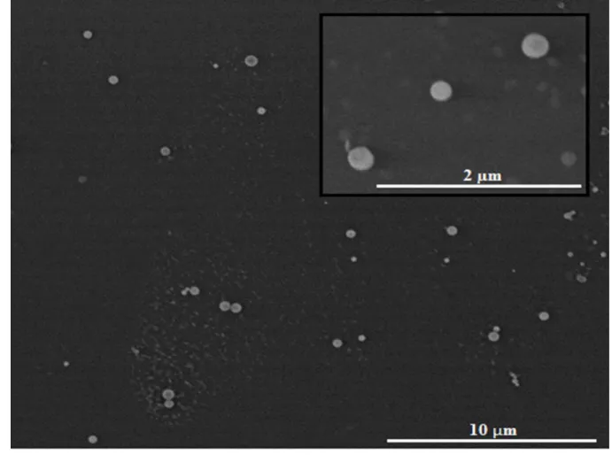
Moreover, the zeta potential values of the nanoparticles increased slightly from + 31.4 mV (± 0.793) to + 35.7 mV (± 1.418) after the addition of curcumin. This increase in the surface charge is directly related to the increase in size, whereby, bulkier particles hinder the knitting effects of STPP with the free –NH3+ groups of chitosan. This leaves relatively more of the free –NH3+ in the curcumin loaded nanoparticles with a consequent increase in zeta potential. The morphology (Figure 2) of the nanoparticles revealed that they are spherical and well-distributed with the size analysis consistent with the DLS studies.
Figure 2: FESEM image of MCPCNPs 241
242
243
244
245
246
247
248
249
250
251
252
253
254
255 256
The encapsulation efficiency of the MCPCNPs was 69.43 % (± 1.4), asserting that the nanoparticles retained considerable amount of curcumin despite a drug loading of 0.03 % (± 0.001).
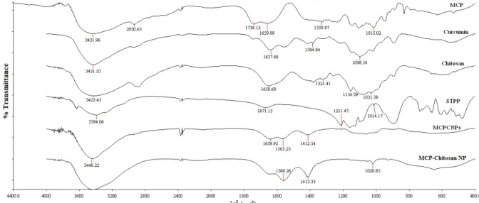
The FTIR spectra of the MCPCNPs and the raw materials are presented in Figure 3.
Figure 3: FT-IR spectra of chitosan , STPP , MCP, curcumin, MCP-chitosan nanoparticles and MCPCNPs
The disappearance of the amine group of chitosan at 1650.68 cm-1, the phosphate group of STTP at 1211.47 cm-1 and the D-galacturonic acid peak of MCP at 1015.02 cm-1 in the MCPCNPs spectrum can be attributed to the crosslinking of chitosan with MCP and STPP, respectively.The relatively sharper peaks at 3448.22 cm-1 in the spectra of the MCPCNPs and MCP-chitosan nanoparticles indicate a higher density of hydroxyl moieties. The intense peaks at 1560.31 cm-1 and 1412.57 cm-1 in both spectra presents shifted amine peaks of the raw materials from the 1600-1700 cm−1 region. This corresponds to the –NH deformation due to the interaction between the carboxylic groups of MCP and amino groups of chitosan. The only observed difference between the MCPCNPs spectrum and that of the MCP-chitosan nanoparticles is the presence of the peak at 1638.92 cm-1 in the former, which ascertains the incorporation of curcumin in the formulation.
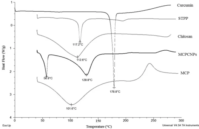
Figure 4 presents the thermal responses from the MCPCNPs and the raw materials. The melting points of STPP, chitosan and MCP were found to be at 117.2 0C, 112.6 0C and 101.6 0C, respectively. Curcumin exhibited a melting peak at 178.8 °C, however this peak was not 258
259
260
261
262
263
264
265 266 267 268 269
270
271
272
273
274
275
276
277
278
279
280
281
282
observed in the thermogram of the MCPCNPs. There were two melting episodes displayed by the MCPCNPs at 56.9 °C and 128.8 0C. The peak appearing at 128.8 0C is mainly attributed to chitosan.
Figure 4: DSC profiles of chitosan , STPP , MCP, curcumin and MCPCNPs
The shift in the melting point of chitosan is due to its interactions with STPP and MCP as part of the crosslinking, whilst the endothermic at 56.9 0C is manifestation of complexes within the MCPCNPs. These complexes are formed from possible weak van der Waal type interactions between the MCP and curcumin thereby destroying the specific structural arrangement of the particular functional head group regions [20]. Interestingly, the sharp curcumin peak at 178 0C is completely lost in the thermogram of the nanoparticles, affirming the presence of curcumin within the particles in amorphous conformation. The MCP peaks disappear in the MCPCNPs thermogram due to the disruption of their joint intermolecular hydrogen bonds by curcumin molecules.
3.3. Swelling of MCPCNPs matrices 284
285
286
287
288
289
290
291
292
293
294
295
296
297
298
299
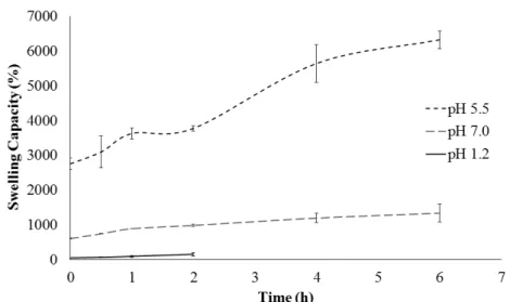
The crucial prerequisite of mucoadhesion between dosage form and epithelia is the ability of the former to swell effectively. Therefore, the swelling ratios of the MCPCNPs were assessed as a function of the three pH buffers (pH 1.2, 5.5 and 7.0) for 0 h, 30 minutes, 1 h, 2 h, 4 h and 6 h using the particle size analyser (Zeta Sizer Nano Series). In all three pH media, the MCPCNPs swelled rapidly (within 30 minutes), with the highest swelling ratio in pH 5.5 and lowest in pH 1.2 (Figure 5).
Figure 5: Swelling profile of MCPCNPs in pH 1.2, 5.5 and 7.0
Free chitosan swells in acidic media; however, the swelling of the MCPCNPs was restricted due to the formation of polyelectrolyte complex between the amino groups of chitosan and the carboxylate group of MCP. At pH 5.5, the swelling ratio increased significantly due to the complete protonation of the amine groups in chitosan and the ionization of the carboxyl groups in MCP, thus yielding a significant increase in charge density. This increase in charge density results in repulsion between polymer segments, creating voids that facilitates the penetration of water within the polymer matrix and causing an increase in the size of the particles. The high swelling ratios obtained at pH 7.0 can be related to the weak interactions between chitosan and MCP due to the lower cationic nature and anionic nature of chitosan 301
302
303
304
305
306
307
308
309
310
311
312
313
314
315
316
317
318
and MCP at this pH, respectively. This makes the linkage between MCP and chitosan susceptible to dissociations with a consequent reduction in matrix rigidity.
3.4. Mucoadhesion Studies
Several in vitro and in vivo methodologies have been proposed for studying mucoadhesive properties of dosage forms [21]. In the present study, the micro-BSA assay was adopted and validated using the everted caecal sac technique.
In the micro-BSA staining colorimetric assay, the amount of mucin adsorbed on to the MCPCNPs was used as a measure of the extent of mucoadhesion of the MCPCNPs. The advantage of this method over other methods is that it presents lesser batch-to-batch
variability thus providing more reproducible data. According to Khutoryanskiy’s report [22] the pH values in the three distinct sections of the colon range from 5.20 - 7.02, therefore three broadly isosmotic buffers representing these different sections of the colon were used.
Furthermore, mucoadhesion within the pH limits expected in the stomach was also
considered. The spectrophotometric analysis utilised provided measurements at very dilute concentrations of mucin (25–200 µg/mL) and displayed a linear relationship with respect to the concentration of mucin and the absorbance at 562 nm, registering a linear regression of 0.999 for the assay in pH 1.2, 5.5, 6.25 and 7.0. Clearly, the extent of adsorption of mucin on the MCPCNPs was pH-dependent (Figure 6) due to the influence of the latter on the degree of ionisation of MCPCNPs and mucin. Thus, the lower percentage of adsorption of mucin on MCPCNPs at pH 1.2 was due to the lower magnitude ionisation of mucin at this pH.
320
321
322
323
324
325
326
327
328
329
330
331
332
333
334
335
336
337
338
339
340
Figure 6: Mucin adsorption of MCPCNPs at different pH
This effectively reduced the extent of interactions between chitosan moieties within
MCPCNPs and mucin. On the other hand, higher mucin adsorption capacities were observed at higher pH values (pH 7.0, 5.5 and 6.25). This pH range approximate physiological values within the colon and because mucin has sialic acid residues and a pKa of 2.6, a negative charge condensation manifests within mucin at such pH values. Consequently, electrostatic interactions are most favourable at this pH range from the cationic nature of MCPCNPs and the negative charge of the colon mucosa.
Furthermore, a good correlation can be observed between the swelling indices of MCPCNPs and mucoadhesion, suggesting that swelling of the MCPCNPs is a precursor to the
electrostatic interaction with mucin explained above. Mucoadhesion was further enhanced through physical entanglement between mucin and MCPCNPs. Physical entanglement is chiefly promoted by MCP within the MCPCNPs due to ionisable carboxyl groups of pectin (pKa of 4.0) which enhances its swelling and physical entanglement with mucin chains. The
above data is in agreement with those by Bigucci et al. where polyelectrolyte complexes composed of chitosan and pectin for delivering vancomycin displayed the best in
vitro mucoadhesion in colonic pHs with an efficiency of 58-60% [23]. In another report, Gadalla et al. showed that zinc-pectinate chitosan microparticles for colonic delivery of progesterone exhibited high mucoadhesion in colonic pH as well (≈ 80%) [24]. They both attributed this observation to the open expanded conformations in pectin and the cationic 342
343
344
345
346
347
348
349
350
351
352
353
354
355
356
357
358
359
360
361
362
nature of chitosan which promoted physical entanglement and electrostatic interaction of the formulation with the negatively charged sialic acid residues in the mucus, respectively. The mucoadhesion tendency of the MCPCNPs was further validated via the everted caecal sac set-up. In this technique, no external force is applied to the particles, thus it is true representation of the passive nature of the adhesion that occurs between the particles and the caecal tissue. The percentage of MCPCNPs bound to the rat caecum was found to be 66.76 % (±3.86). This adequate mucoadhesion percentage of the MCPCNPs is expected to be due to the electrostatic attraction between the positive amine groups of chitosan and the negative sialic acids of mucin [25], as well as due to the formation of covalent bonds between the carboxylic side chains of the pectin and the cysteine-rich subdomains of the mucus glycoprotein [26]. The results from the two-caecal mucoadhesion set-ups accord and therefore either of them can be used to ascertain the extent of mucoadhesion.
3.5. Curcumin release from the MCPCNPs
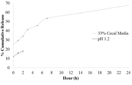
Orally administered drug delivery systems intended for the colon should ideally yield minimal release of the payload in gastric media but release maximally in colonic conditions. The in vitro release of curcumin from the MCPCNPs at conditions mimicking the pH and times likely to be encountered in the stomach and colon (2 h in pH 1.2 and 24 h in 33 % (w/v) caecal media) is presented in Figure 7.
Figure 7: In vitro drug release profile of curcumin from MCPCNPs in pH 1.2 for 2 h and 33% caecal medium for 24 h
364
365
366
367
368
369
370
371
372
373
374
375
376
377
378
379
380
381
382
The release of curcumin reached ≈ 18 % over 2 h in acidic conditions due to the poor solubility of curcumin at acidic pH, coupled with the protonation of the charged carboxylic groups (COO-) of pectin which minimises columbic repulsions between polymer chains and therefore keeps the matrix secluded. Furthermore, at this pH, the amino groups of chitosan also become protonated, which favours a closely-knit gel network with MCP that impedes the diffusion of curcumin within the matrices. On the other hand, a significant release of
curcumin was observed in the caecal media over 24 hr (≈ 68 %), pointing to the profound effects colonic enzymes exhibit on the integrity of the chitosan-MCP composite matrix, which tends to loosen the polymer matrix network allowing free diffusion of curcumin. The obtained release results correlate with the swelling capacities profiles presented previously, further approving the released amount of curcumin from the MCP-chitosan composite matrix. In order to investigate the mechanism of curcumin release from the MCPCNPs in caecal medium, the data were fitted to the zero-order, first-order, Higuchi and Korsmeyer–Peppas models [27] and results are presented in Table 1.
Model r2 for complete profile
Korsmeyer-Peppas 0.986 Higuchi 0.954 First-order 0.920 Zero-order 0.847
Table 1: Correlation coefficients of best fits to studied models
The models were not fitted to the release profile in pH 1.2 medium because less than 15% of curcumin was released within the 2hr study period (stipulated gastric resident time) and aptly, the release of curcumin in pH 1.2 (stomach) is not very relevant to the present pursuit.
Curcumin release was best fitted to the Korsmeyer-Peppas model with a correlation of 0.986 and an exponent (n) of 0.27, (Table 1). This indicates that the release mechanism followed a quasi-Fickian diffusion mechanism. We believe that this mechanism is borne by a natural propensity of curcumin to diffuse passively from the porous matrix of the MCPCNPs. Furthermore, diffusion was prompted by degradation of the pectin chains, especially at surface of the particle matrices by enzymes within the caecal medium. A closer study of the profile in the caecal medium reveals a bi-phasic release pattern, with the first inflection (1-7 h) steeper than the second (7-24 h), (Figure 7). During the initial phase (first inflection), diffusion is limited to the outer or near outer surfaces of the MCPCNPs matrix, reinforced by 387
388
389
390
391
392
393
394
395
396
397
398
399
400
401
402
403
404
405
406
407
408
409
410
411
412
413
414
415
enzymatic effects. The second inflection involves release from the deeper domains of the MCPCNPs with lesser enzymatic effects on the matrices and more due to passive diffusion of curcumin. We may conclude that curcumin release is largely governed by diffusion, dictated by enzymatic effects on the MCPCNPs matrix that can best be described as a quasi-Fickian diffusion mechanism.
4. Conclusion
The MCPCNPs have demonstrable potential for colon delivery. The data suggest that the formulation survives acidic pH milieu, with a limited amount of curcumin release, but releases curcumin significantly at media representing the colon. This presents a possible model for colon delivery of curcumin following oral delivery whereby significant curcumin payload is retained within the nanoparticles until colon arrival. Furthermore, the
nanoparticles are mucoadhesive at pH representing the colon region. The combined effect of the curcumin payload and galactin moiety from the pectin carrier and the mucoadhesion presented at colonic condition, provide a unique strategy aimed at achieving efficient anticancer agent delivery to the colon. Therefore, we propose that the system is an excellent candidate for further in vivo studies.
Acknowledgment
We thank Professor Ting Kang Nee for providing the sacrificed rats from which caecal content was used so that no rat was sacrificed solely for the purpose of this investigation.
References
[1] Jemal, A., Bray, F., Center, M. M., Ferlay, J., Ward, E., Forman, D. (2011). Global cancer statistics, CA Cancer J. Clin. 61 (2011) 69–90.
[2] Ferlay, J., Soerjomataram, I., Dikshit, R., Eser, S., Mathers, C., Rebelo, M. Bray, F., Cancer incidence and mortality worldwide : Sources , methods and major patterns in GLOBOCAN 2012, Int. J. Cancer. 136 (2015) E359–E386.
[3] Hua, S., Karl, G., Nell, H., Orally administered liposomal formulations for colon targeted drug delivery, Front Pharmacol. 5 (2014) 1-4.
417
418
419
420
421
422
423
424
425
426
427
428
429
430
431
432
433
434
435
436
437
438
439
440
441
442
443
444
445
[4] Chuah, L. H., Billa, N., Roberts, C. J., Burley, J. C., Manickam, S., Curcumin-containing chitosan nanoparticles as a potential mucoadhesive delivery system to the colon, Pharm. Dev. Technol. 18 (2011) 1–9.
[5] Anand, P., Kunnumakkara, A. B., Newman, R. A., Aggarwal, B. B., Bioavailability of curcumin: Problems and promises, Mol. Pharm. 4 (2007) 807–818.
[6] Prasad S., Gupta S., Tyagi A., Aggarwal B., Curcumin, a component of golden spice: From bedside to bench and back, Biotechnol. Adv. 32 (2014) 1053-1064.
[7] Rampino, A., Borgogna, M., Blasi, P., Bellich, B., Cesàro, A., Chitosan nanoparticles: Preparation, size evolution and stability, Int. J. Pharm. 455 (2013) 219–228.
[8] Jonassen, H., Kjoniksen, A. L., Hiorth, M., Stability of chitosan nanoparticles cross-linked with tripolyphosphate, Biomacromolecules. 13(2012) 3747–3756.
[9] Mishra, R. K., Banthia, A. K., Majeed, A. B. A., Pectin based formulations for biomedical applications: A review, Asian J. Pharm. Clin. Res. 5 (2012) 1–7.
[10] Glinsky, V. V., Raz, A.,Modified citrus pectin anti-metastatic properties: one bullet, multiple targets, Carbohydr. Res. 344 (2009) 1788–1791.
[11] Leclere, L., Cutsem, P. Van, & Michiels, C, Anti-cancer activities of pH- or heat-modified pectin, Front Pharmacol. (2009) 1–8.
[12] Venzon, S. S., Canteri, M. H. G., Granato, D., Demczuk, B., Maciel, G. M., Stafussa, A. P., Haminiuk, C. W. I., Physicochemical properties of modified citrus pectins extracted from orange pomace, JFST. 52 (2015) 4102–4112.
[13] Barrow, H., Rhodes, J. M., Yu, L. G.,The role of galectins in colorectal cancer progression, Int. J. Cancer. 129 (2011) 1–8.
[14] Wong, T. W., Colombo, G., Sonvico, F., Pectin matrix as oral drug delivery vehicle for colon cancer treatment, AAPS PharmSciTech. 12 (2011) 201–214.
[15] Munjeri, O., Collett, J. H., Fell, J. T. , Hydrogel beads based on amidated pectins for colon-specific drug delivery: The role of chitosan in modifying drug release, J. Control. Release. 46 (1997) 273–278.
[16] Chang, K. L. B., Lin, J., Swelling behavior and the release of protein from chitosan-pectin composite particles, Carbohydr. Polym. 43 (2000) 163–169.
447 448 449
450
451
452
453
454
455
456
457
458
459
460
461
462
463
464
465
466
467
468
469
470
471
472
473
474
[17] Andriani, Y., Grastianto, Siswanta, Mudasir, Glutaraldehyde-Crosslinked Chitosan-Pectin Nanoparticles as a Potential Carrier for Curcumin Delivery and Its In Vitro Release Study, Int. J. Drug Deliv. 7 (2015) 167–173.
[18] Chuah, L. H., Roberts, C. J., Billa, N., , Abdullah, S., Rozita, R., Cellular uptake and anticancer effects of mucoadhesive curcumin-containing chitosan nanoparticles, Colloids Surf. B. 16 (2014) 228–36.
[19] Urias-orona, V., Rascón-chu, A., Lizardi-mendoza, J., Carvajal-Millán, E., Gardea A., A., Ramírez-Wong, B., A Novel Pectin Material : Extraction , Characterization and Gelling Properties, Int. J. Mol. Sci. 11 (2010) 3686–3695.
[20] Chen, Y., Wu, Q., Zhang, Z., Yuan, L., Liu, X., Zhou, L., Preparation of curcumin-loaded liposomes and evaluation of their skin permeation and pharmacodynamics, Molecules. 17 (2012) 5972–5987.
[21] Smart, J. D., The basics and underlying mechanisms of mucoadhesion, Adv. Drug Deliv. Rev. 57 (2005) 1556–1568.
[22] Khutoryanskiy, V. V., Supramolecular materials: Longer and safer gastric residence, Nat Mater. 14 (2015) 963–964.
[23] Bigucci, F., Luppi, B., Cerchiara, T., Sorrenti, M., Bettinetti, G., Rodriguez, L., Zecchi, V., Chitosan/pectin polyelectrolyte complexes: Selection of suitable preparative conditions for colon-specific delivery of vancomycin, Eur. J. Pharm. Sci. 35 (2008) 435–441.
[24] Gadalla, H. H., Soliman, M. G., Mohammed, A. F., El-Sayed M. A., Development and in vitro/in vivo evaluation of Zn-pectinate microparticles reinforced with chitosan for the colonic delivery of progesterone, Drug Deliv. 23 (2016) 2541-2554.
[25] Deacon, M. P., Mcgurk, S., Roberts, C. J., Williams, P. M., Tendler, S. J. B., Davies, M. C. Harding, S. E., Atomic force microscopy of gastric mucin and chitosan mucoadhesive systems, Biochem. J. 348 (2000) 557–563.
[26] Pengpong, T., Sangvanich, P., Sirilertmukul, K., Muangsin, N., Design, synthesis and in vitro evaluation of mucoadhesive p-coumarate-thiolated-chitosan as a hydrophobic drug carriers, Eur. J. Pharm. Biopharm. 86 (2014) 487–497.
476
477
478
479
480
481
482
483
484
485
486
487
488
489
490
491
492
493
494
495
496
497
498
499
500
501
502
[27] AbouAitah, K. EA., Farghali, A. A., Swiderska-Sroda, A., Lojkowski, W., Razin, A. MF., PH-controlled Release System for Curcumin based on Functionalized Dendritic Mesoporous Silica Nanoparticles, J. Nanomed. Nanotechnol. 7 (2016) 1-11
504
505