SYNAPTIC LEARNING RULES FOR LOCAL
SYNAPTIC INTERACTIONS:
Theory and Application to Direction Selectivity
Thesis by Chunhui Mo
In Partial Fulfillment of the Requirements for the Degree of
Doctor of Philosophy
California Institute of Technology Pasadena, California
2003
© 2003
Chunhui Mo
Acknowledgements
I would like to thank my advisor, Dr. Christof Koch, whose constant support has
made this research work possible. He provided me with the critical guidance to
advance my research in the right direction and stay focused on important
questions. He sets high standards for research quality and scientific ethics. I can
always count on his encouragement when I venture into creative areas and his
patience when I make slow progress.
I am very grateful to my thesis committee, Drs. Scott Fraser, Gilles Laurent,
Steven Quartz, and Erin Schuman, who have provided me with invaluable
feedback over the years. Special thanks go to Gilles for stimulating scientific
discussions and Erin for valuable insights in the experimental side of learning
and calcium dynamics.
I thank all Koch lab members for tolerating my big background jobs on their
desktop machines. All my friends, fellow BMB and CNS students, and Techers
have made my stay at Caltech an enjoyable experience. Thank you.
This “reverse-phi” modeling project originated in a joint discussion with Tomaso
Poggio and Margaret Livingstone. I would like to thank both for their help and
criticism. I would also like to thank Drs. Kevin Archie, Bartlett Mel and Terry
Finally, I would like to thank my parents for motivating me to go through the
graduate school and my friend Ying Gong for constant encouragement. Thanks
for the critical reading of my thesis!
This research was supported by grants from the NSF-sponsored Engineering
Research Center at Caltech, the National Institutes of Health, and the National
Abstracts
This thesis is organized in two parts, both concerned with local synaptic
interactions within the dendritic tree. The first part is focused on how specific
synaptic arrangements that can be used to compute direct ion selecti vity can be
learned in a n unsupervised manner. The second part consists of a double
synaptic veto model that can account for the observed reverse-phi selectivity of
direction-selective cells. We propose an activity-based, local learning model that
may account for the direction selectivity in neurons in the visual cortex based on
the local veto operation among excitation and inhibition. We implement the
learning rule with local calcium concentration changes and a BCM type learning
curve (Bienenstock, Cooper and Munro, 1982). Our biophysical simulations
suggest that a model cell implementing our learning algorithm develops direction
selectivity organically after unsupervised training. The learning rule is also
applicable to cells with multiple direction-selective subunits on dendrites and is
stable under a number of starting conditions.
Reverse-phi motion is the illusory reversal of perceived direction of movement
when the stimulus contrast is reversed in successive frames. Livingstone (2000)
showed that direction-selective cells in striate cortex of the alert macaque
monkey showed reversed excitatory and inhibitory regions when two different
contrast bars were flashed sequentially during a two -bar interaction analysis. We
implementing a synaptic shunting scheme. Our results suggest that a simple
synaptic-veto mechanism with strong direction selectivity for normal motion
cannot account for the observed reverse phi-motion effect. A direct interaction
between the ON and OFF pathway, missing in the original shunting-inhibition
model, is essential to account for the reversal of response. We propose a double
synaptic-veto mechanism in which ON excitatory synapses are gated by both
delayed ON inhibition at their null side and by delayed OFF inhibition at their
preferred side. The converse applies to OFF excitatory synapses. Mapping this
scheme onto the dendrites of a direction-selective neuron permits the model to
respond best to normal motion in its preferred direction and to reverse-phi motion
Chapter Six: A Learning Rule for the Reverse-Phi Selective Synaptic
Placement 106
6.1 Introduction 106
6.2 The reverse-phi learning scheme 106
6.3 The background firing of inhibitory inputs can facilitate the “double
veto” synaptic placement 109
Chapter Seven: Summary 113
Chapter One
Introduction
This thesis consists of two major parts. The first part, a detailed description of a
synaptic learning rule for local synaptic interactions between excitation and
shunting inhibition on a direction-selective cell’s dendrite is given in Chapters
Two and Three. The second part, a double synaptic veto mechanism that can
account for the observed “reverse-phi” selectivity of cortical direction-selective
cells, is introduced in Chapter Four which has been published as Mo and Koch
(2003). This chapter serves as a guided tour to give readers a quick overview of
each chapter and the motivation behind.
The ability to detect motion direction is arguably one of the most important
functions of all vision systems. Direction-selective cells in the retina of the rabbit
(Barlow and Levick, 1964; reviewed by Vaney et al., 2001), the pretectal nucleus
of the optical tract (NOT) of the wallaby (Ibbotson and Price, 2001), the lobular
plate of the fly (Egelhaaf and Borst, 1992), and the visual cortex of the cat and
monkey (Goodwin et al., 1975; Emerson and Gerstein, 1977; Ganz and Felder,
1984; Emerson et al., 1993;Jagadeesh et al., 1993; Livingstone, 1998) have
been extensively studied (reviewed by Clifford and Ibbotson, 2003). Over the
years, both feed-forward (Koch and Poggio, 1985; Livingston, 1998; Anderson et
Orban, 1996; Ernst et al., 2001; Rao and Sejnowski, 2001) schemes have been
proposed. Recent experimental evidence suggests the asymmetrical delayed
inhibition is likely to be one of the mechanisms that underlie cortical direction
selectivity (Livingstone, 1998). Such a mechanism may be based on shunting
inhibition, i.e., an increase in a chlorine based GABA_A conductance that
reverses close to the cell's resting position, as proposed by Koch and Poggio
(1985).
One important open question is: how can the required synaptic specific
interaction among excitations and inhibitions be obtained in an unsupervised
manner? The wiring requirement for such a scheme is the following: excitation in
visual space can reside on either side of the inhibitory zone, but not on both
sides, in which case the model receives symmetric input in space-time and is
thus not direction-selective. Cats reared in a stroboscopically illuminated
environment develop normal orientation-selective neurons in cortex but
direction-selective neurons are virtually abolished. This effect remains after long periods of
normal visual exposure (Cynader and Chernenko, 1976; Humphrey and Saul,
1998; Saul and Feidler, 2002; rabbit visual cortex: Grigonis et al., 1988). The
results suggest the establishment of direction-selective properties of cortical cells
is likely to go through a visual-experience driven, activity-based learning phase.
The learning process for direction selectivity may be independent of the
establishment of orientation selectivity. In this study, we propose an
or other types of computations based on local veto operation among excitation
and inhibition. We assume that the inhibitory connection is fixed and the learning
process only occurs at excitatory synapses in chapter two and through most of
chapter three, because very little experimental evidence exists for inhibitory
learning. A global, activity-based inhibitory learning scheme that works with our
local excitatory learning rule is also addressed in the last section of chapter three.
Given the fact no calcium flow through a n inhibitory channel when it is open, it is
unclear if our calcium based local learning mechanism can be applied to the
learning of inhibitory synapse. We here adopt a global inhibitory learning
mechanism as described by Soto-Trevino et al. (2003). In cat area 17, 40% of
cells are found to have their inhibition tuning preference different from their spike
outputs in an intracellular study investigating synaptic mechanisms of orientation
and direction selectivity (Monier et al, 2003). The excitation tuning preference of
these cells is the same as either the spiking output or the inhibition. The lack of
homogeneities in input combinations might reflect the result of an activity based,
excitation-inhibition differential learning process during development.
Various modified versions of the original Hebbian learning rule that can account
for the development of direction selectivity have been proposed (Bienenstock et
al., 1982; Feidler et al., 1997; Blais et al., 2000; Rao and Sejnowski, 2001). Our
learning rule is based on the peak calcium concentration change at spines
following a synaptic input. We use a BCM type learning curve (Bienenstock et al.,
experimentally verified. Direction-selective cells in the monkey V1 can respond to
movement within 0.1 degree, which is much smaller than their receptive field size
(Livingstone, 1998), suggesting cortical selective cells have
direction-selective subunit structures on their dendrites (Emerson, 1997). We use
compartmental simulations and a model cell with very simple geometry to test
our learning rule, different from all previous studies that treat the post synaptic
cell as a point-neuron or simply an integration unit (Koch and Segev, 2000; Koch
et al., 2003). This allows us to investigate the development of direction-selective
subunit structures at the dendritic level.
The effect of post-synaptic calcium concentration on LTP and LTD has been
shown by (Yang and Zucker, 1999) in their photolysis experiments. Fast rising
calcium concentration changes in dendritic spines mediated by action potential
and long sustained rising mediated by synaptic inputs have been observed in
calcium imaging experiment (Sabatini and Svoboda, 2002). A comparison of the
calcium dynamics that is discovered in their study and that is implemented in our
model is given in the discussion section of Chapter two. We assume
calcium-dependent synaptic weight changes can be achieved biochemically through the
calcium -dependent phosphorylation of
a-amino-3-hydroxy-5-methyl-4-isoxazolepropionic acid (AMPA) subtype of glutamate receptors (AMPARs) as
described by Castellani et al. (2001).
a continuous spatial and temporal sequence. Reverse-phi motion was first
demonstrated by Anstis (Anstis, 1970; Anstis and Rogers, 1975). Subjects
perceive the reverse direction of motion when the contrast of a moving object
reverses in the second frame of a two-frame shift experiment. Reverse-phi like
effects have also been reported during electrophysiological experiments from
direction-selective complex cells in the cat striate cortex (Emerson et al., 1987),
the H1 cell in the fly's lobula plate (Egelhaff and Borst, 1992), and the optical
tract of the wallaby (Ibbotson and Clifford, 2001). Recent recordings from
direction-selective cells in the alert monkey show that cells in both cortical areas
V1 and MT reverse facilitation and suppression regions in the 2-bar interactions
map when two different contrast bars are presented (Livingstone et al., 2000;
Conway and Livingstone, 2001). This implies that these cells respond to
reverse-phi motion in the reversed direction. In Chapter Four, we show how the circuitry
for the normal direction selectivity can be adapted to account for the reverse-phi
selectivity. Our results suggest that a simple synaptic-veto mechanism with
strong direction selectivity for normal motion cannot account for the observed
reverse phi-motion effect due to the fact that a direct interaction between the ON
and OFF pathways is missing in the original shunting inhibition model. We show
a double synaptic-veto mechanism, derived from the traditional asymmetrical
delayed shunting inhibition model, can account for both normal and reverse-phi
motion direction selectivity. In such a scheme, ON excitatory synapses are gated
by both delayed ON inhibition at their null side and by delayed OFF inhibition at
this scheme onto the dendrites of a direction-selective neuron permits the model
to respond best to normal motion in its preferred direction and to reverse-phi
motion in its null direction.
Both the learning model and the reverse-phi model use shunting inhibition to
achieve dendritic specific veto of excitatory inputs. The shunting inhibition’s local
“gating” effect is discussed in Chapter Five, as well as the optimal inhibition and
excitation input locations in our model. Recent experiments in the retina provide
evidence in favor of at least some nonlinear interactions between excitatory and
shunting inhibitory inputs that take place within the dendrites of
direction-selective ganglion cells (Taylor et al., 2000; for a dissenting view, see
Borg-Graham 2001). Large conductance changes that reverse around the cell's resting
potential have been observed in V1 during visual stimuli (Anderson et al., 2000;
Borg-Graham et al., 1998). Our results suggest shunting inhibition may be
important for forming direction-selective synaptic connections as well as the final
direction selectivity.
Finally, in Chapter Six we discuss whether our learning rule described in
chapters two and three can account for the “double -veto” synaptic placement we
proposed in chapter four. Reverse-phi motion stimuli do not appear to be a
common feature of natural spatiotemporal scenes. It therefore remains unclear
why cortical cells should invert their direction selectivity for reverse-phi motion.
has to be established as a by-product of developing normal direction selectivity,
rather than a stand -alone training process. We show such connections may be
established due to the background firing of inhibitory input cells, which causes
Chapter Two
A Synaptic Learning Rule for Local Synaptic Interactions
between Excitation and Shunting Inhibition
2.1 Introduction
The ability to distinguish object movement directions is important to all motion
processing systems. In the cat and monkey, V1 is the first stage in the visual
pathway where direction-selective (DS) cells are encountered. Cats reared in a
stroboscopically illuminated environment develop normal orientation-selective
neurons in cortex but direction-selective neurons are virtually abolished. This
effect remains after long periods of normal visual exposure (Cynader and
Chernenko, 1976; Humphrey and Saul, 1998; Saul and Feidler, 2002). Therefore,
direction selectivity is likely to require structured synaptic input during early
developmental stages. These neurons are thus excellent targets for the study of
activity-dependent synaptic weight changes. Hebb proposed his famous learni ng
rule based on the correlation between pre- and post-synaptic activities (Hebb,
1949). Long-term potentiation (LTP) and the long-term depression (LTD) are
likely to be among the synaptic manifestations of such a learning rule (Bliss and
Lomo, 1973; Dudek and Bear, 1992; Reviewed by Malenka and Nicoll, 1999).
However, a computational study showed that the simple Hebbian learning rule
performs poorly in direction-selective synapse placement (Feidler et al., 1997).
non-linear interaction between two different inputs in space-time. In a natural
environment, as many stimuli are expected to move in the preferred than in the
null direction. While there exists a correlation between pre- and post-synaptic
firing in the preferred direction, this is not the case for motion in the opposite, null,
direction.
There are various modified versions of the original Hebbian learning rule that can
account for the development of direction selectivity. The principal component
analyzer model proposed by Oja in 1982 adds a non-linear decay term to
achieve stability (Oja, 1982), although itself is not a direction selective learning
rule. A synaptic “gating” rule links synaptic weight changes with
post-synaptic activities (Feidler et al., 1997). The BCM learning rule, originally
proposed to account for orientation selectivity and binocular interactions , has
been extended to the generation of direction-selective units (Bienenstock et al.,
1982; Feidler et al., 1997; Blais et al., 2000).
Intracellular calcium concentration is one of the biophysical variables critical to
LTP induction, suggesting it may be an important messenger bridging the gap
between firing frequency changes and synaptic modifications (Lynch et al., 1983;
Yang et al., 1999; Zucker, 1999; Kalikulov et al., 2002). NMDA receptors are
likely to be the entry points of calcium at spines since an antagonist of NMDA
receptors inhibits the induction of LTP (Collingridge et al., 1983). The
account for the age-dependent decline of visual cortical plasticity (Carmignoto
and Vicini, 1992). Glutamate receptors may involve in regulating spine structure
plasticity (Fischer et al., 2000). Calcium influx through NMDA channels can
cause a large, localized and transient increase in the postsynaptic calcium level
(MacDermott et al., 1986; Gamble and Koch, 1987; Zador et al., 1990; Helmchen
et al., 1999; Sabatini et al., 2001; Sabatini et al. 2002). The existence of
voltage-gated voltage-gated calcium channels in hippocampal cells and their contribution to
synaptic plasticity have been reported (Dingledine, 1982; Cummings et al.). A
simulation study by Castellani et al. (2001) suggests a BCM type learning curve
can be achieved by calcium-dependent protein kinases and phosphatases
activity changes that in turn modify the phosphorylation state of AMPA receptors
and thus their conductance. Enzymes involved in this process reside in the
postsynaptic density (reviewed by Kennedy, 2000) and may involve with the
long-term calcium -dependent regulation in neuronal gene expression (reviewed
by Bito et al., 1997).
A great deal is known about direction-selective cells through experimental,
modeling a nd theoretical investigations. The motion energy model (Adelson and
Bergen, 1985), as well as the equivalent Reichardt model (Reichardt, 1961;
Santen and Sperling, 1985), interprets observed physiological and
psychophysics data well. At the biophysical le vel, network models that rely on
excitatory feedback (Douglas et al., 1995; Maex and Orban, 1996; Rao and
1998; Anderson et al., 1999) have been proposed to account for
direction-selective synaptic arrangements in the cortex. The asymmetry in the summation
of excitatory inputs at dendrites alone is not sufficient to account for the
directional response based on modeling study by Anderson et al. (1999).
Asymmetrical delayed inhibition is likely to be one of the mechanisms that
underlie direction selectivity. Such a mechanism based on shunting or silent
inhibition was proposed for the cortex by Koch and Poggio (1985). A bar moving
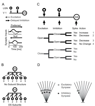
in the preferred direction reaches the excitatory input before the inhibitory one,
which only acts after an additional delay (Fig. 2-1a). The excitatory input reaches
the soma and causes the cell to spike because of the temporal offset between
the two inputs. In the opposite, null direction, the excitato ry input is “vetoed” by
the inhibition if the bar’s speed is approximately matched to the delay. The wiring
requirement for such a scheme is simple: excitation in visual space can reside on
either side of the inhibitory zone, but not on both sides, in which case the model
receives symmetric input in space-time and is thus not direction-selective. Large
conductance changes that reverse around the cell's resting potential have been
observed in V1 during visual stimuli (Anderson et al., 2000; Borg-Graham et al.,
1998). Given the local effect of shunting inhibition, it is interesting to investigate
its possible role in synaptic learning.
Both retinal and cortical direction-selective cells have subunit structures within
their receptive fields (Barlow and Levick, 1965; Emerson et al., 1987; Livingstone
subunit structures is shown in Fig. 2-1b. Both cells receive inputs from the same
group of LGN cells but the one with subunit structures utilizes its resources more
efficiently and has superior position-invariant direction tuning. Synaptic logic
models involving complex, branch-specific synapse placements had long been
proposed (Poggio, 1982; Koch and Poggio, 1987). Recent work by Mel and
colleages suggests that the large dendritic tree of cortical pyramidal neurons may
function as a two-layer neural network (Poirazi et al., 2003). The standard form of
Hebbian learning rule and all of its variations describing changes only in the
overall connection strength between the pre- and post-synaptic neurons cannot
account for structure plasticity (Mel, 2002) and local learning, for they do not
distinguish among post-synaptic connection locations. We here demonstrate,
using a highly idealized compartmental simulation of a single post-synaptic
neuron, how a learning rule for local synaptic interactions between excitation and
shunting inhibition could, in principle, account for direction selectivity and
2.2 Methods
All compartmental simulations were carried out using the program NEURON
(Hines and Carnevale, 1997). The idealized cell morphology of a
direction-selective neuron included eight dendrites (width 0.5 µm, length 100 µm) that
were directly connected to the soma (width 16 µm, length 16 µm). Each dendrite
was unbranched and had 20 compartments, for a total of 180 compartments. The
dendrites were passive except for an N-type calcium conductance, while the cell
body contained sodium and potassium conductance that gives rise to fast
Hodgkin-Huxley-like action potentials. Given our model cell’s low L value /large
space constant (L=0.45 at the tip of the dendrite), large sodium conductance is
not needed at dendrites to sustain back-propagating spikes as opposed to a
simulation study by Tsay and Yuste (2002) using a big layer five pyramidal
neuron model. There was no spike adaptation mechanism in our model. The
biophysical parameters included: Ri=250 Ω•cm, Cm=0.5 µF/cm2
, Eleak=-60 mV,
Rm=10 kΩ•cm2
, gNa=0.030 S/cm2, gK=0.028 S/cm2, ENMDA=0 mV, gNMDA=0-2 nS,
τNMDA on=0.1 ms, τNMDA off=80 ms, EAMPA=0 mV, gAMPA=0-2 nS, τAMPA on=0.1 ms,
τAMPA off=2 ms, EGABA=-60 mV, gGABA=5.0 nS, τGABA on=1 ms, τGABA off=80 ms.
Synaptic input was modeled using the point process in NEURON (adopted from
Archie and Mel, 2000). The input resistances of the model cell were 570 MΩ,
I008 MΩ (L=0.22, transfer resistance 519 MΩ), and 1448 MΩ (L=0.45, transfer
resistance 503 MΩ), at the soma, the middle of the dendrite and the tip of the
synaptic input around 2ns to elicit spikes at the soma. Real cells may have much
lower input resistance at soma, but they may also require several simultaneous
excitatory inputs to elicit spikes. The large input resistance was due to our cell’s
limited size (dendrite: 100 µm in length, 0.5 µm in diameter. Soma: 16 µm in
diameter). Increasing the dendrite length to 200 µm and diameter to 1.5 µm and
connecting a large compartment that was 500 µm in length and 5 µm in diameter
decreased the model cell’s input resistance to around 70 MΩ. Our learning
model converged as expected using this alternative geometry setting when we
increased the excitatory and inhibitory input connection strength by ten folds to
make them large enough to elicit spikes.
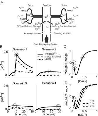
The N-type voltage-gated calcium conductance was taken from (Benison et al.,
2001) and mapped to dendrites with a density of 1 mS/cm2. We assume that
excitatory synapses are located to dendritic spines and that the rapid rise in
intracellular calcium concentration at the postsynaptic site inside the spine
following synaptic activation have two main sources (Fig. 2-2a): calcium current
through the NMDA synapse, ICa_NMDA ,, and calcium current, ICaN, that is located in
the dendrite and a certain amount of which diffuses up into the spine. ICa_NMDA
was calculated as one third of the total current through the NMDA synapse but
with a reversal potential ECa=130mv. Of the ICaN entering the dendritic
compartment associated with the spine, 5% was assumed to contribute to the
calcium concentration at the spine (Koch, 1999). These scaling factors were
2.3 A local learning scheme for direction selectivity
We considered what kinds of information were available to an excitatory,
geniculate synapse that just landed on a dendrite of a cortical neuron to correctly
judge whether its activity contributed or degraded the direction selectivity (DS) of
the host cell, assuming the inhibitory input was already connected and fixed.
There are three major pieces of information accessible to local mechanisms: the
state of the excitatory input, the state of the local inhibitory input and whether or
not the host cell generates a somatic action potential within a small time window
and this spike propagates back into the dendrite to the postsynaptic site of
excitation. Assuming binary states (e.g., excitatory input is either on or off), this
gives rise to eight possible scenarios (for instance, both excitation and inhibition
are active and the host cell spikes). We assume the excitatory synapse can only
be modified when it is active; this reduces the combinations to four scenarios (Fig.
2-1c). In scenario one, there is no inhibition and the cell spikes after the
excitatory synapse opens. The assumption is that the excitatory synapse directly
contributes to the cell’s direction selectivity and thus its connection strength
should increase. In scenario two, there is no inhibition and no spike when the
excitatory synapse opens. The fact that the DS cell isn’t spiking suggest that the
stimulus moves in the null direction; yet the excitatory input is not gated by the
inhibition and counteracts the cell’s direction selectivity. Its connection strength
should decrease. In scenario three, both the excitation and the inhibition are
corresponds to the null direction motion and that the excitatory synapse lands on
a spot with incorrect matching of inhibition. Its connection strength should
therefore decrease. In scenario four, the excitation is successfully blocked by
inhibition, suggesting a null direction movement, in which case the blocking by
inhibition is legitimate. On the other hand, this could also correspond to a
preferred direction movement. The cell generates an action potential in the
presence of inhibition, yet this spike, propagating back from the soma into the
dendrite, is blocked by inhibition from reaching the site of excitation. Given this
ambiguity, the best possible action is to do nothing and keep the excitatory
weight constant.
An excitatory synapse can adjust its weight if it can distinguish these four
scenarios. We next mapped out local calcium concentration changes during
these scenarios and used this biophysical variable to “inform” the excitatory
synapse about which action it should take during the learning process.
2.4 Calcium dynamics at spine and the local learning rule
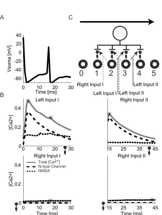
Fig. 2-2a illustrates a dendritic branch with two spines with independent
excitatory inputs. Inhibition was mapped to a dendritic compartment located
between the excitatory inputs and the soma, fulfilling the “on-the-path”
requirement (Koch et al., 1982). We mapped the two excitatory synapses into
one, electrical equivalent, dendritic compartment (Koch, 1999). Calcium can
through the N-type voltage gated calcium channels inserted into the dendritic
membrane. We reason that calcium can enter the spine from the dendrite but
cannot exit to the dendrite from the spine; given the large volume difference
between the spine head and the dendrite , and given various calcium pumps
along the thin neck of the spine. Therefore, the two spines are chemically
independent, although they are electrically equivalent (Zador and Koch, 1990).
The excitatory and inhibitory inputs and the back-propagating spike from the
soma affect the total calcium concentration at the spine in their own way. The
excitatory input directly correlates with the calcium current entering through
NMDA channels, which is spine/synapse specific. Its time course is mainly
determined by the conductance change of the local NMDA synapse. Back-
propagating action potentials signal the global activation state of the DS cell.
N-type voltage-gated calcium channels are mainly activated when there is a
back-propagating spike. Given the NMDA synapse’s reversal potential (set to zero),
the synaptic input current alone cannot elevate the membrane potential high
enough to cause significant activation of the N-type voltage gated calcium
channel. The on-the-path shunting inhibition affects the time course of both
calcium currents. It clamps the membrane voltage when open, thereby reducing
the amount of calcium current entering through NMDA channels. Furthermore, it
completely blocks back-propagating spikes, which in turn blocks calcium
entrance through N-type voltage gated calcium channels. The clamping and
The total calcium concentration at the synapse results from the addition of the
two currents assuming instant diffusion of the intracellular free calcium from
dendrites into spine. We modeled all internal calcium buffers and calcium pumps
using a single decay constant.
We implemented the above calcium scheme and computed the resultant
changes in free, intracellular calcium concentration ([Ca2+]) at the spine for the
four scenarios considered earlier (Fig. 2-2b). In scenario one, there is no local
inhibition and the cell spikes after the excitatory synapse opens. Calcium enters
through both channels. Changes of [Ca2+] are high. In scenario two, there is
neither local inhibition nor a back-propagating spike immediately after the
excitatory synapse opens. Calcium mainly enters through NMDA channels.
Changes of [Ca2+] fell into an intermediate range. In scenario three, the excitatory
input is blocked by the inhibition but the cell spikes. The amount of calcium
entering through NMDA channels is reduced by inhibition. Meanwhile residual
calcium enters through voltage N-type gated calcium channels due to the
back-propagating action potential immediately before the synapse opens, adding to
the total calcium concentration. Changes of [Ca2+] again fall into an intermediate
range. In scenario four, excitation is blocked by inhibition in the absence of any
action potential. Only a limited amount of calcium enters through the NMDA
synapse due to the clamping effect of shunting inhibition. Changes of [Ca2+] are
As we mentioned earlier, the proper action for scenario one is to increase the
weight of the excitatory synapse. Changes of [Ca2+] in this case are high. The
proper action for scenario two and three is to decrease the synaptic weight.
Changes of [Ca2+] in these cases are medium. The proper action for scenario
four is to keep the synaptic weight unchanged. Changes of [Ca2+] are low. In
order to link the synaptic weight change with maximum calcium exposure, we
map the peak calcium exposures to a BCM type learning curve (Fig. 2-2c). The
amplitude of each excitatory synapse from the geniculate input to the target cell
is changed in accordance with the maximum calcium concentration just below
the synapse reached within 30 ms of synaptic activation. Following the BCM
learning curve (Fig. 2-2c), this is associated with the following change in synaptic
weight:
weightchange=a ec(−[Ca2+]−d)−ec(−[Ca 2+]−d)
2π +b
With a=2.63, b=1, and c=14. d is a constant that varies continuously between
-0.10 and -0.22 as gNMDA varies from 0 to 2 ns. The learning curve is chosen to
give a negative output at a medium calcium concentration and a positive one at
high calcium concentration (Fig. 2-2c). The parameters a and b are chosen to
restrict the function’s output to between -1 and 1. The parameter c is a scaling
factor that determines the width of the curve and parameter d is a sliding
threshold that linearly shifts the curve according to different values of gNMDA (Fig.
Instead of implementing a non-linear threshold that will narrow or broaden the
learning curve with respect to the average [Ca2+] change during training, we use
a linear sliding-threshold to shift the learning curve without changing its shape as
shown in Fig. 2-2d. We link the sliding threshold directly to the excitatory
synaptic connection strength. This prevents runaway excitation and helps
stabilize the synapse (Bienenstock et al., 1982; Abbott and Nelson, 2000). The
larger the connection strength, the more [Ca2+] accumulates over time,
independent of learning scenarios.
2.5 Direction-selective single-unit learning
We first tested our learning rules in a model cell without subunit structures. The
connection scheme is shown in Fig. 2-3a. The model initially receives balanced
excitatory inputs from both left and right LGN neurons. Two excitatory inputs are
mapped into the same compartment on the dendrite. The initial connection
strength is 1ns each. Delayed inhibition is fixed at 5 ns and is mapped to a
compartment between the excitatory inputs and the soma. During each trial, a
bright bar moving to either the left or to the right is randomly presented and the
maximum change of [Ca2+] at each excitatory synapse within 30 ms of its
opening during the trial is recorded. After each trial, the synaptic weight change
is calculated based on the learning curve shown in Fig. 2-2d.
A synapse can only be rewarded if the host cell spikes and this action potential
direction selectivity if none of the excitatory inputs is strong enough to drive the
cell to spike. Therefore, we need to impose a “competition” rule to control the
model’s excitability. We implemented this by holding the total excitatory
connection strength over each local dendrite constant during simulations. This
rule is of the “subtraction” type (Abbott and Nelson, 2000); that is, after each trial,
half of the value of the synaptic weight above or below the total connection
strength is subtracted from or added to both excitatory synapses. If the two
excitatory synaptic weights are ge1 and ge2 , the rule specifies ge1 new = ge1 old -(ge1
old+ge2 old-2 ns)/2 (for details see Chapter 3). This competition rule prevents both
inputs from slipping to zero. We assume there is no initial bias and the model cell
receives balanced input from the left and the right. The outcome is dependent on
the training sequence. The synaptic weight changes for both inputs during a
simulation run that lasted 300 trials are shown in Fig. 2-3b. Before training, the
model cell responds equally for motion in either direction (Fig. 2-3d). During the
initial training period, if a rightward moving bar is present, the left excitatory
synapse opens first and causes the host cell to spike (scenario one for the left
synapse). Then the inhibitory synapse opens and blocks the excitatory input from
the right excitatory synapse (scenario three for this synapse). After the trial, the
left connection is strengthened and the right one weakened. If a leftward moving
bar is shown to the model, the right synapse opens first and causes the model
cell to spike (scenario one). Then the inhibitory synapse opens and blocks
excitation from the left excitatory input (scenario three). After the trial, the right
consisted of alternating left and right stimuli, we would expect the synaptic
strengths of both sides to oscillate within a range close to the learning step size,
but never converge. However, a random training sequence contains consecutive
left or right trials and thus causes the oscillation to be larger than one learning
step. The longer the training sequence, the larger the oscillation we can expect
the model to encounter. Once the oscillation reaches a large enough value such
that the connection strength of one excitatory input, say the right input, drops
below a value that is enough to elicit a somatic spike, the oscillation stops. Now
during its preferred direction motion, a bar moving from the right to left, its weight
is decreased according to the scenario two instead of being increased according
to scenario one. The right input thus enters a downward spiral and gradually
decreases its weight to zero while its counter part, the left input gradually
increases its weight to the maximum allowed value. In the simulation shown in
Fig. 2-3b, this transition occurs around 80 trials. After training, only the excitatory
input to the left side of inhibition remains. The cell acquired direction selectivity
for rightward motion, with DI = 1 (Fig. 2-3d). Fig. 2-3c shows another simulation
run during which the right excitatory input cell won the competition and the prefer
direction of motion is reve rsed. We ran 100 simulations with a 0.032 ns learning
step size, during all of which the model cell converged to a DS cell within 200
trials (in 52 out of these 100 the preferred direction was rightward). An index of
direction selectivity (DI) is computed as (preferred direction response – null
direction response) / (preferred direction response + null direction response). DI
extent of selectivity yields DI = 1. In all above cases, the model cell reached DI=1
after training.
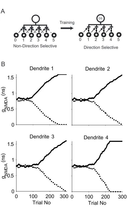
2.6 Direction-selective multiple subunits learning
How can our learning rule assure that direction selectivity in different dendritic
subunits of the host neuron is the same? To answer this question, we tested our
DS learning rule in a model cell with four direction-selective subunits on its
dendrites. The model connection scheme is shown in Fig 2-4a. Each of the
middle four LGN cells (1-4) provides delayed inhibitory input to one dendrite (1-4
from the left) of the model cell. LGN cells 0-3 each provides a left excitatory
input to dendrite 1-4 respectively. We refer to this group as the left input
connection group. LGN cells 2-5 each provide a right excitatory input to dendrite
1-4 respectively. We refer to this group as the right input connection group and to
the connections within a group as “friends” and the connections between groups
as “competitors”. The model cell initially has four potential DS subunits on four of
its eight dendrites. The learning goal is to have all members within one group
out-compete their competitors after training. If the four subunits were completely
independent and all received the exact same sequence of visual stimuli, we
would expect them to converge to the same direction selectivity. Unfortunately,
neither of these two conditions is true. Although at each trial the same moving
bar is presented to each subunit, the exact timing of the bar reaching the
receptive field of each geniculate cell is different. Both NMDA and GABA
status of subunits. The shunting inhibition mostly affects local connections but it
also has a more global effect. The same curve as in the single-unit learning case
is used in our simulations without being tailored to each subunit. These
differences can cause different branches to learn to respond to opposite
directions of motion. The model cell is thus not direction-selective . The problem
can be solved if there are internal links between group members and competition
between the groups. The links between group members indeed exist in our
model through somatic spikes. For example, the LGN cell 1’s input connection to
the dendrite 2 and the LGN cell 2’s input connection to the dendrite 3 belong to
the same left input group. In a rightward movement trial, a late spike caused by
LGN cell 1’s input will also be counted as a spike caused by LGN cell 2 given the
overlap of their input time courses. In case LGN cell 2’s connection streng th
drops below the transition threshold, this would “rescue” it from scenario two to
scenario one. The same spike, however, will not help the LGN cell 2’s connection
to dendrite 1, which belongs to the right input group. The inhibitory input to
dendrite 1 always opens before LGN cell 2 on dendrite 1 during a rightward
movement trial, and thus blocks any back-propagating spike from reaching LGN
cell 2’s connection point. So this “link forward” effect only benefits group friends
but not competitors. A similar “link backward” effect also exists. An early spike
caused by LGN cell 2 will also be registered as LGN cell 1’s own spike if it
happens within 30 ms of LGN cell 1’s firing. The “rescue” effort only occurs
during the preferred direction movement while there are no linkages among
far away from the divergent point, long consecutive same direction trials are
required to increase its connection strength above the spiking threshold. To
speed up convergence, we imposed an additional “majority” rule. We linearly
scaled the learning step size of each trial with respect to the total number of
action potentials generated at the soma during that trial. In such a setting, a
group member is increased more in its preferred direction and decreased less in
its null direction, once its group responds with more spikes than the other one.
This creates a direct competition between groups and thus facilitates
convergence.
Initially, the model cell receives balanced inputs at each of its dendrites as shown
in Fig. 2-4b and is not direction-selective. After 300 trials training, the entire left
input group wins over the right input group and the model cell develops four DS
subunits on its dendrites sensitive to rightward motion. Each direction-selective
subunit reaches its divergent point at different trials and goes through different
weight change paths. Note initially, LGN cell 2 provides excitatory input to both
dendrite 1 and 3. After training, only the connection to dendrite 3 remains.
Therefore the learning process is indeed branch specific. We carried out 100
simulations with a 0.032 ns learning step size, which was increased linearly with
the number of action potentials generated according to our majority rule. The
model cell converged to achieve uniform DS subunit structures within 200 trials in
all cases. DI equals 1 in all cases. The model converged to a right
the remaining runs.
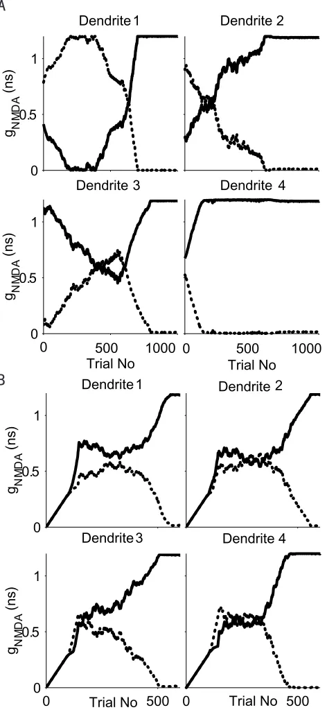
In the above stimulations, all dendrites receive balanced input from each side.
We further tested our learning model in a “random start” configuration. The total
input connection strength to a dendrite is still fixed but the relative contribution
from the left input cell and from the right input cell is randomly assigned. Such a
simulation run is shown in Fig. 2-5a. After 200 training trials, the right input group
won at dendrite one while the left input group was leading at dendrite three and
four. The competitions between the two groups were about even at dendrite two.
Because of the “majority” rule we imposed and the “link forward” and “link
backward” effect, the left input group finally increased its “friends’”connection
strength at dendrite one and two and destabilized its “competitors”. After 1000
training trials, the model cell developed four rightward motion selective subunits.
To test the stability of our learning model in the random start condition, we ran 10
simulations, each at 4 different learning step sizes: 0.1 ns, 0.032 ns, 0.01 ns and
0.003 ns. Training periods are 100 trials, 500 trials, 1000 trials and 2000 trials
respectively. DI converged to 1 for all conditions. Another possible scenario
during development is that initially all the excitatory geniculate inputs to the V1
cell are very weak and not enough to drive the cell to spike. Gradually, as the
input connections are strengthened, the cell starts to spike and competition
among input synapses begin. We tested our learning model in such a
“developmental” configuration. Initially all connection weight are zero. The
The total excitatory connection strength to a dendrite is below the desired value,
so at each trial, both inputs are increased by the maximum allowed learning step
size (Fig. 2-5b). During the initial 100 trials, there are no spikes and all the input
connections increase at each trial. Once the input connection strength reaches
about 0.3 ns, the model cell starts to spike and the connection strength of
different input groups starts to diverge. After 600 training trials, the model cell
develops four rightward motion selective subunits. To test the stability of our
learning model in the development condition, we carried out 10 simulations each
at 4 different learning step sizes. The model cell converged to DI=1 under all
conditions.
2.7 Discussion
Our learning scheme makes several testable predictions. Firstly, intra-cellular
calcium concentration changes should correlate with LTP (high calcium
concentration increase) and LDP (moderate calcium concentration increase) at
the synaptic level. Postsynaptic calcium elevation experiments using photolysis
of caged EGTA in CA1 hippocampal slices suggest such a relationship (Yang
and Zucker, 1999). Shunting inhibition can be mimicked using the dynamic
clamp (Chance and Reyes, 2002). This, together with two-photon calcium
imaging, can be used to determine if shunting inhibition indeed can direct local
synaptic modifications via local calcium concentration change. Secondly, our
“competition” rule suggests neurons with direction-selective subunit structures on
connection strengths to each of their major dendrites. Evidence for such a
mechanism, albeit operating at the whole cell level, has been provided by
Turrigiano and Nelson (1998). Local protein synthesis (reviewed by Schuman,
1997) may play a n important part in this process. In addition, we predict that cats
reared in an environment with little motion in one particular direction---achieved
by having the animals wear LCD goggles---will show a deficit in
direction-selective cells tuned for that direction relative to the opposite direction of motion.
In our learning model, random motion plays a key role in breaking the balance
between the left and right input cells. Symmetry could also be broken by a bias in
the initial connection strength between the left and right input.
Our learning scheme makes a few key assumptions. Delayed inhibition is of the
shunting type, pre-connected to fulfilling the on-path condition, and fixed. The
training stimuli move at a speed that matches the delay factor of the inhibition.
Shunting inhibition is crucial to our multiple subunits learning model in achieving
the branch specific veto of excitation and the branch specific blocking of back
propagating spikes. We compared the single-unit and the multiple subunits
model with four, six and eight subunits in the standard case with hyperpolarizing
inhibition with EGABA=-60mV - 90mV (model cell’s resting potential is -60mV). The
single-unit learning is not dependent on the shunting inhibition while the more
subunits the learning model has, the more it relies on shunti ng inhibition (for
In our simulations, the inhibitory connection strength is fixed. However, the
strength of inhibition close to the cell body may be related to calcium levels there,
as recently considered (Soto-Trevino et al., 2001). We implemented such a
global inhibitory learning scheme together with our local excitatory learning
mechanism and achieved differential excitation-inhibition learning (for details see
Chapter Six).
Fast rising calcium concentration changes in dendritic spine mediated by action
potential and long sustained rising mediated by synaptic inputs have been
observed in calcium imaging experiments (Sabatini and Svoboda, 2002). The
measured calcium decay constant is 12 ms at spines and 15 ms at small dendrites.
We used a single decay constant of 15 ms for both calcium sources and assumed
instantaneous dendrite-to-spine diffusion. We have no evidence to suggest that a
more sophisticated treatment of calcium dynamics will change our conclusion
appreciably. Experimental evidence suggests voltage-gated calcium channels
exist in spines, while little calcium diffuses between the spine and the dendritic
shaft in either directions (Sabatini and Svoboda, 2002; for a dissent view see
Majewska et al., 2000a; Majewska et al., 2000b; Holthoff et al., 2002). Such a
scheme is computationally equivalent to our model setting given we used
instantaneous dendrite to spine uni-direction calcium diffusion and single
compartment for the spine and the dendritic shaft. We choose the N-type voltage
gated calcium channel to have a voltage sensitive calcium dynamics different
gated calcium channels should also serve our purpose.
We exploit calcium gain-control mechanisms which dynamically shift the learning
curve according to the average, local activity level. The key to the stability of the
original BCM learning rule (Bienenstock et al., 1982) is a non-linear threshold
that decreases and increases faster than the average response. Such a sliding
threshold control requires that the tuning curve be narrowed for small responses
and broadened for large responses. For the sake of simplicity, we implemented a
linear sliding of the learning curve without changing its shape. The linear sliding
can be implemented in many ways, such as by an increased calcium pump within
the spine with respect to time-averaged calcium exposure or adaptation of
calcium -dependent enzyme activities. Both the duration and the amplitude of
post-synaptic calcium concentration have been shown to affect LTP and LTD
(Yang and Zucker, 1999). Brief and large postsynaptic calcium concentration
changes lead to LTP while sustained and moderate changes cause LTD.
However, brief and moderate calcium concentration change can lead to either
LTP or LTD. A calcium gain-control mechanism/sliding learning threshold can
explain such phenomena. Sustained calcium concentration elevation may shift
the learning curve as well as the LTD/LTP transition threshold toward high
calcium concentration and thus increase the probability of LTD formation.
Spike-time-dependent plasticity (STDP) is a temporal asymmetry Hebbian
pre-synaptic input and the back-propagating spike. STDP was shown in a
modeling study to automatically balance synaptic strengths and reduce the
spiking latency of the post-synaptic neuron (Song et al., 2000). A network of
neocortical neurons implementing STDP developed direction selectivity after
training (Rao and Sejnowski, 2000; Rao and Sejnowski, 2001). Our learning rules
are different from the "prediction and sequence learning" mechanism (Montague
and Sejnowski, 1994; Montague et al., 1995; Markram, 1997). In our learning
scenario one, the excitatory connection is increased if there are
back-propagating spikes within a certain period following synaptic activation; while in
learning scenario three, the excitatory connection is decreased if there is a spike
a few milliseconds before its opening and the back-propagating spike is clamped
by inhibition at opening. These fit into the general framework of the STDP with
the exception that our learning rules not only take the temporal sequence
between the input and the output into account but also the states of local
inhibition. The difference is critical for the learning of branch-specific DS subunits.
We assume the direction selectivity of V1 cells is derived from feed-forward
connections only. The learning is also restricted to feed-forward connection.
There are extensive feedback interactions among V1 cells and these feedback
currents are likely to be important for sharpening directional tuning (Douglas et
al., 1995; Maex and Orban, 1996). It will be important to see if the same learning
principle can be used to establish mutual excitation between cells selective to the
directions of motion. Such network learning may be used to generate two
direction-selective cell groups with roughly equal members during the
Chapter Three
A Detailed View of Critical Parameters and Constraints T hat
Affect the Learning Model
3.1. Introduction
We described a local synaptic learning model for local synaptic interactions
between excitation and shunting i nhibitions in the chapter two. Here we take a
detailed look at several important model parameters and additional constrains
that affect the stability of the learning model and its convergence speed. The first
such parameter is the learning step size, which directly affects the number of
training trials required to reach direction selectivity. The smaller the learning step
size, the longer the required training period. The subtractive type of “competition”
rule affects the stability of both the single -unit learning model and the
multi-subunits learning model. It helps to control the post-synaptic cell’s excitability.
“Linkages” between subunits are needed to have all subunits converge to the
same direction selectivity. The “link -back” and “link-forward” effect connect
subunits through back propagating spikes. The “majority” rule facilitates the quick
convergence of subunits. Here we compare the simulations with and without
The inhibitory input connection strengths are fixed in all previously described
simulations. In section 3.6, we propose a simply global inhibitory synapse
learning scheme that links the average spikes at soma with the inhibitory
synaptic connection strength. The goal of inhibitory learning is to achieve a
targeted average spike number given random direction motion stimuli. We tested
the inhibitory learning rule in simulations together with our excitatory synapse
learning rule. Our results show such a scheme can achieve excitation-inhibition
differential learning.
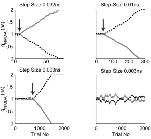
3.2 The model convergent speed and learning step size
We tested the relationship between the learning step size and the model
convergent speed (Fig. 3-1). In our learning model, the synaptic weight change at
each step is calculated from the learning curve and then scaled with learning
step size set for the simulation. The larger the learning step size, the larger
synaptic weight change after each trial. At 0.032ns (maximum allowed
conductance is 2ns), the learning model reaches divergent point with 10 trials.
About 60 trials are required to reach the divergent point when the learning step is
decreased to 0.01ns. Two simulations at learning step size 0.003ns are shown in
Figs. 3-1 C and D. The model cell reaches the divergent point around 800 trials
in one simulation, but fails to converge within 2000 trials in the other one. In our
learning model, the oscillation of synaptic weights is caused by the incoming
stimuli sequence. The smaller the learning size, the more consecutive
inputs to the divergent point. If our learning curve were a step function, such that
a synapse were increased or decreased by exactly one learning step size, we
would expect the number of consecutive same direction trials needed to be
N = (g initial synaptic weight – g divergent point)/step size
2N random trials are thus required to reach the divergent point. The actual
situation in our model is more complicated because we use a continuous BCM
type learning curve. The synaptic weight changes at each trial are not exactly
one learning step, but we would expect the number of training trials needed to
reach the divergent point still scales non-linearly with respect to the learning step
size. For simulations shown in Fig. 3-1, 10, 60, 800 trials are needed to reach the
divergent point for learning step size 0.032ns, 0.01ns, 0.003ns, not far from the
8-fold (23) increase in trial length for the every 3 fold decrease in the step size.
The learning model converges much faster (only N trials are required), if the
stimuli are same direction motions. The bursting spiking waves discovered in
developing mammalian retina (Meister et al, 1991; Wong et al, 1993) may be
equivalent to such same direction stimuli.
3.3 The “competition” rule
The Hebbian learning rule is a positive feedback learning rule. Correlated input
connections are increased and uncorrelated input connections are decreased
during the training process. Such a learning rule causes fluctuations in the
excitability of the post synaptic cell. Various constraints have been proposed to
use a subtractive type constraint in our learning model. In each trial, the total
excitatory input connection strength to each dendrite is held constant. Half the
amount that is over/under the targeted value after learning is then subtracted
from/added to all synapses on that dendrite. This has the benefit of creating a
direct competition between synapses on a dendrite while keeping the total
excitability of the model cell constant during simulations.
We first investigated the “competition” rule’s effect on the single -unit learning
model. Synaptic weight changes for several simulation runs without imposing
such a “competition” rule are shown in Fig. 3-2. In our learning model, a synapse
can only be rewarded if there is a back propagating spike. Simulations show
both connections gradually decrease toward zero during training if neither
connection’s initial strength is large enough to cause a spike (Fig. 3-2A). If both
initial connections are well above the spiking threshold, the learning model is
stable and became direction-selective after training as shown in Fig. 3-2B.
Although we did not impose the “competition” rule in this case, we still limited the
highest synaptic conductance a synapse can reach to 2ns and the lowest to zero.
Another simulation run with both initial connection strengths just above the
spiking threshold is shown in Fig. 3-2C. To test the stability of our learning model
in the same condition as Fig. 3-2C, that is, with initial values strong enough to
elicit spikes, we ran 10 simulations each at 4 different learning step sizes: 0.1ns,
0.032 ns, 0.01 ns and 0.003 ns. The training length for each step size was 100
completed, a leftward movement trial a nd a rightward movement trial were
presented to the model cell and the resulting spike numbers were recorded. The
cell converged to DI=1 i n all trials as shown in Fig. 3-2D.
We then tested our multi-subunits learning model without imposing the
“competition” rule. The challenge facing the multi-subunits model is to coordinate
the learning between subunits. We used the “random start” initial condition to put
different subunits into different domain of direction selectivity initially and then
tested the model’s ability to converge without the “competition” rule. In most
cases, the model cell failed to become direction-selective after training as shown
in Fig. 3-3A. Note again, in the multi-subunits model, the competition rule was
imposed onto each dendrite. We ran 10 simulations each at 4 different learning
step sizes: 0.1 ns, 0.032 ns, 0.01 ns and 0.003 ns. Model cell’s average DI was
close to 0 after trainings. Synaptic weight changes for the left input cells and the
right input cells at each dendrite during one simulation run are shown in Fig. 3-3B.
Initially, dendrite one is not direction-selective . Dendrite two and three a re
rightward motion selective while dendrite four is leftward motion selective. After
training, connections to dendrite one and four all drop below the spiking threshold
while connections to dendrite two and three all reaches maximum. The model
cell is thus not direction-selective.
Our results suggest both the single -unit and the multi-subunits learning model
without the “competition” rule if either initial connection strength is above the
spiking threshold but not if both initial connections are weak. This is expected
given that we do not implement a sliding threshold that scales non-linearly with
respect to the average response of the cell as in the original BCM model. Our
linear sliding threshold and the “competition” constraints are easy for real
neurons to imple ment than a non-linear sliding threshold.
3.4 Linkages between subunits
The model cell initially has four potential DS subunits on its dendrites. The
learning goal is to have all members of one group win over their competitors after
training. If the four subunits are completely independent, all receive the exact
same sequence of visual stimuli and have the same initial condition, we would
expect them to converge to the same direction selectivity. Unfortunately, neither
of these conditions is true. Although at each trial the same moving bar is
presented to each subunit, the exact timing of the bar reaching the receptive field
of each excitatory input is different. Both the NMDA synapses and the GABA
synapses we use in simulations have long off ramps. Their late currents cause
difference in the status of subunits. The shunting inhibition mostly affects local
connections but it also had some global effects. We do not tailor our learning
curve to each subunit but rather use the same curve as in the single-unit learning
case. In addition, we would expect different subunits to receive different stimuli in
a natural setting. Therefore the difference in subunits can cause different
not direction-selective. The problem would be solved if there are internal links
between group members and there is competition between the groups. The link
between group members indeed exists in our model through back-propagating
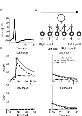
spikes. We first considered the “link back” effect as show in Fig. 3-4. The LGN
cell 2’s inp ut connection to the dendrite 3 (left input I) and the LGN cell 3’s input
connection to the dendrite 4 (left input II) belong to the same “left input group”.
LGN cell 2’s input connection to dendrite 1 (right input I) and LGN cell 3’s input
connection to dendrite 2 (right input II) belong to the same “right input group” (Fig
3-4A). Synaptic calcium concentration changes during a rightward movement trial
for all four synapses are shown in Fig. 3-4B. LGN cell 2 spikes at time zero while
LGN cell 3 spikes 15ms later. Left input 1’s connection strength is below the
spiking threshold. In a rightward movement trial, this corresponds to scenario 2
for the left input I: no inhibition, no back propagating spike and the connection
strength should be further weakened after the trial. However, 15 ms after left
input I’s opening, left input II opens . Left input II causes a back propagating spike
which also propagates back close to left input I’s connection site (dendrite 3).
This switches left input I from scenario 2 to scenario 1. The connection strength
of left input I increases after the trial instead of decreases. The same spike,
however, does not affect right input I and right input II. The inhibitory synapses
on dendrite 1 and 2 opens before those two right input connections and thus
blocks the back-propagating spike. So this “link forward” effect only benefits
inputs (adjacent in the visual space) in our model given the various time course
settings.
A similar “link forward” effect also exists in our model as shown in Fig. 3-5. In this
case the LGN cell’s firing rate is elevated to make them spike multiple times
during a bar sweep. The connection strength of left input I and left input II a re
reversed. Left input I now causes the model cell to spike twice during its opening
while left input II alone is too weak to cause a spike. During a rightward
movement trial, the second spike caused by left input I “rescues” left input II. The
spike does not cause either right input I or right input II to increase their
connection strength because the inhibitory connections to dendrite 1 and 2
blocks the back-propagating spike. However, if the LGN cell array’s firing
freque ncy is held low, the left input I can only cause one spike during its opening.
In our model setting, such a spike occurs before left i nput II’s opening and thus
can not help increase left input II (Fig. 3-6). In the real case, we expect overlap
between each LGN cell’s receptive fields. The noise and jitter in spike timing can
also help create such a linkage.
3.5 The “majority” rule
The “rescue” effort mentioned above only happens during the preferred direction
movement for a group and there are no links between group members during the
null direction movement. If a group member is stuck far away from the transition