EXPLORING AND HARNESSING PEG-IMMUNE SYSTEM INTERACTIONS TO ENGINEER TARGETED STEALTH NANOPARTICLES
Qi Yang
A dissertation submitted to the faculty at the University of North Carolina at Chapel Hill in partial fulfillment of the requirements for the degree of Doctor of Philosophy in the Department
of Pharmaceutical Sciences in the School of Pharmacy.
Chapel Hill 2016
Approved by: Samuel K. Lai Leaf Huang
ii
© 2016 Qi Yang
iii
ABSTRACT
Qi Yang: Exploring and harnessing PEG-immune system interactions to engineer targeted stealth nanoparticles
(Under the direction of Samuel K. Lai)
iv
v
ACKNOWLEDGEMENTS
Completion of this work was made possible through the contributions and support of many people. First, I would like to thank my advisor, Dr. Samuel Lai, who has offered a deftly balanced mixture of freedom and guidance as I pursued my Ph.D. and has helped to nurture and push me as a scientist. Next, I would like to acknowledge Dr. Leaf Huang, who, in addition to serving as the chair of my committee, gave me an opportunity to work in his lab as an
undergraduate and served as my first research mentor, for which I am truly grateful. I would also like to thank my other committee members Dr. Steven Park, Dr. Rudolph Juliano, and Dr. Andrew Wang for their time, advice, and feedback. Special thanks go to the UNC small animal imaging, flow cytometry, and animal studies cores, as well as to all of the other labs in
vi
TABLE OF CONTENTS
LIST OF TABLES... xi
LIST OF FIGURES... xii
LIST OF ABBREVIATIONS... xiv
CHAPTER 1: INTRODUCTION... 1
1.1 Nanoparticles and MPS clearance... 1
1.2 Nanoparticle PEGylation... 2
1.3 Humoral immune responses... 3
1.4 Nanoparticle drug delivery to tumors... 6
1.5 Thesis overview... 7
CHAPTER 2: BACKGROUND ON HUMORAL ANTI-PEG IMMUNITY... 9
2.1 Introduction... 9
2.1.1 Advantages and physicochemical properties of effective stealth PEGylation...10
2.2 PEG-specific immunity in animal models... 12
2.2.1 The first report of anti-PEG Abs in vivo... 12
2.2.2 Accelerated blood clearance of PEGylated systems is attributed to anti-PEG Abs... 13
2.2.3 Immunological mechanism(s) of anti-PEG Ab induction... 17
2.2.4 Properties of the anti-PEG Ab epitope... 19
vii
2.3 Anti-PEG immunity in humans... 22
2.3.1 Pre-existing anti-PEG Abs in the general population... 22
2.3.2 Induction and effects of anti-PEG Abs in individuals treated with PEGylated therapeutics... 24
2.3.3 Clinical implications of and strategies to overcome anti-PEG Abs... 28
2.4 Ongoing questions regarding anti-PEG antibodies... 30
2.5 Detection of anti-PEG Abs by validated ELISA methods... 32
2.6 Conclusions... 34
CHAPTER 3: BACKGROUND ON HETEROGENEOUS TUMOR TREATMENT AND BISPECIFIC PROTEIN-MEDIATED PRETARGETED DRUG DELIVERY... 40
3.1 Introduction... 40
3.2 Conventional cancer targeting strategies: passive targeting... 42
3.3 Conventional cancer targeting strategies: active targeting... 44
3.4 Tumor heterogeneity and implications for targeted drug delivery systems... 46
3.5 Pretargeted radioimmunotherapy (PRIT)... 50
3.6 Pretargeted drug delivery to heterogeneous tumors...53
3.7 Biological and pharmaceutical aspects and considerations of pretargeted drug delivery... 55
3.7.1 Binding pairs... 56
3.7.2 Target antigen(s) ... 60
3.7.3 Pharmacokinetics and biodistribution... 62
3.8 Challenges and unknowns...64
3.9 Conclusion... 66
viii
4.1 Introduction... 68
4.2 Materials and methods... 70
4.2.1 PS-PEG synthesis and characterization... 70
4.2.2 Direct fluorescent quantification of the PEG coating density... 71
4.2.3 PEG coating density quantification by PDAM assay... 72
4.2.4 PEG conformational regime calculations... 73
4.2.5 THP-1 culture and uptake assay... 73
4.2.6 Primary human leukocyte culture and uptake assay... 74
4.2.7 Intravital imaging of particle circulation... 75
4.2.8 PS-PEG biodistribution... 75
4.2.9 Extended circulation and biodistribution of densely PEGylated particles... 76
4.2.10 Statistical analysis... 76
4.3 Results... 77
4.3.1 Synthesis and characterization of PS-PEG nanoparticles... 77
4.3.2 Influence of PEG coating characteristics on particle uptake by cultured macrophage cells... 79
4.3.3 Influence of PEG coating characteristics on particle uptake by primary human peripheral leukocytes... 81
4.3.4 Influence of PEG coating characteristics on particle circulation kinetics in vivo... 83
4.4 Discussion... 85
4.5 Conclusions... 89
CHAPTER 5: ANALYSIS OF PRE-EXISTING ANTI-PEG ANTIBODIES IN THE GENERAL POPULATION... 92
ix
5.2 Materials and methods... 94
5.2.1 Human plasma and serum samples... 94
5.2.2 Chimeric anti-PEG antibody standards... 95
5.2.3 Anti-PEG Ab ELISA... 96
5.2.4 Human antibody isotyping quantification... 97
5.2.5 Statistical analyses... 98
5.3 Results... 98
5.3.1 Validation and specificity of ELISA assays for measuring anti-PEG Ab levels... 98
5.3.2 Anti-PEG Ab levels in the contemporary population... 101
5.3.3 Anti-PEG Ab levels in historical samples... 108
5.4 Discussion... 113
5.5 Conclusions... 117
CHAPTER 6: PRETARGETING WITH BISPECIFIC FUSION PROTEINS TO FACILITATE DELIVERY OF NANOPARTICLES TO MOLECULARLY DISTINCT TUMORS... 121
6.1 Introduction... 121
6.2 Materials and methods... 124
6.2.1 Preparation and characterization of PS-PEG-biotin nanoparticles... 124
6.2.2 Cell culture and cell uptake assay... 125
6.2.3 Pharmacokinetics and biodistribution of biotinylated nanoparticles... 126
6.2.4 Biodistribution of pre-targeted nanoparticles in mouse models containing single and dual tumors... 126
6.2.5 Statistical analysis... 127
x
6.3.1 Synthesis and characterization of PS-PEG-biotin...127
6.3.2 Pretargeted delivery of PS-PEG-biotin nanoparticles in vitro... 129
6.3.3 PS-PEG-biotin circulation kinetics and tissue biodistribution...131
6.3.4 Biodistribution and tumor accumulation of pretargeted PS-PEG-biotin nanoparticles in single tumor mouse model…...132
6.3.5 Biodistribution and tumor accumulation of pretargeted PS-PEG-biotin nanoparticles in dual tumor mouse model... 133
6.4 Discussion... 135
6.5 Conclusions... 139
CHAPTER 7: CONCLUSIONS AND PERSPECTIVES... 141
xi
LIST OF TABLES
Table 2.1 Examples of anti-PEG antibodies and/or accelerated blood
clearance responses to PEGylated systems... 35
Table 2.2 Human studies and clinical trials demonstrating anti-PEG Ab responses... 38
Table 4.1 PS-PEG density, hydrodynamic diameter, ζ-potential, and theoretical RF/D and PEG conformation values...90
Table 4.2 PK model and parameters for various PS-PEG5 kDa particles ... 91
Table 5.1 Summary of patient demographics for contemporary and historical samples... 118
Table 5.2 ELISA assay details for anti-PEG IgG1, IgG2, IgG3, IgG4, and IgM... 118
Table 5.3. Binding kinetics of chimeric anti-PEG IgG and IgM... 119
Table 5.4 Prevalence of anti-PEG IgG and IgM in contemporary human plasma samples... 119
Table 5.5 Prevalence of anti-PEG IgG1-4 in contemporary human plasma samples... 119
xii
LIST OF FIGURES
Figure 1.1 Antibody structure... 5
Figure 2.1 Impact of PEG density and conformation... 12
Figure 2.2 Accelerated blood clearance and anti-PEG antibodies in animal models... 16
Figure 2.3 Proposed type-2 T-cell independent (TI-2) response mechanism for the formation of anti-PEG Abs and the ABC effect...19
Figure 2.4. Preliminary studies of mouse anti-PEG IgG and IgM binding to poly(methacrylate [P(OEG300)]...21
Figure 2.5 Anti-PEG antibodies in human patients... 28
Figure 2.6 ELISA methods for detection of anti-PEG antibodies... 33
Figure 3.1 Strategies for the delivery of nanoparticle drug carriers and/or radioisotopes to tumor cells... 42
Figure 3.2 Different types of tumor heterogeneity... 49
Figure 3.3 Clinical tumor heterogeneity... 50
Figure 3.4 Pretargeted delivery of nanoparticles (NPs) to heterogeneous tumors... 54
Figure 3.5 Diagnostic magnetic resonance profiling of human tumor cell lines, fibroblasts, and leukocytes using a pretargeted approach in vitro...55
Figure 3.6 Internalization of pretargeted single-walled carbon nanotubes... 62
Figure 4.1 Direct and indirect characterization of PS-PEG density... 78
Figure 4.2 Differentiated THP-1 cell uptake of PS-PEG beads...80
Figure 4.3 Phase diagram mapping particle uptake by differentiated THP-1 cells... 81
Figure 4.4 Primary human peripheral blood leukocyte uptake of PS-PEG beads... 82
Figure 4.5 Circulation and biodistribution of PS-PEG beads... 84
xiii
Figure 5.1 Confirmation of anti-PEG ELISA specificity... 100
Figure 5.2 Anti-PEG IgG and IgM in the general population... 103
Figure 5.3 Anti-PEG IgG1-4 in the general population... 104
Figure 5.4 Anti-PEG IgM, IgG, IgG1, and IgG2 levels in healthy individuals by age group, gender, and race...105
Figure 5.5 Relationship between anti-PEG Ab levels and age. ... 106
Figure 5.6 Anti-PEG IgM, IgG, IgG1, and IgG2 prevalence in healthy individuals by age group, gender, and race...107
Figure 5.7. Anti-PEG IgG and IgM in historical samples... 109
Figure 5.8 Anti-PEG IgG1-4 levels in historical samples... 110
Figure 5.9 Anti-PEG IgG1-4 prevalence in historical samples... 111
Figure 5.10. Total antibody levels in contemporary and historical samples... 112
Figure 6.1. Pretargeted nanoaparticle delivery to a heterogeneous population of lymphoma cells... 123
Figure 6.2 PS-PEG-biotin nanoparticle characterization... 128
Figure 6.3 Dot blot confirming the relative biotin density on PS-COOH, PS-PEG, and PS-PEG-biotin beads... 129
Figure 6.4 Pretargeted nanoparticle delivery to B- and T-cell lymphomas in vitro... 130
Figure 6.5 Circulation kinetics and tissue biodistribution of fully biotinylated nanoparticles... 132
Figure 6.6 Organ biodistribution of pretargeted PS-PEG-biotin nanoparticles in single tumor-bearing mice... 133
xiv
LIST OF ABBREVIATONS
Ab antibody
ABC accelerated blood clearance
Ag antigen
ANOVA analysis of variance APC allophycocyanin ASNase asparaginase
AUC area under the curve BsP bispecific protein BSA bovine serum albumin CA clearing agent
CDR complementarity-determining region CI confidence interval
COOH carboxyl
D PEG grafting density
DSPE 1,2-distearoyl-sn-glycero-3-phosphoethanolamine EDC 1-ethyl-3-(3-dimethylaminopropyl)carbodiimide ELISA enzyme-linked immunosorbent assay
EPR enhanced permeability and retention FP fusion protein
xv
HCV hepatitis C virus HRP horseradish peroxidase ID/g injected dose per gram
IFN interferon
IHC immunohistochemistry i.m. intramuscular
i.v. intravenous IVIM intravital imaging MAb monoclonal antibody
MPS mononuclear phagocyte system
MW molecular weight
MYO myoglobin
NH2 amine
NHL non-Hodgkin’s lymphoma
NP nanoparticle
PAL phenylalanine ammonia lyase PDAM 1-pyrenylyldiazomethane PEG polyethylene glycol
PK pharmacokinetics
PL phospholipid
PRIT pretargeted radioimmunotherapy
PS polystyrene
xvi
RIT radioimmunotherapy S-NHS N-hydroxysulfosuccinimide
SA streptavidin
s.c. subcutaneous
scFv single-chain variable fragment TCO trans-cyclooctene
TI T cell-independent
TMB 3,3',5,5'-tetramethylbenzidine
Tz tetrazine
1
CHAPTER 1: INTRODUCTION 1.1 Nanoparticle and MPS clearance
The application of nanotechnology to delivery of therapeutic and/or diagnostic agents has become an important area of pharmaceutical science, with several nanoparticle formulations already on the market and many more in development [1]. Nanoparticles are commonly defined as being 1-100 nm in diameter, though submicron-sized particles are often also included in this category. Nanoparticles can protect their cargo from the biological microenvironment, alter drug solubility, and provide controlled drug release. Furthermore, owing to their unique
physicochemical properties (e.g., size, shape, surface charge), nanoparticle formulations can also significantly improve an encapsulated drug’s biodistribution and bioavailability, and
consequently its pharmacokinetic and pharmacodynamics profile, compared to free drug [2]. In addition, by reducing the level of free drug in the blood, nanoparticles can reduce toxicity associated with drug accumulation in nontarget tissue and broaden the drug’s therapeutic window. It is important to note, however, that the use of nanoparticles can also introduce new problems. For example, liposomal formulations of doxorubicin exhibit reduced cardiotoxicity compared to the free drug but increase the likelihood of side effects such as hand-foot syndrome [3].
2
foreign materials, including bacteria, viruses, and fungi. Similar to these naturally occurring particles, nanoparticles can be rapidly opsonized by adsorption of plasma proteins such as serum albumin, complement factors, apolipoproteins, and immunoglobulins, generating an abundant protein corona that marks the nanoparticles for receptor-mediated phagocytosis by MPS cells [4]. The process of opsonization and sequestration by MPS cells is extremely efficient: unmodified nanoparticles can be fully eliminated from systemic circulation within minutes [5, 6].
1.2 Nanoparticle PEGylation
Surface modification of nanoparticles with polyethylene glycol (PEG) was first introduced in the 1990s to reduce the rapid clearance of nanoparticles by MPS cells [5]. Currently, PEG remains the most popular compound within the genre of so-called “stealth” polymers—polymers that, when grafted on the surface of nanoparticles, can reduce their
opsonization and subsequent immune cell-mediated clearance. In the case of PEG, these effects are largely due to its hydrophilicity and high flexibility, which generates a thick, amorphous hydration shell that repels nonspecific protein adsorption and improves colloidal stability [4, 6, 7]. Thus, modification with PEG (i.e., PEGylation) can significantly prolong nanoparticle circulation times in the blood, increasing the half-life of nanoparticles to several hours or even days [8, 9].
Although PEGylation is a frequently exploited nanoparticle modification strategy, the extent to which PEGylation improves nanoparticle circulation times remains highly variable [9]. The diverse classes of nanoparticle systems (e.g., liposomal, micellar, metallic, polymeric, silica, and carbon nanoparticles) have necessitated the development of a variety of PEGylation
3
adsorption of PEG-containing surfactants, and particle formulation using PEG copolymers. Nanoparticle composition and formulation processes not only affect the PEGylation method(s) that can be used but also introduces different limitations on the density of PEG grafting that can be achieved. As a result, PEG coating characteristics (e.g., PEG density, PEG MW, potential PEG shedding) can vary greatly. An additional complicating factor is the lack of methods to accurately measure the extent of PEG grafting on most nanoparticles. Overall, the precise characteristics of PEG coatings that influence opsonization and clearance of PEG-coated nanoparticles by MPS cells remain not well-understood.
1.3 Humoral immune responses
Adaptive immunity is critical for immune defense against reinfection by foreign
pathogens. In contrast to innate immune responses that rely on recognition of shared features of pathogens, adaptive immune responses are mediated by immune cells and humoral
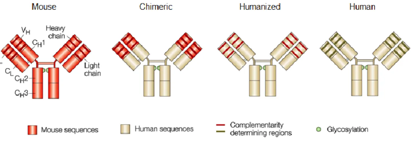
macromolecules, particularly antibodies, that can bind specific epitopes on individual foreign molecules (i.e., antigens). Antibodies are Y-shaped proteins with complementarity-determining regions (CDRs) at the ends of their two arms that can bind target epitopes (Fig. 1.1).
4
elimination of infected cells through antibody-dependent cell-mediated cytotoxicity. Generally, antibodies are produced by B cells after activation by helper T cells, although certain antigens (i.e., T cell-independent antigens) are able to activate B cell antibody production without stimulation from T cells. Upon initial exposure to an antigen, IgM antibodies are produced after a lag phase of 1-2 weeks, typically followed by a peak of IgG antibodies as the differentiated B cells undergo class-switching. Long-lived plasma cells and memory B cells generated after the primary response can result in more rapid secondary antibody responses, which are
predominantly IgG. However, the features of induced antibody responses can vary depending on the specific antigen, dose, route of exposure, and individual host genetics and environmental exposure [11]. For example, IgG1 (~60% of all IgG) is typically associated with responses to protein antigens and memory B cell induction, whereas IgG2 (~30% of all IgG) is commonly induced by polysaccharide antigens, often in a T cell-independent manner that may not generate a meaningful memory response [12].
5
completely abrogate drug efficacy, but also because they can induce serious, potentially life-threatening, side effects such as anaphylactic and hypersensitivity reactions [16-18].
Antibodies can potentially form against foreign components of nanoparticle carriers, including PEG. Although PEG was long assumed to be immune inert due to its ability to resist protein binding, growing evidence indicates that both animal subjects and human patients treated with PEGylated drugs can develop antibodies to PEG, leading to a marked reduction in the efficacy of PEGylated systems and/or unexpected anti-PEG antibody-associated side effects [19, 20]. Interestingly, PEG-specific antibodies have even been observed among “treatment-naïve” donors [21, 22]. Unfortunately, the overall prevalence, systemic concentration, and potential clinical impact of these anti-PEG antibodies is currently unclear. A thorough characterization of anti-PEG antibodies among the general population will likely have important implications for the future clinical use of the many PEGylated drugs on the market and in development.
Figure 1.1. Antibody structure. Antibodies are Y-shaped macromolecules composed of two heavy and two light chains. Within these chains, the variable heavy and light domains (VH and VL, respectively)
contain the complementarity-determining regions (CDRs) responsible for antigen binding, and the effector functions of the antibody are determined by the constant domains (CL, CH1, CH2, and CH3), in
particular the CH2 and CH3 domains, which comprise the Fc region that is recognized by complement
proteins and various receptors on immune cells. Chimeric antibodies contain nonhuman VH and VL
6
1.4 Nanoparticle drug delivery to tumors
The application of nanomedicine to oncology has been largely driven by the unique properties of tumor tissues. Once tumors develop beyond a critical size, typically 1-2 mm2, angiogenesis is required to supply adequate nutrients to support further tumor growth [24]. This neovascularization is induced through the release of vascular endothelial growth factor (VEGF) and other proangiogenic factors by the tumor cells. However, the unbalanced secretion of these factors, results in abnormal and heterogeneous vessels with irregular function and structure, characterized by fenestrations in the endothelium that range from hundreds of nanometers to several micrometers in size [25, 26]. Combined with a lack of adequate draining lymphatic vessel formation for most tumors, the “leaky” vasculature allows the preferential extravasation and accumulation of macromolecules and nanoscale materials in tumor tissues, compared to normal tissue with tight endothelial vessel linings. This phenomenon, termed the enhanced permeability and retention (EPR) effect, is the basis for “passive” targeting of tumors using nanoparticles, as any sufficiently long-circulating nanoparticle smaller than the tumor vessel fenestrations should be able to exploit the EPR effect to accumulate in tumor tissues [27].
7
optimized for each nanoparticle formulation to balance the stealth properties imparted by PEGylation and the ability to specifically target cancer cells.
1.5 Thesis overview
In this thesis, my goal is to provide a blueprint for the engineering of nanoparticle systems that can better target specific cells and tissues by rigorously characterizing the
interactions of PEGylated nanoparticles with cells and antibodies of the immune system. This goal is divided into the following three aims:
Aim 1: Elucidate interactions between PEGylated nanoparticles and innate immune
system. I synthesized a series of PEGylated nanoparticles with carefully tuned physicochemical properties comprised of a range of PEG molecular weights and densities. Next, I developed an indirect fluorogenic probe-based assay to quantify the number of PEG groups conjugated onto the nanoparticles. I further assessed the influence of PEG MW and grafting density on
nanoparticle uptake by cultured human macrophage-like cells and primary human leukocytes using flow cytometry and monitored the systemic circulation time of various PEGylated nanoparticles in BALB/c mice using intravital imaging and pharmacokinetics analysis.
Aim2: Investigate human adaptive immune responses against PEG (anti-PEG
antibodies). To enable quantitative analysis of pre-existing anti-PEG antibody responses in humans, I engineered a variety of chimeric anti-PEG monoclonal Ab standards. Using these standards in combination with rigorously validated competitive ELISAs, I was able to quantify the levels of anti-PEG IgM and different subclasses of anti-PEG IgG (IgG1-4) in both
8
Aim 3: Evaluate the use of a pretargeting strategy for nanoparticle delivery to
9
CHAPTER 2: BACKGROUND ON HUMORAL ANTI-PEG IMMUNITY* 2.1 Introduction
Extended circulation of proteins and nanoparticle therapeutics is often necessary to achieve adequate drug concentrations in target tissues [9, 28, 29]. Unfortunately, many peptide and protein drugs are rapidly degraded and/or cleared from the systemic circulation due to their small size [30], and nanoparticulate drug carriers are readily eliminated by the cells of the mononuclear phagocyte system (MPS) [9, 31]. To overcome these challenges, proteins and nanoparticles are frequently conjugated to various hydrophilic polymers, which can significantly reduce degradation and opsonization, consequently extending the circulation half-lives of the modified therapeutics [28, 32]. These polymers are frequently referred to as “stealth” polymers, reflective of their ability to render proteins and particles inert to the biological environment.
Polyethylene glycol (PEG) has been, and continues to be, the most widely used stealth polymer in drug delivery, with over a dozen PEGylated pharmaceuticals currently on the market and many more in clinical testing [9, 29]. PEG has a long history of safe use in humans, and the polymer is classified under the Generally Recognized As Safe (GRAS) category by the FDA. Despite the frequent use of PEG to extend circulation kinetics, a number of investigators have observed the rapid clearance of some PEGylated systems upon repeated administration [19, 33]. This “accelerated blood clearance” phenomenon was ultimately attributed to the formation of PEG-specific antibodies [34]. Indeed, animals that receive repeated doses of PEGylated systems often generate a potent IgM antibody response to PEG, which causes the complete elimination of
10
subsequent doses of PEGylated agents from the circulation within minutes to a few hours [19]. The induction of anti-PEG antibodies (anti-PEG Abs) in humans was also observed in recent clinical trials of PEGylated proteins and has been correlated with poor drug efficacy [35, 36]. Interestingly, there is emerging evidence that anti-PEG Abs can be found in the general population in individuals who likely have never received PEGylated therapeutics injected systemically [21, 37]. As many more PEGylated protein and nanoparticle therapeutics are expected to enter the market over the next several years, an improved understanding of the prevalence, induction, and effects of anti-PEG immunity is undoubtedly critical for the continued clinical use of PEGylated systems.
2.1.1 Advantages and physicochemical properties of effective stealth PEGylation
The stealth properties of PEG are rooted in several distinctive molecular and physical characteristics. First, PEG is exceedingly hydrophilic, with each ethylene glycol subunit (-CH3
-CH3-O-) surrounded by a minimum of 2-3 water molecules [38, 39]. Thus, PEG coatings
generate a hydration shell with a large excluded volume that sterically prevents
biomacromolecules from penetrating into the polymer layer and binding to the underlying core via hydrophobic or electrostatic interactions [7, 40, 41]. Second, PEG is highly flexible and exhibits high chain mobility, which results in an exceedingly large number of polymer chain conformations. As a result, any substantial reduction in the conformational freedom of PEG, including the displacement of PEG chains by intruding biomacromolecules, is
11
For proteins, PEG conjugation decreases enzymatic degradation, opsonization, and immunogenicity of the protein core [30]; PEGylation can also improve stability and solubility [29]. Additionally, the resulting increase in the hydrodynamic diameter can reduce renal elimination and improve the biodistribution and pharmacokinetics of PEGylated proteins [30]. For nanocarriers, PEGylation reduces opsonization and MPS cell clearance, resulting in significantly prolonged circulation kinetics [31, 32]. For oncological applications, this effect often leads to greater tumor distribution via the enhanced permeability and retention (EPR) effect, while decreasing accumulation in non-targeted organs [9]. PEGylation can also improve nanocarrier stability and minimize the premature release of cargo therapeutics. Finally, PEG coatings have been shown to decrease nanocarrier association with structural components of mucus and extracellular matrix, thereby improving distribution and delivery to regions such as mucosal surfaces and brain tissues [45, 46].
Naturally, the effectiveness of PEG as a nanoparticle coating polymer is critically dependent on the density and resulting conformations assumed by conjugated PEG chains. The thickness of the PEG coating is dictated by its Flory radius (RF; a function of the molecular
weight) and the distance between two neighboring PEG chains (D; a function of the PEG coating density) [47]. When neighboring PEG chains are sparsely packed and do not overlap, PEG occupies a diffuse volume generally termed a “mushroom” conformation (RF/D ≤ 1). As more
PEG polymers are introduced, the excluded volume and repulsion by neighboring PEG chains cause the polymer to transition from a diffuse conformation to a more extended “brush”
conformation (RF/D > 1) [4, 9], eventually reaching a “dense brush” regime, where the height of
the PEG layer exceeds the RF by at least two-fold (at RF/D > 2.8) (Fig. 2.1a) [43, 48, 49]. The
12
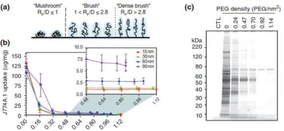
begins to exhibit stealth polymer functions. However, we and others have recently found that both rigid polymeric and metallic nanoparticles require PEG grafting densities far exceeding the minimum for brush conformation to demonstrate effective stealth nanoparticle behavior [49, 50]. Indeed, maximal reduction of uptake by mouse and human phagocytes in vitro required at least a dense brush PEG coating (Fig. 2.1b), and PEG grafting densities extending well into the dense brush conformation were necessary for the evasion of serum protein adsorption (Fig. 2.1c), as well as to achieve sustained circulation in vivo.
Figure 2.1. Impact of PEG density and conformation. a) The conformation adopted by PEG chains at various grafting densities. At low grafting densities (RF /D ≤ 1), the PEG chains adopt a diffuse
“mushroom” conformation. At higher densities, the PEG chains are increasingly able to repel
opsonization and cell uptake as they transition into a more extended “brush” conformation (RF /D > 1)
and eventually reach a “dense brush” regime (RF/D > 2.8). b) Uptake of PEG5k-grafted gold NPs by
mouse J774A.1 macrophage-like cells. A PEG5k density of 0.16 PEG/nm 2
corresponds to brush
conformation; all other PEG densities correspond to dense brush conformation. c) Qualitative analysis of the serum proteins adsorbed onto 30 nm gold NPs modified with varying amounts of PEG5k. A PEG5k
density of 0.24 PEG/nm2 corresponds to brush conformation; all other PEG densities correspond to dense brush conformation. Panels B and C were reprinted with permission from Ref [50] (copyright 2012 ACS), and panel A was adapted with permission from Ref [49] (copyright 2014 ACS).
2.2 PEG-specific immunity in animal models
2.2.1 The first report of anti-PEG Abs in vivo
13
convenient and intuitive to assume that PEG should be immunologically inert and escape binding by antibodies. However, in 1983, less than a decade after the introduction of protein PEGylation, Richter and Akerblom reported the generation of PEG-specific antibodies following
intramuscular (i.m.) or subcutaneous (s.c.) injections of various PEG-modified proteins in Complete Freund’s Adjuvant [51]. In contrast, they found that free PEG (MW 10-5.9 x 10³
kDa) administered under similar conditions exhibited little to no immunogenicity. This landmark study demonstrated for the first time that antibodies can be formed against PEG polymers. Later
studies confirmed that not only can anti-PEG Abs be elicited by immunization with PEGylated proteins [52, 53], but also that the induction of PEG-specific immunity can occur in the absence of adjuvants [54, 55].
2.2.2 Accelerated blood clearance of PEGylated systems is attributed to anti-PEG Abs
While single doses of PEGylated therapeutics often demonstrate extended system circulation times in vivo, some PEGylated systems exhibit rapid elimination upon repeated administration. For example, Moghimi and Gray reported in 1997 that when long-circulating polystyrene particles coated with poloxamine 908 (a PEG-containing surfactant) were administered 3-4 days after an initial dose, the particles were swiftly cleared from systemic circulation by MPS cells in rats [56]. Similarly, Dams et al. and several other groups observed that repeated weekly dosing of empty PEG liposomes also significantly reduced the circulating half-lives of the subsequent doses (Fig. 2.2a), with a corresponding increase in liver
accumulation and hepatic clearance (Fig. 2.2b), as well as moderate increases in splenic
14
unclear for a number of years after its discovery. Because the infusion of “naïve” mice with plasma from animals pre-dosed with poloxamine-coated polystyrene beads failed to generate an ABC effect, Moghimi and Gray suggested that the observed phenomenon was not due to plasma factors but rather potentially resulted from an change in phagocyte receptor expression and/or activity elicited by the initial particle dose [56]. In contrast, Dams et al. reported that the
transfusion of blood or serum from rats pre-treated with PEGylated liposomes generated an ABC effect and observed that this effect was dependent on the presence of a heat-labile, 150-kDa serum factor. Because ABC was observed for serum depleted of IgG or IgM, they proposed that the observed effect was likely due to complement protein(s) [33]. However, because the extent of IgM depletion appeared incomplete, the involvement of residual IgM could not be discounted. Although Laverman et al. did not identify the specific immune factors responsible, they observed that the ABC phenomenon occurs in two phases: the induction phase, when the immune system is primed by the initial injection, and the effectuation phase, when the pharmacokinetics and biodistribution of the PEGylated therapeutics are affected by the resulting immune response [60].
15
into naïve mice resulted in the rapid clearance of PEGylated therapeutic proteins (e.g., 38-fold reduction in systemic concentration compared to uninjected control) [55, 64]. Numerous other groups have subsequently corroborated the relationship between anti-PEG Abs and the ABC phenomenon [65-70]. The observed anti-PEG Ab response is predominantly IgM [34, 69, 71, 72], although the development of anti-PEG IgG has also been reported (Fig. 2.2c and d, Table 2.1) [53, 65, 73].
Anti-PEG Ab-mediated complement activation may also be involved in the MPS clearance of repeatedly dosed PEGylated therapeutics. Antibodies, particularly IgM, can efficiently activate the complement system, and opsonization by complement proteins such as C3b facilitates particle phagocytosis and clearance. Serum from rats generating an ABC response demonstrated complement activation upon incubation with PEGylated liposomes [74], and heat-treatment (complement inactivation) of this serum abrogated the first-pass hepatic clearance of PEGylated liposomes [75]. A proteomics analysis indicated that, after the induction of anti-PEG Abs, PEGylated liposomes are predominantly bound by plasma IgM and complement proteins (i.e., C1, C3) in mice [76]. Additionally, complement proteins can disrupt liposomal membranes; indeed, the leakage of cargo epirubicin from PEG-liposomes was associated with complement activation [77] Altogether, these results suggest that complement can play an important role in the ABC of PEGylated liposomes, although the role of complement in the ABC of various non-liposomal PEGylated systems (e.g., polymeric nanoparticles, proteins) remains to be further investigated.
16
including polymeric nanoparticles, micelles, adenovirus, and proteins (Table 2.1) [54, 68, 79]. Across fifteen studies, the presence of anti-PEG Abs reduced the circulation half-lives of PEGylated agents by 2- to 10-fold on average and increased the hepatic and splenic accumulation by roughly 2- to 5-fold and 1- to 2-fold, respectively. These results clearly underscore the potency and impact of anti-PEG immunity, which represents a particularly important concern in light of increasing number of PEGylated therapeutic proteins and
nanomedicines that are FDA-approved or currently in clinical development. Indeed, recent FDA guidelines recommend screening for anti-PEG Abs when evaluating the potential
immunogenicity of therapeutic proteins [80].
Figure 2.2.Accelerated blood clearance and anti-PEG antibodies in animal models. a) Amount of
99m
17
determined using ELISA. *p < 0.05, ***p < 0.005. d) PEG-specific antibodies responses after an initial injection of PEGylated liposomes (100 μg/animal) in mice, as determined using ELISA. Panels A and B were reprinted from Ref [33]; panel C was reprinted from Ref [63] with permission from Elsevier; and panel D was reprinted from Ref [65] with permission from Elsevier.
2.2.3 Immunological mechanism(s) of anti-PEG Ab induction
Given PEG’s well-documented anti-fouling properties, the induction of PEG-specific antibodies no doubt appears paradoxical, and the precise mechanism(s) underlying the formation of anti-PEG Abs has received much attention. To date, research efforts have primarily focused on elucidating the cellular processes involved in the generation of PEG-specific immunity in rodent models after repeated intravenous (i.v.) dosing of PEGylated liposomes.
In both rats and mice, splenectomy prior to or immediately following the injection of an initial dose of PEGylated liposomes dramatically reduced the extent of anti-PEG IgM responses, whereas splenectomy performed 4 or more days after the initial injection did not eliminate the ABC of PEGylated systems, suggesting that splenic cells serve as the primary site of anti-PEG Ab induction [54, 61, 72, 81]. In addition, the ABC phenomenon appears to involve B cells functioning through T-cell independent (TI) mechanisms, as T cell-deficient nude mice, but not SCID mice (B and T cell-deficient), generated an ABC response to both empty and nucleic acid-containing PEGylated liposomes [72, 81, 82]. In general, marginal zone B cells are involved in immune responses to TI antigens [83]. Consistent with a TI response to PEG, splenic marginal zone B cell depletion in rats eliminated the formation of PEG-specific IgM antibodies, and PEGylated liposomes are initially localized in the marginal zone upon repeat injection [84].
Due to PEG’s structural similarity to other highly repetitive polymeric antigens such as microbial polysaccharides, the research groups of Kiwada and Ishida have explored the
18
contact with marginal zone B cells and crosslinks surface antibodies present on these cells, triggering the production of PEG-specific IgM antibodies. Then, the induced anti-PEG IgM binds to subsequent doses of PEGylated agents in the circulation and activates complement binding, ultimately resulting in hepatic clearance through Kupffer cell uptake [19].
While the majority of published findings on the induction of anti-PEG are consistent with this TI-2 mechanism, there are a small number of studies that present contrasting results. TI responses typically do not induce significant memory or antibody class switching unless there is strong co-stimulation by non-cognate immune cells and/or secreted factors such as cytokines (e.g., IL-1, IL-6, TNFα) [85-87]. While IgM is indeed the dominant anti-PEG Ab isotype observed, a few studies have reported anti-PEG IgG responses [53, 65, 73]. For example, Judge
et al. observed a strong initial IgM response that was replaced by an elevated IgG response (peaks at day 7 and 20, respectively) after a single dose of PEGylated liposomes (Fig. 2.2d) [65]. Whether PEG-specific IgG was formed due to exceptional B cell stimulation that generated class switching or to the induction of anti-PEG Abs through non-TI-2 mechanisms remains unclear. The ABC phenomenon was also elicited after the s.c. injection of PEGylated solid nanoparticles, leading Zhao et al. to suggest that regional lymph nodes can also directly produce anti-PEG immune responses [88]. However, because a minor amount of the s.c. administered nanoparticles were distributed to the spleen, the involvement of splenic lymphocytes cannot be excluded. Additionally, macrophage depletion prior to an initial dose of PEGylated liposomes completely abrogated the ABC of subsequent doses of PEGylated liposomes in rats, suggesting the potential dependence of anti-PEG Ab induction on non-B cell populations as well [60].
19
responses. In many studies, the ABC effect is generated 3-7 days after the initial dose (Fig. 2.2a and b) and diminishes over the period of a couple weeks [67, 72]. Nevertheless, Semple et al.did report an anti-PEG IgM response that persisted for at least 50 days in dogs (1, 2, 3, or 7 d dosing intervals), highlighting the potential for long-term ABC responses in vivo and the need to further evaluate not only acute but also long-term anti-PEG Ab responses [81].
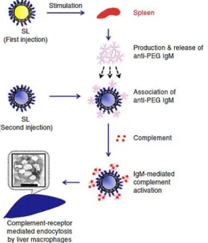
Figure 2.3.Proposed type-2 T-cell independent (TI-2) response mechanism for the formation of anti-PEG Abs and the ABC effect. Splenic B cells are stimulated by an initial dose of PEGylated therapeutic and produce anti-PEG IgM. These antibodies then associate with subsequent doses of
PEGylated systems and activate complement proteins, which then opsonize PEGylated system and lead to its eventual clearance through hepatic MPS cells. Reprinted from Ref [19] by permission from Macmillan Publishers Ltd.
2.2.4 Properties of the anti-PEG Ab epitope
20
typical PEG chain lengths for modified nanoparticles and proteins are approximately 1-5 kDa and 5-40 kDa, respectively, which covers a range from several tens to hundreds of ethylene glycol subunits and could readily and extensively crosslink any receptors capable of binding PEG. Richter and Akerblom reported hapten inhibition of anti-PEG Ab precipitation with PEG of 300 MW, suggesting that the antigenic epitope of PEG may consist of a 6-7 subunit region [51]. This value has been commonly cited as the size of the anti-PEG Ab binding epitope.
Nevertheless, a recent study observed that tri(ethylene glycol) (MW 150-160) was bound by anti-PEG Abs in direct and competitive ELISAs (see section 2.5 for methods of anti-anti-PEG Ab
detection) [89]. We have likewise found that both anti-PEG IgM and IgG can bind to polymers composed of repeating methacrylate PEG300 subunits (Fig. 2.4). Together, these findings suggest
that the anti-PEG Ab binding epitope could be smaller than the proposed 6-7 subunit length. Since free PEG is known to be non-immunogenic, the antigenic determinant for anti-PEG Abs has been suggested to occur at the linkage between PEG and other materials. Based on the observations that anti-PEG Abs induced by hydrophobic PEGylated micelles were able to bind to PEGylated liposomes, and vice versa, whereas hydrophilic PEGylated micelles avoided the induction of and opsonization by anti-PEG Abs, Shiraishi et al. proposed that the anti-PEG Ab epitope is the interphase between a hydrophobic core and conjugated PEG groups [69].
Nevertheless, because free PEG can inhibit anti-PEG Ab binding in competitive ELISAs and hemagglutination assays [51, 52], at least some of the observed anti-PEG Ab responses must be specific to PEG itself. Due to the disparity in the immune responses to free PEG versus
21
Figure 2.4.Preliminary studies of mouse anti-PEG IgG and IgM binding to poly(methacrylate PEG300) [P(OEG300)]. a) Dot blot of mouse anti-PEG IgG binding to unmodified polystyrene beads (COOH) or polystyrene beads coated with PEG5k or P(OEG300). b) Binding of mouse anti-PEG IgM
antibodies to magnetic polystyrene beads coated with PEG5k or P(OEG300) was determined using ELISA.
2.2.5 Factors influencing the formation of anti-PEG immunity and ABC in animals
The ABC phenomenon and immune responses to PEG are affected by a number of factors such as the dosing regimen [67], animal model [78], drug/cargo incorporation [90], nanocarrier/protein identity and composition [51, 59], and PEG structure [54, 91]. For example, the dosing interval that generates a maximal ABC response is dependent on the timing for anti-PEG Ab formation, which typically peaks at 3-7 days post-injection (Fig. 2.2a and b).Additional doses administered after less than 48 h or more than 4 weeks typically exhibit extended
22
immunogenicity of the administered PEGylated agents; however, only a small number of studies reported testing for endotoxin contaminants [56, 65, 81]. The composition and physicochemical properties (e.g., size [68, 97], lipid membrane rigidity [78], curvature, PEG density and terminal groups [52, 54, 66]) of PEGylated systems can also affect anti-PEG Ab induction and ABC responses. For an excellent review of these various factors, please refer to Ref [19].
2.3 Anti-PEG immunity in humans
PEGylation has been critical to the success of numerous therapeutic agents currently on the market, including uricase, interferon-α, and liposomal doxorubicin, as well as many protein and nanomedicines currently drugs in clinical trials [30, 98, 99]. However, a growing body of evidence clearly suggests that the induction of anti-PEG Abs is possible in humans. In contrast to most animal studies, the anti-PEG Ab response in humans is more skewed towards IgG isotype antibodies (Table 2.2). Interestingly, we and others have found that a significant fraction of the normal population actually possesses pre-existing anti-PEG (i.e., the presence of PEG-specific antibodies in the absence of treatment with PEGylated therapeutics), which may become even more prevalent in the years ahead [100]. Both pre-existing and induced anti-PEG Abs present significant challenges to the clinical efficacy of PEGylated therapeutics [37, 100].
2.3.1 Pre-existing anti-PEG Abs in the general population
23
PEG-specific IgG, with 19%, 5%, and 3% of the total individuals possessing IgG only, IgM only, and both IgM and IgG antibodies, respectively. The reasons for the discrepancy in the observed anti-PEG Ab incidence rates are unclear. Both studies utilized passive hemagglutination of PEG-modified RBCs to detect PEG-specific antibodies, so the differences are unlikely to be caused by the method of detection. In light of the decades-long gap between the reports, these variations could reflect a substantial increase in the prevalence of pre-existing anti-PEG Abs in the general population, but this hypothesis has not been carefully assessed.
How pre-existing anti-PEG are generated in individuals who have never received any formal treatment with PEGylated therapeutics remains largely unknown. As a GRAS product, PEG is widely used in cosmetics, processed foods, pharmaceuticals, agriculture, and industrial manufacturing. PEG-containing surfactants, as well as PEG itself, are found in the vast majority of household and hygiene products (e.g., soap, shampoo, toothpaste, lotion, detergent). It is natural to assume that frequent exposure to PEG could lead to the inevitable formation of anti-PEG Abs, but this constant exposure does not offer insight into the actual mechanism(s)
underlying anti-PEG immunity. While we have no direct supportive evidence to date, we wish to offer the following speculation: the human body is frequently subjected to insults (e.g.,
abrasions, lacerations, skin tears) that may result in local inflammatory responses and
24
anti-PEG Abs. Subsequent persistent exposure to PEG-containing products may further induce a robust memory immune response to the polymer.
Beyond the initial reports by Richter and Akerblom and by Armstrong et al., the prevalence of pre-existing anti-PEG Abs has been further reported in both healthy donors and untreated controls of clinical trials (Table 2.2). Tillmann et al. observed an incidence rate of 7%-8% in healthy individuals and in hepatitis and lupus patients, whereas 44% of hepatitis C patients were found to be positive for anti-PEG Abs prior to treatment with PEGylated interferon [102]. Treatment-naïve gout and hemophilia patients and patients with phenylketonuria demonstrated pre-existing anti-PEG Ab incidence rates of 19%, 6%, and 16%, respectively [103-105]. In addition, 38% of pediatric leukemia patients receiving unmodified asparaginase were also found to possess anti-PEG Abs [106]. Importantly, the relatively high incidence rate in this study was observed for patients with a mean age of 8.8 years, suggesting that anti-PEG Abs can be developed relatively early in life.
2.3.2 Induction and effects of anti-PEG Abs in individuals treated with PEGylated therapeutics
Studies of PEGylated therapeutics in humans began nearly three decades ago, but early results indicated that anti-PEG Ab responses were non-existent or clinically insignificant in humans. In a clinical trial of PEG-modified allergens, 50% of allergy patients had high anti-PEG Ab titers after one year of hyposensitization treatment, compared to 3.3% of untreated patients [101]. However, the occurrence of anti-PEG Abs did not appear to prime further immune
PEG-25
modified bovine adenosine deaminase (PEG-ADA, Adagen) generated IgG anti-PEG-ADA antibodies, but competitive ELISAs using ADA and different PEGylated proteins indicated that these antibodies were formed against ADA rather than the PEG moiety [107]. In a study of hepatitis C (HCV) patients, the presence of pre-existing anti-PEG Abs in 44% of the patients did not appear to affect the efficacy of antiviral PEG-interferon therapy [102]. The potential reasons for the apparent lack of anti-PEG Ab effects, including immune impairment and hepatic damage caused by HCV, were not explored.
Unlike most studies that report anti-PEG Ab responses in only a subset of patients, 100% of phenylketonuria patients developed PEG-specific Abs within 6 weeks of a s.c. injection of PEGylated phenylalanine ammonia lyase (PEG-PAL) [103]. Although the authors found that neither pre-existing nor induced anti-PEG Abs appeared to influence the efficacy of a single dose of PEG-PAL, peak therapeutic efficacy was observed on day 6, whereas testing for anti-PEG Abs was performed on days 0, 14, 28, and 42. Thus, the potential effects of the observed anti-PEG responses on the activity of multiply-dosed anti-PEG-PAL is unclear. Importantly, two patients in the study later experienced severe adverse reactions to i.m. injections of medroxyprogesterone acetate, which contains both free PEG and polysorbate as excipients. While there is insufficient data to prove causation or statistical significance, this observation indicates that future studies should also investigate whether anti-PEG responses may impact not only the repeated
administration of PEGylated therapeutics but also the use of pharmaceutical formulations comprising free PEG or PEG-containing chemicals excipients.
26
pediatric acute lymphoblastic leukemia patients treated with PEG-ASNase, anti-PEG IgM antibodies were observed in 46% of the patients, and the presence of anti-PEG Abs was strongly correlated with the rapid clearance of PEG-ASNase and loss of protein activity (Fig. 2.5a) [106]. In contrast, anti-PEG Abs present in patients treated with unmodified ASNase exhibited no effect on therapeutic protein clearance or activity (Fig. 2.5b). Because serum samples were only
collected after treatment, it is not clear whether the observed anti-PEG Abs were induced or pre-existing. However, given that 38% of patients treated with control ASNase also exhibited anti-PEG Abs, the authors suggested that the antibodies observed in the anti-PEG-ASNase group were likely pre-existing.
In the earliest clinical trial of PEG-uricase, 38% of refractory gout patients developed anti-PEG Abs, which was correlated with poor efficacy, after a single s.c. injection of the PEGylated drug [98]. This PEG-specific antibody response demonstrated apparent class switching, with IgM and IgG predominating at days 3-7 and 7-14, respectively, after injection. One patient was later re-challenged with PEG-uricase and demonstrated an anamnestic antibody response to the PEGylated protein [98]. In a separate study, anti-PEG Ab responses were
generated in 35% of gout patients after a single i.v. infusion of PEG-uricase, and anti-PEG Ab formation also associated with rapid protein clearance [108]. Additionally, one patient with a pre-existing anti-PEG Ab response exhibited a correspondingly reduced half-life for PEG-uricase.
27
correlated with the loss of PEG-uricase activity [22]. Similar results were obtained in a study by Hershfield et al., with 37% of treatment-naïve patients establishing an anti-PEG Ab response and non-responsive to PEG-uricase treatment by the end of the clinical trial; half of these anti-PEG Ab responses were pre-existing [104]. Three patients that received PEG-uricase during previous studies (1-3 years prior) were also non-responsive to the new round of treatment, exhibiting loss of PEG-uricase efficacy earlier than the affected treatment-naïve patients (2-7 days vs. ~2 weeks). Interestingly, of the demographic characteristics (e.g., age, gender, BMI, renal function) examined during the repeated dosing studies, only age (>60-70 years) and organ recipient status, both of which involve some level of immunoinsufficiency, were found to be associated with reduced anti-PEG Ab formation [22, 104]. In addition to rapid PEG-uricase clearance, anti-PEG Ab-positive individuals also demonstrated an increased rate of infusion reactions [98, 104, 109], but the precise involvement of anti-PEG Abs in adverse reactions to PEG-uricase and other PEGylated therapeutics remains unclear [35].
In recent clinical trials of pegnivacogin, a PEGylated RNA aptamer, the presence of pre-existing anti-PEG Ab has alarmingly been associated with first-dose allergic reactions to the PEGylated drug. A phase IIb study of pegnivacogin was terminated after 3 patients (out of 640) developed serious allergic reactions, one of which was deemed life-threatening, within less than thirty minutes of a first dose of the drug [17]. Competitive ELISA analysis of the patient samples indicated that all three patients possessed high levels of pre-existing antibodies against PEG (>97th percentile for all analyzed samples), but no antibodies against the aptamer itself. In a subsequent phase III trial, pre-existing anti-PEG Ab were also linked with severe allergic
28
experience any adverse reactions. For patients with anti-PEG IgG levels one- or three-fold above the assay cutoff point, the likelihood of experiencing a severe allergic reaction was 5% and 16%, respectively. The authors suggested that the presence of both high anti-PEG Ab levels and large dose of PEGylated drug (~0.8 PEG mg/kg, i.v. bolus) was a key factor in the observed
anaphylactic responses, although other unidentified contributing factors are also likely involved [17, 110].
Figure 2.5.Anti-PEG antibodies in human patients. Anti-PEG IgM vs. asparaginase (ASNase) activity for patients treated with a) PEG-ASNase and b) ASNase. Flow cytometry was used to detect anti-PEG Abs bound to PEG hydrogel (TentaGel-OH) particles. c) Mean serum uric acid (sUA) levels in patients receiving biweekly i.v. infusions of PEG-uricase. Normal sUA levels are typically defined as ≤6 mg/dL (indicated by gray dashed line). d) Mean anti-PEG Ab titers in patients receiving biweekly i.v. infusions of PEG-uricase. Panels A and B were reprinted from Ref [106], and panels C and D were modified from Ref [22].
2.3.3 Clinical implications of and strategies to overcome anti-PEG Abs
29
PEG-specific immune responses on the growing clinical use of PEGylated therapeutics and underscores the need to incorporate testing for anti-PEG Abs in clinical trials of PEG-containing drugs. Standard laboratory tests (see section 2.5) that can quantitatively and accurately measure anti-PEG Ab levels and determine a patient’s anti-PEG Ab status are crucial to this effort, as suggested by others [21, 35, 111]. Importantly, clinical trial designs must screen for pre-existing anti-PEG immunity, as well as monitor treatment history, since previous exposure to PEGylated therapeutics could prime future responses to subsequent therapy with PEGylated drugs.
Furthermore, the true extent of anti-PEG Abs in the human population and the factors that lead to anti-PEG Ab immunity must be further investigated.
In addition to an improved understanding of the prevalence and development of anti-PEG immunity in humans, strategies to avert or overcome anti-PEG Ab responses must be developed. Unfortunately, the effect of important dosing regimen factors identified in animal studies
remains to be fully evaluated in human subjects. In the clinical trials of PEG-uricase, neither the dose (0.5-24 mg/patient), dosing interval (2-4 weeks), nor route of administration (s.c. or i.v.) appeared to affect anti-PEG Ab induction or its effects [22, 98, 104], but these results must be corroborated for other PEGylated drugs.The use of cleavable or sheddable PEG has been
30
that specifically target immunological pathways related anti-PEG immunity may allow the specific suppression of anti-PEG Ab generation while avoiding unwanted side effects.
The use of alternative stealth polymers such as chitosan, poly(carboxybetaine), poly(2-oxazaline), XTEN peptide, and poly(glycerol) has also received growing attention [114-117]. These polymers are less ubiquitous in everyday household items and thus may not encounter the problem of pre-existing antibodies. Nevertheless, antibodies against various natural and synthetic repeating polymers have been reported [118, 119], suggesting that stealth polymers other than PEG may also prove immunogenic upon repeated administration in humans. In individuals with induced or pre-existing anti-PEG Abs, the elimination of circulating anti-PEG Abs could be achieved through selective plasmapheresis, although the use of such a complicated procedure clearly poses additional cost burdens and may not be warranted if alternative strategies to remove anti-PEG Abs are available. Additionally, it may be possible to overwhelm PEG-specific
immune responses by simply administering a much greater dose of the PEGylated therapeutic [60, 81, 120]. Nevertheless, dosage increases will obviously be limited by the maximum
tolerated dose and potential toxicity to various clearance organs. A conceptually similar but more desirable approach would be to first saturate pre-existing anti-PEG Abs with free, low molecular weight PEG. Indeed, Moghimi reported that the administration of free PEG and PEG-containing molecules 1-3 h prior to a second dose of poloxamine-modified polystyrene beads reduced the ABC of these particles in rats [121]. Further animal and human studies are needed to confirm the safety and efficacy of such a strategy.
2.4 Ongoing questions regarding anti-PEG antibodies
31
to human patients, that are of great interest to the scientific and clinical communities. While a full discussion of these questions is beyond the scope of this review, we wish to highlight a few of them below:
How are anti-PEG Abs able to specifically bind PEG polymers? Due to PEG’s flexible, neutral, and hydrophilic character, the precise antibody-polymer interactions that allow anti-PEG Abs to specifically bind to such an amorphous target in the absence of
hydrophobic and electrostatic interactions are of interest.
What is the immunological pathway of anti-PEG Ab formation in humans? The features of human anti-PEG Ab responses (i.e., pre-existing anti-PEG Abs, high prevalence of IgG, and memory responses to PEGylated products) suggests that the mechanisms underlying PEG-specific immunity may differ greatly between humans and animal models currently used to study anti-PEG immunity. Due to the difficulty of performing mechanistic studies in humans, the use of animal models that more accurately
recapitulate human anti-PEG Ab responses are necessary to improve our understanding of anti-PEG immunity, including that elicited by long-term exposure to PEG and PEG-containing products.
What factors predispose individuals towards anti-PEG Ab formation and are certain
portions of the human population more, or less, inclined towards anti-PEG immunity?
32
identify other factors that affect PEG-specific immunity, as well as reveal additional strategies to manage anti-PEG Ab responses.
What is the current and likely future prevalence of pre-existing anti-PEG Abs? The reported prevalence of anti-PEG Abs varies significantly, with values ranging from as low as 5% to over 40%. Thus, a precise estimate of the level of anti-PEG Abs in both the general and special populations is sorely needed. Additionally, given the disparity in the incidence rate between early and more recent studies, the potential for further increases in the prevalence of pre-existing anti-PEG Abs must be explored.
How can anti-PEG immunity be efficiently and effectively managed in a clinical setting?
Strategies to overcome pre-existing and/or induced anti-PEG Abs, including the
administration of an excess dose of PEGylated therapeutic or prior injection of free PEG polymer, should be further investigated.
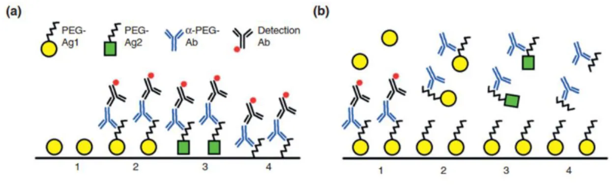
2.5Detection of anti-PEG Abs by validated ELISA methods
33
that was injected; thus the possibility that induced antibodies were actually bound to the carrier rather than PEG itself cannot be fully discounted. As noted by others, there is a critical need for more rigorous, validated anti-PEG Ab detection methods [35, 100]. In our opinion, both direct and competitive ELISAs should be used in combination to confirm the PEG-specificity of anti-PEG Abs with the application of proper controls and conditions (see Fig. 2.6), as was carried out in some recent human trials [22, 98, 104]. Standard curves can be generated using commercially available anti-PEG Abs (e.g., mouse, rat, rabbit, chicken, goat, and monkey host Abs) to quantify induced or pre-existing anti-PEG Abs. Additionally, the validation of ELISA protocols (e.g., determination of precision, sensitivity, reagent interference) must be performed and reported [22, 104]. Importantly, the use of Tween and other PEG-containing detergents must be avoided, as they can significantly reduce the sensitivity of anti-PEG Ab detection assays [52]. The
development of standardized laboratory tests that can quantitatively and accurately measure anti-PEG Ab levels is crucial to furthering our understanding of anti-anti-PEG immunity.
34
2.6 Conclusions
Because of its ability to significantly prolong the circulation of nanoparticles and proteins, as well as its presumed lack of immunogenicity, PEG has been widely used to modify various therapeutic agents. However, a growing body of evidence indicates that potent and specific antibody responses can be generated against the PEG polymer, and anti-PEG Ab
Table 2.1. Examples of anti-PEG Ab and/or accelerated blood clearance responses to PEGylated systems. Type of PEGylated
system
Animal model Dose*,† Dosing interval*
Parameters of initial or control dose
Parameters of subsequent dose(s)
Fold change in parameters‡
Anti-PEG Ab response#
Ref
PEGylated liposome Balb/c mice 0.01-1 μmol PL/kg 5-7 d N.D. N.D. - IgM + (no to
weak) IgG
[72]
PEGylated liposome Std:ddY mice 25 μmol PL/kg 10 d t1/2,β12.9 h
Cl 0.07 mL/h
AUC0-24h
486%dose∙h/mL
t1/2,β6.3 h
Cl 0.2 mL/h
AUC0-24h
221%dose∙h/mL
0.5 (t1/2,β)
2.9 (Cl)
0.5 (AUC0-24h)
N.D. [57]
PEGylated liposome KM mice 0.1 μmol PL/kg (initial); 5
μmol PL/kg (subsequent)
6 d 44%IDblood,4h 13%IDliver,4h 12%IDspleen,4h
14%IDblood,4h 28%IDliver,4h 18%IDspleen,4h
0.3 (%IDblood,4h)
2.2 (%IDliver,4h)
1.5 (%IDspleen,4h)
0.2 (t1/2), 0.2 (AUC)
º
IgM§ [78]
PEGylated liposome Wistar rats 0.001 μmol PL/kg - N.D - - IgM + (weak)
IgG
[63]
PEGylated liposome Wistar rats 5 μmol PL/kg 7 d t1/2,α 2.4 h
52.5%IDblood,4h 8.1%IDliver,4h 2.2%IDspleen,4h
t1/2,α 0.1 h 0.6%IDblood,4h 46.4%IDliver,4h 6.3%IDspleen,4h
0.04 (t1/2,α)
0.01 (%IDblood,4h)
5.7 (%IDliver,4h)
2.9 (%IDspleen,4h)
N.D. [33]
PEGylated liposome Wistar rats 0.001 μmol PL/kg (initial);
5 μmol PL/kg (subsequent)
4-6 d t1/214.8 h
Clh<1 mL/min 8%IDliver,24h 8%IDspleen,24h
t1/20.3-1.8 h
Clh25-55 mL/min 67-72%IDliver,24h 8-12%IDspleen,24h
0.02-0.12 (t1/2) >25-55 (Clh) 8.4-9.0 (%IDliver,24h) 1.0-1.5 (%IDspleen,24h)
IgM [71]
PEGylated liposome Wistar rats 0.001 μmol PL/kg (initial);
5 μmol PL/kg (subsequent)
5 d 51%IDblood,24h 6%IDliver,24h
<2%IDblood,24h 68%IDliver,24h
<0.02 (%IDblood,24h)
11 (%IDliver,24h)
IgM + (weak) IgG
[61]
PEGylated liposome Wistar rats 5 μmol PL/kg 7 d 76.4%IDblood,4h
15%IDliver,4h
0.6%IDblood,4h 68%IDliver,4h
0.01 (%IDblood,4h)
4.5 (%IDliver,4h)
N.D. [60]
PEGylated liposome Sprague-Dawley rats
7 μmol PL/kg 7 d t1/216.7 h
Cl 1.7 mL/h
AUC 856 μg∙h/mL
t1/20.2 h
Cl 74.3 mL/h
AUC 17 μg∙h/mL
0.01 (t1/2)
43.7 (Cl)
0.02 (AUC)
IgM§ [68]
PEGylated liposome Dunkin-Hartley guinea pigs
0.1 μmol PL/kg (initial); 5 μmol PL/kg (subsequent)
6 d 34%IDblood,4h 12%IDliver,4h <2%IDspleen,4h
12%IDblood,4h 37%IDliver,4h <2%IDspleen,4h
0.4 (%IDblood,4h)
3.1 (%IDliver,4h)
~1 (%IDspleen,4h)
0.6 (t1/2), 0.6 (AUC)º
IgM§ [78]
PEGylated liposome Rhesus monkey 5 μmol PL/kg 7 d t1/2 87.5 h
17.6%IDliver,4h
t1/2 14.2 h 41.2%IDliver,4h
0.2 (t1/2)
2.3 (%IDliver,4h)
N.D. [33]
Type of PEGylated system
Animal model Dose*,† Dosing interval*
Parameters of initial or control dose
Parameters of subsequent dose(s)
Fold change in parameters‡
Anti-PEG Ab response#
Ref
PEGylated liposome Rabbits 9 mg PL/animal 7 d N.D. N.D - IgG¶,∆ [73]
PEGylated liposome Japanese white rabbits
0.1 μmol PL/kg (initial); 5 μmol PL/kg (subsequent)
6 d 47%IDblood,4h 15%IDliver 4%IDspleen
13%IDblood,4h 35%IDliver 6%IDspleen
0.3 (%IDblood,4h)
2.3 (%IDliver,4h)
1.5 (%IDspleen,4h)
0.5 (t1/2), 0.4 (AUC)º
IgM§ [78]
PEGylated pDNA liposome
ICR mice 100 μg pDNA/animal 7 d 80%IDblood,1h
8%IDliver,1h 2%IDspleen,1h
23%IDblood,1h 41%IDliver,1h 4%IDspleen,1h
0.3 (%IDblood,1h)
5.1 (%IDliver,1h)
2.0 (%IDspleen,1h)
IgG + IgM∆ [65]
PEGylated ODN vesicles
ICR mice 50 mg PL/kg, 10 mg
ODN/kg
7 d 70%IDblood,1h 6%IDblood,1h 0.1 (%IDblood,1h) IgM§ [81]
PEGylated Gd liposome
C57BL/6 and Balb/c mice
5 μmol PL/kg 7 d 11%IDblood,6h
9%IDliver,6h 10%IDspleen,6h
<0.5%IDblood,6h 31%IDliver,6h 1%IDspleen,6h
<0.05(%IDblood,6h)
3.4 (%IDliver,6h)
0.1 (%IDspleen,6h)
IgM∆ [69]
PEGylated Hb vesicles
ddY mice 0.1 mg Hb/kg 7 d t1/2 2.7 h
Cl 3.7 mL/h
AUC 27.1%dose∙h/mL
t1/2 1.3 h
Cl 22.3 mL/h
AUC 4.5%dose∙h/mL
0.5 (t1/2 )
6.0 (Cl)
0.2 (AUC)
IgM [122]
PEGylated EPI liposome
Wistar rats 1 μmol PL/kg, 0.08 EPI/kg
(initial); 5 μmol PL/kg, 0.4 mg EPI/kg (subsequent)
7 d 52%IDblood,4h 16%IDliver,4h 8%IDspleen,4h
8%IDblood,4h 36%IDliver,4h 15%IDspleen,4h
0.2 (%IDblood,4h)
2.3 (%IDliver,4h)
1.9 (%IDspleen,4h)
IgM§ [77]
PEGylated DXR liposome
Beagle dogs 0.67 μmol PL/kg and 2 mg
DXR/m2
3 wk t1/224.1 h
Cl 1.5 mL/h/kg
AUC0-∞76.0 μg∙h/mL
t1/21.5 h
Cl 127.8 mL/h/kg
AUC0-∞0.6 μg∙h/mL
0.06 (t1/2)
85.2 (Cl)
0.01 (AUC0-∞)
IgM§ [120]
PEGylated TOPO liposome
Beagle dogs 0.5 mg TOPO/kg 7 d Cmax7.9 mg/L
Cl 0.4 mL/min/kg
AUC0-t1.4 mg∙min/mL
Cmax1.7 mg/L
Cl 6.7 mL/min/kg
AUC0-t0.1 mg∙min/mL
0.2 (Cmax)
16.8 (Cl)
0.07 (AUC0-t)
IgM§ [66]
PEGylated solid lipid nanoparticle
Wistar rats 5 μmol PL/kg (initial s.c.,
subsequent i.v.)
7 d AUC0-4h27.3 mg∙h/L
8 μg/g (liver, 4 h)
9 μg/g (spleen, 4 h)
AUC0-4h6.6 mg∙h/L
25 μg/g (liver, 4 h)
26 μg/g (spleen, 4 h)
0.2 (AUC0-4h)
3.1 (liver, 4 h)
2.9 (spleen, 4 h)
IgM§ [88]
PEGylated solid lipid nanoparticle
Kunming mice 10 μmol PL/kg 7 d 71.3%IDblood,0.5h
5.4%IDliver,0.5h 4%IDspleen,0.5h
42.6%IDblood,0.5h 23.3%IDliver,0.5h 9%IDspleen,0.5h
0.6 (%IDblood,0.5h)
4.3 (%IDliver,0.5h)
2.3 (%IDspleen,0.5h)
N.D. [70]
PEGylated solid lipid nanoparticle
Beagle dogs 2 μmol PL/kg 7 d t1/2,β3.4 h
Cl 0.2 mL/min/kg
AUC0-24h90.6 mg∙h/L
t1/2,β1.6 h
Cl 0.4 mL/min/kg
AUC0-24h34.0 mg∙h/L
0.5 (t1/2,β)
2.0 (Cl)
0.4 (AUC0-24h)
IgM§ [70]

![Figure 2.4. Preliminary studies of mouse anti-PEG IgG and IgM binding to poly(methacrylate PEG 300 ) [P(OEG 300 )]](https://thumb-us.123doks.com/thumbv2/123dok_us/8329152.2209052/37.918.196.715.114.332/figure-preliminary-studies-mouse-anti-peg-binding-methacrylate.webp)