A Case Study on Mathematical Morphology
Segmentation for MRI Brain Image
Senthilkumaran N, Kirubakaran C
Department of Computer Science and Application, Gandhigram Rural Institute, Deemed University, Gandhigram, Dindigul-624302.
Abstract— Medical image processing has already become an important component of clinical analysis. Because it is an interdisciplinary research domain of medicine, mathematics and computer skills. MRI has good impact on analysis and treatment of many health problems, although the effect on improved health effects is uncertain. MRI brain image has complexity due to its gray level color values. The gray scale pictures are difficult to analysis because of its color ranges are slightly varied. Using mathematical morphology the grayscale images are enhanced and segmented. Mathematical morphology is a new mathematical theory which can be utilized to examine and process MRI images. A shape concept from set theory is an alternative approach to image processing also provided by mathematical morphology. This article presents a work on MRI brain segmentation and filtering techniques on mathematical morphology. In particular, the results are related to spatially variant mathematical morphological operations and enhance the image detail. These edge detection and Watershed segmentation methods are measured on MRI brain image data.
Keywords— MRI, Mathematical Morphology, Edge detection, Watershed segmentation.
I. INTRODUCTION
Image segmentation is a fundamental process, which is usually practiced in many image processing applications to hold data from image [1] [16]. Medical image segmentation is an important work for object analysis of the human organs such as brain, blood vessels and retinal images. It is an important pre-processing work in medical image processing [3]. Segmenting the various brain tissues is very significant in the analytic thinking of brain MRI data, and it delivers a vast range of application. If the data analysis is mistaken, the comparable treatment and the decision also wrong [4]. Designing of clinical systems that need an image segmentation process are available with a huge scope of practical techniques and method [16]. To assist with this, several researchers have suggested methods for evaluating or validating the strength of various segmentation algorithms [5]. One of the segmentation and the object extraction method is mathematical morphology.
The morphology technique was originally established by Matheron and Serra at Ecoledes mines [1]. Mathematical morphology theory treats the image as mathematical sets and morphological transformations which developed from Minkowski addition and subtraction are defined to extract features in images [3]. Mathematical morphology is an image processing tool for dividing the image elements that are useful analysis the data. Such as boundaries, skeletons and convex hull are the region shape that is represented and descript by morphology [1]. Mathematical morphology is a
nonlinear filter method, which could be utilized for image processing, including image segmentation, edge detection, noise suppression, texture analysis, feature extraction, shape recognition, image restoration, image compression and reconstruction [6].
The morphological edge detection operator selects suitable structuring element and makes utilize the basic operations of morphology, including erosion, dilation, opening and closing. All image enhancement and segmentation process are processed using morphological operators, which are dilation and erosion. Other operators are synthesisation of these two operations. These operations are carried out in a correct sequence to find a neat image edge [7]. Watershed transformation is an effective segmentation method. The foundation of this method is to consider the image as a topographical relief. This relief is flooded from its minima. A dam is built, when two lakes joined. The group of all dams is called the watershed line. The watershed separates the image in catchment basins [12]. The above mentioned edge detection and segmentation methods are implemented on MRI brain image.
This paper presents the execution of some mathematical morphology segmentation methods for image segmentation. This paper is organized as follows, section II is for the purpose of presenting information about basics of mathematical morphology. Edge detection using morphology operations is explained, detailed in section III. Section IV focused the segmentation using watershed transform. Section V contains the visual results of section III and IV. Conclusion takes place in section VI.
II. BASIC MATHEMATICAL MORPHOLOGICAL OPERATIONS Mathematical morphology is a non-linear imaging diagnostic technique that separates image part as information. Using the geometric structuring it works. This work briefly revises some mathematical morphology operators and the corresponding operations related [10]. There are two data are considered as an input by Mathematical operators. An image to be processed and a suitable structuring element, which is projected by the physique and size of the neighborhood pixels [10]. Founded on this medical image processing, basic mathematical morphology operations are acquired and used in higher-level [11].
move the structure elements within the image by pixel by pixel, and at the same time will do basic operations on sets of intersection and union [2] [6].
Fig 1. Structuring element
Dilation and erosion are the tow basic mathematical morphological operators. The other morphological operations are the combination of the two basic operations in a sequential manner. In the pursuit, we present just about basic mathematical morphological operators of grayscale images [14].
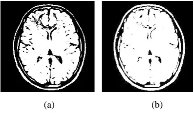
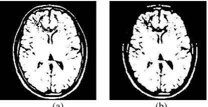
Binarization is the operation that changes a gray level image into a binary image [10]. This process performs examining the gray-level value of each pixel in the enhanced image, and if the value is larger than the threshold, then the picture element value is going down to a binary value on; otherwise it is set to zero is expressed in Fig. 2.
(a) (b)
Fig 2. (a) Original Image (b) Binary image
A. Dilation:
Dilation is a transformation of expanding, which increases the grayscale value of the image [2] [3]. Dilation operation on an image containing labels 0 and 1, with a structuring element B, changes the value of pixel i in the image from 0 to 1, if the result of convolving B with input image, centered as i, is more than some predetermined value. We have set this value to be zero. The structuring element (as well recognized as the dilation kernel) determines the details of how a particular dilation grows boundaries in an image [10] [1]. Dilation is shown in Fig.3. Let F (x, y) denotes a gray-scale two dimensional image, B
denote structuring element. Dilation of a grayscale image F (x, y) by a grayscale structuring element B (s, t) is denoted by [3]
)}
,
(
)
,
(
max{
)
,
)(
(
F
⊕
B
x
y
=
F
x
−
s
y
−
t
+
B
s
t
(a) (b)
Fig 3. (a) Original Image (b) Dilated image
B. Erosion:
Erosion is a transformation of reducing, which decreases the gray-scale value of the image [2] [3]. Erosion operation on an image containing labels 0 and 1, with a structuring element B, changes the value of pixel i in the image from 1 to 0, if the result of convolving B with the input image, centered as i, is less than some predetermined value [1]. We have set this value to be the area of B, which is essentially the number of pixels that are 1 in the structuring element itself. The structuring element (as well recognized as the erosion kernel) determines the inside information of how particular erosion thins boundaries [10] are shown in Fig.4.
(a) (b)
Fig 4. (a) Original Image (b) Eroded image
Erosion of a grayscale image F (x, y) by a grayscale structuring element B (s, t) is denoted by [3] [13]
( ⊖ )( , ) = min { ( + , + ) − ( , )}
C. Opening:
An opening operation contains of erosion followed by dilation with the same structuring element. Opening operator consists of an erosion followed by a dilation and can be applied to eliminate all pixels in regions that are too small to contain the structuring element [10]. The result of opening operation is presented in Fig.5. In this example the structuring element is frequently shouted out a probe, because it is probing the image looking for little objects to filter away the image [1] [3]. Opening of gray scale image
F (x, y) by gray scale structuring element B (s, t) are denoted respectively by
(a) (b) Fig 5. (a) Original Image (b) By opening
D. Closing:
A closing operation shown in Fig.6 consists of a dilation followed by erosion with the same structuring element. The morphological closing operator tends to smooth the contours of objects; it joins narrow breaks, fills holes smaller than the structuring element [10]. Closing of gray scale image F (x, y) by gray scale structuring element B (s, t)
are denoted respectively by [3]
⋅
= ( ⨁ ) ⊝
(a) (b)
Fig 6. (a) Original Image (b) By closing
III.SEGMENTATION USING MATHEMATICAL MORPHOLOGY Regarding segmentation of the particular object of an image, we consider that there is some contrast different between brain tissue (gray matter) and cerebro-spinal fluid (white matter), which separates the brain [10]. The morphological edge detection algorithm selects appropriate structuring element 3*3 to the input image and makes use of the basic theory of morphology, including erosion, dilation, opening and closing operation and the combined operations [7]. The style of the operation stands for thinking over the relation between the processed image and origin image (original picture), and the option of structuring element decides the turning and refined effect [10] [7].
The morphological gradient clearly shows the sharp gray level transition in the input image F. By morphology, erosion and dilation operations satisfy the operation features:
⊖
⊆ ⨁
Opening and closing operations satisfy:
∘
⊆
⊆ ∙
The sharpnesses of the image F (x, y), which is denoted by Ed (F), are determined as the difference set of the
dilation domain of F (x, y). This is likewise known as dilation residue edge detector [7]:
( ) = ( ⨁ ) −
Opening and closing operations are better for filtering. But they use the inventory of erosion and dilation, the answer of the processed image is only correlative with the inside and outside of the object boarder [3] [10]. The accurate object edge presents in the image will be obtained by working, the difference between the processed image by above process and the image before dilation. The chase is the morphological steps are producing the edges of the objects present in the input image [7].
( ∙ )⨁ −
∙
Where
= ( ∙ ) ∘
IV.SEGMENTATION USING WATERSHED TRANSFORM The watershed transform is a more popular image segmentation method from the discipline of mathematical morphology. It was introduced by Digabel and Lantuejoul in the domain of image processing to use analyze sample binary images [8]. Water precipitating onto the real landscape naturally collects to form pools in lower catchment basins, where its obstructed by dams or ridges. The input image can be separated by partitioning it into which areas are come under the catchment basins in the watershed [5] [14].
Normally the watershed transformation is employed to a boundary mapping in gray scale function. That gray scale function derived from the input image, which has low values within regions and high values along region boundaries [5].
Let f be the input image: D → N, with minimum and upper limit of gray level h. The minimum and maximum value of f define a recursion with the gray level h increases from hminto hmax [8]. Xh refers the union of the set of basins
computed at level h. The watershed Wshed (f) of f is the
complement of X in D:
( ) = /
The following processes employedto segment the image by watershed transformation. A set of pixels belonging to regional minima. Set of pixels in which a drop of water shall slide towards a single minimum. For a paid minimum, this set of pixels is called a basin [14]. Set of pixels in which a drop of water could fall into more than one basin. The points satisfying this condition from the tip line of the surface and are called watershed lines.
Marker-controlled watershed segmentation follows this basic process:
1. Work out a segmentation function. This is an image whose dark regions are the objects you are trying to segment.
2. Compute foreground markers. These are connected blobs of pixels inside each of the targets [15].
3. Compute background markers. These are pixels that are not part of any object.
5. Compute the watershed transform of the modified segmentation function [15].
There are two faults in the watershed transform. First the disappearance of the border brings about miss segmentation or over-segmentation. Second demerit is to various targets of image segmentation, many small targets may draw up a large target with different characteristics [8].
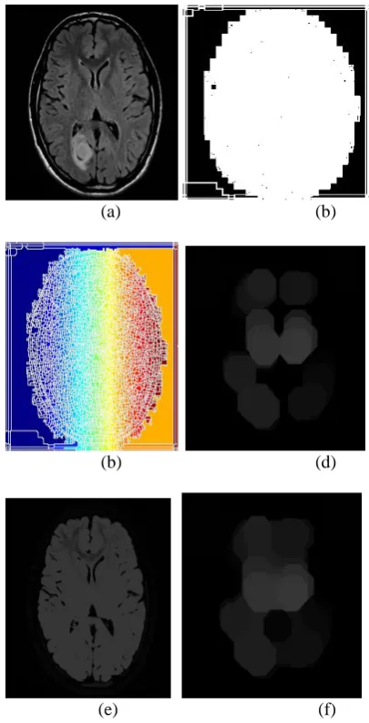
V. RESULTS AND ANALYSIS
In this paper is realized in Matlab, to have an experiment on a group of MRI brain image. The final segmentation result in which morphological gradient image is applied as referenced image. Morphological gradient images treated with opening-closing filters are used. The morphological gradient operators and dilation residue edge detector are succeeding in MRI brain edges detection, and the detected edges are clearly in Fig.7.
(a) (b)
(c) (d)
(e) (f)
(g)
Fig 7. (a) Original Image (b) binary gradient mask (c) dilated gradient mask (d) binary image with filled holes (e) cleared border image (f)
segmented image (g) outlined original image
When medical image segmentation applies to concern clinical situations, its accurateness will affect the diagnosis results and treatment. The waters shall flood the topographical surface from the marks and shall create as many basins. This way, we can solve the over segmentation problem. The watershed segmentation is implemented here. It shows the correct result in Fig.8.
(a) (b)
(b) (d)
(e) (f)
Fig 8. (a) Original Image (b) Gradient Magnitude (c) Mark the foreground objects (d) opening-by-reconstruction (e) complement the image (f)
closing-by-reconstruction
(g)
VI.CONCLUSION
In this paper, the basic operations of mathematical morphology, edge detection and watershed transformations are studied and MRI medical image segmentation. The implementation shows that by applying morphological opening-closing filters to deal with gradient images with the corresponding structure element union are eliminated and produced the segmented regions in the MRI brain image. The experimental results show that the mathematical morphology is more efficient for medical image analysis and segmentation. The success of this combined operation morphology is however, fully dependent upon the morphological gradient image.
ACKNOWLEDGMENT
This research work is supported by University Grant Commission, India, through a Major Research Project, Grant (UGC-F.No:42-131/2013(SR)).
REFERENCES
[1] Reecha Sharma and Beant Kaur, “Detection of Edges Using Mathematical Morphology for X-Ray Images”, International Journal of Engineering Sciences, Issue Dec. 2011, Vol. 5, pp 230-238.
[2] I. Balan, “Using Mathematical Morphology to Detect the Imperfections of the Printed Circuit Boards”, Journal of Applied Computer Science, no.1 (2) /2008, pp 9-14.
[3] Zhao Yu-qian, Gui Wei-hua, “Medical Images Edge Detection Based on Mathematical Morphology”, Engineering in Medicine and Biology 27th Annual Conference, Sep 2005, pp 6492-6495.
[4] C. Tsai, B. S. Manjunath and R. Jagadeesan, “Automated Segmentation of Brain MR Images”, Pattern Recognition, Elsevier, Vol. 28, No. 12, 1995, pp. 1825-1837.
[5] Joshua E. Cates, Ross T. Whitaker, “Case study: an evaluation of user-assisted hierarchical watershed segmentation”, Medical Image Analysis, Elsevier, 2005, pp 566–578.
[6] Zhao Fang and Ma Yulei, “Medical Image Processing Based on Mathematical Morphology”, International Conference on Computer Application and System Modeling, 2012, pp 948-950.
[7] J.Mehena, “Medical Images Edge Detection Based on Mathematical Morphology”, International Journal of Computer & Communication Technology (IJCCT), Volume-2, Issue-VI, 2011, pp 49-53. [8] CHEN Jia-xin and LIU Sen, “A Medical Image Segmentation
Method Based on Watershed Transform”, International Conference on Computer and Information Technology (CIT’05) IEEE, 2005. [9] J.A. Jiang and C.L. Chuang, “Mathematical-morphology-based
edge detectors for detection of thin edges in low-contrast regions”, IET Image Process., 2007, 1, (3), pp. 269–277.
[10] D.Selvaraj and R.Dhanasekaran, “Novel approach for segmentation of brain magnetic resonance imaging using intensity based thresholding”, ICCCCT, 2010, pp 502-507.
[11] A. Dufour and O. Tankyevych, “Filtering and segmentation of 3D angiographic data: Advances based on mathematical morphology”, Medical Image Analysis 17, Elsevier, 2013, pp 147–164.
[12] Benoit Naegel, “Using mathematical morphology for the anatomical labeling of vertebrae from 3D CT-scan images”, Computerized Medical Imaging and Graphics 31, Elsevier, 2007, pp 141–156. [13] Anjan Bikash Maity and Sandip Mandal, “Edge Detection Using
Morphological Method and Corner Detection Using Chain Code Algorithm”, International Journal of Computer Science Issues, Vol. 8, Issue 4, No 1, July 2011, pp 583-588.
[14] R. Kiruthikaa, “An Implementation of Watershed Based Image Segmentation Algorithm Using FPGA Processor”, International Journal of Science and Research (IJSR), Volume 2 Issue 12, Dec 2013, pp 399-402.
[15] Mandeep Kaur and Gagandeep Jindal, "Medical Image Segmentation using Marker Controlled Watershed Transformation", International Journal of Computer Sci ence And Technology, Vol. 2, Issue 4,2011, pp 548-551.