doi: 10.3978/j.issn.2305-5839.2015.03.16
View this article at: http://dx.doi.org/10.3978/j.issn.2305-5839.2015.03.16
Clinical data
History
The patient, a 59-year-old man, was found to be with lesion in the right upper lobe of the lung. He received coronary artery bypass graft in the department of cardiac surgery of our hospital due to angina pectoris 3.5 months ago. The pre-operative CT showed a nodular shadow in the right upper lobe of the lung. PET-CT findings were highly suggestive of lung cancer. A second chest CT after the surgery showed that the lesion in the right upper lobe of the lung did not change remarkably. The patient then visited our hospital again and was admitted due to “space-occupying lesion in the right upper lobe of the lung”. He had a history of hypertension for over 20 years. Ten years ago, he received a surgery for the gallbladder stones.
Physical examination
No bilateral supraclavicular lymph node enlargement was detected. Chest examination showed no positive sign.
Auxiliary examination
(I) Chest CT showed a lobulated and spiculated nodular shadow in the anterior segment of the right upper lobe, with vesicles visible inside it. No remarkable change was observed when compared with the
previous CT findings (Figure 1).
(II) Metastasis was not detected on head CT, bone ECT, and abdominal ultrasonography.
Surgical procedures
Anesthesia and body position
After the induction of general anesthesia, the patient was under double-lumen endotracheal intubation and underwent left-sided one-lung ventilation.
The patient was placed in the left lateral decubitus position and in a Jackknife position (Figure 2).
Procedures
(I) Incisions: a 1.5 cm camera port was created in the 8th intercostal space at right posterior axillary line, and two 1.0 cm working ports were separately made in the 5th intercostal space at anterior axillary line and the 8th intercostal space at scapular line. A 4 cm auxiliary port was made in the 7th intercostal space at midaxillary line (Figure 3);
(II) Connection of robot manipulators: the robot patient cart is positioned directly above the operating table and then connected. Its left hand was attached to bipolar cautery forceps, and its right hand was attached to a unipolar cautery hook. Incision protector was applied in the auxiliary port;
(III) Intra-operative inspection showed that the lesion was located at the anterior segment of the right upper lobe, along with pleural indentation. Thus, wedge resection of the lesion was decided, during which a single-use endoscopic linear cutter/stapler (two golden reloads and one blue reload) was used (Figures 4-6);
Figure 1 Chest CT.
Figure 2 Surgical position.
Figure 3 Surgical incisions.
Figure 4 Wedge resection of the lesion.
Figure 5 Wedge resection 2.
(V) Dissociate the upper pulmonary vein behind the phrenic nerve (Figure 8);
(VI) Pull the upper pulmonary vein using an elastic cuff (Figure 9);
(VII) Cut off the upper pulmonary vein, and then the vein of the right upper lobe of the lung was clamped and divided using the single-use endoscopic linear cutter/ stapler (white reload) (Figure 10);
(VIII) Dissect the lymph nodes in the pulmonary hilum (Figure 11);
(IX) Dissociate the apical and anterior branches of arteries in the superior lobe of right lung (Figure 12); (X) Pull the apical and anterior branches of arteries
using elastic cuffs (Figure 13);
(XI) Clamp and divide the posterior segmental artery in the right upper lobe using a white reload (Figure 14)
and the hypoplastic horizontal fissure using a golden
reload (Figure 15);
Figure 7 An extraction bag was inserted to harvest the completely resected lobe via the incision.
Figure 8 Dissociate the superior pulmonary vein.
Figure 9 Pull the superior pulmonary vein using an elastic cuff.
Figure 10 Single-use endoscopic linear cutter/stapler (white). Figure 6 Wedge resection 3.
Superior pulmonary vein
Figure 11 Remove the hilar lymph nodes.
(XII) Clamp and divide the right upper lobe bronchus using a golden reload (Figure 16);
(XIII) An extraction bag was inserted to harvest the
Figure 12 Dissociate the apical-anterior branch of the right upper pulmonary artery.
Figure 13 Pull the apical-anterior branch of the right upper pulmonary artery using elastic cuff.
[image:4.595.74.258.277.419.2]Figure 14 Clamp and divide the posterior segmental artery in the right upper lobe using a white reload.
Figure 15 Clamp and divide the horizontal fissure using a golden reload.
Figure 16 Clamp and divide the right upper lobe bronchus using a golden reload.
[image:4.595.339.522.277.419.2] [image:4.595.337.523.469.610.2] [image:4.595.74.258.470.611.2]potential leakage of the bronchial stumps (Figure 20); (XVII) Wash the thoracic cavity with warm saline. The robotic
arms were withdrawn after the bleeding was stopped. A closed chest drainage tube was placed in the 5th intercostal space at the anterior axillary line, reaching the top of pleura. Close the chest after a closed chest drainage tube was placed at the camera port.
Postoperative treatment
Postoperative treatment was similar to that after the conventional open lobectomy.
Pathological diagnosis
A moderately-well differentiated adenocarcinoma sized 2.0 cm × 1.5 cm × 1.0 cm in the right upper pulmonary lobe, with visceral pleural invasion. No metastasis was seen at the bronchial stump or the sampled lymph nodes. The post-operational pathological stage: pT2aN0M0.
Acknowledgements
None.
Footnote
Conflicts of Interest: The authors have no conflicts of interest
[image:5.595.338.523.83.221.2]to declare. Figure 18 Remove the lymph nodes in the inferior pulmonary ligament.
[image:5.595.75.258.86.225.2]Figure 19 Remove the subcarinal lymph nodes.
Figure 20 Bronchial stump leak test showed negative result.
[image:5.595.73.258.271.412.2]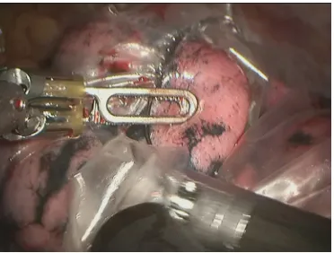

![2 [(Dimethylamino)methylidene]propanedinitrile](data:image/gif;base64,R0lGODlhAQABAIAAAP///wAAACH5BAEAAAAALAAAAAABAAEAAAICRAEAOw==)