IJEDR1502024
International Journal of Engineering Development and Research (www.ijedr.org)130
Identifying Abnormalities in Lung CT Images Based
On Honeycomb Texture Segmentation
D.Janagan,
Final year M.Tech student,
Department of Computer Science and Engineering, SRM University, Kattankulathur, Kancheepuram DT – 603203, India
Abstract - The honeycomb texture based on the lung diseases are diagnosed based on the part of lung tissue in CT images, since the texture of segmentation is the most essential part of Computer Aided Diagnosis (CAD) systems. In this paper abnormality in lung tissues shown in CT imageis segmented by wavelet transform method. Wavelet transform uses honeycomb texture in lung region for segmentation. High resolution areas are extracted from the vertical sub-image of lung region. The regions that have low pixel size of intensities are placed in separate region and it is grown for segmentation.
Index Terms - Lung segmentation, Computer Aided Diagnosis, Region growing technique, Lung abnormalities
I. INTRODUCTION
Advances in Computed Tomography (CT) technology has been used widely in diagnosing and quantifying different diseases. Several diseases in lung are diagnosed by investigating the patterns of the lung tissue in pulmonary CT images, therefore texture segmentation and analysis plays major role in CAD systems. For detecting interstitial lung diseases, Quantitative computerized analysis of diffuse lung disease was developed by Uchiyama in high-resolution computed tomography a CAD Scheme. Here a composite approach is proposed which is useful for detection of abnormalities in the lung tissue and has a better success rate in pediatric CT images. Our main focus is to find the abnormal texture named Honeycomb Texture, which is the sign of interstitial lung diseases. Our method is composed of multi-resolution techniques and intensity similarities. Lung regions are first segmented using watershed transform. The wavelet representation” is utilized to decompose lung image into its directional detailed sub- images. In the transformed image athreshold is set to extract the regions that have high resolution. The regions with blood vessel branches in most cases have the same resolution as honeycomb areas, these regions are also extracted. The pixels with lower intensities in original image are kept in order to separate honeycomb region from areas with vessel branches.We apply morphological operations on these objects to obtain accurate seeds and mask. The last step is the extraction of region of interest using 2D region growing technique.
II. RELATED WORK
Literature survey is the most important step in the research oriented project. Before developing the system, it is necessary to determine time factor and cost which will denote the efficiency of the project. In this few reference papers are studied and analyzed, so that the problems in these approaches can be enhanced. The research orientation in this project is done to improve and to enhance the quality and efficiency of the present system. The time factor and cost factor can also be defined well and also the database with quality and trust will be made available so the extraction will provide quality results.
Stephane G. Mallat et al proposed a theory of multiresolution signal decomposition [9]. They developed multi-resolution representations for analyzing the information content of images effectively. They studied the properties of the operator which approximates a signal at a given resolution. The difference of information between the approximation of a signal at a resolution, can be extracted by the decomposition of this signal on a wavelet orthonormal basis of L z ( R n ) . In L z ( R ) , a wavelet orthonormal basis is a family of functions ( @ \v (2 /x - n ) ) , J , n ) E z which is built by dilating and translating a unique function I+I (x). This decomposition defines an orthogonal multi-resolution representation called a wavelet representation. It is computed using an algorithm called pyramidal algorithm which is based on convolutions with quadrature mirror filters.The wavelet representation of images differentiates several spatial orientations.
IJEDR1502024
International Journal of Engineering Development and Research (www.ijedr.org)131
The watershed transform is a region based segmentation approach. Buecher and Lantu´ejoul formalized the concept and later Vincent and Soille turned it into an “immersion-based” algorithm. The algorithm is used to find catchment basins and watershed ridge lines in an image by treating it as a surface. In this surface the light pixels are high and dark pixels are low. Several definitions of the watershed transform have been declared. Marker-based watershed algorithms on discrete images are considered in this paper.Cornelis, J. De Becker et al proposed a techniques for Cardiac Image Segmentation [7] Two algorithms for medical image processing are discussed: CAVITY DETECTOR, which solves the segmentation problem of regions which are not completely surrounded by walls and EDGMENTATION, which is used for (i) preprocessing before segmentation, (ii) boundary refinement before e d g e detection, (iii) Segmentation based on a modified split-and- merge approach, and (iv) data reduction. In this paper, the principles of the algorithms are described and illustrate their generic properties by discussing the results of applications in 2-D and 3-D cardiac MRI and 3-D and 4-D cardiac SPECT.
III. WAVELET TRANSFORM TECHNIQUES
Recently, multi-resolution-based approaches have drawn increasing attention in the field of texture analysis. Several successful applications of this approach to automatic texture segmentation have been proposed. Wavelet transform is useful in multi-resolution-based approaches. Wavelet analysis is the process of breaking up of an original signal or original image into shifted and scaled versions of the original wavelet. In the proposed approach discrete wavelet transform is utilized to decompose lung image into its directional sub images. The discrete wavelet transform of image f(x,y) of size M by
( )
√ ∑ ∑ ( )
( )( )
( )
√ ∑ ∑ ( )
( )(2)
* +
( ) ( )
( ) ( ) ( )
( ) ( )( )
I={H,V,D}
Discrete wavelet transform corresponds to multiresolution approximation expressions. Multiresolution analysis is carried out using 2 channel filter banks which is composed of a low-pass and a high-pass filter and each of the filter bank is then sampled at a half rate of the previous frequency. It is possible to obtain wavelet transform of any order,by repeating this procedure. The filtering process is applied in a manner that it is separable. This achieved by filtering the lines and columns so the original image can be transformed into approximation, horizontal, vertical and diagonal sub-images.
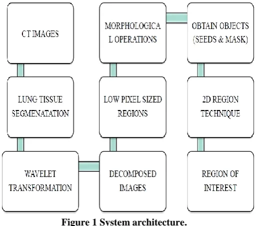
IV. ARCHITECTURE
In proposed System, the main focus is to find the abnormal texture called honeycomb texture by using multi-resolution technique and similarity intensities. The lung region is processed by using watershed transform.The watershed transform has interesting properties.These properties help in different image segmentation applications. The watershed transform is simple and intuitive, can be parallelized, and always produces a complete division of the image. Watershed transform has few drawbacks over segmentation, such as sensitivity to noise and poor detection of thin or low signal to noise ratio structures. An improved version of the watershed transform that enables the introduction of prior information in its calculation is proposed. Figure 1.
IJEDR1502024
International Journal of Engineering Development and Research (www.ijedr.org)132
V. DESIGN FOR REMOVAL OF UNDESIRABLE IMAGES1. Lung Region Segmentation. 2. Decomposing method.
3. Extracting Honeycomb from other texture 4. 2D region growing technique.
VI. DESIGN IMPLEMENTATION:
Lung Region Segmentation.
The preprocessing step is lung segmentation, which is a commonly used preprocessing technique in CAD. We present a lung segmentation technique which uses watershed transform, which is fast and accurate. We first mark the lungregion precisely with internal and external markers. The markers are combined with the gradient image of the original data. We apply watershed transform is on the combined data to find the lung borders. We use a filter called rolling ball filter which is used to smoothen the contour and fill the cavities while preserving the original borders. The proposed method doesn’t require us to find an optimal threshold and separating the attached left and right lungs, which are common in lung segmentation methods and time consuming. This approach has been applied on several pulmonary CT images and the results are quicker, robust and has a high rate of accuracy.
Decomposing method.
IJEDR1502024
International Journal of Engineering Development and Research (www.ijedr.org)133
The original image can be transformed into approximation, horizontal, vertical and diagonal sub images.Extracting Honeycomb from other texture
In a lung CT image, honeycombing is appears as dark holes but vessel branchesappear as brighter area. Using this feature we can distinguish honeycomb pattern from other high resolution areas. To extract the low intensity regions with high resolution from the CT image, the threshold images are used as a mask on the original image.
The process of extraction can be provided by two objects as,
Seed.
Mask.
Seeded-region growing with 8-connected neighborhoods is used for the extraction of the exact parts of the honeycomb tissue. The darkest high resolution objects are dilated by a disk structuring element of size to obtain an accurate mask for thegrowingregion,
Region growing technique
The Regions Of Interest is extracted using2D region growing technique. In these techniques the researchers use2D abundance of statistical measures which makes the process very slow. Comparing our Composite method with the techniques elucidates that the proposed technique is very promising.
VIII. CONCLUSIONS
A composite technique is proposed to segment honeycomb lung tissue in pediatric CT images. In this method wavelet transform is utilized combined with intensity similarities. This technique has been tested on several datasets and compared with other methods based on speed, sensitivity and specificity. The result reveals the robustness of the proposed approach.The proposed approach can be further improved to detect all possible abnormalities with different types of textures in lung tissue. Multilevel thresholding can be utilized to separate different patterns with different resolutions. Automatic lung disease quantification is another field of research which is done on segmented abnormal region in CT images. It can also be used to monitor the healing process or the improvement of the disease. Optimization methods can also be utilized to increasethe specificity.
REFERENCES
IJEDR1502024
International Journal of Engineering Development and Research (www.ijedr.org)134
[2] M. Bister,J. Cornelis, A. Cornelis,G. Demonceau,J. De Becker, and C. Vanhove “Techniques for car-diac imagesegmentation,” in Engineering in Medicine and Biology Society (EMBS), Paris, France, 1992, IEEE, vol. 14, pp. 1906– 1908.
[3] J. Alirezaie,P. Babyn,andR. Shojaii, “Automatic lung segmentation using watershed transform,” in International Conference of Image Processing (ICIP), Genova, Italy, September 2005, IEEE, vol. II, pp. 1270–1273.
[4] S.G. Mallat, “A theory of multi resolution signal decomposition: The wavelet representation,” IEEE Transactions Pattern Analysis and Machine Intelligence, vol. 11, no. 7, pp. 674–693, 1989.
[5] K. Doi, “Current status and future potential of computer-aided diagnosis in medical imaging,”British Journal of Radiology, vol. 78, no. 1, pp. 3–19,January 2005.
[6] K. Ashizawa,K. agaoki,K. Doi, A. Egawa,A. Fukushima, H. Hayashi,K. Hayashi, S. Honda Y. Imafuku,I. Kida, S. Kimura, and S. Katsuragawa,T. Yamaguchi, and N. Matsuyama “Application of an artificial neural network to high-resolution ct: Usefulness in differential diagnosis of diffuse lung disease,”American Journal of Roentgenology, vol. 183, pp. 297–305, 2004.
[7] G.W HunninghakeE.A. Hoffman, P.G. Hartley,M. Sonka, G. McLennan,R. Uppaluri, and “Computer recognition of regional lung disease patterns,” American Journal of Respiratory and Critical Care Medicine,vol. 160, no. 2, pp. 648– 654, August 1999.
[8] E.A. Hoffman,G. McLennan,T. Mitsa,R. Uppaluri, and M. Sonka, “Quantification of pulmonary emphysema from lung computed tomography images,” American Journal of Respiratory and Critical CareMedicine, vol. 156, no. 1, pp. 248– 254, July 1997.
[9] J. R. Galvin,T. Mitsaand ,R. Uppaluri, “Fractal analysis of high-resolution ct images as a tool for quantification of lung disease,” in Medical Imaging, 1995, vol. 2433, pp. 133–142.
[10] N. L. Muller, D. P. Naidichand W. R. Webb,“High- Resolution CT of the Lung”, Lippincott Williams & Wilkins, Univ. of California, San Francisco, 3rd edition,January 2001.